天津医科大学实验动物科学部
- 格式:doc
- 大小:78.00 KB
- 文档页数:3
天津医科大学实验动物使用审查表
填写说明
一、根据天津医科大学实验动物管理和使用委员会要求,2021年开始不在天津医科大学动物实验平台进行全程动物实验的不予审查,动物伦理审查收取审查费。
二、进驻动物实验平台进行实验前填写动物伦理审查表,该表代替动物实验申请书。
三、各种费用计算按填写内容进行核定,按规定执行。
四、按规定未交齐动物实验期间费用的不给予动物伦理审查结论。
五、仔细看申请书填写要求,并按要求详细填写内容。
六、动物实验期间设施兽医有权对动物实验进行检查,凡发现实验和申请书不符的情况,兽医有权终止实验,责令项目人补充或完善动物福利伦理审查。
七、实验动物使用和管理委员会审查意见页中项目基本情况由项目人填写,并应和申请书中的一致。
八、如果项目人对审查意见有英文需求,可在审查意见中参照现有格式提供中英对照。
九、动物伦理审查不接受电话咨询。
十、初审合格的申请表以“当前年月(如:202101)-项目名称-项目人-项目单位”格式提交到IACUC@邮箱。
未经初审合格不要提交。
十一、正式提交不提交填写说明页
天津医科大学实验动物使用审查表2021年动物实验版(代动物实验申请书)
实验动物使用和管理委员会审查意见(2021年动物实验版)
项目基本情况
编号:TMUaMEC 2021001。
天津医科大学的前身天津医学院创建于1951年,是新中国成立后国家政务院批准新建的第一所高等医学院校,著名内分泌学家、医学教育家朱宪彝教授为首任校长。
1993年,天津市市委、市政府决定天津医学院与天津第二医学院合并组建天津医科大学,同年12月得到国家教委批准。
1994年6月,天津医科大学正式组建成立。
学校现任党委书记为张连云教授,校长为著名生物化学与分子生物学家、中国科学院院士尚永丰教授。
学校为国家“211工程” 重点建设院校。
1医大简介天津医科大学是国家“211工程”重点建设院校,为仅有的两所进入国家“211工程”建设的独立设置西医院校(天津医科大学、北京协和医学院)之一。
在网大公布的2007年中国大学排名中天津医科大学在独立设置的75所医学院校中与北京中医药大学并列为第2名,综合排名为第67[2]名。
另外在网大2010年公布的1999年至2010年进步最显著的大学名单中,天津医科大学榜上有名,与中国政法大学、华东师范大学、苏州大学、西北大学、北京中医药大学这五所重点高校共同评为进步最显著的大学。
在武书连公布的2011年中国大学评价中,天津医科大学被评为2011中国大学医学最强的23所学校之一,等级为A级。
2011年6月1日上海交通大学高等教育研究院世界一流大学研究中心(教育部战略研究基地)校史板报(18张)完成的“中国两岸四地大学排名”中,在众多港澳台高校参评的情况下,天津医科大学综合排名列第67名,摘得中国内地独立设置医学院校中的第一。
2013年发布的中国·两岸四地大学排名中,在两岸四地高校中列第62位,大陆地区第37位。
[3]学校现有全日制本科以上在校生9894人,其中,本科生5329人,硕士生2871人,博士生428人,留学生1266人。
天津医科大学是继协和医学院后,国家最早批准试办八年制的医学院校之一,首批试办七年制的15所院校之一。
自建校以来,学校已培养了40000余名各层次医学人才。
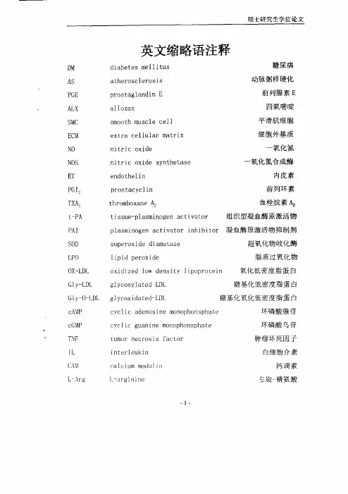
英文缩略语注释diabetesmellitus糖尿病atherosclerosiS动脉粥样硬化prostaglandinE前列腺素Ealloxan四氧嘧啶smoothmusclecell平滑肌细胞extracellularmatrix细胞外基质nitricoxide一氧化氮nitricoxidesynthetase一氧化氮合成酶endothelin内皮素prostacyclin前列环素thromboxaneA2血栓烷素A2tissue—plasminogenactivator组织型凝血酶原激活物plasminogenactivatorinhibitor凝血酶原激活物抑制剂superoxidediSmutase超氧化物岐化酶lipidperoxide脂质过氧化物oxidizedlOWdensitylipoprotein氧化低密度脂蛋白glycosylatedLDL糖基化低密度脂蛋白glycoxidated—LDL糖基化氧化低密度脂蛋白cycliCadenosinemonophonsphate环磷酸腺菅cyc¨cguaniFiemon。
phonsphate环磷酸鸟苷tumornecrosiSfacfor肿瘤坏死因子interleukin白细胞介素calciummodulirl钙调素l,一argjniDe左旋精氨酸。
●凹叱加,,曙EXCMSIAPIDO—V。
V.ⅥⅥFⅥ1附脶眦似㈣吲№懈盯哪m忡呲㈣啪噼吣m洲删肿儿吣㈨中文摘要目的:在以往实验的基础上,研究大剂量PBE:及PGE。
对糖尿病性动脉粥样硬化家兔病理形态学、血管内皮依赖性舒张功能、氧化应激、血凝纤溶状态及血栓形成的影响,探讨PGE抗糖尿病性动脉粥样硬化的作用机理,从而为临床防治糖尿病大血管并发症提供科学依据。
{方法:I.先采用静脉注射ALX法复制DM模型,再饲以高胆固醇饲料复制DM性AS家兔模型,按血脂血糖水平随机分成3组。
2.利用大体和光镜标本观察PGE对DM性As家兔主动脉病变的影响。
糖尿病牙周炎昆明系模型小鼠牙周组织中脂联素的表达赵博;王永兰【摘要】BACKGROUND:Adiponectin and periodontal disease are closely related with type 2 diabetes. OBJECTIVE:To detect the expression of adiponectin in periodontal tissue of Kunming mice of diabetes periodontitis, and explore the effects on periodontal tissue pathology and prognosis. METHODS:Sixty 3-week-old Kunming mice were divided into three groups according to the different modeling methods:control group (normal feeding), diabetes group (diabetic model was established by injection of al oxan), diabetes and periodontitis group (periodontitis model was established by local ligation and bacteria developed). Each group contained 20 rats. RESULTS AND CONCLUSION:At 20 days after modeling, the blood glucose, fasting insulin and insulin resistance index of mice in the diabetes group were higher than that in the control group (P<0.05). ELISA, RT-PCR and western blot analysis results showed that, adiponectin gene and protein expression levels in periodontal tissues were the highest in the control group, then in the diabetes group, and the lowest in the diabetes and periodontitis group, at 1, 3, 6, months postoperatively, respectively (P<0.05). The periodontal gum tissue inflammation and histopathological changes were observed at 1, 3, 6, months postoperatively. The results showed that, the inflammation in periodontal tissues was the most obvious in the diabetes and periodontitis group, and was visible in the diabetes group, but the inflammation was not present inthe control group. Experimental findings indicate that, the down-regulation of adiponectin expression in the periodontal tissue is one of the important factors that trigger and aggravate the onset of periodontitis.%背景:脂联素与牙周炎和2型糖尿病发病有密切关系。
缺血预适应对肝脏热缺血再灌注损伤保护作用的机制研究金涛;胡宇;刘超【摘要】Objective To explore the pathogenesis of ischemic preconditioning to warm ischemia reperfusion injury of hepatocytes in rats. Methods Ninety SD rats were randomly divided into three experimental groups:sham operation group (group A), warm hepaticischemia/reperfusion group(group B and group C). Group C was given ischemic preconditioning treatment. Serum levels of alanine aminotransferase (ALT) and aspartate aminotransferase (AST) were detected 0 h, 2 h, 6 h, 12 h and 24 h after ischemia reperfusion injury. Levels of TNF-α and IL-1β were tested detected by ELISA. Levels of malondialdehyde (MDA) and superoxide dismutase (SOD) of hepatocytes were detected at the same time points. Mitochondrial membrane potential was examined to assess ischemia reperfusion injury of hepatocytes in rats using chart of intensity of JC-1 in mitochondria. Results The serum levels of ALT, TNF-α, IL-1β, and MDA were significantly higher in hepatic warm ischemia reperfusion group and ischemic preconditioning group than those in sham operation group (P<0.05). Values of prothrombin activity and cholinesterase were significantly lower in liver warm ischemia reperfusion group and ischemic preconditioning group than those of sham operation group (P<0.05). The SOD level of liver was significantly lower in warm ischemia reperfusion group and ischemic preconditioning group than that in sham operation group. The indexes were better in ischemicpreconditioning group than those of warm ischemia reperfusion group(P<0.05). The mitochondrial membrane potential level of liver cells reached the lowest value 0 hours after ischemia and reperfusion, and then increased gradually within 24 hours (P<0.05). And the level of mitochondrial membrane potential of liver cells was significantly higher in ischemic preconditioning group than that in warm ischemia reperfusion group (P<0.05). Conclusion Ischemic preconditioning may play a protective role in warm ischemia-reperfusion injury in rats. Ischemic preconditioning may significantly decrease the levels of ALT, AST, TNF-α,IL-1βand MDA, and increase the SOD activity in hepatocytes. Thedamageof mitochondrial membrane potential is decreased after ischemic preconditioning, which might be the pathogenesis of ischemic preconditioning to warm ischemia reperfusion injury of hepatocytes in rats.%目的:探讨缺血预适应对肝脏热缺血再灌注损伤保护作用的分子机制。
唑来膦酸局部应用对大鼠拔牙创软硬组织的影响崔彩雯;张健;马婷;汪铭【摘要】Objective: To investigate the effects of locally application of zoledronic acid (ZA) on hard and soft tissue in rats that were pretreated with ZA during tooth extraction. Methods: Fifty-four 7-week old male Wistar rats were divided into ZA group, gelatin sponge group, and blank control group, hi each group, left mandibular first molars were extracted under general anesthesia. Rats were euthanized two weeks after tooth extraction, and mandibular bones were collected. Bone mor-phometric analysis using X-ray images was performed and the gingival around the extraction socket was harvested for HE staining and Masson staining to observe the histopathology effect. Results: New bone formation in the extraction socket was promoted and alveolar ridge absorption was delayed in the ZA group compared to the control group. The alveolar ridges of the ZA group were higher than those of the control group (2.60± 0.l0mmVSl .99± 0.22mm, P0.001; 2.60± 0.10mm VS 2.12± 0.24mm, PO.001). Histological images in ZA group showed that the mucous about extraction socket was getting thinner, the fiber was slender and disordered, but no differences of blood ressels were observed. Conclusions: Local application of zoledronic acid could inhibit bone resorption and promote new bone formation of the tooth extraction socket, also it might inhibit the formation of collagen fibers. These actions of ZA may be relevant to the pathogenesis of BRONJ.%目的:观察唑来膦酸(zoledronic acid,ZA)局部应用对大鼠下颌磨牙拔除后剩余软硬组织的影响.方法:将54只雄性Wistar大鼠随机分为三组,即空白对照组、明胶海绵组和唑来膦酸治疗组,麻醉下完整拔除左侧下颌第一磨牙并做相应处理,术后2周同期处死所有大鼠.用X线片测量大鼠拔牙窝剩余牙槽嵴的高度,取拔牙窝周围牙龈组织制作石蜡切片行HE染色和Masson染色,观察大鼠下颌磨牙拔牙窝的愈合情况.结果:唑来膦酸治疗组牙槽嵴高度显著高于空白对照组和明胶海绵组(2.60±0.10mmVS1.99±0.22mm,P<0.001;2.60±0.10mm VS 2.12±0.24mm,P<0.001).组织学观察拔牙创周围黏膜层上皮变薄,胶原较为纤细且排列疏松,血管变化不明显.结论:唑来膦酸局部应用抑制牙槽嵴骨吸收,促进拔牙窝新骨形成,对软组织作用不显著,可能抑制牙龈黏膜中胶原纤维的合成.【期刊名称】《口腔颌面修复学杂志》【年(卷),期】2011(012)005【总页数】4页(P261-264)【关键词】唑来膦酸;局部用药;牙槽骨;软组织【作者】崔彩雯;张健;马婷;汪铭【作者单位】天津医科大学口腔医院颌面外科天津300070;天津医科大学口腔医院颌面外科天津300070;天津医科大学口腔医院颌面外科天津300070;南京市中西医结合医院江苏210000【正文语种】中文【中图分类】R782.11双磷酸盐(bisphosphonate,BP)用于治疗骨代谢异常类疾病和恶性肿瘤的骨吸收抑制剂,有报道[1]发现BP治疗的癌症患者拔牙后发生下颌骨坏死(bisphosphonate-related osteonecrosisof thejaw s,BRONJ)其中以唑来膦酸所占比例高。
骨髓间充质干细胞移植脑梗死模型后的CT、MRI特征分析杨廷双【摘要】背景:研究表明骨髓间充质干细胞可通过多种途径发挥促进脑组织功能恢复的作用。
目的:分析骨髓间充质干细胞移植脑梗死模型大鼠的CT、MRI特征。
<br> 方法:将40只脑梗死模型大鼠随机分为2组,梗死组通过尾静脉注射1 mL PBS、移植组通过尾静脉注射1 mL细胞浓度为2.0×10⁹ L-1的细胞悬液。
于移植后1,2,3周进行神经功能缺损评分(mNSS),评价神经功能恢复情况;于移植后的6 h,1,3,5,7 d对各组大鼠行CT及MRI扫描,观察脑梗死区TIWI、T2WI、FLAIR、DWI序列的信号改变特征,梗死体积大小。
测量脑梗死区域T1WI、T2WI、FLAIR、DWI序列的信号强度比(SIR)及其相对变化率(∆SIR),并与正常对侧相应解剖区域进行比较。
<br> 结果与结论:移植后1,2,3周,移植组的神经功能缺损评分明显低于梗死组(P<0.05);移植后3,5,7 d时移植组脑梗死体积较梗死组显著减少(P<0.05),移植组T1WI序列SIR均明显高于梗死组,T2WI、FLAIR序列SIR较梗死组显著降低,差异有显著性意义(P<0.05);移植后7 d时移植组DWI序列SIR较梗死组明显降低(P<0.05)。
移植组T1WI序列∆SIR与梗死组比较差异无显著性意义(P>0.05),移植组T2WI、FLAIR及DWI序列∆SIR较梗死组显著增高,差异有显著性意义(P<0.05)。
结果显示MRI可显示大脑任意角度的切面像,对骨髓间充质干细胞移植治疗脑梗死的疗效评价起重要作用。
%BACKGROUND:Studies have shown that bone marrow mesenchymal stem cel s can play a catalytic role in brain function recovery through a variety of ways. OBJECTIVE:To analyze the CT and MRI features of rats with cerebral infarction undergoing bone marrow mesenchymal stem cel transplantation. METHODS:Forty rats with cerebral infarction wererandomized into two groups:model group was treated with 1 mL PBS and transplantation group was injected with 2.0×10⁹/L cel suspension. Modified neurological severity score was detected at 1, 2, 3 weeks after transplantation for neurological function evaluation. CT and MRI scans were performed at 6 hours, 1, 3, 5, 7 days after transplantation to observe the changing features of T1WI, T2WI, FLAIR, DWI sequences in the infracted area as wel as infarction size. Signal intensity ratios (SIR) of T2WI, FLAIR, DWI sequences and relative rates of change (∆SIR) were determined and compared with the normal values. RESULTS AND CONCLUSION:After transplantation 1, 2 and 3 weeks, the modified neurological severity scores were significantly lower in the transplantation group than the model group (P<0.05). After transplantation 3, 5, 7 days, the infracted size was reduced significantly in the transplantation group compared with the model group (P<0.05). Compared with the model group, SIR value of T1WI sequence was higher, but the SIR values of T2WI and FLAIR sequences were lower in the transplantation (P<0.05). There were significant differences in the∆SIR values of T2WI, FLAIR and DWI (P<0.05) rather than T1WI (P>0.05) between the two groups. These findings indicate that MRI can show the brain sections at any angle, and play an important role to assess the therapeutic efficacy of bone marrow mesenchymal stem cel transplantation on cerebral infarction.【期刊名称】《中国组织工程研究》【年(卷),期】2015(000)036【总页数】5页(P5795-5799)【关键词】干细胞;移植;骨髓间充质干细胞;大鼠;脑梗死;CT;MRI;特征分析;细胞移植【作者】杨廷双【作者单位】天津医科大学总医院滨海医院放射科,天津市 300480【正文语种】中文【中图分类】R394.2文章亮点:1骨髓间充质干细胞移植对恢复脑梗死大鼠神经功能,减轻脑组织水肿和脑梗死体积起到促进作用。
2019年11期教海探新高教学刊《实验动物学基本实验技术》选修课的设计与探讨*刘爱丽,刘迢迢*(天津医科大学生物医学工程与技术学院,天津300070)实验动物学是生命科学领域内的一门综合性新兴学科,以实验动物和动物实验为研究对象,是生命科学研究的重要支撑条件[1]。
旨在培养学生的科研素质和实验操作能力,为其从事动物实验相关的科学研究奠定基础[2,3]。
目前已成为众多高等医学院校中研究生的一门基础课程,但相关课程很少在本科阶段开设。
生物医学工程是生物医学与理工科的交叉学科,以医工结合为基础,培养具备生命科学、电子技术及信息科学的理论知识以及医学与工程相结合的科学研究能力,能够从事教学、研究以及管理等相关工作的专业人才。
目前此专业本科生的生命科学相关知识仅限于《生理学》和《系统解剖学》两门课程的学习,而实验动物学方面的实验操作能力有所缺乏。
根据学院现有的仪器设备和实验室条件,阐述为生物医学工程专业本科生开设《实验动物学基本实验技术》选修课的必要性和可行性,并详细探讨拟开设课程的内容设置和考核方式。
一、开设《实验动物学基本实验技术》选修课的必要性实验动物作为现代生命科学实验研究的四个基本要素AEIR之一(即动物、试剂、设备和信息),被称为活的试剂和活的精密仪器[4],是生物科学实验研究的重要工具和基础条件,在科学研究和实验教学中具有突出贡献[5]。
动物实验是指借助实验动物,采用特定的实验方法来检验或探索相关科学问题而进行的实验研究。
动物实验作为科学领域内必不可少的一种研究手段,也是实验教学的重要形式,生物医学科学的发展离不开动物实验[6]。
为了响应国家教育部高等教育本科阶段培养创新型人才的号召,学校鼓励在校本科生积极申报大学生创新创业训练计划项目,并进入科研实验室参与教师的科研课题[7,8]。
实验动物和动物实验基本知识和实验技能的缺乏却成为了很多生物医学工程专业本科生的阻碍。
坚持教学和科研相结合的原则,为满足该专业本科生对动物实验的实际需求,培养本科生的科研素质,拟开设《实验动物学基本实验技术》选修课。
枢及外周神经系统和胃肠道中。
V I P释放后与特异受体结合发挥生理功能,可增加ACTH分泌,有较强的扩血管作用,参与休克、再灌注损伤等病理过程的发生发展[5]。
本实验显示,H I E组血浆V I P明显高于非H I E组,说明V I P参与了窒息新生儿H I E的发病过程。
纳洛酮是阿片受体的特异性拮抗剂,纳洛酮与阿片受体结合后,及时阻断β2EP的病理效应,切断疾病的恶性循环,改善呼吸循环功能,稳定溶酶体膜,发挥抗脂质过氧化物作用[6]。
同时降低血浆中V I P水平,阻止病程的发展,具有良好的干预作用。
参考文献[1] 侯 琳,濮海平,邵伯芹.银杏提取物对新生鼠缺血缺氧性脑损伤NSE2100mRNA表达的影响[J].中国药理学通报,2003,19(1):100-2.[2] 刘 玮,吴华璞,祝晓光,明 亮.白芍总苷对全脑缺血再灌注损伤的保护作用[J].中国药理学通报,2004,20(2):211-4.[3] Johnst on MV.Neur otrans m itter alterati ons in a model of perinatalhypoxic2ische m ic brain injury[J].A nn N eurol,1983,13:511-3.[4] Pasaoglu H,I nciKE,Kurts oy A.Endogenous neur opep tides in pa2tients with acute traumatic head injury,I:Cerebr os p inal fluid be2 ta2endor phin levels are increased with in24hours f oll owing the trauma[J].N europeptides,1996,30(1):47-51.[5] 罗晓阳,张 翔,王嘉军.纳洛酮对脑损伤大鼠血浆及海马匀浆中血管活性肠肽的影响[J].中国危重病急救医学,1996,8(11):641-2.[6] 罗星照.纳洛酮在儿科临床的应用[J].中国新药与临床杂志,1998,17(6):379-80.二色桌片参皂苷i n tercedensi de A的抗真菌和抗肿瘤活性邹峥嵘1,2,3,易杨华1,姚新生3,杜力军2,周大铮1,张淑瑜1(1.上海第二军医大学药学院海洋药物研究中心,上海 200034;2.清华大学生物学博士后流动站,北京 100084;3.深圳清华大学研究院博士后科研工作站,广东深圳 518057)Stud i es on the an ti funga l and an titu m or acti v iti es of the i n terceden si de A fro m M en s amar i a i n terce2 den s Lam pertZ OU Zheng2r ong1,2,3,YI Yang2hua1,Y AO Xin2 sheng3,DU L i2jun2,ZHOU Da2zheng1,ZHANG Shu2 yu1(1.Research Center of M arine D rugs,School of Phar m acy,Sec2 ond M ilitary M edical U niversity,Shanghai 200034,China;2. Postdoctoral S tation of B iology in Tsinghua U niversity B eijing 100084,China;3.Postdoctoral Research W orkstation of R ITS Shenzhen 518057,China)中国图书分类号:R2332;R282171;R28411;R730152; R97815;R97911文献标识码:A文章编号:1001-1978(2005)06-0761-02关键词:二色桌片参;intercedenside A;抗肿瘤;抗真菌Key words:mensa maria intercedens la mpert;intercedenside A;antifungal;anticancer收稿日期:2004-07-28,修回日期:2004-12-27基金项目:上海市科技发展攻关资助项目(No02DZ19101)作者简介:邹峥嵘(1970-),男,医学博士,博士后,研究方向:中药二次开发和海洋药物,E2mail:zhr wl m@t ;易杨华(1946-),男,教授,博士生导师,研究方向:海洋生物活性成分,通讯作者,Tel/Fax:021*********,E2mail:yiyanghua@hot m ;姚新生(1934-),男,博士,中国工程院院士,Tel/Fax:0755226036131,E2mail:yaoxinsheng@ 二色桌片参(Mensa maria intercedens La mpert)属枝手目(Dendrchir otida)瓜参科(Cucu mariidae)棘皮动物[1],从我国福建东山到海南岛的海域均有分布,资源十分丰富。
天津医科大学科研实验动物资源需求情况调查表为更高地为学校科研服务,以满足科研条件的需要,科技处组织此次有关科研实验动物需求的问卷调查工作。
希望您在百忙之中填写此调查表,非常感谢您对此项工作的支持!一、基本资料学院(系)名称:教研室(实验室/项目课题组)名称:填表人:职称:联系电话:E-mail:目前从事动物试验分类:□医学研究类:基础医学、临床医学、预防医学、解剖生理学、病理/传染病等□药学试验类:药理学试验、药动学试验、临床前(毒理)试验、药物安全试验□新型治疗/药物类:基因治疗、细胞治疗、药物治疗等□动物药品试验类:动物疫苗、抗生素、生长素、饲料添加物等□医疗器材试验:CT、核磁、超声等□疫苗类:人用疫苗、动物疫苗等□中草药/健康食品研发类:□生农研究类:生物试剂、农药等□其他研究:二、对科研实验动物的需求选择说明:(1)购买来源:选择: A国家实验动物资源中心 B北京华阜康公司 C中国军事医学科学院D北京维通利华公司 E天津实验动物生产单位 F国外科研机构G国外公司(例如JAX、TACONIC等) H其他(2)饲养地点:选择:I校实验动物科学部 J学校其它地方 K 学校外(3)饲养环境:选择:M 屏障设施内 N 非屏障设施 O 不知道是什么环境(4)遗传来源:选择:P 常规大、小鼠 Q 转基因大、小鼠 R 基因突变鼠(KO或CKO) S 大、小鼠以外的其他动物1. 最近三年(2010年、2011年、2012年)实际使用实验动物情况:(不够,可以加栏)2. 未来三年内(2013年、2014年、2015年)计划使用的实验动物品系名称及数量需求:(不够,可以加栏)3. 您希望实验动物科学部引进繁殖的实验动物品系及需求:(可加页)说明:饲养地点:选择:A校实验动物科学部 B学校其它地方 C 学校外饲养环境:选择:A 屏障设施内 B 非屏障设施 C 不知道是什么环境三、您对实验动物设施的需求:1. 希望提供GLP实验室(Good Laboratory Practice,实验室优良操作规范):□是,说明□否2. 希望提供下列功能(可复选):□一般动物提供□实验室出租□动物寄养□动物试验合作□疾病模式动物提供□教育训练□胚胎冷冻服务□动物健康诊断服务□动物遗传监测服务□其他3.根据您目前的实验需求,您最希望提供的与实验动物相关的实验仪器是:A、小动物可见光活体成像系统□B、Micro-CT(活体或离体)□C、Micro SPECT-CT □D、激光共聚焦显微镜系统□E、小动物核磁共振系统(MRI)□F、生物辐照仪□G、流式细胞仪(分选/不分选)□ H、DNA合成仪□I、高速冷冻离心机□G、超速冷冻离心机□ K、全自动生化分析仪□ L、全自动核酸提取仪□M、全自动血细胞分析仪□ N、全自动血凝分析仪□ O、气相色谱仪□P、液相色谱仪□ Q、代谢笼(进口)□其它仪器:四、您对实验动物机构服务的需求□1. 无需求;□2. 提供实验动物□3. 提供实验场所□4. 健康监测服务(微生物检测);□5. 遗传背景检测;□6. 寄养服务;□7. 净化服务;□8. 冷冻胚胎服务;□9. 辅助生殖技术;□10. 疾病诊断服务;□11. 病理表型分析;□12. 行为表型分析;□13. 影像分析服务(包括荧光、CT、PET、X线等);□14. 动物进出口服务;□15. 毒性/安全性/功效(GLP)服务;□16. 基因工程技术服务;□其他:五、其它具体建议:(可附页)。
天津市科学技术局关于发放天津医科大学实验动物使
用许可证的通知
文章属性
•【制定机关】天津市科学技术局
•【公布日期】2020.12.31
•【字号】津科基〔2020〕160号
•【施行日期】2020.12.31
•【效力等级】地方规范性文件
•【时效性】现行有效
•【主题分类】检验监管
正文
天津市科学技术局关于发放天津医科大学实验动物使用许可
证的通知
津科基〔2020〕160号
天津医科大学:
你单位报送的《实验动物使用许可证申请书》收悉。
市科技局根据国家及天津市关于实验动物许可证管理的相关规定,组织有关专家对你单位所申请事项进行了审查和现场验收。
根据验收情况及专家意见,按照《天津市实验动物许可证管理办法(试行)》(津科财〔2005〕103号)》的有关规定,经研究决定,准予发放天津医科大学实验动物使用许可证,许可证设施坐落在天津市和平区气象台路22号(实验动物科学部基础医学研究中心楼1-3层),许可证适用范围:屏障环境小鼠、大鼠。
许可证编号:SYXK(津)2020-00010,许可证自发证之日起五年有效,每年进行年检。
请你单位按照国家及天津市关于实验动物许可证管理规定的要求,认真做好相
关工作。
2020年12月31日。
天津医科大学实验动物科学部动物实验福利伦理审查申请表-增补表
(请至少在预计引进、订购或领用动物之前一周申请)
此处供委员会使用
计划调整编号
批准日期
终止日期
本实验调整计划使用范围:
•增加同一种类其他品系;
•改变动物性别
•动物数量改变不超过20%
•改变或增加非存活性手术
•增加样品采集次数和与此protocol相关的测试,前提是该方法在动物福利委员会已批准的范围内•改变麻醉剂和镇静剂,前提是该试剂在动物福利委员会已批准的范围内
•改变安乐死的方法,前提是该方法在动物福利委员会已批准的范围内
•改变项目的标题
•增加或减少实验人员
一、原研究计划编号:
项目标题:
二、申请调整的具体内容:
A.追加动物数量(不得超过原申请的20%):
C . 添加或减少动物实验操作人员:
注:职责可为课题设计、主要操作、辅助操作、手术操作、小鼠管理等
列出每个人相对于职责,已有的实验经验和资格
实验室负责人(PI)签名:日期:
三、校福利伦理委员会审阅意见:
□批准□不批准
主要负责人:________________ 签名:________________ 日期:_________________
注:
1) 为节省审批时间,请首先e-mail本申请书电子版至天津医科大学实验动物使用和管理委员会邮箱(yddwkxb@)和PI本人。
2)预定动物时请递交一式一份纸质版给实验动物科学部。
一、工程概况:本次招标的天津市医药科学研究所动物实验楼项目内容如下:工程地址:天津市和平区多伦道79号天津市医药科学研究所院内,原制剂楼。
建筑面积:约为663M2。
建筑层数:三层砖混框架结构。
建筑层高:一层3.9米,二、三层为3米。
室内层高:实验用房一层2.4米;走廊为2.4米;二、三层室内和走廊均为2.2米。
结合天津市医药科学研究所建设规划,为加快科研条件建设,提升科技创新能力和科技竞争力,在上级单位的支持下,天津市医药科学研究所决定改建动物实验楼,使其成为药物研发的重要支撑平台。
该所的主要的研究方向包括药理学、药代动力学、临床医学、预防医学、医学检验、药学、药物制剂等专业。
改造后的动物实验室不仅要满足本所科技研发工作的需要,还要更多地承接社会各界的技术开发、技术合作及委托项目,并且为本所今后的发展预留拓展空间。
因此,在动物实验室改造方案设计上要瞄准国内先进水平,符合国家相关标准规范,在动物实验楼的功能配置上既要保证科研实验条件,又要具备一定的前瞻性,确保科研实验质量,增加实验的可靠性。
现将本动物实验楼内各功能实验室平面工艺布局介绍如下:实验用房:一层:动物接收室、检疫隔离室、预备兔室、实验兔室、热原实验室、仪器室、实验室、洗刷消毒室、走廊、更衣室、以及饲料库、处置及废弃物储存室、更衣淋浴室、值班室、管理控制室、卫生间、配电室、两间空调机房;二层:一间豚鼠饲养室、六间实验室、无菌室和实验准备室,以及走廊、洗刷室、库房、清扫用品室等;三层:两间小鼠饲养观察室、一间大鼠饲养观察室、瘤源保种及传代室、手术室、洗刷消毒室、内准备间、笼器具库、洗衣室、垫料饲料库、清洁走廊、缓冲间以及一更、二更等。
二.工程特点本方案是一个中型、功能较齐备的动物实验综合性设施。
在既满足自身使用,又符合相关规范,充分利用有效面积,尽量减少一次性投入和降低日常运转费用的前提下,设计出多功能、多品种的综合实验和科研用房条件。
内部可容纳的实验类别和功能基本满足医药科学研究所科研工作的实际需要。