
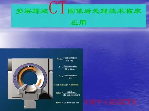
多层螺旋CT图像后处理技术临床应用
- 格式:ppt
- 大小:4.37 MB
- 文档页数:107
World Notes on Antibiotics,20 1 3,Vo1.34,No.5
多层螺旋CT及图像后处理技术在隐匿性肋骨骨折中的应用价值
吴宇 (成都市第一人民医院医学放射科,成都 61 0000)
摘要: 目的探讨多层螺旋CT及图像后处理技术在隐匿性肋骨骨折中的诊断价值,提高正确诊断率。方法回顾性分析40例 有明确胸部外伤史,临床症状明显但胸部X线平片检查阴性患者的CT检查资料。结果40例患者共检出肋骨骨折58处,均无明显的 骨折移位;轴位像检出骨折50处、VR像检出骨折43处、MIP像检出骨折16处、MPR像检出骨折56处及CPR像检出骨折58处。结论 多层螺旋CT及图像后处理技术是诊断隐匿性肋骨骨折的理想手段,值得临床推广应用。 关键词: 体层摄影术;X线计算机;隐匿性肋骨骨折;图像处理 中图分类号:R45 文献标识码:A 文章编号:1001.8751(2013)05—0216.02
The Clinical Application of Postprocessing Technique of Multi- Slice Spiral CT Images in Occult Rib Fracture
弭 (Department ofRadiology,the First People’S Hospital ofChengdu,Chengdu 610000)
Abstract:0bjective To evaluate the clinica1 application of postprocessing technique of multi.slice spira1 CT images in occult rib fracture.Methods The CT data of 40 cases with history of thoracic iniury.obvious clinical symptoms and negative X—ray plain film were analyzed retrospectivel Results 58 fractured ribs were checked out in 40 cases,without obvious fracture displacement;50 fractures detected with axial view images.43 fractures detected with VR images,1 6 fractures detected with MIP images,56 fractures detected with MPR images.and 5 8 fractures detected with CPR images.Conclusion Postprocessing technique of multi.slice spiral CT images was an effective waV to diagnose occult rib fracture.it is worthy of clinical application. Key words:computerized tomography;X—ray;occult rib fracture;image processing

・2464・
多层螺旋CT重建
临床应用
李 刚 Journal of Qiqihar University of Medicine,2012,Vo1.33,No.18
后处理技术在骨关节骨折中的
【摘要】 目的 探讨多层螺旋CT三维重建与多平面重建诊断骨关节骨折的临床观察。方法 回
顾性分析本院于2010~2011年期间收治的52例普通平片检查后未发现骨折、可疑骨折和明确骨折的
患者行螺旋CT扫描,后给予三维重建及多平面重建,综合分析普通平片与16层螺旋cT影像,比较两
种图像显示骨折详细情况的能力。结果在显示各种骨折、脱位情况上,三维重建及多平面重建比普通
平片更为明确、直观,减少了误诊、漏诊的发生。结论 对于隐匿性骨折,多层螺旋CT比普通平片诊断
更为明确,三维重建与多平面重建能够更好了观察骨折的变化和移位情况,两者相互补充有利于临床诊
断和治疗骨关节骨折。 【关键词】 骨关节 骨折 体层摄影术X线计算机 图像重建
在外伤中,骨关节骨折是常见的改变,x线、CT、MRI等
是其影像学检查方法,其中重要的检查是CT,多层螺旋CT
后处理技术能更直观地显示骨折情况[1]。本次研究选用本院
于2010 ̄2011年期间收治的52例普通平片检查后未发现骨
折、可疑骨折和明确骨折的患者行螺旋CT扫描,后给予三维
重建及多平面重建,观察比较普通平片与16层螺旋CT影像
显示骨折情况的能力,现将报道如下。
1资料与方法
1.1 一般资料本组是本院于2010 ̄2011年期间收治的典
型骨关节骨折患者52例,年龄3~73岁,男33例,女19例。
受伤的原因有车祸伤,摔伤。其中16例踝关节,12例膝关
节,1O例髋关节,8例肘关节,6例腕关节。上述患者的临床 表现主要是患者疼痛、肿胀、畸形。
1.2 检查方法 使用美国生产的GEB排螺旋CT(1ight-
speed)进行检查。给予患者普通平片检查后采取16层螺旋
实用医技杂志2012年8月第19卷第8期Journal of Practical Medical TechniⅡues,Au st 2012,V0I_19,No.8
多层螺旋CT三种图像后处理技术
在肺栓塞诊断中的应用评价
广东省汕头市中心医院(515031)
黄朝华杨江爽翁泽生吴先衡陈瑟燕林时勖
【摘要】 目的 比较不同重建层厚的各种图像后处理技术对肺动脉栓子的显示及诊断价值。方法收集40
例经多层螺旋CT肺动脉成像检查并经临床及实验室检查确诊的肺动脉栓塞(PE)图像资料,利用原始数据分别行层
厚0.625、1.25、2.5、5.0 mm重建图像,然后运用多平面重组(MPR)、最大密度投影(MIP)及容积成像(VR)3种图像后
处理方法对于在不同层厚的各级肺动脉显示及其栓子分布情况进行观察分析。结果本组40例PE均为双肺多发
肺栓塞,共累及各级肺动脉分支409处,范围从肺动脉主干至亚段动脉。40例PE中,对亚段动脉栓子检出率,0.625、
1.25、2.5 mm MPR图像和MIP图像的差异无统计学意义,5 mm MPR、5 mm MIP及VR图像对亚段动脉栓子显示较
0.625、1.25、2.5 mm MPR及MIP图像差异有统计学意义。结论肺栓塞的诊断主要依靠薄层横断图像,对亚段动脉
栓子的观察至少要用2.5 mm重建层厚的MPR或MIP图像,两者任选择一项即可。而1.25 mm层厚对亚段动脉栓子
不易漏诊,且能大大地提高观察者阅片速度,故1.25 mm层厚的MPR或MIP为最佳成像方法。
【关键词】栓塞,胆固醇;体层摄影术,螺旋计算机;放射性核素血管显像术;图像处理,计算机辅助
Evaluate the clinical value of three-dimensional reconstruction imaging of 64-slice spiral CT in pulmonary em・ bolism HUANG Chao—hua.YANG Jiang-shuang,WENG Ze-sheng.WU Xian.heng CHEN Se。yah,LIN Shi-xu. Shantou Central Hospital,Guangdong 515031,China 【Abstract】 Objective To compare the rate of embolus of wel1.visualized pulmonary arteries according to the anatomic level by using different reconstruction slice thickness with three.dimensional reconstruction imaging and analyze its diagnostic valHe.Methotis F0nv patients with pulmonary embolism(PE)defined by 64 slice spiral CT pulmonary angiography,clinical and the laboratory test were enrolled in this study.The multi.planar reformation (MPR)and maximum intensity projection (MIP)technique with slice thicknesses of 0.625,1.25,2.5,5.0 mm and volumetric rendering(VR)technique were obtained from each patient。then compared the abilities of showing the embolus in the pulmonary and the branches of four deferent slice thickness tIlree.dimensional reconstructing imaging through MPR。MIP and VR.Results F0rtv cases were mani-rested as multiple pulmonary embolisms in bilateral lung,involved about 409 places in all levels of pulmonary artery branch,ranging from main pulmonary artery to subsegmental artery.Difference detection rate with 0.625,1.25,2.5 mm MPR,MIP image in subsegmental artery embolism was not statistically significant.Compared with the 5 mm MPR,5 mm MIP,VR and 0.625,1.25,2.5 mm MPR,MIP,subsegmental artery embolus were bad visualized with tlle 5 mm MPR,5 mm MIP,VR image that it has statistics difference.Conclusion Diagnosis of PE mainly depended on thin slice traverse imaging,at least 2.5 mm slice thickness or the thinner slice thickness with MPR or MIP imaging should be taken to ensure the subsegmental artery embolism is detected,and choose one of both is appropriate.1.25 mm MPR or 1.25 mm MIP imaging are found not only to improve the rate of the subsegmental artery embolism,but also to enhance reading speed of observer,which are the best technology。 【Key words】 Embolism,cholesterd;Tomograhy,spiral computed; Radionuclide angiography; Imaging processing,computer-assisted

多层螺旋CT血管成像在主动脉夹层诊断中的应用价值分析
目的 分析多层螺旋CT血管成像(MSCTA)在主动脉夹层(AD)诊断中的应用价值。 方法 回顾性分析48例AD患者的MSCTA影像学资料。图像经多平面重组(MPR)、曲面重建(CPR)、最大密度投影(MIP)、容积再现(VR)技术后处理,由2名经验丰富的影像医师对资料进行评估分析。 结果 MSCTA在主动脉夹层(AD)中均能明确诊断撕裂内膜片及真假腔。真腔强化CT 值显著高于假腔(P < 0.05),真腔多被假腔螺旋状包绕,横断面图像上可见“鸟嘴征”样特征,轴位图像上可见“夹心饼”样特征。图像能明确显示初始破口位置、再破口数目、腔内血栓和主要分支血管受累情况。 结论 MSCTA可作为AD诊断和术前评估的可靠影像学检查方法之一,具有无创、快速、安全、准确等优点。
标签: 体层摄影术;血管成像;主动脉夹层;价值
主动脉夹层(aortic dissection,AD)是临床最常见的主动脉疾病之一,是指各种原因致使主动脉内膜破裂,腔内血液通过内膜的破裂口进入主动脉壁中层,导致中膜撕裂、剥离形成套管,具有起病急、进展快和病死率高的特点。其发病率约为每年5/100万~10/100万,若得不到及时诊治,约3/4患者于患病后1个月内死亡,严重危害患者身体健康[1]。近年我国AD发病率逐年增高,如何及时有效治疗降低AD的死亡率亟待解决。有报道称采用合理的诊断方式、早期正确诊断是提高疗效和降低死亡率的关键[2]。数字减影血管造影是AD诊断的金标准,但其费用较高,且该检查具有一定的创伤性,限制了其在AD临床应用。多层螺旋CT血管成像(MSCTA)是一种无创的血管成像技术,具有安全、简便、快速、经济及强大后处理功能等优点,已成为诊断全身血管性疾病的重要手段[3],目前,已有文献分析了MSCTA在主动脉夹层诊断中的作用,但其仍有争议。本研究回顾性分析了48例AD患者的MSCTA影像学资料,探讨其在AD诊断中的应用价值。现报道如下: