1113-1世纪天使13
- 格式:pdf
- 大小:322.12 KB
- 文档页数:8
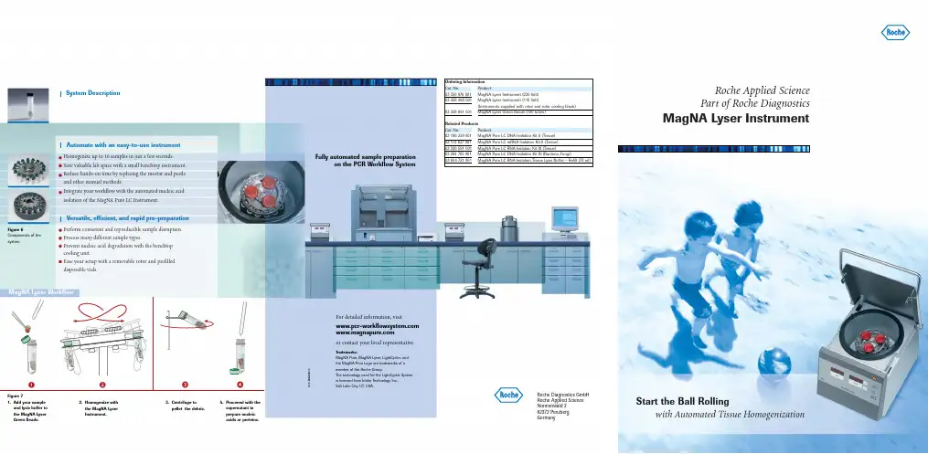
Ordering Information Cat. No. Product ***********MagNA Lyser Instrument (230 Volt)***********MagNA Lyser Instrument (110 Volt)(Instruments supplied with rotor and rotor cooling block)***********MagNA Lyser Green Beads (100 tubes)Related Products Cat. No. Product***********MagNA Pure LC DNA Isolation Kit II (Tissue)***********MagNA Pure LC mRNA Isolation Kit II (Tissue)03 330 591 001MagNA Pure LC RNA Isolation Kit III (Tissue)***********MagNA Pure LC DNA Isolation Kit III (Bacteria, Fungi)***********MagNA Pure LC RNA Isolation Tissue Lysis Buffer – Refill (70 ml)System DescriptionHomogenize up to 16 samples in just a few seconds.Save valuable lab space with a small benchtop instrument.Reduce hands-on time by replacing the mortar and pestle and other manual methods.Integrate your workflow with the automated nucleic acid isolation of the MagNA Pure LC Instrument.Perform consistent and reproducible sample disruption.Process many different sample types.Prevent nucleic acid degradation with the benchtop cooling unit.Ease your setup with a removable rotor and prefilled disposable vials.Automate with an easy-to-use instrumentVersatile, efficient, and rapid pre-preparationFigure 71. Add your sample and lysis buffer to the MagNA Lyser Green Beads.2. Homogenize with the MagNA Lyser Instrument.3. Centrifuge to pellet the debris.4. Proceeed with the supernatant to prepare nucleic acids or proteins.For detailed information,visit or contact your local representative.Trademarks:MagNA Pure, MagNA Lyser, LightCycler, and the MagNA Pure Logo are trademarks of a member of the Roche Group.The technology used for the LightCycler System is licensed from Idaho Technology Inc., Salt Lake City, UT, USA.Fully automated sample preparationon the PCR Workflow SystemRoche Diagnostics GmbH Roche Applied Science Nonnenwald 282372 Penzberg Germany0000Roche Applied Science Part of Roche DiagnosticsMagNA Lyser InstrumentStart the Ball Rollingwith Automated Tissue HomogenizationᕤᕣᕢᕡFigure 6Components of the system.The MagNA Lyser InstrumentAutomated tissue homogenizationProcessing conditionsRefer to the following tables for guidelines on setting up your homogenizationSample material(10 mg)*Time settings(seconds)Cooling(between the runs)Speed Average yield(µg)***Average purity(OD 280/260 nm)***Spleen 2 x 25 906,00030–40 1.9Liver 25-6,00016–18 1.8Lung 2 x 25906,00025 1.8Kidney25-6,000201.8Maize leaves **20-5,00010n.d.Maize polenta **20-5,0008n.d.Tortilla chips **20-5,0001n.d.*Aliqout containing 10 mg sample material (here mouse and food samples) was taken for the DNA purificationusing the MagNA Pure LC DNA Isolation Kit II (Tissue), (see pack insert)**Centrifugation after the homogenization for 5 minutes at 2,200 x g*** Yield and purity strongly depend on the condition of the sample material n.d.not determinedData kindly provided by Dr. Peterhänsel, RWTH Aachen, GermanyFigure 1Gel electrophoresis from genomic DNA isolated from tissue homogenized with the MagNA LyserInstrument, using the MagNA Pure LC DNA Kit II (Tissue).Marker: DNA Marker III*Aliquot containing 10 mg sample material (here mouse and human research samples) was taken to purify RNAeither with the MagNA Pure LC RNA Isolation Kit III (Tissue) or the MagNA Pure LC mRNA Isolation Kit II (Tissue) homogenized with the MagNA Lyser Instrument.** Yield and purity strongly depend on the condition of the sample material. The yield for mRNA was not determined.Sample material(10 mg)*Time settings(cycles/seconds)Cooling(between/afterthe runs in seconds)SpeedAverage yield (mg)(total RNA)**Average purity(OD 280/260 nm)**RNA/mRNARarely expressed targets in small numbers of target cells,as seen in experiments about minimalresidual diseases,are difficult to detect.Increasing the cell number can improve sensitivity and lead to accurate results.Without the MagNA Lyser pre-processing,the MagNA Pure mRNA HS Kit can efficiently obtain mRNA from a maximum of 1 x 10⁷white blood cells (WBCs),as shown in research studies with human samples.However,using greater cell numbers results in a saturation effect with quantitative assays (Figure 3).Homogenization of the lysate with the MagNA Lyser Instrument prior to the purification eliminatesthe amplification saturation at 1 x 10⁷cells and allows the use of up to 2.5 x 10⁷WBCs (Figure 4 and 5),enhancing the analytical sensitivity of the assay.Eliminate sensitivity barriers with increased sample inputFigure 3mRNA was purified from different amounts of human white blood cells with the MagNA Pure mRNA HS Kit. G6PDH was amplified using the LightCycler t(9;22) Quantification Kit (see text beside).Figure 4mRNA was purified from different amounts of human white blood cells with the MagNA Pure mRNA HS Kit. The lysates from 2.5 x 10⁷cells and 5 x 10⁷cells were homogenized with the MagNA Lyser Instrument (2x50 seconds with 90 seconds cooling in between) prior to the mRNA purification. G6PDH was amplified using the LightCycler t(9;22) Quantification Kit (see text beside).Figure 5Scalability from 1 x 10⁶cells to 2.5 x 10⁷cells is represented in the graph and the table of the relationship between crossing points and cell numbers. The limitation of cell input is indicated by no change in crossing point with increased cell number (see text beside).Cell number 5 x 1072.5 x 1071 x 1075 x 1061 x 106Log (cell number)7.77.47.06.76.0Crossing point 20.320.321.822.424.4crossingpointLog(cell number)252423222120195.86.36.87.37.8Figure 2Gel electrophoresis from total RNA isolated from tissue homogenized with the MagNA Lyser Instrument, using the MagNA Pure LC RNA Kit III (Tissue).Ma r k e rS p l e e nL i v e rL u n gK i d n e yM a r k e rMa i z e l e a v e sMa i z e l e a v e sS p l e e nL i v e r11 kb5 kb5 kb28 S rRNA 18 S rRNASpleen 2 x 50 90 6,500–7,000 30–40 1.9Liver 50 - 6,500–7,000 13–17 2.0Thymoid tissue60906,500n.d.n.d.Heart 60 90 6,500 n.d. n.d.Abdominal fat 60 90 6,500 n.d. n.d.Aorta 60 90 6,500 n.d. n.d. Other samples1+n x 50 90 6,500–7,000- -1 x 105 x 101 x 10- 5 x 101 x 105 x 105 x 10- 5 x 102.5 x 10 5 x 10。
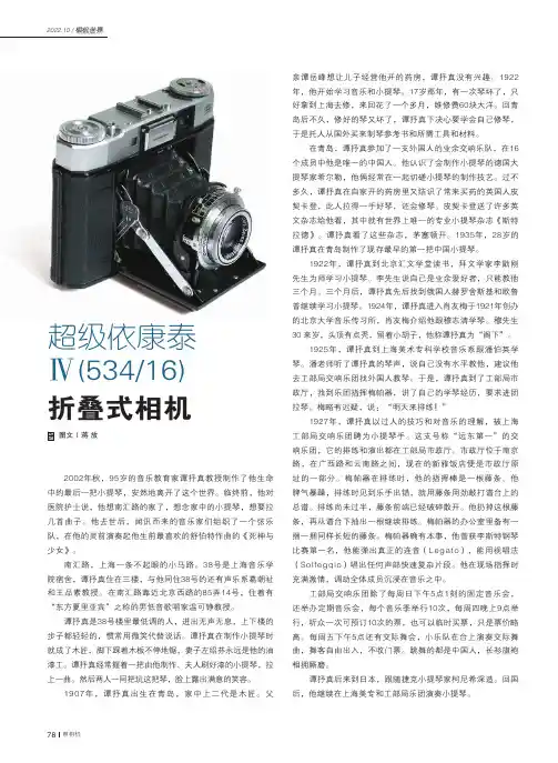
78 照相机2022.10 /折叠式相机2002年秋,95岁的音乐教育家谭抒真教授制作了他生命中的最后一把小提琴,安然地离开了这个世界。
临终前,他对医院护士说,他想南汇路的家了,想念家中的小提琴,想要拉几首曲子。
他去世后,闻讯而来的音乐家们组织了一个弦乐队,在他的灵前演奏起他生前最喜欢的舒伯特作曲的《死神与少女》。
南汇路,上海一条不起眼的小马路。
38号是上海音乐学院宿舍,谭抒真住在三楼,与他同住38号的还有声乐系葛朝祉和王品素教授。
在南汇路靠近北京西路的85弄14号,住着有“东方夏里亚宾”之称的男低音歌唱家温可铮教授。
谭抒真是38号楼里最低调的人,进出无声无息,上下楼的步子都轻轻的,惯常用微笑代替说话。
谭抒真在制作小提琴时就成了木匠,脚下踩着木板不停地锯,妻子左绍芬永远是他的油漆工。
谭抒真经常握着一把由他制作、夫人刷好漆的小提琴,拉上一曲。
然后两人一同把玩这把琴,脸上露出满意的笑容。
1907年,谭抒真出生在青岛,家中上二代是木匠。
父图文|蒋 放超级依康泰 Ⅳ(534/16)亲谭岳峰想让儿子经营他开的药房,谭抒真没有兴趣。
1922年,他开始学习音乐和小提琴。
17岁那年,有一次琴坏了,只好拿到上海去修,来回花了一个多月,维修费60块大洋。
回青岛后不久,修好的琴又坏了,谭抒真下决心要学会自己修琴,于是托人从国外买来制琴参考书和所需工具和材料。
在青岛,谭抒真参加了一支外国人的业余交响乐队,在16个成员中他是唯一的中国人。
他认识了会制作小提琴的德国大提琴家希尔勒,他俩经常在一起切磋小提琴的制作技艺。
过不多久,谭抒真在自家开的药房里又结识了常来买药的英国人皮契卡登,此人拉得一手好琴,还会修琴。
皮契卡登送了许多英文杂志给他看,其中就有世界上唯一的专业小提琴杂志《斯特拉德》。
谭抒真看了这些杂志,茅塞顿开。
1935年,28岁的谭抒真在青岛制作了现存最早的第一把中国小提琴。
1922年,谭抒真到北京汇文学堂读书,拜文学家李勖刚先生为师学习小提琴。
目录1.肝肾胰功能 (3)2.心脑血管/糖脂病 (9)3.风湿过敏/免疫功能 (13)4.肝炎/病毒标志物 (15)5.凝血/血液病项目 (18)6.肿瘤标志物 (21)7.甲功/激素项目 (23)8.血气分析项目 (26)9.血细胞分析、骨髓细胞学检测项目 (27)11.尿液、体液检测项目 (29)12.细菌学检验项目 (31)13.检验项目分类、代码及收费一览表 (33)序实验医学的飞速发展以及循证医学在临床医学中的广泛应用,检验学科已经从”医学检验”发展成了”检验医学”,检验科不再是传统意义上的辅助科室,已成为临床医学的重要组成部分。
它与临床联系更加密切,正凭借全新的检验理念、现代化的检测技术及科学严谨的工作作风定格为临床医学的专业科室,在疾病的诊断、治疗、预防发挥着重大作用,并成为衡量一所医院医疗水平的重要标志之一。
临床医师是患者诊疗方案的制订人,从检验项目的选择到检验结果的合理应用都贯穿于整个医疗过程。
如何合理选择检验项目,使其发挥临床最大的功效是每个医师必考虑的问题之一。
在具体诊疗过程中,一方面要求医师有针对性地申请检验项目,尽量选择对某种疾病有特异性诊断价值的项目,另一方面还应对该检验的方法学原理、临床意义及干扰检验的生理、病理、药理等深入理解,掌握检验结果在不同时间、不同环境、不同疗程的变化。
考虑到临床一线医师工作的繁重及实验医学的快速发展,本院检验科编写了这本《检验项目的临床应用手册》,手册涵盖了本院检验科开展的大部分检验项目,尤其最近开展的新的检验项目,对项目的具体临床应用情况以及临床意义作了较为详尽的介绍,希望该手册有助于本院临床医师选出最佳、合理、经济实用的检验项目和合理应用检验结果诊疗疾病,同时也希望该手册有助于本院“精品医学中心”的建设。
1.肝肾胰功能项目参考范围(方法)检验项目的临床应用丙氨酸氨基转移酶(ALT)<40U/L血清ALT 95%的参考范围是5-35u/L,有三个医学决定水平,分别为20、60、300u/L。
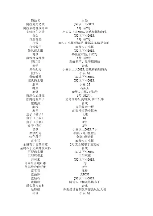
物品名出处阿拉克尼之线J NZ以下小BOSS.阿拉米德合成纤维1号,或2号.安特洛尔之徽小安以上大BOSS,盗贼和福加的头白金J NZ以下小BOSS.白金合金1号,或2号.白银50左右小怪或精灵.我都是杀精灵来的.白银骰子50级左右小怪暴风雨之眼J NZ以下小BOSS.薄纱40级左右怪,1号2号薄纱合成纤维1号,或2号.彩虹石彩虹葫芦、黑平原蚂蚁赤铜合成赤铜配方小安以上大BOSS,盗贼和福加的头蛋白石小安,GJ毒蜘蛛丝J NZ以下小BOSS.肥沃的土壤J NZ以下小BOSS.翡翠小安,GJ蜂巢石头人府绸40级左右怪,1号2号府绸合成纤维1号,或2号.伽姆提的爪子熊岛的那只美加多,和三只牛橄榄油合成海沙多的象米一样海星幻影沙漠的小鱿鱼盒子(裤子)飞熊盒子(上衣)GJ盒子(手套)3号盒子(鞋)2号黑铁小安以上BOSS,7号黑铁配方牛熊,7号,德里怪红色种子金猪.或亚根黄宝石50级左右小怪金属布丁史莱姆皮2号或金属布丁史莱姆金属布丁史莱姆皮衣料合成巨型麻雀蛋J NZ以下小BOSS.巨型麻雀爪巨型麻雀开司米J NZ以下小BOSS.开司米合成纤维1号凯尔维合成纤维2号蓝宝石亚根蓝晶体大BOSS蓝钻石 J NZ以下小BOSS.硫磺粉隧道1、2和训练场布丁绿尖鼠皮衣料合成绿磨茹你要是没看到说明你没玩过天使玛瑙小安,GJ猫眼新手村,转身任务送的不少吧美人鱼的眼泪 三只牛密斯里训练场羊以上的怪、实验室密斯里合金1号棉花玛米奇、羊、盗贼枪手新兵、石头人木头新手村兔子尼龙玛米奇、鳄鱼铅熊青玉组合粉1号或2号肉包子安城杂货任务三角绵羊的角三角绵羊沙漠的沙子码字要时间的沙漠玫瑰花瓣大BOSS,70级以上怪沙漠玫瑰纱线牛熊直接出.或合吧.纱线新手村兔子、布丁森林玛米奇珊瑚碎片J NZ以下小BOSS.伤心的布丁史莱姆国王的眼泪伤心的布丁史莱姆国王,GJ,JNZ 水晶40级怪出.水晶粉40级怪出、训练场熊丝绸合成纤维1号四叶苜蓿35-56级怪出.松果隧道1的葫芦太阳石葫芦差不多的怪都有出.炭再问打死你.天鹅绒合成纤维2号天空陨石牛熊,盗贼和福加的头天使的羽毛 布丁森林玛米奇铁矿石打死你还问铜黑布丁乌金赤铜10个合熊钢合金2号,GJ.熊角 三只熊熊皮熊啊大哥,还有2号熊皮衣料合成血石红兔子,血石组合粉1号或2号送到你不想要.鸭嘴怪皮衣料合的啊.老大亚麻石头人亚麻合成纤维2号羊皮杀了羊就有了.羊皮衣料合成耀眼的阳光大BOSS出的比米还要多.叶笛JNZ,精灵优秀双重恢复剂大BOSS或征途2任务粘土小孩子都知道珍珠贝城厨师行会苎麻布合成纤维1号.灼热的沙子大鱿鱼紫水晶小石人一只一个吧.组合金粉大BOSS.或自已合.怎么合看BBS 钻石组合粉大BOSS.或自已合.怎么合看BBS 转生材料出处纱线尼龙亚麻羊毛丝绸棉花毒蘑菇绿蘑菇名目蘑菇转职材料战士:铁矿石太阳石洞笋B OSS出灼伤的石头60级怪密斯里B BS去熊钢钻石BOSS出,还有熊月球金属组合粉1号技师:木头铜黄金白银密斯里熊钢暴风雨之眼白金合金厨师:B BS去看上面新鲜西红柿紫菜肉干料理王盒饭转生材料任务.药师:新鲜西红柿新鲜无花果欢乐洋葱50~60级怪无污染西红柿B城任务换.怎么换我也不知道,BBS去.裁缝:新鲜西红柿新鲜无花果棉花尼龙亚麻看BBS去.百科里有.凯尔维亚麻纤维阿拉米德纤维羊毛合成纤维1号城市NPC任务绿豆土豆芝麻茄子扫帚头树皮看BBS去.百科里有.爆率10只一个还可以.1只一个大BOSS一只好几个,其它的就少了自已去试,精灵掉的还可以、大陆尽头的巨型麻雀也掉自已去试,主要在月球和深红.10只一个BOSS出的还可以.1只一个10只一个实验室出的多.10个黑铁+1黑铁配方合来.够你用就是.多的不敢捡.10只一个10只一个多的不敢捡.一只一个BOSS出的还可以.1只一个还可以.1只一个10只有一个吧三个橄榄合来的.有上天使的人不知道是笨蛋打晕了也出不了几个最多.少少少GJ出的多BOSS出的多,小怪现在出的少了.多的是自已试还是2号去吧.看公式多少.大家有多的用不完给我点出的多也用的多.现在出的好少哦.不够用.还行有任务在手好多.谁要说下,下次我就不N店了.可以卖好多钱的哦有上天使的人不知道是笨蛋看公式多的不敢捡.还可以多,没有我送你不多.看到记的捡.我K不够用就是,多捡点.2只黑熊有一个吧.推荐布丁森林还可以BOSS一只有一个吧.少啊.你有多的丢给我吧.用的不多不多.用的少.够用就行用的不多吧.只用一两个我那有爆率还可以,就是转职用量大的惊人.BOSS出的多.够你用就是.不多.够用.够你用就是.大哥,送我吧.传说中爆率最低的东东不多.千分之一的爆率,杀1000只羊有可能得绿箭鼠皮布丁森林玛米奇布丁森林玛米奇隧道1的枪手、新手村的羊、布丁森林玛米奇羊、布丁森林玛米奇羊、布丁森林玛米奇隧道4螳螂、四转后去杀鳄鱼到处都是隧道4螳螂你要只要这就把熊猫送我.我不反对的.够用.布丁森林玛米奇新手村的羊新手村的羊满地都是月球遗迹、深红高原月球遗迹、深红高原。

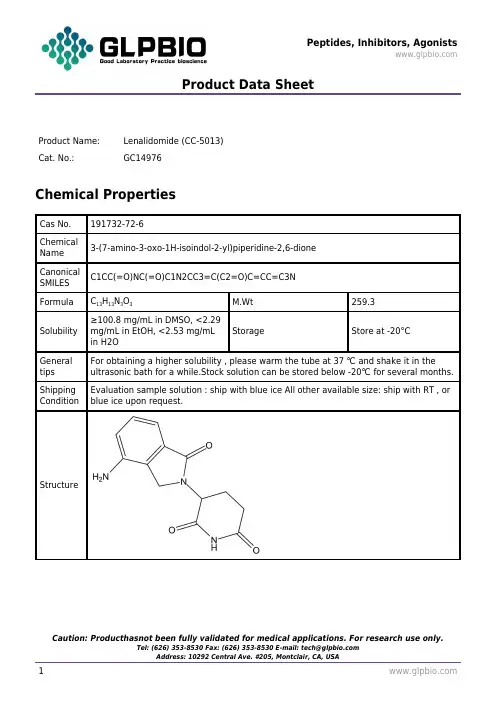
Product Data SheetProduct Name:Lenalidomide (CC-5013)Cat. No.:GC14976Chemical PropertiesCas No.191732-72-6ChemicalName3-(7-amino-3-oxo-1H-isoindol-2-yl)piperidine-2,6-dioneCanonicalSMILESC1CC(=O)NC(=O)C1N2CC3=C(C2=O)C=CC=C3NFormula C13H13N3O3M.Wt259.3Solubility ≥100.8 mg/mL in DMSO, <2.29mg/mL in EtOH, <2.53 mg/mLin H2OStorage Store at -20°CGeneral tips For obtaining a higher solubility , please warm the tube at 37 ℃ and shake it in the ultrasonic bath for a while.Stock solution can be stored below -20℃ for several months.Shipping Condition Evaluation sample solution : ship with blue ice All other available size: ship with RT , or blue ice upon request.StructureProduct Data Sheet实验参考方法Cell experiment: [1]Cell lines Peripheral blood mononuclear cells (PBMCs)Preparation method The solubility of this compound in DMSO is >10 mM. General tips for obtaining a higher concentration: Please warm the tube at 37 °C for 10 minutes and/or shake it in the ultrasonic bath for a while.Stock solution can be stored below -20°C for several months.Reaction Conditions10 μM, 7 daysApplications The cells were incubated with the dye at 37°C for 10 min and treated for 7 days in RPMI culture medium with lenalidomide. Cells were surface stained with anti-CD4-PerCP and anti-CD25-APC, followed by intracellular staining with anti-FOXP3-PE. Lenalidomide inhibited the expression of CD4+CD25high CTLA-4+FOXP3+ cells. Incubation with lenalidomide significantly decreases expression of the T regulatory cell population after 7 days of culture. The drug decreased the percentage of CD4+CD25high cells expressing both CTLA-4 and FOXP3 from 25 to 12%.Animal experiment: [2]Animal models Male Sprague–Dawley ratsDosage form Oral administration, 50 mg/kg or 250 mg/kgApplications In the rat mesenteric window assay (RMWA), representative differences between vehicle and 50 or 250 mg/kg lenalidomide-treated rats were visualized by staining with an antibody against rat endothelium in bFGF-induced angiogenic windows. The induction of angiogenesis by bFGF was significantly inhibited by oral treatment of lenalidomide in a dose-dependent manner. Lenalidomidesignificantly decreased the percentage of vascularized area from 5.16% in the control group to 2.58 and 1.69 in the 50 and 250 mg/kg group, respectively.Other notes Please test the solubility of all compounds indoor, and the actual solubility may slightly differ with the theoretical value. This is caused by an experimental system error and it is normal.References:[1] Galustian C, Meyer B, Labarthe M C, et al. The anti-cancer agents lenalidomide and pomalidomide inhibit the proliferation and function of T regulatory cells. Cancer Immunology, Immunotherapy, 2009, 58(7): 1033-1045.[2] Dredge K, Horsfall R, Robinson S P, et al. Orally administered lenalidomide (CC-5013) is anti-angiogenic in vivo and inhibits endothelial cell migration and Akt phosphorylation in vitro. Microvascular research, 2005, 69(1): 56-63.Product Data SheetBackgroundLenalidomide (also known as CC-5013), an oral derivative of thalidomide, is an antineoplastic agent exhibiting antitumor activity through a variety of mechanisms, including immune system activation, angiogenesis inhibition, and direct antineoplastic effects. It has been extensively studied for the treatment of multiple myeloma and myelodysplastic syndrome as well as lymphoproliferative disorders including chronic lymphocytic leukemia (CLL) and non-Hodgkin lymphoma. According to recent studies, Lnalidomide promotes and restores immune system function in CLL patients by inducing an overexpression of costimulatory molecules in leukemic lymphocytes to restore the humoral immunity and immunoglobulins production as well as improving the ability of T cells and leukemic cells to form synapses with T lymphocytes.Reference[1].Ana Pilar Gonzalez-Rodriguez, Angel R. Payer, Andrea Acebes-Huerta, Leticia Hergo-Zapico, Monica Villa-Alvarez, Esther Gonzalez-Garcia, and Segundo Gonzalez. Lenalidomide and chronic lymphocytic leukemia. BioMed Research International 2013.。

cas9 cas12 cas13原理CRISPR-Cas系统是一种广泛应用于基因编辑领域的工具,其中包括Cas9、Cas12和Cas13等多个种类的蛋白质。
这些蛋白质能够与特定的RNA序列结合,并通过剪切DNA或RNA来实现基因组的改变或调控。
本文将分别介绍Cas9、Cas12和Cas13的原理和应用。
Cas9是CRISPR-Cas系统中最早被发现和研究的蛋白质。
它由两个重要的功能域组成,即RNA导向的DNA切割功能域和RNA识别功能域。
Cas9与一种名为crRNA的RNA分子结合后,能够通过碱基互补配对的方式准确地识别并结合到目标DNA序列上。
随后,Cas9通过其DNA切割功能域对目标DNA进行剪切,进而引起DNA双链断裂。
这个过程激活了细胞内的DNA修复机制,从而实现了基因组的改变。
Cas9的应用广泛,可以用于基因敲除、基因敲入、基因修复等多种基因编辑操作。
通过设计合适的引导RNA序列,可以将Cas9定向到目标基因的特定位置,实现对该基因的靶向编辑。
这项技术在基础科学研究、农业育种以及人类疾病治疗等领域都有重要的应用潜力。
与Cas9相比,Cas12和Cas13是近年来被发现的新型CRISPR-Cas系统蛋白质。
它们在结构和功能上与Cas9有所不同,但同样具有基因编辑的能力。
Cas12和Cas13也能够通过与特定的RNA 序列结合来实现对DNA或RNA的剪切。
Cas12与Cas9的主要区别在于其剪切靶标的特异性。
相对于Cas9只能识别和结合PAM序列附近的DNA,Cas12能够在缺乏PAM 序列的情况下识别和结合目标DNA序列。
这一特性使得Cas12在某些特定基因组中的靶向编辑更加灵活和高效。
Cas13则是一种专门用于剪切RNA的CRISPR-Cas蛋白质。
与Cas9和Cas12不同,Cas13主要针对RNA分子进行编辑。
它能够通过与目标RNA序列结合并剪切该RNA,从而实现对基因表达的调控。
中 文 说 明 书适用产品目录号:J1341、J1342和J13432020 版 CTM467原英文技术手册TM467Glucose Uptake-Glo™AssayG9711, G9712 and G9713普洛麦格(北京)生物技术有限公司Promega (Beijing) Biotech Co., Ltd 地址:北京市东城区北三环东路36号环球贸易中心B座907-909电话:************网址:技术支持电话:800 810 8133(座机拨打),400 810 8133(手机拨打)技术支持邮箱:*************************TM4672020制作1Glucose Uptake-Glo™ Assay所有技术文献的英文原版均可在/ protocols 获得。
请访问该网址以确定您使用的说明书是否为最新版本。
如果您在使用该试剂盒时有任何问题,请与Promega 北京技术服务部联系。
电子邮箱:*************************1. 产品描述 (2)2. 产品组分和储存条件 (4)3. 一般注意事项 (5)3.A Assay Buffer中的葡萄糖 (5)3.B 摄取时间 (5)3.C 2-脱氧葡萄糖的浓度 (5)3.D 对比生物反应和检测灵敏度 (5)4. 测量葡萄糖摄取 (6)4.A 试剂配制 (6)4.B 操作步骤 (7)4.C 检测对照 (8)4.D Glucose Uptake-Glo™ Assay的不同规格培养板 (8)5. 细胞分化和多重检测的附加操作步骤 (9)5.A 培养基的成分 (9)5.B 3T3L1-MBX纤维母细胞:分化为脂肪细胞 (10)5.C L6成肌细胞:分化为成肌细胞 (12)5.D 使用Glucose Uptake-Glo™ 和CellTiter-Glo® Assay 多重检测癌细胞 (13)5.E 使用RealTime-Glo™ Assay多重检测脂肪细胞 (14)6. 附录 (16)6.A 计算葡萄糖摄取率 (16)6.B 其他数据 (17)6.C 相关产品 (19)7. 内容变更总结 (21)普洛麦格(北京)生物技术有限公司Promega (Beijing) Biotech Co., Ltd地址:北京市东城区北三环东路36号环球贸易中心B 座907-909电话:************网址:技术支持电话:800 810 8133(座机拨打),400 810 8133(手机拨打)技术支持邮箱:*************************TM4672020制作21.Description The Glucose Uptake-Glo™ Assay is a non-radioactive, plate-based, homogeneous bioluminescent method for measuring glucose uptake in mammalian cells based on the detection of 2-deoxyglucose-6-phosphate (2DG6P). When 2-deoxyglucose (2DG) is added to cells, it is transported across the membrane and rapidly phosphorylated in the same manner as glucose. However, enzymes that further modify glucose-6-phosphate (G6P) cannot modif 2DG6P, and thus this membrane-impermeable analyte accumulates in the cell. After a brief period of incubation an acid detergent solution (Stop Buffer) is added to lyse cells, terminate uptake and destroy any NADPH within the cells. A high-pH buffer solution (Neutralization Buffer) is then added to neutralize the acid. A DetectionReagent containing glucose-6-phosphate dehydrogenase (G6PDH), NADP +, Reductase, Ultra-Glo™ RecombinaLuciferase and proluciferin substrate is added to the sample wells. G6PDH oxidizes 2DG6P to6-phosphodeoxygluconate and simultaneously reduces NADP + to NADPH. The Reductase uses NADPH to convethe proluciferin to luciferin, which is then used by Ultra-Glo™ Recombinant Luciferase to produce a luminescensignal that is proportional to the concentration of 2DG6P.Figure 1. Schematic diagram of the Glucose Uptake-Glo™ Assay technology. 2-deoxyglucose (2DG) is transported into cells and phosphorylated to produce 2-deoxyglucose-6-phosphate (2DG6P). Adding Stop Buffe stops 2DG transport, lyses cells, and inactivates proteins. The acid is neutralized by the Neutralization Buffer before the addition of the 2DG6P Detection Reagent. The glucose-6-phosphate dehydrogenase (G6PDH) within the 2DG6P Detection Reagent oxidizes 2DG6P to 6-phosphodeoxygluconate (6PDG) and reduces NADP + to NADPH. The reductase uses NADPH to convert the proluciferin to luciferin, which is then used by Ultra-Glo™ Recombinant Luciferase to produce light.2DG6P6PDGStep 1.Step 2.Step 3.Add 2DG6P Detection Reagent.G6PDHNADP +NADPH + reductase proluciferin luciferin ATPUltra-Glo™rLuciferase1.DescriptionThe Glucose Uptake-Glo™ Assay is a non-radioactive, plate-based, homogeneous bioluminescent method for measuring glucose uptake in mammalian cells based on the detection of 2-deoxyglucose-6-phosphate (2DG6P). When 2-deoxyglucose (2DG) is added to cells, it is transported across the membrane and rapidly phosphorylated in the same manner as glucose. However, enzymes that further modify glucose-6-phosphate (G6P) cannot modify 2DG6P, and thus this membrane-impermeable analyte accumulates in the cell. After a brief period of incubation, an acid detergent solution (Stop Buffer) is added to lyse cells, terminate uptake and destroy any NADPH within the cells. A high-pH buffer solution (Neutralization Buffer) is then added to neutralize the acid. A Detection Reagent containing glucose-6-phosphate dehydrogenase (G6PDH), NADP +, Reductase, Ultra-Glo™ Recombinan Luciferase and proluciferin substrate is added to the sample wells. G6PDH oxidizes 2DG6P to 6-phosphodeoxygluconate and simultaneously reduces NADP + to NADPH. The Reductase uses NADPH to conver the proluciferin to luciferin, which is then used by Ultra-Glo™ Recombinant Luciferase to produce a luminescent signal that is proportional to the concentration of 2DG6P.Figure 1. Schematic diagram of the Glucose Uptake-Glo™ Assay technology. 2-deoxyglucose (2DG) istransported into cells and phosphorylated to produce 2-deoxyglucose-6-phosphate (2DG6P). Adding Stop Buffer stops 2DG transport, lyses cells, and inactivates proteins. The acid is neutralized by the Neutralization Buffer before the addition of the 2DG6P Detection Reagent. The glucose-6-phosphate dehydrogenase (G6PDH) within the 2DG6P Detection Reagent oxidizes 2DG6P to 6-phosphodeoxygluconate (6PDG) and reduces NADP + to NADPH. The reductase uses NADPH to convert the proluciferin to luciferin, which is then used by Ultra-Glo™Recombinant Luciferase to produce light.2DG2DG 2DG6PStep 1.Step 2.Add 2DG to cells.Add Stop and Neutralization Buffers to end reactions, lyse cells and eliminate cellular NADPH.2DG6P1. 产品描述Glucose Uptake-Glo™ Assay 是一种非放射性且基于培养板的均质生物发光检测方法。
GeneCopoeia TMExpressway to DiscoveryGeneCopoeia Inc.9620 Medical Center Drive, Suite 101Rockville, MD20850, USATel: +1(301)762-0888;To l l f r e e:+1(301)762-3888Fax:+1(301)762-3888Web: E.coli Competent Cells stbl3™产品套装编号:CC003产品内容产品编号包装规格Competent Cells stbl3 CC001-01 100 μl×10Control DNA(pUC19,10 pg/μl) CC003-02 50 μl×1储存条件:-80℃保存 保存时间: 12 个月■ 产品概述:常规质粒制备的宿主菌,可进行蓝白斑筛选;复制慢病毒载体系统推荐使用的菌株,对慢病毒载体等较大的质粒转 化的效果好,有效减低错误重组的可能性。
■ Genotype:F- mcrB mrr hsdS20 (rB-, mB- ) recA13 supE44 ara-14 galK2 lacY1 proA2 rpsL20 (Strr ) xyl -5 - leu mtl-1.■ 细胞种类:大肠杆菌Stbl3™菌株能有效地抑制长片段末端重复区的重组,非常适合于含有同向重复序列的慢病毒和其他反转录 病毒载体的复制。
此外,细胞非常稳定,能提高慢病毒载体或其他不稳定载体的克隆效率。
■ 转化效率:我们提供的感受态的转化效率>1 X 10⁸ transformants/1 μg PUC19 Plasmid也就是,当您以100 μl Competent Cells Stbl3/ 10 pg pUC19 Plasmid 进行转化,产生的菌落数>1000 trans formants■ 使用步骤:1. 把感受态细胞置于冰中解冻;2. 把50-100 µl的感受态细胞移至灭菌处理的试管内;3. 加入用于转化的DNA或反应产物(10 ng以下);4. 冰中放置30分钟;5. 42℃ 放置30~60秒;6. 冰中放置2~3分钟;7. 加入37℃预温好的SOC培养基,使终体积为1 ml;8. 37℃振荡培养1小时(160~225 rpm);9. 取适量涂布琼脂平板培养基;10. 37℃过夜培养。
Jun1 - Jun15,2021WORLD VISION 2021.NO.11专题FEATURES档案1-3.位于开罗的埃及国家博物馆最深处,静静地摆放着埃及的国宝:图坦卡蒙黄金面具与金棺。
考古人员小心地对陪葬品进行修护。
2131.在高管和病人眼里,托帕活泼开朗,天天笑容满面,人们亲切地叫她“快乐的简”。
2.托帕幼年时被送进了社会福利院。
3.托帕用于杀人的药品是吗啡和阿托品2种药品的大量混合制剂。
2134.托帕受到了谋杀指控,她自己坦白说20年间共杀死了31人,但最终她并没有受到法律的惩罚。
5.托帕的父亲酗酒并患有精神疾病。
他因精神崩溃而被关在精神病院。
档案RecoRd file4Jun1 - Jun15,2021WORLD VISION 2021.NO.11此时的托帕过着一种双面人生。
在高管和病人眼里,托帕活泼开朗,天天笑容满面,人们亲切地叫她“快乐的简”。
然而,对待身边的同学,她则两面三刀,暗藏狡诈,常常散播那些与她关系差的同学的谣言,致使几位同学被开除,而且她还偷窃别人的财物和医院物品。
1890年,托帕顺利通过专业测试,但因擅自离开病房而被学校开除,也没获得学位证书。
几个月后,她再次因偷盗病人财物、擅自篡改病历和伪造学位证书而被另外一家医院开除。
之后,托帕放弃了在医院谋职,靠做私人护理谋生,毅然决然地在双面人生的道路上越走越远。
而这是一条不归路。
“天使”的复仇在接下来的10年里,得益于众多医生的高度赞赏和大力推荐,托帕逐渐走上了自己的人生巅峰:成尽管托帕受到了谋杀指控,她自己也坦白说20年间共杀死了31人,但她并没有受到法律的惩罚。
她人生的最后一站是精神病院,在那里度过了剩余的36年。
文 |江南柳 图 | 枫月在“天使”面具之后双面女护士1885年,对托帕来说,是一个关键的人生转折点。
这一年,她离开了生活近20年的领养之家,搬出了那栋老房子。
离开的原因要从她领养家庭的姐姐伊丽莎白的新婚说起。
武汉普诺赛生命科技有限公司 Procell Life Science&Technology Co.,Ltd.使用前请仔细阅读说明书。
如果有任何问题,请通过以下方式联系我们:全国免费电话:400-650-3656销售电话:************销售邮箱:*****************.cn官方网站:CL-0378MC3T3-E1 Subclone 14(小鼠颅顶前骨细胞亚克隆14)武汉普诺赛生命科技有限公司——您身边的细胞培养专家Copyright ©2020-2021 Procell Life Science&Technology Co.,Ltd. All Rights Reserved 细胞株培养扩增技术服务申明本公司受贵单位委托,进行细胞株的技术服务工作,并收取相应细胞株技术服务费用,细胞株技术服务具体项目清单见订购合同。
本公司提供完善的技术支持及售后服务,收到产品后处理方式及相应售后条款参见《细胞售后条例》。
收到常温细胞后如何处理?(细胞培养详细操作步骤请参照《普诺赛细胞培养操作指南》)1. 收到常温细胞后,及时拍照记录有无漏液/瓶身破损现象。
2. 用75%酒精擦拭细胞培养瓶表面,显微镜下观察细胞状态。
先不要打开培养瓶盖,将细胞置于细胞培养箱内静置培养2-4小时,以便稳定细胞状态。
3. 仔细阅读细胞说明书,了解细胞相关信息,如贴壁特性(贴壁/悬浮)、细胞形态、所用基础培养基、血清比例、所需细胞因子、传代比例、换液频率等。
4. 静置完成后,取出细胞培养瓶,镜检、拍照,记录细胞状态(所拍照片将作为后续服务依据);建议细胞传代培养后,定期拍照、记录细胞生长状态。
5. 若细胞生长密度超过80%1:2~1:3(按实际收货细胞密度决,移除细胞培养瓶内培养基,预留6mL 左右原瓶培养基继续培养,直至细胞密度达左右再进行传代操作,注意拧松瓶盖或更换透气瓶盖。
6. 传代(参考邮件操作指南),或及时联系技术支持进行指导传代。