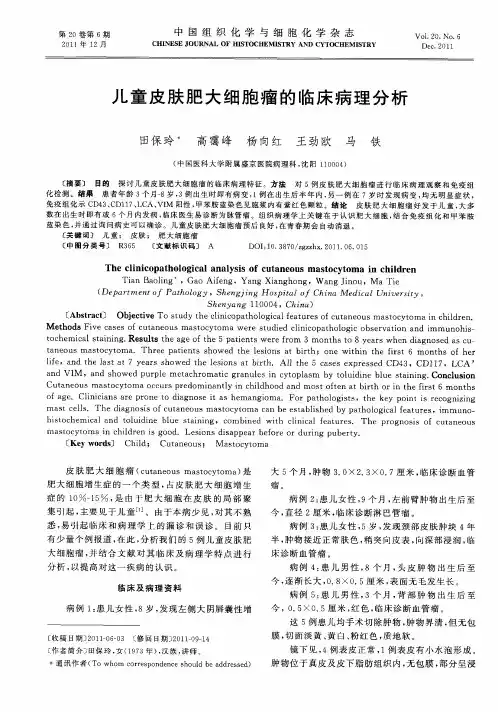
皮肤肥大细胞瘤1例
- 格式:pdf
- 大小:349.46 KB
- 文档页数:1

皮肤肥大细胞增生症的临床特点及病理分析李卉;林达;郭泽;杜文辉;张书梅【摘要】目的探讨皮肤肥大细胞增生症(CM)的临床及病理特征和误诊情况.方法回顾性分析安徽医科大学第一附属医院2009 ~ 2016年收治的29例CM患者的临床特点、组织病理改变和误诊情况.结果 29例患者年龄40 d ~ 49岁,≤2岁16例;皮疹形态多样,斑丘疹型CM(MPCM) 22例(75.86%),肥大细胞瘤3例(10.35%),弥漫型CM 4例(13.79%);2例伴有大疱,1例成人持久性发疹性斑状毛细血管扩;临床误诊13例(44.83%).组织病理发现肥大细胞真皮弥漫性浸润13例,其中2例见表皮下水疱;浅层血管周围浸润16例.29例患者Giemsa染色均见异染颗粒.结论CM多见于儿童,皮损形态多样,其中MPCM最为常见,临床容易出现误诊,特别是成年人.临床需加强对不典型CM的关注,借助组织病理学检查,可以减少误诊的发生.%Objective To study the clinical,pathological features and misdiagnosis of the cutaneous mastocytosis (CM).Methods The clinical characteristics,histopathologic changes (HE and Giemsa staining) and misdiagnosed cases of 29 cases of CM were retrospectivelyanalyzed.Results The age of the patients ranged from 40 days to 49 years.Sixteen patients were aged two years or younger.The clinical signs of CM were variant.There were 22 cases of maculopapular CM (MPCM) (75.86%),3 cases of mastocytoma (10.35 %),4 cases of diffuse CM(DCM) (13.79%),respectively.Two patients suffered from bulla and one adult patient had telangiectasia macularis eruptiva perstans.There were 13 (44.83%) cases of clinical misdiagnosis.Histologically,diffuse infiltration of the mast cells in the dermis was found in 13 cases,including 2 cases ofsubepidermal blister;16 cases demonstrated subtle infiltration within the papillary dermis.Purple metachmmafic granules in mast cells were shown by Giemsa staining in all cases.Conclusion CM is more often seen in children,especially in infants with varied lesions.MPCM is the most common form.CM is prone to misdiagnosis,especially in adults.To reduce the misdiagnosis,doctors should strengthen the ability to identify atypical CM with help of the histopathologic examination and dye.【期刊名称】《安徽医学》【年(卷),期】2017(038)009【总页数】3页(P1143-1145)【关键词】皮肤肥大细胞增生症;临床特点;病理;误诊【作者】李卉;林达;郭泽;杜文辉;张书梅【作者单位】230022合肥安徽医科大学第一附属医院皮肤性病科;230022合肥安徽医科大学第一附属医院皮肤性病科;230022合肥安徽医科大学第一附属医院皮肤性病科;230022合肥安徽医科大学第一附属医院皮肤性病科;230022合肥安徽医科大学第一附属医院皮肤性病科【正文语种】中文肥大细胞增生症是以肥大细胞在多种组织或器官内增生为特征的一组疾病,从出生到成人均可发病,临床表现复杂。