
无菌检查法-PPT课件
- 格式:ppt
- 大小:525.00 KB
- 文档页数:23
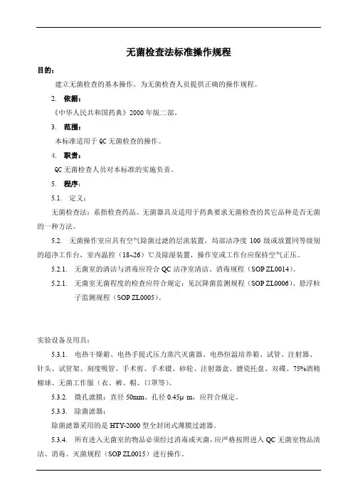
无菌检查法标准操作规程
目的:
建立无菌检查的基本操作,为无菌检查人员提供正确的操作规程。
2. 依据:
《中华人民共和国药典》2000年版二部。
3. 范围:
本标准适用于QC无菌检查的操作。
4. 职责:
QC无菌检查人员对本标准的实施负责。
5. 程序:
5.1. 定义:
无菌检查法:系指检查药品、无菌器具及适用于药典要求无菌检查的其它品种是否无菌的一种方法。
5.2. 无菌操作室应具有空气除菌过滤的层流装置,局部洁净度100级或放置同等级别的超净工作台,室内温控(18~26)℃及除湿装置,操作室或工作台应保持空气正压。
5.2.1. 无菌室的清洁与消毒应符合QC洁净室清洁、消毒规程(SOP ZL0014)。
5.2.1. 无菌室无菌程度的检查应符合规定:见沉降菌监测规程(SOP ZL0006)、悬浮粒子监测规程(SOP ZL0005)。
实验设备及用具:
5.3.1. 电热干燥箱、电热手提式压力蒸汽灭菌器、电热恒温培养箱、试管、注射器、
针头、试管架、刻度吸管、手术剪、手术镊、砂轮、注射器盒、搪瓷托盘、双碟、75%酒精棉球、无菌工作服(衣、裤、帽、口罩等)。
5.3.2. 微孔滤膜:直径50mm、孔径0.45μm,应符合规定。
5.3.3. 除菌滤器:
除菌滤器采用的是HTY-2000型全封闭式薄膜过滤器。
5.3.4. 所有进入无菌室的物品必须经过消毒或灭菌,应严格按照进入QC无菌室物品清洁、消毒、灭菌规程(SOP ZL0015)进行操作。
5.4. 稀释剂:灭菌注射用水。
5.5. 培养基:
5.5.1. 需气菌、厌气菌培养基(流体硫乙醇酸盐培养基);真菌培养基。
5.5.2. 培养基的使用,配制、管理及灵敏度检查应按照培养基管理规程(SMP ZL0036)、培养基灵敏度检查规程(SOP ZL0019)、培养基配制规程(SOP ZL0016)进行操作。
..
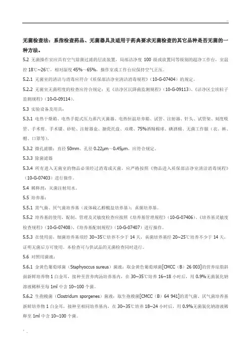
’. 无菌检查法:系指检查药品、无菌器具及适用于药典要求无菌检查的其它品种是否无菌的一种方法。
5.2 无菌操作室应具有空气除菌过滤的层流装置,局部洁净度100级或放置同等级别的超净工作台,室温控18℃~26℃,相对湿度45%~65%,操作室或工作台应保持空气正压。
5.2.1 无菌室的清洁与消毒应符合《质保部洁净室清洁消毒规程》(10-G-07404)的规定。
5.2.2 无菌室无菌程度的检查应符合规定:见《洁净区沉降菌监测规程》(10-G-09113)、《洁净区尘埃粒子监测规程》(10-G-09114)。
5.3 实验设备及用具:
5.3.1 电热干燥箱、电热手提式压力蒸汽灭菌器、电热恒温培养箱、试管、注射器、针头、试管架、刻度吸管、手术剪、手术镊、砂轮、注射器盒、搪瓷托盘、双碟、75%酒精棉球、碘酒棉、无菌工作服(衣、裤、帽、口罩等)。
5.3.2 微孔滤膜:直径50mm、孔径0.22μm~0.45μm,应符合规定。
5.3.3 除菌滤器
5.3.4 所有进入无菌室的物品必须经过消毒或灭菌,应严格按照《物品进入质保部洁净室清洁消毒规程》(10-G-07403)进行操作。
5.4 稀释剂:灭菌注射用水。
5.5 培养基:
5.5.1 需气菌、厌气菌培养基(流体硫乙醇酸盐培养基);真菌培养基。
5.5.2 培养基的使用,配制、管理及灵敏度检查应按照《培养基管理规程》(10-G-07406)、《培养基灵敏度检查规程》(10-G-07408)、《培养基配制规程》(10-G-07407)进行操作。
5.5.3 在使用前,细菌培养基须经30~35℃培养不少于14天,真菌培养基经20~25℃培养不少于14天,证明无菌后方可使用。本检查可与供试品的无菌检查同时进行。
5.6 对照用菌液:
5.6.1 金黄色葡萄球菌(Staphyoccus sureus)菌液:取金黄色葡萄球菌[CMCC(B)26 003]的营养琼脂斜面新鲜培养物1白金耳,接种至营养肉汤培养基内,在30~35℃培养16~18小时后,用0.9%无菌氯化钠溶液稀释至每1ml中含10~100个菌。

detecting Mycoplasmas. Test the effectiveness of the cells tobe used by applying the procedure shown below and inocu-lating not more than 100 cfu or ccu microorganisms of suit-able reference strains of M. hyorhinis and M. orale. The cellsare suitable if both reference strains are detected. The indi-cator cells must be subcultured without an antibiotic beforeuse in the test.Test MethodNOTE—The following is provided for information.SOLUTIONSPhosphate Buffered Saline—2.0 M Monobasic Potassium Phosphate—Dissolve 13.61 gof anhydrous monobasic potassium phosphate in 50 mL ofwater.2.0 M Dibasic Potassium Phosphate—Dissolve 17.42 g ofanhydrous dibasic potassium phosphate in 50 mL of water.Phosphate Buffered Saline Solution (pH 7.4)—Combine 3.6mL of 2.0 M Monobasic Potassium Phosphate, 16.4 mL of 2.0M Dibasic Potassium Phosphate, 8g of sodium chloride, and1 L of water. Mix thoroughly. Adjust the pH if necessary.Bisbenzimide Stock Solution—Dissolve 5 mg of bisben-zimide in water, and dilute with the same solvent to 100mL. Store in the dark.Bisbenzimide Working Solution—Immediately beforeuse, dilute 100 mL of Bisbenzimide Stock Solution with Phos-phate Buffered Saline Solution (pH 7.4) to 100 mL.Phosphate-Citrate Buffer Solution pH 5.5—Mix 56.85mL of a 28.4-g/L solution of anhydrous disodium hydrogenphosphate and 43.15 mL of a 21-g/L solution of citric acid.METHOD1.Seed the indicator cell culture at a suitable density (forexample, 2×10⁴ to 2×10⁵ cells/mL, 4×10³ to 2.5 ×10⁴ cells/cm2) that will yield confluence after3 days of growth. Inoculate 1 mL of the product to beexamined into the cell culture vessel, and incubate at36±1°.2.After at least 3 days of incubation, when the cells havegrown to confluence, make a subculture on cover slipsin suitable containers or on some other surface (for ex-ample, chambered slides) suitable for the test proce-dure. Seed the cells at low density so that they reach50% confluence after 3–5 days of incubation. Com-plete confluence impairs visualization of Mycoplasmasafter staining and must be avoided.3.Remove the medium and rinse the indicator cells withphosphate buffered saline, pH 7.4, then add a suitablefixing solution (a freshly prepared mixture of 1 volumeof acetic acid, glacial, TS and 3 volumes of methanol, issuitable when bisbenzimide is used for staining).4.Remove the fixing solution and wash the cells with ster-ile Purified Water. Dry the slides completely if they areto be stained more than 1 hour later (particular care isneeded for staining of slides after drying owing to arti-facts that may be produced).5.Add a suitable DNA stain and allow standing for a suit-able time (bisbenzimide working solution and a stand-ing time of 10 minutes are suitable).6.Remove the stain and rinse the monolayer with PurifiedWater.7.Mount each coverslip, where applicable (a mixture ofequal volumes of glycerol and Phosphate-Citrate BufferSolution pH 5.5 is suitable for mounting). Examine byfluorescence (for bisbenzimide stain a 330 nm/380 nmexcitation filter and an LP 440 nm barrier filter are suit-able) at 400× magnification or pare the microscopic appearance of the test cul-tures with that of the negative and positive controls,examining for extranuclear fluorescence. Mycoplasmasproduce pinpoints or filaments over the indicator cellcytoplasm. They may also produce pinpoints and fila-ments in the intercellular spaces. Multiple microscopicfields are examined according to the protocol establish-ed during validation.Interpretation of ResultsThe product to be examined complies with the test if flu-orescence typical of Mycoplasmas is not present. The test isinvalid if the positive controls do not show fluorescence typ-ical of Mycoplasmas. The test is invalid if the negative con-trols show fluorescence typical of Mycoplasmas.á71ñ STERILITYTESTSuPortions of this general chapter have been harmonizedwith the corresponding texts of the European Pharmacopeiaand/or the Japanese Pharmacopeia. Those portions that arenot harmonized are marked with symbols (uu) to specify thisfact.uThese Pharmacopeial procedures are not by themselvesdesigned to ensure that a batch of product is sterile or hasbeen sterilized. This is accomplished primarily by validationof the sterilization process or of the aseptic processing pro-cedures.The test is applied to substances, preparations, or articleswhich, according to the Pharmacopeia, are required to be ster-ile. However, a satisfactory result only indicates that no con-taminating microorganism has been found in the sample ex-amined under the conditions of the test.PRECAUTIONS AGAINST MICROBIALCONTAMINATIONThe test for sterility is carried out under aseptic condi-tions. In order to achieve such conditions, the test environ-ment has to be adapted to the way in which the sterility testis performed. The precautions taken to avoid contaminationare such that they do not affect any microorganisms thatare to be revealed in the test. The working conditions in

1 <71>无菌检查(USP29-NF24)
◆这个章节的大部分内容都和欧洲药典或日本药典相应检测部分保持一致。有一些不一致的地方已用特殊标记(◆◆)注明。◆
下述步骤被用于检查药典中规定要求无菌的是否满足其专论中相应的无菌要求。供试品的性状允许时,可以采用供试品的无菌检查中的薄膜过滤法来检测。如果薄膜过滤方法不能使用,则采用供试品的无菌检查中的直接接种法进行检测。所有的器具,除了标明的无菌器皿外,都采用直接接种法进行检测。重新检测的规定条款见结果的观测和分析。
因为无菌检测是一项非常严格的操作过程(必须保证无菌操作才能得到一个准确的检测结果),所以对操作人员进行相关的培训和鉴定是非常重要的。无菌检查即在无菌状态下实行。为了达到这个状态,检测的环境必须适合无菌操作。要防止检验过程中暴露的微生物受污染。要对无菌检验的操作环境进行适当地取样检测和合适的控制来监控。
不仅仅通过这些药典中的步骤就能确保一批产品无菌或已经灭菌。确认在无菌状态下操作或按无菌操作步骤操作则是首先需要的。
当用药典中合适的药典方法检测产品中出现了微生物污染,其结果宣判该无菌检测所需方法无效,甚至是更换了操作步骤得到了不同的结果。◆额外的无菌检测信息,参见<1211>灭菌和确保无菌概略。◆
培养基
按以下处方制备培养基,或使用按该处方生产的符合规定的脱水培养基,其能满足需气菌,厌气菌,霉菌的增殖培养。培养基采用有效的程序灭菌。
下面培养基适合用于无菌检查。硫乙醇酸盐流体培养基主要用于厌气菌的培养。还用于需气菌的培养。大豆粉-酪蛋白消化物培养基适合霉菌和需气菌的培养。
硫乙醇酸盐流体培养基
L-光胺酸 2.5g
氯化钠 5.5g/5.0g
葡萄糖(C6H12O6.H2O) 0.75g
琼脂,颗粒状(含水量不低于15%) 5.0g
酵母浸出物(可溶于水) 15.0g
酪蛋白胰酶消化物 0.5g
巯基乙酸钠或巯基乙酸酯 0.3ml