Core_shell structured silica spheres
- 格式:pdf
- 大小:1.43 MB
- 文档页数:7

Biomimetic materialsAbstract :this article is about the biomimetic materials, including what is the bionics , Lotus leaves and their biomimetic materials, Rice leaves and their biomimetic materials, Butterfly wings and their biomimetic materials, Water strider legs and their biomimetic materials, The Study of Biomimetic Materials about Conch Nacre Structure. What’s more, there is also talking about the design criteria for tissue engineering and the Bio-inspired ceramics processing. In the final, the future about biomimetic materials is presented.Key words :Biomimetic materials, bionics, Lotus leaves, Rice leaves , Butterfly wings, Water strider legs, Conch Nacre Structure, design criteria, ceramics processing1 IntroductionBionics is a term mad e by the Steele according to the Latin word “BIOS” (meaning the way of life) and the suffix "NIC" Bionics is the science which studys the structure and properties of biological systems in order to provide the designing idea and working principle。
读书报告我们组的研究课题是:多孔SiO2复合微球的制备与吸附性能研究。
旨在研究制备出具有规整孔道结构和连续可调的孔径、较大的比表面积和孔容、良好的热稳定性和化学稳定性的多孔二氧化硅复合微球,并探究其在吸附与分离、大分子催化等方面的作用,从而推广应用到污水治理的过程中。
前期我们主要是充分阅读文献并互相讨论,交流心得体会,理清实验思路。
下面我简单介绍一下我在阅读文献过程中的一些收获及心得体会。
二氧化硅复合微球单分散微球是指不但组成、形状相同,而且粒子尺寸较为均匀的微球。
微球按照直径大小分为纳米微球和微米微球。
其中粒径小于500nm的为纳米微球,粒径介于500nm~500um 的成为微米微球。
纳米态的二氧化硅为无定形的白色粉末,是一种无毒、无味、无污染的非金属材料,微结构为球形,呈絮状和网状的准颗粒结构,具有对抗紫外线的光学性能,掺入材料中可提高材料的抗老化性和耐化学性,还具有吸附色素粒子、降低色素衰减的作用。
单分散球形SiO2由于比表面积大、密度小、分散性好,同时又具有良好的光学以及力学特性,因而在生物医学、催化、功能材料、高性能陶瓷、涂料、复合材料、记录材料、传感器、催化剂、吸附剂、化妆品、药物、色谱柱填料、结构陶瓷原料、油墨的添加剂、光电学,数据存储、医学诊断以及免疫测定等相关材料和研究领域有着重要应用。
中空介孔的 SiO2球具有很高的比表面积和空容,可以作为封装时的干燥剂使用,也可用于催化剂载体SiO2无毒性以及生物相容性使其被用作药物载体。
介孔二氧化硅微球示意图二氧化硅复合微球的制备方法1.溶胶-凝胶法溶胶- 凝胶法一般是先制备表面功能化的模板颗粒或者加入表面活性剂, 利用有机硅烷的水解/缩合反应, 在模板的表面形成二氧化硅壳层。
聚合物胶束和乳胶粒虽然都可被应用做模板。
但一般来讲, 乳胶粒作为模板粒径较大; 在亚微米到微米范围, 胶束作为模板粒径较小, 大多低于100 nm。
胶束作为模板的优点是: 通过调整聚合物的尺寸、聚集情况以及溶剂可以实现对胶束的尺寸和形貌的控制。

Preparation of solid,hollow,hole-shell and asymmetric silica microspheres by microfluidic-assisted solvent extractionprocessMinhua Ju,Xiaobo Ji,Chongqing Wang,Ruwei Shen,Lixiong Zhang ⇑State Key Laboratory of Materials-Oriented Chemical Engineering,College of Chemistry and Chemical Engineering,Nanjing University of Technology,Nanjing 210009,PR Chinastructures are prepared microfluidic device.a r t i c l e i n f o Article history:Received 2January 2014Received in revised form 25March 2014Accepted 2April 2014Available online 13April 2014Keywords:MicrofluidicSilica microspheres Solvent extraction Hollow spheres Interfaciala b s t r a c tPresent work demonstrated the facile preparation of silica microspheres with various structures (solid,hollow,hollow with a hole and filbert-like solid).These were prepared by first forming monodisperse silica sol droplets in a simple microfluidic device,followed by extracting the solvent from the droplets in an extractant or at the interface between the extractant and liquid paraffin at different conditions.The effect of different extractants and extracting temperature was investigated.The products were characterized by optical microscope and scanning electron microscope.Extraction in fatty acid methyl ester (FAME)at room temperature led to formation of solid silica microspheres,while extraction at the interface between FAME and liquid paraffin at 60°C resulted in formation of hollow silica e of mixture of castor oil (CO)and dimethyl carbonate (DMC)as extractant resulted in formation of hollow silica microspheres with a hole on the surface,whereas increase in the DMC content in extracting medium led to formation of filbert-like silica solid microspheres.Change in size of cavity and hole was studied by changing the extracting temperature.The formation process and mechanism of these silica microspheres are proposed based on the diffusion rate.The relationship between the size of the microspheres and the state of the droplet at the interface is correlated.Ó2014Elsevier B.V.All rights reserved.1.IntroductionInorganic microspheres have attracted much attention due to their wide applicationsin many areas,such as catalyst supports,adsorbents,sensors,drug carriers,and photoelectric materials [1–5].According to their internal structure,they can be cataloged as solid and hollow ones.The inorganic solid microspheres can be synthesized by the Stöber method [6],spray drying [7],hydrothermal synthesis [8],emulsion polymerization [9]and so on.The resulting products always exhibit good sphericity./10.1016/j.cej.2014.04.0081385-8947/Ó2014Elsevier B.V.All rights reserved.⇑Corresponding author.Tel.:+862583172265;fax:+862583172261.E-mail address:lixiongzhang@ (L.Zhang).However,asymmetricalfilbert-like inorganic solid microspheres have not been reported yet.On the other hand,inorganic hollow microspheres have recently been paid much more attentions than their solid counter-parts because of their lower density,larger specific surface area and higher capacity.They are most commonly prepared by the template methods[10–12],although other template-free methods, such as Ostwald ripening process[13],self-assembly[14],emul-sion polymerization[15],and chemical selective etching[16],are also reported.These methods are either dependent on the uniform sacrificial templates,or careful controlling of synthesis conditions, or use of multisteps.Recently,the microfluidic technology has shown remarkable capabilities for preparing monodisperse parti-cles[17–21],inorganic TiO2,carbon,SiO2hollow microspheres based on interfacial polymerization[22–25],chitosan and chito-san/silica hybrid microspheres[26,27],Fe3O4@ZIF-8magnetic core–shell microspheres[28],however,it is still a challenge to eas-ily tune the internal structure of the microspheres between hollow and solid.Another type of the inorganic hollow microspheres,i.e.,the one with a hole in the surface of the microsphere,is recently developed.This type of microspheres exhibits‘‘lock–key’’effect and are more useful for loading objects such as cells and confining a microreaction[29].Up to now,SiO2and TiO2are the mainly inorganic components which exhibit this structure,although it has been seen on some organic components,including poly(o-methoxyaniline)[30],polystyrene(PS)[31–33],poly (methyl methacrylate)(PMMA)[32],poly-(L-lactide)(PLLA)[32], poly(acrylamide-ethylene glycol dimethacrylate)[34],polymethylsilsesquioxane(PMSQ)[35],[36],PS/polydivinylbenzene(PDVB)[37],thylolpropane triacrylate)(PETPTA)[29],andrylamide)[38].Mainly,particle orare used to prepare these organic hollowin the shell[37].Similarly,the SiO2hollowhole in the shell can be also prepared by themethods[39–42].On the other hand,the TiO2with a hole are prepared by hydrothermal synthesisthese methods are difficult to control the hole sizesize distribution of the microspheres is quitepreparation of inorganic microspheres withspherical morphology is still a challenge.Herein,we report a versatile method tospheres with solid,hollow,hollow with a hole andsolid structures in a simple microfluidic device.process is based on solvent extraction in a silicathat is suspended on the interface between thethe non-extractant.Although the dropletmethod has been applied to prepare solidTiO2[44],SiO2[45],ordered mesoporous silica[46–48]and hollow colloidal crystals microspheresducted by immersing the droplet containing thein the extractant,leading to equal solventtion.To the best of our knowledge,this is thefirstaration of materials based on interfacialbetween extractant and non-extractant.Therates of the two parts of the microdroplets resultof hollow structure,which can be further tunedtures with a hole in the shell.When theenough,filbert-like microspheres can be obtained.ing the operating mode and the composition ofsilica microspheres with several kinds of internalmorphology can be produced.Thesepotentially used as catalyst supports,for cells[29].2.Materials and methods2.1.MaterialsTetraethoxysilane(TEOS),Span80,and liquid paraffin were all purchased from Sinopharm Chemical Reagent Co.,Ltd.Absolute ethanol and methylene blue were acquired from Wuxi Yasheng Chemical Co.,Ltd.(Wuxi,China)and Shanghai SSS Reagent Co., Ltd.(Shanghai,China),respectively.Fatty acid methyl esters (FAMEs)were prepared in lab by transesterification of cottonseed oil with methanol in a microreactor with the assistance of catalyst KOH[50].Castor oil was bought from Wuxi Zhanwang Chemical Co.,Ltd.(Wuxi,China).2.2.The microfluidic deviceA microneedle and a PTFE tube were vertically arranged and sandwiched between two PMMA plates.The needle with110l m i.d.and2cm length was used for transferring the dispersed phase, and the PTFE tube with500l m i.d.and4cm length was used for collecting the microdroplets.The continuous-phase was intro-duced from an inlet of the PMMA plate perpendicular to the needle (Fig.1).The solvent-assisted thermal press technique was used to bond the two PMMA plates.Prior to use,the bonded microchip was cleaned by distilled water.Schematic illustration of the microfluidic device and the preparationM.Ju et al./Chemical Engineering Journal250(2014)112–1181132.4.Preparation of silica microspheresMonodisperse silica sol microdropletsthe above mentioned microfluidic device uous oil phase containing liquid paraffin a dispersed phase containing the silica sol glass capillaries through two syringe 5mL h À1and 0.5mL h À1,respectively.were collected in a 50mL polypropylene or other certain extraction solvent.During lets were solidified into the precursor and water in the droplets were extracted.ing was positioned at different depths in the structure of the obtained shown in Fig.1.Finally,the precursor formed to silica microspheres by 5h.The air flow rate was 12mL min À11°C min À1.Table 1lists the preparation of silica microspheres.2.5.CharacterizationMicrodroplets and precursor microspheres were observed by optical microscope (Olympus CX31).Scanning electron microscope (SEM,Philips Quanta 200and Hitachi-S4800)analyses were used to observe the morphology of silica microspheres.The samples were coated with Au.The typical acceleration voltage was 5–15kV.3.Results and discussionThe preparation of silica microspheres through the solvent extraction is based on shrinkage/hardening of the silica sol micro-droplets by a sol–gel process.Probably,difference in diffusion rate of the solvent out of the microdroplets and silica nanoparticles in the microdroplets determines the formation of solid or hollow microspheres.Therefore,efforts were made to adjust the solvent diffusion rate by choosing suitable extraction solvent and opera-tion mode.In the beginning,FAME was used as the extraction solvent.The silica sol microdroplets containing water,silica nanoparticles and ethanol from hydrolysis of TEOS were prepared in the microfluidic device.The optical microscope observation (Fig.2a)shows their quite uniform particle size of ca.320l m.The coefficient variation (CV)value was calculated to be 3%,indicating the formation of monodisperse silica sol microdroplets.The droplets were collected by submerging the outlet tubing in the FAME solution.The end of the tubing was about 4cm below the liquid surface.Under such a circumstance,the microdroplets did not float up to the surface.During the collecting process,the paraffin floated up quickly and dissolved in FAME completely at the same time,while the droplets floated up for about 1.5cm first and then settled down to the bot-tom of the beaker.This phenomenon can be ascribed to the lowerdensity of liquid paraffin (0.84g cm À3)and to the increased density of the microdroplets resulting from diffusion of the inside solvent (ethanol)to the extraction solvent.The obtained precursor micro-spheres (sample 1)are ca.100l m in particle size (Fig.2b).The size of these microspheres is much smaller than that of original micro-droplets,resulting from shrinkage of the microdroplets.Consistent transparency under optical microscope indicates homogenous internal structure.After calcination,the precursor microspheres transformed to the silica microspheres and further shrank to ca.90l m in diameter (Fig.2c).The SEM picture of a broken silica microsphere shows solid homogenous internal structure of the microspheres (Fig.2d),consistent with the observation by the opti-cal microscope.When the end of the outlet tubing was positioned 2cm below the surface of the FAME solvent,the liquid paraffin floated up and reached to the surface of the FAME.The interface between paraffin and FAME was not very distinct because of the dissolution of liquid paraffin and FAME.A few microdroplets floated up to the surface of FAME along with paraffin.Close observation revealed that these floating droplets stayed at the interface between FAME and paraffin,with part immersed in FAME and the other in paraffin.After about 1s,the droplets began to settle down to the bottom of the beaker.The optical microscope picture of the precur-sor microspheres (sample 2,Fig.3a)shows spherical morphology with ca.106l m in diameter.While the particle size of the calcined silica microspheres are ca.95l m (Fig.3b).Most of the precursor microspheres transmit light uniformly,while shadows are observed on several precursor microspheres obviously (arrows in Fig.3a).The broken silica microsphere in Fig.3c reveals that there are some cavities inside the silica microspheres.Thus,the shadows observed by the optical microscope indicate the internal hollowTable 1Properties and synthesis parameters of silica microspheres from the microdroplets with ca.320l m in diameter.Sample T (°C)Extraction solvent Diameter of the precursor microspheres (l m)Diameter of silica microspheres (l m)b Diameter of the cavity (l m)120FAME 9890–220FAME 10695–320FAME 11011060460FAME 135********Castor oil138133656a 60Castor oil +20wt%DMC 172165957a 60Castor oil +35wt%DMC 187185125860Castoroil +50wt%DMC118108–a Hollow silica microspheres with a hole on the shell.bAll the silica microspheres are shrunk by 13±3%in diameter compared with the precursor microspheres.Fig.2.The optical microscope pictures (a,b)and SEM pictures (c,d)of microdro-plets (a),precursor microspheres (b)and silica microspheres (c,d)of sample 1.114structure in the pared with sample1,the silica microspheres of sample2are a little bit larger,probably resulting from their loose internal structure.When the end of the outlet tubing was positioned right at the surface of FAME,almost all the droplets could stay at the interface between the FAME and liquid paraffin before they settled down to the bottom of the beaker after about1s.The collected precursor microspheres and the corresponding silica microspheres(sample 3)are quite uniform in size,with mean sizes of ca.120and 110l m,respectively(Fig.4).The shadow on each precursor micro-sphere can also be observed(Fig.4a),implying the internal hollow structure.The corresponding silica microspheres exhibit integral spherical structure(Fig.4b).However,one big cavity(ca.60l m) inside the microsphere can be seen from the SEM picture of a bro-ken silica microsphere(Fig.4c).Thus,the observations by SEM andoptical microscope are consistent.The cavity is not in the center of the microsphere,and the thinnest shell of the silica sphere is about l m in thickness.The sizes of both the precursor microspheres and the shadows for sample3are larger than those of sample2. These results indicated that the internal structure of the micro-sphere can be tuned by adjusting the diffusion rate of the solvent inside the droplets.To further increase the diffusion rate of solvent,the tempera-ture of FAME was raised from previous room temperature to 60°C.The end of the outlet tubing was still positioned right at the surface of FAME.On this occasion,these microdroplets stayed at the interface between the FAME and liquid paraffin for less than one second before they settled down quickly.The obtained precursor microspheres(sample4)exhibit a much bigger shadow than sample3on each microsphere,as observed by the optical microscope(Fig.5a).This indicates the enlargement of the cavities. The SEM picture of the corresponding silica microspheres(Fig.5b) still shows integral spherical structure with a uniform particle size distribution.The diameters of the precursor and silica micro-spheres are about135and123l m,respectively.Some of the silica microspheres possess a hole on the surface,probably resulting from damage of shell by calcination.The SEM picture of a broken silica microsphere(Fig.5c)clearly shows a hollow internal struc-ture with small wall thickness.The diameter of the cavity is ca. 90l m,larger than that in the silica microspheres of sample3.Previous work have demonstrated that mass diffusion of the solvent encapsulated in a polymeric shell could open up a hole on the shell,leading to formation of a hollow polymer microsphere with a single hole[26–36].By keeping this in mind,we further raised the FAME temperatures to80and90°C,and found that the formed microdroplets settled down quickly.This phenomenon may be attributed to the enhancement of the diffusion rate of eth-anol because of boiling of ethanol at this temperature.Thus the residence time of microdroplets on the interface between liquid paraffin and FAME was shortened,and the expected hole on the shell did not appear yet.To keep the microspheres on the interface for a longer time,we substituted castor oil for FAME as the density of the castor oil is higher and ethanol is easily solved in castor oil.Furthermore,it can form a distinct interfacial surface with liquid paraffin because of their immiscibility.The temperature of the castor oil was also kept at60°C.Under such a circumstance,the droplets could stay for about7s before they settled down.The optical microscope picture of the collected precursor microspheres(sample5, Fig.6a)shows a spherical-like morphology with a dark-spot(ca.Fig.3.The optical microscope picture(a)and SEM pictures(b,c)of precursor microspheres(a)and silica microspheres(b,c)of sample2.Fig.4.The optical microscope picture(a)and SEM pictures(b,c)of precursor microspheres(a)and silica microspheres(b,c)of sample3.Fig.5.The optical microscope picture(a)and SEM picture(b,c,d)of precursor microspheres(a)and silica microspheres(b,c,d)of sample4.Journal250(2014)112–118115(Fig.6d).The diameter of the cavity is ca.65l m.The thickness of the thinnest shell is ca.100nm,much thinner than that of sample 4.These results suggest that formation of the hollow microsphereswith a hole is quite possible when the droplets can stay longer at the interface before they settle down.This means the solvent inside the droplets can be extracted in a fast and complete way.To further enhance the solvent diffusion rate,we thus added certain amount of dimethyl carbonate(DMC)into the castor oil because of its higher density(1.069g cmÀ3)and higher solubility for ethanol and water[51].Therefore,addition of DMC in castor oil can prolong the residence time of microdroplets at the interface and increase the diffusion rate for ethanol and water.The droplets stayed at the interface between liquid paraffin and the extractant for ca.7s before they settled down.Fig.7a and b shows,respec-tively,the optical microscope picture of collected precursor micro-spheres and the SEM picture of the silica microspheres obtained by adding20wt%DMC in castor oil(sample6).Both of them show spherical morphology,with diameters of188and165l m,respec-tively.The photopermeability of this sample is different from those of other samples,exhibiting a shadow on the shell and a circle on the sphere.There is a hole(ca.35l m)on the surface of the silica microspheres(Fig.7c).Silica microspheres also exhibit a hollow structure,with a big cavity of ca.95l m(Fig.7d).Fig.8a and c shows,respectively,the optical microscope picture of the precursor microspheres and SEM picture of the correspond-ing silica microspheres obtained with35wt%DMC in castor oil (sample7).Similar photopermeability to that of sample6was observed.The diameters of the precursor and silica microspheres are200and185l m,respectively.The diameter of the hole on the microsphere is ca.80l m,a little bit larger than that on sample 6.The size of the inside cavity is ca.125l m(Fig.8e).Thus,by add-ing certain amount of DMC in castor oil,hollow silica microspheres with a hole on the surface can be prepared.In addition,the diam-eters of the hole and the cavity can also be easily adjusted by con-trolling the amounts of DMC.When50wt%DMC was added into castor oil,the droplets could stay at the interface between paraffin and the extraction solvent for a longer time and did not settle down until the precursor microspheres were obtained.The optical microscope picture of the precursor microspheres(sample8, Fig.8b)shows uniform photopermeability,suggesting solid inter-nal structure.However,silica microspheres with asymmetric spherical shape,filbert-like morphology,were formed(Fig.8d). They are comprised of an integrated hemisphere and a1/5sphere with a distinct boundary between them.The diameters of the hemisphere for the precursor and silica microspheres are120 and110l m,respectively,and those of the1/5sphere for the pre-cursor and silica microspheres are160and140l m,respectively.Fig.6.The optical microscope picture(a)and SEM pictures(b,c,d)of precursor microspheres(a)and silica microspheres(b,c,d)of sample5.Fig.7.The optical microscope picture(a)and SEM pictures(b,c,d)of precursor microspheres(a)and silica microspheres(b,c,d)of sample6.Fig.8.The optical microscope picture(a,b)and SEM pictures(c,d,e,f)of precursor microspheres(a,b)and silica microspheres(c,d,e,f)of sample7(a,c,e)and8(b, Journal250(2014)112–118can be proposed,as shown in Fig.9.Once the droplet is collected in the extractant,the extracting process starts with diffusion of etha-nol in droplets to the extractant,leading to shrinkage of the droplet surface.At the same time,the silica nanoparticles in the droplets migrate to the surface of the droplet driven by the diffusion of eth-anol.Also,they exchange for volume phase and epiphase[52], leading to the inward migration of the nanoparticles.If the diffu-sion rate of ethanol is fast enough,this inward migration of the precursor nanoparticles can be ignored.Thus,the nanoparticles are considered to move to the surface of the droplet.In the case that the droplets are totally immersed in FAME,ethanol in droplets diffuses to FAME from all directions uniformly,resulting in the uni-form shrinkage of the droplets and formation of silica solid micro-spheres.Consequently,the diameter of the resultant precursor microsphere is much less than that of the droplets(row1,Fig.9). This process is the same as those for the preparation of TiO2and SiO2solid microspheres[40,41].When the droplet is collected on the surface of FAME,the lower part of the droplet is immersed in FAME and the upper part is covered by liquid paraffin.On this occasion,ethanol in the lower part diffuses to FAME from mainly the lower part,leading to the accumulation of the precursor nano-particles at the lower part of the droplet.This will decrease the par-ticle concentration in the upper part,resulting in heterogeneous concentration of the silica nanoparticles in the droplet.Conse-quently,microspheres with inconsonant wall thickness and the eccentric hollow structure are formed after the droplet settles down in FAME.Raising the temperature of the extraction solvent can accelerate the diffusion rate of ethanol and decreases the den-sity of the extraction solvent,leading to the increase in the area of the lower part immersed in FAME and the decrease in the silica nanoparticle concentration in the upper area covered by liquid par-affin.This results in the formation of thin wall in the upper part and large cavity in the microspheres.Additionally,the faster diffu-sion of ethanol makes the precursor nanoparticles easier to gelati-nize,resulting in the production of the larger silica microspheres (row2,Fig.9).This can explain the increase in the diameters of further reduced,facilitating the formation of silica microspheres with very thin wall.Under certain conditions,the formed wall in the liquid paraffin-covered upper area is so thin that it cannot stand itself and collapses to form a hole on the surface(row3, Fig.9).In fact,we observed the inward bending of the wall around the hole in some silica microspheres of sample6(Fig.S1in the sup-porting information).When a large amount of DMC is added into castor oil,the droplet can stay at the interface between the extract-ant and paraffin for a long time(more than7s).Ethanol is extracted in a fast and complete way,leading to fast shrinkage of the droplet which results in the formation of a small microsphere with solid internal.Even under this circumstance,the diffusing dif-ference between the liquid paraffin-covered upper part and the lower part immersed in the extraction solvent is still obvious,lead-ing to the formation of asymmetrical microspheres(row4,Fig.9).Based on the above proposed mechanism,we correlated the diameters of the silica microspheres and the cavity with thefloat-ing state of the droplet on the interface based on force balance (supporting information).We chose samples5,6and7for the cor-relation because the preparation conditions for these three sam-ples are almost the same and cavity is formed in these samples. We thus obtained linear relationship between the diameter of the precursor microspheres(d p)and the diameter of the cavity (d c)with the height of the silica sol microdroplet immersed in the extractant(h)with high correlation coefficient as follows.D p¼À1:65hþ257d c¼À1:84hþ1974.ConclusionSilica microspheres with various structures(solid,hollow, hollow with a hole andfilbert-like solid)were prepared based on solvent extraction of silica sol microdroplets formed in a simple microfluidic device.The type of the extractant and the extracting temperature determined the structure and morphology of the obtained silica microspheres.By immersing the droplets in the extractant with low density,solid silica microspheres were formed. Keeping the droplets on the interface between this extractant and liquid paraffin at a high temperature led to the formation of hollow silica microspheres.This process could be further used to prepare hollow silica microspheres with a hole on the surface orfilbert-like silica solid microspheres by substituting the extractant with high density and high ethanol and water solubility for the one used before.The above formation process was related to the diffusion rate and amount of the solvent in the droplets to the extractant, and can be controlled through adjusting the time for droplets stayed at the interface between the extractant and liquid paraffin. The size of the microspheres has linear relation with the height of the droplet immersed in the extractant.This preparation method is versatile to adjust the morphology and the internal structure and the particle size of the silica microspheres by changing the extrac-tion condition and the channel size of the microfluidic device.It can be easily scaled up by numbering up the microfluidic chips and is expected to prepare other kinds of microspheres(such as TiO2)with similar inner structure and morphology. AcknowledgementsThis work is supported by Natural Science Key Project of the Jiangsu Higher Education Institutions(12KJA530002),the Priority Academic Program Development of Jiangsu Higher Education Institutions,and the Research and Innovation Program for College Postgraduates of Jiangsu Province(No.CXZZ11_0356).Fig.9.The proposed formation mechanism of different structures of the silicamicrospheres.M.Ju et al./Chemical Engineering Journal250(2014)112–118117Appendix A.Supplementary materialSupplementary data associated with this article can be found,in the online version,at /10.1016/j.cej.2014.04.008. References[1]W.T.Hu,B.C.Liu,Q.Wang,Y.Liu,Y.X.Liu,P.Jing,S.L.Yu,L.X.Liu,J.Zhang,Amagnetic double-shell microsphere as a highly efficient reusable catalyst for catalytic applications,mun.49(2013)7596–7598.[2]Z.C.Zhang,L.M.Zhang,L.Chen,L.G.Chen,Q.H.Wan,Synthesis of novel porousmagnetic silica microspheres as adsorbents for isolation of genomic DNA, Biotechnol.Prog.22(2006)514–518.[3]P.Y.Cai,W.Bai,L.C.Zhang,H.J.Song,Y.Y.Su,Y.Lv,Hierarchical hollowmicrosphere andflower-like indium oxide:controllable synthesis and application as H2S cataluminescence sensing materials,Mater.Res.Bull.47 (2012)2212–2218.[4]W.Wei,G.H.Ma,G.Hu,D.Yu,T.Mcleish,Z.G.Su,Z.Y.Shen,Preparation ofhierarchical hollow CaCO3particles and the application as anticancer drug carrier,J.Am.Chem.Soc.130(2008)15808–15810.[5]L.Chevallier,A.Bauer,S.Cavaliere,R.Hui,J.Rozière,D.J.Jones,Mesoporousnanostructured Nb-doped titanium dioxide microsphere catalyst supports for PEM fuel cell electrodes,ACS Appl.Mater.Interfaces4(2012)1752–1759. [6]W.Stöber,A.Fink,E.J.Bohn,Controlled growth of monodisperse silica spheresin the micron size range,J.Colloid Interface Sci.26(1968)62–69.[7]J.Bahadur,D.Sen,S.Mazumder,P.U.Sastry,B.Paul,H.Bhatt,S.G.Singh,One-step fabrication of thermally stable TiO2/SiO2nanocomposite microspheres by evaporation-induced self-assembly,Langmuir28(2012)11343–11353.[8]M.M.Titirici, A.Thomas,M.Antonietti,Replication and coating of silicatemplates by hydrothermal carbonization,Adv.Funct.Mater.17(2007)1010–1018.[9]M.Peer, A.Qajar,R.Rajagopalan,H.C.Foley,On the effects of emulsionpolymerization of furfuryl alcohol on the formation of carbon spheres and other structures derived by pyrolysis of polyfurfuryl alcohol,Carbon51(2013) 85–93.[10]I.Yamaguchi,M.Watanabe,T.Shinagawa,M.Chigane,M.Inaba,A.Tasaka,M.Izaki,Preparation of core/shell and hollow nanostructures of cerium oxide by electrodeposition on a polystyrene sphere template,ACS Appl.Mater.Interfaces5(2009)1070–1075.[11]J.M.Gu,S.H.Li,M.L.Ju,E.B.Wang,In situ carbon template-based strategy tofabricate ferrite hollow spheres and their magnetic property,J.Cryst.Growth 320(2011)46–51.[12]J.U.Park,H.J.Lee,W.Cho,C.Jo,M.Oh,Facile synthetic route for thickness andcomposition tunable hollow metal oxide spheres from silica-templated coordination polymers,Adv.Mater.28(2011)3161–3164.[13]H.G.Yang,H.C.Zeng,Preparation of hollow anatase TiO2nanospheres viaOstwald ripening,J.Phys.Chem.B11(2004)3492–3495.[14]M.Mo,J.C.Yu,L.Zhang,S.-K.A.Li,Self-assembly of ZnO nanorods andnanosheets into hollow microhemispheres and microspheres,Adv.Mater.6 (2005)756–760.[15]Z.G.Teng,Y.D.Han,J.Li,F.Yan,W.S.Yang,Preparation of hollow mesoporoussilica spheres by a sol–gel/emulsion approach,Micropor.Mesopor.Mater.127 (2010)67–72.[16]S.Shi,M.Wang, C.Chen, F.Lu,X.Zheng,J.Gao,J.Xu,Preparation ofhydrophobic hollow silica nanospheres with porous shells and their application in pollutant removal,RSC Adv.3(2013)1158–1164.[17]L.Yu,Y.C.Pan,C.Q.Wang,L.X.Zhang,A two-phase segmented microfluidictechnique for one-step continuous versatile preparation of zeolites,Chem.Eng.J.219(2013)78–85.[18]P.F.Dong,J.H.Xu,H.Zhao,G.S.Luo,Preparation of10l m scale monodispersedparticles by jettingflow in coaxial microfluidic device,Chem.Eng.J.214(2013) 106–111.[19]J.Fidalgo,Â.Dias,A.M.Mendes,F.D.Magalhães,Production of monodispersemultivesiculated polyester particles with a T-junction microfluidic device, Chem.Eng.J.2323(2013)323–330.[20]Y.H.Li,D.G.Yamane,S.N.Li,S.Biswas,R.K.Reddy,J.S.Goettert,K.Nandakumar,C.S.S.R.Kumar,Geometric optimization of liquid–liquid slugflow in aflow-focusing millifluidic device for synthesis of nanomaterials,Chem.Eng.J.217 (2013)447–459.[21]X.Chen,M.Arruebo,K.L.Yeung,Flow-synthesis of mesoporous silicas andtheir use in the preparation of magnetic catalysts for Knoevenagel condensation reactions,Catal.Today204(2013)140–147.[22]T.H.Eun,S.H.Kim,W.J.Jeong,S.J.Jeon,S.H.Kim,S.M.Yang,Single-stepfabrication of monodisperse TiO2hollow spheres with embedded nanoparticles in microfluidic devices,Chem.Mater.21(2009)201–203. [23]C.X.Zhao,A.P.J.Middelberg,One-step fabrication of titania hollow spheres bycontrolled interfacial reaction in a droplet-based microfluidic system, Microfluid.Nanofluid.14(2013)703–709.[24]D.Y.Li,Z.C.Guan,W.H.Zhang,X.Zhou,W.Y.Zhang,Z.X.Zhuang,X.R.Wang,C.J.Yang,Synthesis of uniform-size hollow silica microspheres through interfacial polymerization in monodisperse water-in-oil droplets,ACS Appl.Mater.Interfaces10(2010)2711–2714.[25]Y.C.Pan,M.H.Ju,C.Q.Wang,L.X.Zhang,N.P.Xu,Versatile preparation ofmonodisperse poly(furfuryl alcohol)and carbon hollow spheres in a simple microfluidic device,mun.46(2010)3732–3734.[26]H.Zhao,J.H.Xu,P.F.Dong,G.S.Luo,A novel microfluidic approach formonodispersed chitosan microspheres with enhanced autofluorescence, Chem.Eng.J.215–216(2013)784–790.[27]H.Zhao,J.H.Xu,n,T.Wang,G.S.Luo,Microfluidic production of porouschitosan/silica hybrid microspheres and its Cu(II)adsorption performance, Chem.Eng.J.229(2013)82–89.[28]T.Zhang,X.F.Zhang,X.J.Yan,L.Y.Kong,G.C.Zhang,H.O.Liu,J.S.Qiu,K.L.Yeung,Synthesis of Fe3O4@ZIF-8magnetic core–shell microspheres and their potential application in a capillary microreactor,Chem.Eng.J.228(2013) 398–404.[29]W.Wang,M.J.Zhang,R.Xie,X.J.Ju,C.Yang,C.L.Mou,D.A.Weitz,L.Y.Chu,Hole-shell microparticles from controllably evolved double emulsions,Angew.Chem.Int.Ed.52(2013)8084–8087.[30]J.Han,G.P.Song,R.Guo,Synthesis of polymer hollow spheres with holes intheir surfaces,Chem.Mater.19(2007)973–975.[31]S.H.Im,U.Jeong,Y.N.Xia,Polymer hollow particles with controllable holes intheir surfaces,Nat.Mater.4(2005)671–675.[32]U.Jeong,S.H.Im,P.H.C.Camargo,J.H.Kim,Y.N.Xia,Microscalefish bowls:anew class of latex particles with hollow interiors and engineered porous structures in their surfaces,Langmuir23(2007)10968–10975.[33]H.N.Yow,A.F.Routh,Colloidal buckets formed via internal phase separation,Soft Matter4(2008)2080–2085.[34]G.J.Guan,Z.P.Zhang,Z.Y.Wang,B.H.Liu,D.M.Gao,C.G.Xie,Single-hole hollowpolymer microspheres toward specific high-capacity uptake of target species, Adv.Mater.19(2007)2370–2374.[35]M.W.Chang,E.Stride,M.Edirisinghe,A new method for the preparation ofmonoporous hollow microspheres,Langmuir7(2010)5115–5121.[36]M.Li,J.M.Xue,Facile route to synthesize polyurethane hollow microsphereswith size-tunable single holes,Langmuir27(2011)3229–3232.[37]H.Minami,H.Kobayashi,M.Okubo,Preparation of hollow polymer particleswith a single hole in the shell by SaPSeP,Langmuir13(2005)5655–5658. [38]C.L.Mou,X.H.He,X.J.Ju,R.Xie,Z.Liu,L.Liu,Z.B.Zhang,L.Y.Chu,Change in sizeand structure of monodisperse poly(N-isopropylacrylamide)microcapsules in response to varying temperature and ethyl gallate concentration,Chem.Eng.J.210(2012)212–219.[39]H.J.Zhang,J.Wu,L.P.Zhou, D.Y.Zhang,L.M.Qi,Facile synthesis ofmonodisperse microspheres and gigantic hollow shells of mesoporous silica in mixed water–ethanol solvents,Langmuir23(2007)1107–1113.[40]Y.T.Lim,J.K.Kim,Y.W.Noh,M.Y.Cho, B.H.Chung,Multifunctional silicananocapsule with a single surface hole,Small3(2009)324–328.[41]X.Y.Fu,X.H.He,Y.Wang,Facile preparation of silica hollow microspheres byprecipitation-phase separation method,Colloid Surf.A380(2011)241–249.[42]X.Y.Fu,X.H.He,X.Hu,Preparation of single-hole silica hollow microspheresby precipitation-phase separation method,Colloid Surf.A396(2012)283–291.[43]S.J.Ding,T.Q.Lin,Y.M.Wang,X.J.Lu,F.Q.Huang,New facile synthesis of TiO2hollow sphere with an opening hole and its enhanced rate performance in lithium-ion batteries,New.J.Chem.37(2013)784–789.[44]D.Schunk,S.Hardt,H.Wiggers,F.Marlow,Monodisperse titania microspheresvia controlled nanoparticle aggregation,Phys.Chem.Chem.Phys.14(2012) 7490–7496.[45]A.P.Fang,C.Gosse,C.Gaillard,X.Zhao,J.Davy,Tuning silica particle shape atfluid interfaces,Lab Chip12(2012)4960–4963.[46]I.Lee,Y.Yoo,Z.D.Cheng,H.K.Jeong,Generation of monodisperse mesoporoussilica microspheres with controllable size and surface morphology in a microfluidic device,Adv.Funct.Mater.18(2008)4012–4014.[47]N.J.Carroll,S.B.Rathod,E.Derbins,S.Mendez,D.A.Weitz,D.N.Petsev,Droplet-based microfluidics for emulsion and solvent evaporation synthesis of monodisperse mesoporous silica microspheres,Langmuir24(2008)658–661.[48]W.C.Jeong,M.Choi,C.H.Lim,S.M.Yang,Microfluidic synthesis of atto-literscale double emulsions toward ultrafine hollow silica spheres with hierarchical pore networks,Lab Chip12(2012)5262–5271.[49]K.Xu,J.H.Xu,Y.C.Lu,G.S.Luo,Extraction-derived self-organization of colloidalphotonic crystal particles within confining aqueous droplets,Cryst.Growth Des.13(2013)926–935.[50]P.Y.Sun,B.Wang,J.F.Yao,L.X.Zhang,N.P.Xu,Fast synthesis of biodiesel athigh throughput in microstructured reactors,Ind.Eng.Chem.Res.3(2010) 1259–1264.[51]E.Rondeau,J.J.Cooper-White,Biopolymer microparticle and nanoparticleformation within a microfluidic device,Langmuir24(2008)6931–6937. [52]G.Derkachov,K.Kolwas,D.Jakubczyk,M.Zientara,M.Kolwas,Drying of amicrodroplet of water suspension of nanoparticles:from surface aggregates to microcrystal,J.Phys.Chem.C112(2008)16919–16923.118M.Ju et al./Chemical Engineering Journal250(2014)112–118。
1. Large scale synthesis of Janus nanotubes and derivative nanosheets by selective etching Li, Cuiping (Department of Applied Chemistry, School of Chemical Science and Technology, Yunnan University, Kunming 650091, China); Wang, Jiaqiang; Luo, Xia; Ding, Shujiang Source:Journal of Colloid and Interface Science, v 420, p 1-8, April 15, 2014Database: CompendexCompilation and indexing terms, Copyright 2016 Elsevier Inc.Data Provider: Engineering Village2. In situ assembly of well-dispersed Ni nanoparticles on silica nanotubes and excellent catalytic activity in 4-nitrophenol reductionZhang, Shenghuan (Key Laboratory of Material Science and Chemical Engineering, College of Material Science and Chemical Engineering, Harbin Engineering University, Harbin, China); Gai, Shili; He, Fei; Ding, Shujiang; Li, Lei; Yang, Piaoping Source:Nanoscale, v 6, n 19, p 11181-11188, October 7, 2014Database: CompendexCompilation and indexing terms, Copyright 2016 Elsevier Inc.Data Provider: Engineering Village3. Low-temperature synthesis of heterogeneous crystalline TiO 2-halloysite nanotubes and their visible light photocatalytic activityLi, Cuiping (Department of Applied Chemistry, Ministry of Education, Yunnan University, Kunming 650091, China); Wang, Jiaqiang; Feng, Siquan; Yang, Zhenglong; Ding, Shujiang Source:Journal of Materials Chemistry A, v 1, n 27, p 8045-8054, 2013Database: CompendexCompilation and indexing terms, Copyright 2016 Elsevier Inc.Data Provider: Engineering Village4. The facile synthesis of hierarchical NiCoO2 nanotubes comprised ultrathin nanosheets for supercapacitorsXu, Xin (Department of Applied Chemistry, Sch. of Sci. and MOE Key Laboratory for Nonequilibrium Synthesis and Modulation of Condensed Matter, Xi'An Jiaotong University, Xi'an 710049, China); Zhou, Han; Ding, Shujiang; Li, Jun; Li, Beibei; Yu, Demei Source:Journal of Power Sources, v 267, p 641-647, December 1, 2014Database: CompendexCompilation and indexing terms, Copyright 2016 Elsevier Inc.Data Provider: Engineering Village5. The structure dependent electrochemical performance of porous Co 3O4 nanoplates as anode materials for lithium-ion batteriesLiang, Congcong (MOE Key Laboratory for Nonequilibrium Synthesis and Modulation of Condensed Matter, Schoolof Science, Xi'An Jiaotong University, Xi'an 710049, China); Cheng, Danfei; Ding, Shujiang; Zhao, Pengfei; Zhao, Mingshu; Song, Xiaoping; Wang, Fei Source:Journal of Power Sources, v 251, p 351-356, April 1, 2014Database: CompendexCompilation and indexing terms, Copyright 2016 Elsevier Inc.Data Provider: Engineering Village6. Bowl-like SnO2@carbon hollow particles as an advanced anode material for lithium-ion batteriesLiang, Jin (Department of Applied Chemistry, School of Science, MOE Key Laboratory for Nonequilibrium Synthesis and Modulation of Condensed Matter, State Key Laboratory for Mechanical Behavior of Materials, Xi'An Jiaotong University, Xi'an, China); Yu, Xin-Yao; Zhou, Han; Wu, Hao Bin; Ding, Shujiang; Lou, Xiong Wen Source:Angewandte Chemie - International Edition, v 53, n 47, p 12803-12807, November 1, 2014Database: CompendexCompilation and indexing terms, Copyright 2016 Elsevier Inc.Data Provider: Engineering Village7. One-dimensional CdS/ZnO core/shell nanofibers via single-spinneret electrospinning: Tunable morphology and efficient photocatalytic hydrogen productionYang, Guorui (Department of Environmental Science and Engineering, Xi'An Jiaotong University, Xi'an 710049, China); Yan, Wei; Zhang, Qian; Shen, Shaohua; Ding, Shujiang Source:Nanoscale, v 5, n 24, p 12432-12439, 2013 Database: CompendexCompilation and indexing terms, Copyright 2016 Elsevier Inc.Data Provider: Engineering Village8. Hierarchical NiCO2O4 nanosheets@halloysite nanotubes with ultrahigh capacitance and long cycle stability as electrochemical pseudocapacitor materialsLiang, Jin (Department of Applied Chemistry, School of Science, Xi'an Jiaotong University, Xi'an 710049, China); Fan, Zhaoyang; Chen, Sheng; Ding, Shujiang; Yang, Guang Source:Chemistry of Materials, v 26, n 15, p 4354-4360, August 12, 2014Database: CompendexCompilation and indexing terms, Copyright 2016 Elsevier Inc.Data Provider: Engineering Village9. The preparation of hierarchical tubular structures comprised of NiO nanosheets with enhanced supercapacitive performanceXu, Xin (MOE Key Laboratory for Nonequilibrium Synthesis and Modulation of Condensed Matter, Department of Applied Chemistry, Xi'An Jiaotong University, Xi'an 710049, China); Liang, Jin; Zhou, Han; Ding, Shujiang; Yu, Demei Source:RSC Advances, v 4, n 7, p 3181-3187, 2014Database: CompendexCompilation and indexing terms, Copyright 2016 Elsevier Inc.Data Provider: Engineering Village10. Preparation of scale-like nickel cobaltite nanosheets assembled on nitrogen-doped reduced graphene oxide for high-performance supercapacitorsDong, Bitao (Department of Applied Chemistry, School of Science, Xi'an Jiaotong University, Xi'an 710049, China); Zhang, Xue; Xu, Xin; Gao, Guoxin; Ding, Shujiang; Li, Jun; Li, Beibei Source:Carbon, May 12, 2014 Article in Press Database: CompendexCompilation and indexing terms, Copyright 2016 Elsevier Inc.Data Provider: Engineering Village11. A facile one-step synthesis of three-dimensionally ordered macroporous N-doped TiO2 with ethanediamine as the nitrogen sourceWang, Ting (Department of Chemical Engineering, School of Chemical Engineering and Technology, Xi'an Jiaotong University, Xi'an, 710049, China); Yan, Xiaoqing; Zhao, Shishun; Lin, Bo; Xue, Chao; Yang, Guidong; Ding, Shujiang; Yang, Bolun; Ma, Chuansheng; Yang, Guang; Yang, Guorui Source:Journal of Materials Chemistry A, v 2, n 37, p 15611-15619, October 7, 2014Database: CompendexCompilation and indexing terms, Copyright 2016 Elsevier Inc.Data Provider: Engineering Village12. The preparation of mesoporous SnO2 nanotubes by carbon nanofibers template and their lithium storage propertiesZhang, Xue (Department of Applied Chemistry, School of Science, Xi'An Jiaotong University, Xi'an 710049, China); Liang, Jin; Gao, Guoxin; Ding, Shujiang; Yang, Zhenglong; Yu, Wei; Li, Ben Q. Source:Electrochimica Acta, v 98, p 263-267, May 30, 2013Database: CompendexCompilation and indexing terms, Copyright 2016 Elsevier Inc.Data Provider: Engineering Village13. Significantly improving dielectric and energy storage properties via uniaxially stretching crosslinked P(VDF-co-TrFE) filmsTan, Shaobo (Department of Applied Chemistry, School of Science, Xi'An Jiaotong University, Xi'an 710049, China); Hu, Xin; Ding, Shujiang; Zhang, Zhicheng; Li, Huayi; Yang, Lanjun Source:Journal of Materials Chemistry A, v 1, n 35, p 10353-10361, September 21, 2013Database: CompendexCompilation and indexing terms, Copyright 2016 Elsevier Inc.Data Provider: Engineering Village14. Controlled preparation of OS@au/Ag nanoshells with chitosan polyelectrolyteJiang, Xinbing (State Key Laboratory for Manufacturing Systems Engineering, Xi'an Jiaotong University, Xi'an 710049, China); Ding, Shujiang; Yang, Guang; Li, Ben Q. Source:Lecture Notes in Engineering and Computer Science, v 3 LNECS, p 1772-1776, 2013, Proceedings of the World Congress on Engineering 2013, WCE 2013Database: CompendexCompilation and indexing terms, Copyright 2016 Elsevier Inc.Data Provider: Engineering Village15. Key synthesis parameters on preparation of PS at Au nanoshells with chitosan polyelectrolyteJiang, Xinbing (State Key Laboratory for Manufacturing Systems Engineering, Xi'an Jiaotong University, Xi'an 710049, China); Yu, Wei; Ding, Shujiang; Li, Ben Q. Source:Colloids and Surfaces A: Physicochemical and Engineering Aspects, v 436, p 579-588, September 5, 2013Database: CompendexCompilation and indexing terms, Copyright 2016 Elsevier Inc.Data Provider: Engineering Village16. Preparation and electrochemical characteristics of porous hollow spheres of NiO nanosheets as electrodes of supercapacitorsYu, Wei (State Key Lab for Manufacturing Systems Engineering, Xi'an Jiaotong University, Xi'an, China); Jiang, Xinbing; Ding, Shujiang; Li, Ben Q. Source:Journal of Power Sources, v 256, p 440-448, June 15, 2014Database: CompendexCompilation and indexing terms, Copyright 2016 Elsevier Inc.Data Provider: Engineering Village17. CNTs@SnO2@carbon coaxial nanocables with high mass fraction of SnO2 for improved lithium storageDing, Shujiang (School of Chemical and Biomedical Engineering, Nanyang Technological University, 70 Nanyang Drive, Singapore 637457, Singapore); Chen, Jun Song; Lou, Xiong Wen Source:Chemistry - An Asian Journal, v 6, n 9, p 2278-2281, September 5, 2011Database: CompendexCompilation and indexing terms, Copyright 2016 Elsevier Inc.Data Provider: Engineering Village18. Hierarchically structured Pt/CNT@TiO2 nanocatalysts with ultrahigh stability for low-temperature fuel cellsXia, Bao Yu (School of Chemical and Biomedical Engineering, Nanyang Technological University, 70 Nanyang Drive, Singapore 637457, Singapore); Ding, Shujiang; Wu, Hao Bin; Wang, Xin; Wen, Xiong Source:RSC Advances, v 2, n 3, p 792-796, February 7, 2012Database: CompendexCompilation and indexing terms, Copyright 2016 Elsevier Inc.Data Provider: Engineering Village19. Amphiphilic mesoporous silica composite nanosheetsDing, Shujiang (State Key Laboratory of Polymer Physics and Chemistry, Institute of Chemistry, Chinese Academy of Sciences, Beijing, 100190, China); Liu, Bing; Zhang, Chengliang; Wu, Ying; Xu, Huifang; Qu, Xiaozhong; Liu, Jiguang; Yang, Zhenzhong Source:Journal of Materials Chemistry, v 19, n 21, p 3443-3448, 2009Database: CompendexCompilation and indexing terms, Copyright 2016 Elsevier Inc.Data Provider: Engineering Village20. Controlled synthesis of hierarchical NiO nanosheet hollow spheres with enhanced supercapacitive performanceDing, Shujiang (School of Chemical and Biomedical Engineering, Nanyang Technological University, 70 Nanyang Drive, 637457, Singapore, Singapore); Zhu, Ting; Chen, Jun Song; Wang, Zhiyu; Yuan, Chongli; Lou, Xiong Wen Source:Journal of Materials Chemistry, v 21, n 18, p 6602-6606, May 14, 2011Database: CompendexCompilation and indexing terms, Copyright 2016 Elsevier Inc.Data Provider: Engineering Village21. Amphiphilic patchy composite colloidsDing, Shujiang (State Key Laboratory of Polymer Physics and Chemistry, Institute of Chemistry, Chinese Academyof Sciences, Beijing 100190, China); Zhang, Chengliang; Wei, Wei; Qu, Xiaozhong; Liu, Jiguang; Yang, Zhenzhong Source:Macromolecular Rapid Communications, v 30, n 6, p 475-480, March 19, 2009Database: CompendexCompilation and indexing terms, Copyright 2016 Elsevier Inc.Data Provider: Engineering Village22. Glucose-assisted growth of MoS2 nanosheets on CNT backbone for improved lithium storage propertiesDing, Shujiang (Department of Applied Chemistry, School of Science, Xi'An Jiaotong University, 710049 Xi'an, China); Chen, Jun Song; Lou, Xiong Wen Source:Chemistry - A European Journal, v 17, n 47, p 13142-13145, November 18, 2011Database: CompendexCompilation and indexing terms, Copyright 2016 Elsevier Inc.Data Provider: Engineering Village23. Quaternized polystyrene composite hollow particlesHu, Dalin (State Key Laboratory of Polymer Physics and Chemistry, Institute of Chemistry, Chinese Academy of Sciences, Beijing 100190, China); Zhang, Xue; Zhang, Chengliang; Ding, Shujiang; Yang, Zhenzhong Source: Polymer (United Kingdom), v 53, n 17, p 3802-3806, August 2, 2012Database: CompendexCompilation and indexing terms, Copyright 2016 Elsevier Inc.Data Provider: Engineering Village24. Fabrication of size-controllable hollow nano-spheres based on polyimides composites Jing, Jing (Alan G MacDiarmid Institute, Jilin University, Qianjin Street 2699, Changchun, 130012, China); Ding, Shujiang; Zhang, Chengliang; Chen, Chunhai; Rao, Xianhua; Dang, Guodong; Yang, Zhenzhong; Zhou, Hongwei Source:Materials Chemistry and Physics, v 116, n 2-3, p 330-334, August 15, 2009Database: CompendexCompilation and indexing terms, Copyright 2016 Elsevier Inc.Data Provider: Engineering Village25. Synthesis of micro-sized SnO2@carbon hollow spheres with enhanced lithium storage propertiesDing, Shujiang (Department of Applied Chemistry, School of Sciences, Xi'An Jiaotong University, Xi'an 710049, China); Zhang, Dongyang; Wu, Hao Bin; Zhang, Zhicheng; Lou, Xiong Wen Source:Nanoscale, v 4, n 12, p 3651-3654, June 12, 2012Database: CompendexCompilation and indexing terms, Copyright 2016 Elsevier Inc.Data Provider: Engineering Village26. High-field antiferroelectric behaviour and minimized energy loss in poly(vinylidene-co-trifluoroethylene)-graft-poly(ethyl methacrylate) for energy storage applicationLi, Junjie (Department of Applied Chemistry, School of Science, Xi'An Jiaotong University, Xi'an, China); Tan, Shaobo; Ding, Shujiang; Li, Huayi; Yang, Lanjun; Zhang, Zhicheng Source:Journal of Materials Chemistry, v 22, n 44, p 23468-23476, November 28, 2012Database: CompendexCompilation and indexing terms, Copyright 2016 Elsevier Inc.Data Provider: Engineering Village27. One-dimensional hierarchical structures composed of novel metal oxide nanosheets ona carbon nanotube backbone and their lithium-storage propertiesDing, Shujiang (School of Chemical and Biomedical Engineering, Nanyang Technological University, 70 Nanyang Drive, Singapore 637457, Singapore); Chen, Jun Song; Lou, Xiong Wen David Source:Advanced Functional Materials, v 21, n 21, p 4120-4125, November 8, 2011Database: CompendexCompilation and indexing terms, Copyright 2016 Elsevier Inc.Data Provider: Engineering Village28. An electrochemically formed three-dimensional structure of polypyrrole/graphene nanoplatelets for high-performance supercapacitorsSi, Peng (School of Chemical and Biomedical Engineering, Nanyang Technological University, 70 Nanyang Drive, 637457 Singapore, Singapore); Ding, Shujiang; Lou, Xiong-Wen; Kim, Dong-Hwan Source:RSC Advances, v 1, n 7, p 1271-1278, November 7, 2011Database: CompendexCompilation and indexing terms, Copyright 2016 Elsevier Inc.Data Provider: Engineering Village29. Enhancing heat capacity of colloidal suspension using nanoscale encapsulated phase-change materials for heat transferHong, Yan (NanoScience Technology Center, University of Central Florida, Orlando, FL 32826, United States); Ding, Shujiang; Wu, Wei; Hu, Jianjun; Voevodin, Andrey A.; Gschwender, Lois; Snyder, Ed.; Chow, Louis; Su, Ming Source: ACS Applied Materials and Interfaces, v 2, n 6, p 1685-1691, June 23, 2010Database: CompendexCompilation and indexing terms, Copyright 2016 Elsevier Inc.Data Provider: Engineering Village30. Encapsulated nano-heat-sinks for thermal management of heterogeneous chemical reactionsZhang, Minghui (NanoScience Technology Center, Department of Mechanical, Materials, and Aerospace Engineering, University of Central Florida, Orlando, FL, 32826, United States); Hong, Yan; Ding, Shujiang; Hu, Jianjun; Fan, Yunxiao; Voevodin, Andrey A.; Su, Ming Source:Nanoscale, v 2, n 12, p 2790-2797, November 2010Database: CompendexCompilation and indexing terms, Copyright 2016 Elsevier Inc.Data Provider: Engineering Village31. SBA-15 derived carbon-supported SnO2 nanowire arrays with improved lithium storage capabilitiesDing, Shujiang (School of Chemical and Biomedical Engineering, Nanyang Technological University, 70 Nanyang Drive, Singapore 637457, Singapore); Wang, Zhiyu; Madhavi, Srinivasan; Lou, X.W. Source:Journal of Materials Chemistry, v 21, n 36, p 13860-13864, September 28, 2011Database: CompendexCompilation and indexing terms, Copyright 2016 Elsevier Inc.Data Provider: Engineering Village32. Hierarchically structured one-dimensional TiO2 for protein immobilization, direct electrochemistry, and mediator-free glucose sensingSi, Peng (School of Chemical and Biomedical Engineering, Nanyang Technological University, 70 Nanyang Drive, Singapore 637457, Singapore); Ding, Shujiang; Yuan, Jun; Lou, Xiong Wen; Kim, Dong-Hwan Source:ACS Nano, v 5, n 9, p 7617-7626, September 27, 2011Database: CompendexCompilation and indexing terms, Copyright 2016 Elsevier Inc.Data Provider: Engineering Village33. TiO2 hollow spheres with large amount of exposed (001) facets for fast reversible lithium storageDing, Shujiang (School of Chemical and Biomedical Engineering, Nanyang Technological University, 70 Nanyang Drive, Singapore 637457, Singapore); Chen, Jun Song; Wang, Zhiyu; Cheah, Yan Ling; Madhavi, Srinvivasan; Hu, Xiao; Lou, Xiong Wen Source:Journal of Materials Chemistry, v 21, n 6, p 1677-1680, February 14, 2011 Database: CompendexCompilation and indexing terms, Copyright 2016 Elsevier Inc.Data Provider: Engineering Village34. Synthesis of composite eccentric double-shelled hollow spheresLi, Jianjun (State Key Laboratory of Polymer Physics and Chemistry, Beijing National Laboratory of Molecular Science, Institute of Chemistry, Beijing, 100190, China); Ding, Shujiang; Zhang, Chengliang; Yang, Zhenzhong Source: Polymer, v 50, n 16, p 3943-3949, July 31, 2009Database: CompendexCompilation and indexing terms, Copyright 2016 Elsevier Inc.Data Provider: Engineering Village35. SnO2 nanosheet hollow spheres with improved lithium storage capabilitiesDing, Shujiang (School of Chemical and Biomedical Engineering, Nanyang Technological University, Singapore, Singapore); Wen Lou, Xiong Source:Nanoscale, v 3, n 9, p 3586-3588, September 2011Database: CompendexCompilation and indexing terms, Copyright 2016 Elsevier Inc.Data Provider: Engineering Village36. Porous carbon and carbon composite hollow spheresDing, Shujiang (State Key Laboratory of Polymer Physics and Chemistry, Institute of Chemistry, Chinese Academy of Sciences, Beijing 100080, China); Zhang, Chengliang; Qu, Xiaozhong; Liu, Jiguang; Lu, Yunfeng; Yang, Zhenzhong Source:Colloid and Polymer Science, v 286, n 8-9, p 1093-1096, August 2008Database: CompendexCompilation and indexing terms, Copyright 2016 Elsevier Inc.Data Provider: Engineering Village37. Interpenetration network (IPN) assisted transcription of polymeric hollow spheres: A general approach towards composite hollow spheresZhang, Chengliang (State Key Laboratory of Polymer Physics and Chemistry, Beijing National Laboratory of Molecular Science, Institute of Chemistry, Beijing, 100080, China); Ding, Shujiang; Li, Jianjun; Xu, Huifang; Sun, Lili; Wei, Wei; Li, Cuiping; Liu, Jiguang; Qu, Xiaozhong; Lu, Yunfeng; Yang, Zhenzhong Source:Polymer, v 49, n 13-14, p 3098-3102, June 23, 2008Database: CompendexCompilation and indexing terms, Copyright 2016 Elsevier Inc.Data Provider: Engineering Village38. Phenolic resin and derived carbon hollow spheresYang, Mu (State Key Laboratory of Polymer Physics and Chemistry, Institute of Chemistry, Chinese Academy of Sciences, Beijing 100080, China); Jin, Ma.; Ding, Shujiang; Meng, Zhaokai; Liu, Jinge; Zhao, Tong; Mao, Lanqun; Shi, Yi; Jin, Xigao; Lu, Yunfeng; Yang, Zhenzhong Source:Macromolecular Chemistry and Physics, v 207, n 18, p 1633-1639, September 22, 2006Database: CompendexCompilation and indexing terms, Copyright 2016 Elsevier Inc.Data Provider: Engineering Village39. Template synthesis of hydrogel composite hollow spheres against polymeric hollow latexWei, Wei (State Key Laboratory of Polymer Physics and Chemistry, Institute of Chemistry, Chinese Academy of Sciences, Beijing 100080, China); Zhang, Chengliang; Ding, Shujiang; Qu, Xiaozhong; Liu, Jiguang; Yang, Zhenzhong Source:Colloid and Polymer Science, v 286, n 8-9, p 881-888, August 2008Database: CompendexCompilation and indexing terms, Copyright 2016 Elsevier Inc.Data Provider: Engineering Village40. Aerosol assisted synthesis of silica/phenolic resin composite mesoporous hollow spheresYu, Xianglin (State Key Laboratory of Polymer Physics and Chemistry, Institute of Chemistry, Chinese Academy of Sciences, Beijing 100190, China); Ding, Shujiang; Meng, Zhaokai; Liu, Jiguang; Qu, Xiaozhong; Lu, Yunfeng; Yang, Zhenzhong Source:Colloid and Polymer Science, v 286, n 12, p 1361-1368, December 2008Database: CompendexCompilation and indexing terms, Copyright 2016 Elsevier Inc.Data Provider: Engineering Village41. Template synthesis of tin-doped indium oxide (ITO)/polymer and the corresponding carbon composite hollow colloidsXu, Huifang (State Key Laboratory of Polymer Physics and Chemistry, Institute of Chemistry, Chinese Academy of Sciences, Beijing 100080, China); Ding, Shujiang; Wei, Wei; Zhang, Chengliang; Qu, Xiaozhong; Liu, Jiguang; Yang, Zhenzhong Source:Colloid and Polymer Science, v 285, n 10, p 1101-1107, July 2007Database: CompendexCompilation and indexing terms, Copyright 2016 Elsevier Inc.Data Provider: Engineering Village42. A method for measuring the hydrodynamic effect on the bearing landLiang, Peng (School of Mechanical Engineering, Shandong University, Jinan 250061, China); Lu, Changhou; Ding, Jie; Chen, Shujiang Source:Tribology International, v 67, p 146-153, 2013Database: CompendexCompilation and indexing terms, Copyright 2016 Elsevier Inc.Data Provider: Engineering Village43. The stability's influencing factors and active control of hydrostatic journal bearing Liang, Peng (School of Mechanical Engineering, Shandong University, Jinan, Shandong, 250061, China); Lu, Changhou; Chen, Shujiang; Ding, Jie Source:Proceedings of the 2nd International Conference on Electronicand Mechanical Engineering and Information Technology, EMEIT 2012, p 730-733, 2012, Proceedings of the 2nd International Conference on Electronic and Mechanical Engineering and Information Technology, EMEIT 2012 Database: CompendexCompilation and indexing terms, Copyright 2016 Elsevier Inc.Data Provider: Engineering Village44. Directed orthogonal self-assembly of homochiral coordination polymers for heterogeneous enantioselective hydrogenationYu, Liting (State Key Laboratory of Organometallic Chemistry, Shanghai Institute of Organic Chemistry, Chinese Academy of Sciences, 345 Lingling Road, Shanghai 200032, China); Wang, Zheng; Wu, Jiang; Tu, Shujiang; Ding, Kuiling Source:Angewandte Chemie - International Edition, v 49, n 21, p 3627-3630, May 10, 2010Database: CompendexCompilation and indexing terms, Copyright 2016 Elsevier Inc.Data Provider: Engineering Village45. Preparation of carbon-coated NiCo2O4@SnO2 hetero-nanostructures and their reversible lithium storage propertiesGao, Guoxin (School of Chemical and Biomedical Engineering Nanyang Technological University 62 Nanyang Drive Singapore 637459 Singapore); Wu, Hao Bin; Ding, Shujiang; Lou, Xiong Wen David Source:Small, 2014 Article in PressDatabase: CompendexCompilation and indexing terms, Copyright 2016 Elsevier Inc.Data Provider: Engineering Village46. Erratum: Hierarchically structured Pt/CNT@TiO2 nanocatalysts with ultrahigh stability for low-temperature fuel cells (RSC Advances (2012) 2 (792-796) DOI: 10.1039/c1ra00587a) Xia, Bao Yu; Ding, Shujiang; Wu, Hao Bin; Wang, Xin; Wen, Xiong Source:RSC Advances, v 2, n 33, p 13039, December 21, 2012Database: CompendexCompilation and indexing terms, Copyright 2016 Elsevier Inc.Data Provider: Engineering Village47. Preparation of carbon-coated NiCo2O4@SnO2 hetero-nanostructures and their reversible lithium storage propertiesGao, Guoxin (School of Chemical and Biomedical Engineering Nanyang Technological University 62 Nanyang Drive Singapore 637459 Singapore); Wu, Hao Bin; Ding, Shujiang; Lou, Xiong Wen David Source:Small, 2014 Article in PressDatabase: CompendexCompilation and indexing terms, Copyright 2016 Elsevier Inc.Data Provider: Engineering Village48. Facile synthesis of hierarchical MoS2 microspheres composed of few-layered nanosheets and their lithium storage propertiesDing, Shujiang (Department of Applied Chemistry, School of Sciences, Xi'An Jiaotong University, Xi'an 710049, China); Zhang, Dongyang; Chen, Jun Song; Lou, Xiong Wen Source:Nanoscale, v 4, n 1, p 95-98, January 7, 2012 Database: CompendexCompilation and indexing terms, Copyright 2016 Elsevier Inc.Data Provider: Engineering Village49. A NiCo2O4nanosheet-mesoporous carbon composite electrode for enhanced reversible lithium storageFan, Zhaoyang (Department of Applied Chemistry, School of Science and State Key Laboratory for Mechanical Behavior of Materials, MOE Key Laboratory for Nonequilibrium Synthesis and Modulation of Condensed Matter, Xi'An Jiaotong University, Xi'an, China); Wang, Baorui; Xi, Yingxin; Xu, Xin; Li, Mingyan; Li, Jun; Coxon, Paul; Cheng, Shaodong; Gao, Guoxin; Xiao, Chunhui; Yang, Guang; Xi, Kai; Ding, Shujiang; Kumar, R. Vasant Source:Carbon, v 99, p 633-641, April 1, 2016Database: CompendexCompilation and indexing terms, Copyright 2016 Elsevier Inc.Data Provider: Engineering Village50. Composite colloids and patterningDing, Shujiang (State Key Laboratory of Polymer Physics and Chemistry, Institute of Chemistry, Chinese Academy of Sciences, Beijing, 100190, China); Wei, Wei; Yang, Zhenzhong Source:Polymer, v 50, n 7, p 1609-1615, March 20, 2009Database: CompendexCompilation and indexing terms, Copyright 2016 Elsevier Inc.Data Provider: Engineering Village51. Formation of SnO2 hollow nanospheres inside mesoporous silica nanoreactorsDing, Shujiang (School of Chemical and Biomedical Engineering, Nanyang Technological University, 70 Nanyang Drive, Singapore 637457, Singapore); Chen, Jun Song; Qi, Genggeng; Duan, Xiaonan; Wang, Zhiyu; Giannelis, Emmanuel P.; Archer, Lynden A.; Lou, Xiong Wen Source:Journal of the American Chemical Society, v 133, n 1, p21-23, January 12, 2011Database: CompendexCompilation and indexing terms, Copyright 2016 Elsevier Inc.Data Provider: Engineering Village52. Comprehensive investigation of the reciprocity of structure and enhanced photocatalytic performance in finned-tube structured TiO2/BiOBr heterojunctionsXue, Chao (Department of Chemical Engineering, School of Chemical Engineering and Technology, Xi'AnJiaotong University, Xi'an, China); Xu, Xin; Yang, Guidong; Ding, Shujiang Source:RSC Advances, v 5, n 124, p 102228-102237, 2015Database: CompendexCompilation and indexing terms, Copyright 2016 Elsevier Inc.Data Provider: Engineering Village53. An M/M/1 retrial G-queue with Bernoulli working vacation interruption and non-persistent customersLi, Tao (School of Science, Shandong University of Technology, No. 12, Zhangzhou Road, Zibo, China); Zhang, Liyuan; Gao, Shan; Ding, Shujiang Source:ICIC Express Letters, v 9, n 8, p 2253-2261, July 1, 2015Database: CompendexCompilation and indexing terms, Copyright 2016 Elsevier Inc.Data Provider: Engineering Village54. MnO2nanosheets grown on nitrogen-doped hollow carbon shells as a high-performance electrode for asymmetric supercapacitorsLi, Lei (Key Laboratory of Superlight Materials and Surface Technology, Ministry of Education, College of Material Sciences and Chemical Engineering, Harbin Engineering University, Harbin 150001 (P. R. China)); Li, Rumin; Gai, Shili; Ding, Shujiang; He, Fei; Zhang, Milin; Yang, Piaoping Source:Chemistry - A European Journal, 2015 Article in PressDatabase: CompendexCompilation and indexing terms, Copyright 2016 Elsevier Inc.Data Provider: Engineering Village。
Preparation of PS@SiO2, PS@TiO2,PS@TiO2/SiO2 core-shell composites by a spray drying process and their hollow spheres after removing PScores by calcinationsLing Li1,a, Hongliang Li1,b,*, Yingchun Zhu2,c,*, Aiping Fu1,d, Yong Wan1,e and XiuSong Zhao1,f1 Laboratory of New Fiber Materials and Modern Textile, The Growing Basis for State Key Laboratory,Qingdao University, Qingdao 266071, China2 Shanghai Institute of Ceramics, Chinese Academy of Sciences, Shanghai 200050, Chinab lhl@,c yzhu@Keywords:Hollow spheres, Colloid, SiO2, TiO2, PS spheres, Spray drying.Abstract. Polystyrene (PS) spheres encapsulated core-shell composites of SiO2 or TiO2 nanoparticles were prepared by the spray drying technique and hollow spheres of SiO2 or TiO2 nanoparticles were then derived by removing the PS cores with calcinations. The PS spheres were dispersed into the SiO2 or TiO2 colloids, forming a suspension and then the suspensions were sprayed to form micrometer-sized droplets, as the droplets rush through the drying chamber, the PS spheres were encapsulated into the core of SiO2 or TiO2 particles due to the high temperatures and the instant evaporation, obtaining PS@SiO2, PS@TiO2 or PS@SiO2/TiO2 core-shell composites. After removing the PS core by calcination at 550°C, SiO2 or TiO2 hollow spheres were then derived. The influence of drying temperature, the concentration of the SiO2 or TiO2 particles and the ratio of PS sphere to the particles on the structures and specific surface area of the hollow spheres were studied with scanning electron microscopy (SEM) and nitrogen adsorption-desorption measurements. IntroductionRecently, the core-shell structures have attracted much attention. In addition to the general core-shell structures, there has been an increasing interest in the fabrication of hollow structures since hollow materials have shown promising applications in optical, electronic, magnetic, catalytic, photonic crystals, drug-delivery and nano-reactors.1,2 The wide uses of the hollow materials result from their remarkable properties, such as the high specific surface areas, large pore volumes and versatile morphology3,4.Polystyere (PS) spheres have been considered as an ideal core template in the preapration of hollow spheres, however, the spheres surface should be modified before the application due to the poor electric property. Spray drying has been considered as an indispensable industrial process and been widely used in food, pharmaceutical, ceramic, polymer, chemical, and various other industries to obtain dry particles from solution phase5. In this paper, we described a simple and efficient approach to the fabrication of core-shell structures of PS@SiO2, PS@TiO2, PS@SiO2/TiO2 and the corresponding hollow spheres based on the spray drying technique. The influence of the concentration of the SiO2 or TiO2 colloids on the fabricating of the core-shell composites and on the hollow structures were studied with scanning electron microscopy (SEM), nitrogen adsorption-desorption measurements and X-ray diffraction technique.Experimental SectionMaterials. Colloidal silica (LUDOX AS-40, GRACE Davison), Tetrabutyl titanate (Ti(OC4H9)4), Hydrochloric acid(37%), absolute ethanol (SinoPharm). 2,2′-Azobis(isobutyronitrile) (AIBN) (Damao Chemical Plant, China), PVP([-CH2CH(NCH2CH2CH2CO)-]n (M W10000, Xilong Chemical Plant, China), Styrene (98%, guangcheng Chemical Plant, China).All the chemicals were of AR grade and used without further purification. Distilled water was used in the experiments.Preparation of the PS spheres. The PS spheres were synthesized with the following procedure: 1.5g of PVP was dissolved in 95mL of absolute ethanol with stirring (500 rpm) at room temperature. After the PVP was dissolved completely, 3mL of water was added and the solution temperature was slowly raised to 70°C, then 20mL of styrene and 0.2g AIBN were added into the solution sequently. Stirring was kept until a homogenous mixture was obtained, and then the temperature of the mixture was kept at 70°C for several hours. Finally, the product was isolated by centrifuge and washed with distilled water for several times. The solid were dried at 100°C overnight, obtaining a white powder.Preparation of the TiO2 colloid. TiO2 colloids were prepared according to a documented procedure:6 solution A made of 12.5mL of absolute ethanol, 0.25mL of HCl and 0.5mL of deionized water, was added into solution B consisting of 10mL of Ti(OC4H9)4 and 12.5mL of absolute ethanol with stirring. After the addition, the mixture was stirred at room temperature for several hours, obtaining a transparent TiO2 colloids.Preparation of the hollow SiO2, TiO2 or SiO2/TiO2 spheres. A laboratory-scale SP-1500 spray dryer (ShangHai SunYi tech CO., LTD.) was employed to fabricate the core-shell structures of SiO2 or TiO2 particles coated PS cores by the following procedure: 0.1g of PS spheres were dispersed in 100mL of deionized water. Different volume of SiO2, TiO2 colloids or their mixtures were used in the experiment to tune the ratio of PS spheres to the particles. In order to achieve a homogenous dispersion, the PS spheres dispersed into the colloid suspensions were treated with ultrasound before the spray process. The inlet temperature was set at 180°C and the hot air was pumped into the chamber with a flow rate of 200mL/h. The collected powder were then calcined at 550°C for 5 h in air to remove the templates with a temperature increase rate of 1°C per minute.Characterizations. The morphologies of the samples were examined by a JEOL JSM-6390LV scanning electron microscopy (SEM). The crystal structures were determined on a Bruker D8 powder X-ray diffractometer (Cu Kα=1.540598Å). The specific surface areas were estimated using the Brunauer-Emmett-Teller (BET) method with a TriStar 3000 Surface Area and Pore Analyzer (Micromeritics). UV-Vis spectra were measured through a TU-1901 UV-visible spectrophotometer.. Results and discussionPicture A-1 in Figure 1 shows the SEM image of the pristine PS sphere template. Picture A-2 and A-3 shows the particles of SiO2 (A-2) and TiO2 (A-3) prepared using 1mL SiO2 or 3mL TiO2 colloid, which was diluted with 100mL deionized water, as precursor by the same procedure been described for the preparation of the composites. The SEM images show that the surface of the PS sphere is very smooth and a doughnut shaped SiO2 particles were obtained, but the TiO2 particles shows irregular shape.Fig. 1 SEM images of the pristine PS sphere (A-1), the SiO2 (A-2), and the TiO2 (A-3) particles.When the SiO2 colloid/PS sphere mixture were used as precursors, spherical particles were derived after the spraying dry process, obtaining SiO2 coated PS sphere composites (see Figure 2, B-1 and B-2, the core-shell structures was labeled as PS@SiO2). Silica hollow spheres can then be derived by burning the PS templates in air (see Figure 2, b-1,b-2). However, some of the spheres were broken during the removing of the PS templates, demonstrating the hollow property. By increasing the ratio of the SiO2 nanoparticles in the PS/SiO2 colloid mixture, the morphology of the core/shell composites turns to a bit of irregular, but more undestroyed hollow spheres were obtained. When the ratio of theSiO2 particles reaches to a value, isolated silica spheres appear in the product (see the doughnut shaped SiO2 particles in b-2 of Figure 2). When colloid of TiO2 nanoparticles was used instead of SiO2, similar result was obtained (see Figure 3 C-1,C-2 and c-1,c-2), and more complete hollow spheres were also obtained with the increasing of the TiO2 ratio in the precursor suspension.Fig. 2 SEM images of the spherical composites produced by varying the ratio of SiO2 colloid in the mixtures, (B-1) 0.05mL, (B-2) 0.4mL, and the corresponding products after the removing of the PS cores (b-1 and b-2).Fig. 3 SEM images of the PS@TiO2 composite produced by varying the ratio of the TiO2 colloid. (C-1, 1.6mL; C-2, 5mL), and the corresponding products after the removing of the PS cores (c-1 and c-2).Interestingly, more stable and completed hollow spheres can be obtained when TiO2 and SiO2 colloids mixture were utilized to coat the PS cores (see D-1, D-2 and d-1, d-2 in Figure 4). From pictures d-1 and d-2 one can see the undestroyed hollow spheres after the removing of the PS cores by calcinations. This result can be explained as due to the increasing of the cohesion strength when the two colloids were mixed together. Few broken shells can be observed in the SEM images, revealing the hollow property.The XRD measurements demonstrated that the TiO2 and the SiO2/TiO2 hollow spheres consist of crystalline anatase TiO2 (PDF No. 73-1764). Relatively high specific surface area values between 120~160 m2/g have been obtained based on the Nitrogen adsorption-desorption measurements, which will be of benefit to the adsorption or the separation properties of the hollows spheres. The light absorption properties of the TiO2 and the SiO2/TiO2 hollow spheres have been studied by the UV-Vis spectrometer in diffuse reflection mode (The figure is not included in this text), a similar steep slopewith a threshold at about 400nm has been observed on their DSR spectra. More detailed s tudies aimed at clarifying the relation between the structure and the optical properties, the adsorption ability of the hollow materials are in progress.Fig. 4 SEM images of the PS@SiO2/TiO2 composites made with different PS to colloid ratio, (D-1) 0.4mL colloidal SiO2 and 1.6mL TiO2 colloid, (D-2) 0.2mL colloidal SiO2 and 0.8mL TiO2 colloid, and the corresponding products after the removing of the PS cores (d-1 and d-2).ConclusionsWe have successfully synthesized the PS spheres encapsulated SiO2, TiO2 or SiO2/TiO2 core-shell composites via the simple spray drying process and obtained the hollow SiO2, TiO2 and SiO2/TiO2 spheres after removing the PS template by calcination. The influences of the ratio of PS to SiO2 or TiO2 on the structures, the morphologies and the surface area of the derived core-shell composites and on the hollow spheres have been investigated. Further studies of this method will be useful for the preparation of hollow spheres with more complicated composition, structures and properties. AcknowledgementsThis work is supported by the National High Technology Research and Development Program of China (No.2008AA03Z) and the Natural Science Foundation of China (NO.50602028, NO.20773071). We also thank the Scientific Research Foundation of Department of Science and Technology of Shandong Province for financial support (2008BS06006, ZR2009FM022, Y2008A09). The Taishan Scholars Program of Shandong Province and the Project of Shandong Province Higher Educational Science and Technology Program (J10LD12) are also acknowledged. References[1] K. X. Li, H. L. Li, J. H. Zhao, Y. C. Zhu, X. S. Zhao,Advanced Materials Research 2009, 79-82,525-528[2] F. I. Mikrajuddin and K. Okuyama. NanoLett., 2001(5) , 231-234.[3] H. G. Yang and H. C. Zeng. J. phys. Chem. B 2004,108,3492-3495.[4] H. Shiho, N. kawahashi. Colloid Polym. Sci. 2000,278,270-274.[5] D. Sen, S. Mazumder, J. S. Melo, A. Khan, S. Bhattyacharya and S. F. D’Souza. Langmuir 2009,25(12), 6690–6695.[6] S. Ghoshal, A. Ramaswami and R. G. Luthy, Environ. Sci. Technol. 1996, 30, 1275.。
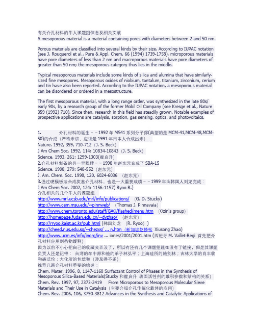
有关介孔材料的牛人课题组信息及相关文献A mesoporous material is a material containing pores with diameters between 2 and 50 nm. Porous materials are classified into several kinds by their size. According to IUPAC notation (see J. Rouquerol et al., Pure & Appl. Chem, 66 (1994) 1739-1758), microporous materials have pore diameters of less than 2 nm and macroporous materials have pore diameters of greater than 50 nm; the mesoporous category thus lies in the middle.Typical mesoporous materials include some kinds of silica and alumina that have similarly-sized fine mesopores. Mesoporous oxides of niobium, tantalum, titanium, zirconium, cerium and tin have also been reported. According to the IUPAC notation, a mesoporous material can be disordered or ordered in a mesostructure.The first mesoporous material, with a long range order, was synthesized in the late 80s/ early 90s, by a research group of the former Mobil Oil Company (see Kresge et al., Nature 359 (1992) 710). Since then, research in this field has steadily grown. Notable examples of prospective applications are catalysis, sorption, gas sensing, optics, and photovoltaics.1. 介孔材料的诞生--1992年MS41系列分子筛(典型的是MCM-41,MCM-48,MCM-50)的合成(严格来讲,应该是1991年日本人合成出来):Nature. 1992, 359, 710-712(J. S. Beck)J Am Chem Soc. 1992, 114: 10834-10843(J. S. Beck)Science. 1993, 261: 1299-1303(霍启升)2.介孔材料制备的另一里程碑--1998年赵东元合成了SBA-15Science. 1998, 279: 548-552(赵东元)J. Am. Chem. Soc. 1998, 120, 6024-6036 (赵东元)3.通过硬模板法合成炭基介孔材料,也是一大重要成绩--1999年由韩国人刘龙完成:J Am Chem Soc. 2002, 124: 1156-1157( Ryoo R.)介孔相关的几个牛人的课题组:/mrl/info/publications/(G. D. Stucky)/~pinnweb/(Thomas J. Pinnavaia)/staff/GAO/flashed/menu.htm(Ozin's group)/~dyzhao/(赵东元)http://rryoo.kaist.ac.kr/pub.html (韩国刘龙(R. Ryoo)).sg/~chezxs/ ... n.htm(新加坡赵修松Xiusong Zhao)http://www.ucm.es/info/inorg/inv ... iones/2001/2001.htm (西班牙M. Vallet-Regi 首先把介孔材料应用到药物缓释)因为以前不小心把自己的收藏夹弄没了,所以有还有几个课题组现在没有了链接,但是其课题负责人还是记得:台湾的牟中原和他的弟子林弘平;上海硅所的施剑林;吉林大学的肖丰收和裘式伦;大化所的包信和(涉及得不多)推荐几篇介孔材料重要的综述:Chem. Mater. 1996, 8, 1147-1160 Surfactant Control of Phases in the Synthesis of Mesoporous Silica-Based Materials(Stucky和霍启升表面活性剂的堆积参数和结构的关系)Chem. Rev. 1997, 97, 2373-2419 From Microporous to Mesoporous Molecular Sieve Materials and Their Use in Catalysis(主要介绍介孔作催化载体的应用)Chem. Rev. 2006, 106, 3790-3812 Advances in the Synthesis and Catalytic Applications of Organosulfonic-Functionalized Mesostructured Materials(有机官能化介孔的合成及在催化中的应用)Acc. Chem. Res. 2002, 35, 927-935Structural and Morphological Control of Cationic Surfactant-Templated Mesoporous Silica(牟中原具体谈论介孔形貌的形成)Angew. Chem. Int. Ed. 2006, 45, 3216–3251 Silica-Based Mesoporous Organic-Inorganic Hybrid Materials(Frank Hoffmann 所著,非常好)Acc.Chem.Res.2005, 38,305-312 Past, Present, and Future of Periodic Mesoporous Organosilicas-The PMOs(O'zin 重点介绍周期性介孔)NATURE 2002 417 813 Ordered porous materials for emergeing application(大牛Davis所著)Chem. Mater. 1999, 11, 2633-2656 Tailored Porous Materials(Thomas J.)介孔分子筛的应用:介孔分子筛吸附氨基酸:Carbon 44 (2006) 530–536 Adsorption of L-histidine over mesoporous carbon molecular sieves(印度人A. Vinu)Separation and Purification Technology 48 (2006) 197–201 Amino acid adsorption onto mesoporous silica molecular sieves (首篇)介孔分子筛吸附蛋白质(酶)Journal of Molecular Catalysis B-Enzymatic 2(1996) 115- 126 Enzyme immobilization in MCM-4 1 molecular sieve(首篇)J. Am. Chem. Soc. 1999, 121, 9897-9898 Mesoporous Silicate Sequestration and Release of Proteins(stucky)J. AM. CHEM. SOC. 2004, 126, 12224-12225 Protein Encapsulation in Mesoporous Silicate-The Effects of Confinement on Protein Stability, Hydration, and Volumetric PropertiesJ. Phys. Chem. B 2003, 107, 8297-8299 Adsorption of Cytochrome C on New Mesoporous Carbon Molecular Sieves(Vinu, A)介孔分子筛负载催化剂J. Phys. Chem. B 2006, 110, 15212-15217 Fabrication and characterization of mesoporous Co3O4 core-mesoporous silica shell nanocomposites(典型的core-shell结构)CHEM. COMMUN., 2003, 1522–1523 Ultra-thin porous silica coated silver–platinum alloy nano-particle as a new catalyst precursorApplied Catalysis A General 308 (2006) 19–30 Carbon oxide hydrogenation over silica-supported iron-based catalysts Influence of the preparation routeChem. Commun., 2005, 348–350 Metallic Ni nanoparticles confined in hexagonally ordered mesoporous silica material介孔分子筛在药物可控释放方面的应用Chem. Mater. 2001, 13, 308-311 A New Property of MCM-41, Drug Delivery System (Vallet-Regi 首篇)Nature 2003, 421, 350 – 353Photocontrolled reversible release of guest molecules from coumarinmodified modified mesoporous silica(第一次只能化)J. AM. CHEM. SOC. 2005, 127, 8916-8917 Fabrication of Uniform Magnetic Nanocomposite Spheres with a Magnetic Core-Mesoporous Silica Shell Structure-support(施剑林所谓的“药物分子运输车”)Angew. Chem. Int. Ed. 2005, 44, 5038 –5044 Stimuli-Responsive Controlled-Release Delivery System Based on Mesoporous Silica Nanorods Capped with Magnetic Nanopartic les(Victor S.-Y. Lin* 在智能介孔药物释放方面是贡献巨大)介孔分子筛吸附废水阳离子Chemosphere 59 (2005) 779–786 Heavy metals removal from electroplating wastewater by aminopropyl-Si MCM-41Environ. Sci. Technol.2000, 34,4822-4827 Surfactant-Templated Mesoporous Silicate Materials as Sorbents for Organic Pollutants in WaterInd. Eng. Chem. Res. 2004, 43, 1478-1484 Highly Selective Adsorption of Pt2+ and Pd2+ Using Thiol-Functionalized Mesoporous Silica介孔分子筛在色谱中的应用Chem. Commun., 2002, 752 - 753Biomolecule separation using large pore mesoporous SBA-15 as a substrate in high performance liquid chromatographyElectrophoresis 2006,27,742–748 Large-pore mesoporous SBA-15silica particles with submicrometer size as stationary phases for high-speed CEC seperation介孔分子筛作为硬模板Adv. Mater. 2001, 13,677-681Ordered Mesoporous Carbons(Ryong Ryoo)Microporous and Mesoporous Materials 63 (2003) 1–9 Synthesis and characterization of spherical carbon and polymer capsules with hollow macroporous core and mesoporous shell structures介孔分子筛复合材料(实现光学性能,比如掺入二氧化钛,量子点)J. Phys. Chem. B 2005, 109, 12309-12315 Synthesis and Characterization of Nano titania Particles Embedded in Mesoporous Silica with Both High Photocatalytic Activity and Adsorption CapabilityChem. Mater. 2005, 17, 1269-1271 Preparation of Mesoporous Titania Thin Films with Remarkably High Thermal StabilityJ. AM. CHEM. SOC. 2006, 128, 688-689Magnetic Fluorescent Delivery Vehicle Using Uniform Mesoporous Silica Spheres Embedded with Monodisperse Magnetic and Semiconductor Nanocrystals。
光催化材料研究进展马晓春;徐广飞【摘要】叙述了近些年来光催化材料的研究进展与核-壳型光催化纳米材料的研究现状,指出光催化材料尤其是纳米材料在治理环境污染方面发挥的巨大作用.同时,结合笔者的工作,简要介绍了纳米SnO2光催化剂的特点、制备方法和复合结构,并对其前景进行了展望.%The paper described the researching progress of photocatalytic materials and photocatalytic nanometer materials of core-shell structure in recent years. And its important role was pointed out in environmental pollution control especially for nanometer materials. At the same time, the characteristics, preparation method and composite structure of nanometer SnO2were introduced briefly according to the works. In addition, the development prospect was also described.【期刊名称】《新技术新工艺》【年(卷),期】2012(000)009【总页数】4页(P58-61)【关键词】光催化材料;TiO2;核-壳结构;SnO2【作者】马晓春;徐广飞【作者单位】浙江工业大学机械工程学院,浙江杭州330014;浙江工业大学机械工程学院,浙江杭州330014【正文语种】中文【中图分类】TQ13随着社会的进步和工业的发展,环境污染逐渐成为威胁人类生存的严重问题。
对此,人们展开了治理污染、保护环境的科学研究,并取得了一定的成效。
纳米硫化铜的制备及光催化性能高杰;李春慧;秦占斌;孙怡;高筠【摘要】In this paper,nano CuS were successfully synthesized at 100℃ in aqueous solution.Copper salts (such as copper chloride,copper sulfate and copper nitrate)and ZnS were used as raw materials.The products were characterized by means of XRD,SEM and UV-Vis techniques.The photocatalytic activity of nano CuS for the degradation of methyl orange was characterized.The results show that the degradation rates of methyl orange under the irradiation of xenon lamp were 17.17%,44.30%and 45.23%respectively.%本文以铜盐(如CuCl2、CuSO4、Cu(NO3)2)和ZnS为原料,在100℃、水溶液条件下成功制备了纳米CuS.产品用XRD、SEM和UV-Vis进行表征.以甲基橙为降解物,测定了CuS产品的光催化活性.结果表明,在氙灯的照射下,纳米CuS产品的光催化效率分别为17.17%,44.30%和45.23%.【期刊名称】《化学工程师》【年(卷),期】2017(031)001【总页数】4页(P7-10)【关键词】纳米硫化铜;硫化锌;光催化性能【作者】高杰;李春慧;秦占斌;孙怡;高筠【作者单位】华北理工大学化学工程学院,河北唐山063009;华北理工大学化学工程学院,河北唐山063009;华北理工大学化学工程学院,河北唐山063009;华北理工大学化学工程学院,河北唐山063009;华北理工大学化学工程学院,河北唐山063009【正文语种】中文【中图分类】O614.12随着人类社会的不断发展,工业全球化快速发展的同时人们的生活水平也有了很大的提高,但是随之带来的严重的环境污染问题已经引发全世界的高度关注。
覆盆子状复合粒子的制备及其作为建筑模块在构筑超亲水涂层中的应用 杜鑫,贺军辉*中国科学院理化技术研究所功能纳米材料实验室,北京 100190关键词:覆盆子状 复合粒子 多孔二氧化硅空心球涂层 超亲水 防雾 近年来,具有阶层结构表面形貌和规整结构的覆盆子状有机@无机复合粒子,由于其高的表面粗糙和潜在的应用,越来越受到人们的关注。
例如,这种复合粒子作为建筑模块,应用于构筑超疏水薄膜(模仿具有自清洁功能的荷叶的表面拓扑结构)和超亲水薄膜(具有多孔结构或/且粗糙表面结构)。
这种复合粒子在胶体和界面科学的基础研究也是非常重要的,因为它们经常被用作模型粒子来研究相行为、流变学和扩散行为。
然而,目前采用一种简单高效的方法来制备结构规整的覆盆子状复合粒子仍是一项挑战。
我们就是针对这方面来开展工作的。
[1][2-5]在本工作中,单分散的覆盆子状聚苯乙烯@二氧化硅复合粒子和具有阶层孔结构的二氧化硅空心球涂层的制备路线图如图1所示。
Scheme 1. Schematic illustration of the fabrication of monodisperse raspberry-like PS@SiO 2 composite nanoparticles (RCNs) using oxygen plasma treated PS spheres as core and hierarchically structured porous film of silica hollow spheres.利用聚乙烯吡咯烷酮作稳定剂,通过无皂乳液聚合的方法制备出单分散的聚苯乙烯微球。
通过调节反应原料中聚乙烯吡咯烷酮的用量,聚苯乙烯微球的粒径在200-1400 nm范围内可以调节。
在下面的实验中,我们选择270和615 nm [6] 的聚苯乙烯微球作为核。
我们采用氧等离子体刻蚀的方法对聚苯乙烯微球表面进[7] 行了羟基功能化。
Colloids and Surfaces A:Physicochem.Eng.Aspects 428 (2013) 79–85Contents lists available at SciVerse ScienceDirectColloids and Surfaces A:Physicochemical andEngineeringAspectsj o u r n a l h o m e p a g e :w w w.e l s e v i e r.c o m /l o c a t e /c o l s u r faCore/shell structured silica spheres with controllable thickness of mesoporous shell and its adsorption,drug storage and release propertiesXuecheng Chen ∗Institute of Chemical and Environment Engineering,West Pomeranian University of Technology,Szczecin ul.Pulaskiego 10,70-322Szczecin,Polandh i g h l i g h t s•Core/shell mesoporous silica sphereswere fabricated by sol–gel method.•The core/shell mesoporous silica spheres exhibited high specific sur-face area.•They showed good adsorption and drug delivery properties.g r a p h i c a la b s t r a cta r t i c l ei n f oArticle history:Received 6September 2012Received in revised form 12March 2013Accepted 22March 2013Available online 1 April 2013Keywords:Core/shell Silica sphere Mesoporous Adsorption Drug deliverya b s t r a c tIn this study,a simple method to coat mesoporous silica layer onto solid silica spheres in a controlled manner via a two-step procedure is reported.Core/shell structured SiO 2@m-SiO 2spheres with different shell thickness have been obtained by the application of the optimized hexadecyl trimethyl ammonium bromide/tetraethylorthosilicate (CTAB/TEOS)ratio.The specific surface areas of the obtained samples increase with the shell thickness increase from 25nm to 90nm.The adsorption ability,storage and release properties related to mesoporous shell thickness and pore sizes were explicitly investigated with Rhodamine B (RhB)as the model molecules.© 2013 Elsevier B.V. All rights reserved.1.IntroductionMesoporous materials,since the first discovery in 1992[1],have attracted great interest due to their applications in cataly-sis,adsorbents,separation and drug-delivery because of excellent chemical,mechanical,and thermal stability [2].Many articles have reported that mesoporous materials either with ordered or disordered pore system can meet the demands of the target appli-cations [3].The synthesis of diameter controlled nanoparticles with∗Tel.:+48506978574.E-mail addresses:xchen@.pl ,chenxuecheng1978@mesopores is a major challenge in advanced materials science.These particles can be also employed as catalytic supports or drug delivery supports [4].The spherical MCM-41type silica particles synthesis with ordered mesopores have been reported by using modified Stöber method [5].However,the resulting diameter of the particles was not fully controllable.The first monodispersed mesoporous spheres were nanometer-size core–shell structured mesoporous silica spheres,which were synthesized from the simultaneous sol–gel polymerization of tetraethylorthosilicate (TEOS)and octadecyltrimethoxysilane followed by the removal of the organic groups [6].Core–shell structured mesoporous silica spheres with orientated and accessible mesopores and high dispersibility are highly valuable for different applications.0927-7757/$–see front matter © 2013 Elsevier B.V. All rights reserved./10.1016/j.colsurfa.2013.03.03880X.Chen/Colloids and Surfaces A:Physicochem.Eng.Aspects428 (2013) 79–85Table1Effects of CTAB,EtOH,H2O,NH3·H2O and TEOS on Formation of core/shell structured silica spheres.SiO2@m-SiO2(n)SiO2(mg)CTAB(mg)EtOH(mL)H2O(mL)NH3·H2O TEOS(mL)25nm10040 3.157.50.1130.07535nm10080 6.3150.2250.14360nm10016012.6300.450.28670nm10016012.6300.450.28690nm10032025.2600.90.572 Notes:SiO2@m-SiO2(70nm)and SiO2@m-SiO2(90nm)were two-step synthesis.Furthermore,the core can be replaced with magnetic nanoparti-cles,gold nanoparticles andfluorescent nanoparticles enabling the core–shell structured spheres with additional functions used e.g. in drug delivery,hyperthermia and cell imaging[7].Recently,Yu et al.reported for thefirst time the synthesis of the monodispersed spherical silica particles with solid core and mesoporous shell featuring mesopore channels perpendicular to the core surface by adopting silica particles as a core component and hexade-cyl trimethyl ammonium bromide(CTAB)as a structure-directing agent for the mesoporous shell[8].Magnetic mesoporous spheres have proved to be a good adsorbent to treat the pollutant water [9].Zhao et al.also developed the high-magnetic spheres with Fe3O4@SiO2core and aligned mesoporous SiO2shell,which were used as a reusable absorbent to remove microcystins from aqueous solution in a fast and highly efficient way[9b].In the current state of the art,the thickness of the mesoporous shell is not designed at will.However,the specific surface area,pore volume of the core/shell structured spheres are related to the shell thickness.Furthermore,the adsorption capability and release rate are more shell thickness dependent.So,in order to fully under-stand the relationships between the mesoporous shell thickness and adsorption ability,the study on drug storage and release behav-ior is much needed.The present work is focused on mesoporous core/shell struc-tured silica spheres using solid silica spheres as the core,CTAB as the surfactant and TEOS as the silica source to construct the meso-porous shell around the bare silica spheres.It showed to be a cost effective,simple,and convenient method for the preparation of well-ordered mesoporous core/shell structured silica spheres with designed shell thickness in the ranged from25to90nm at will. These mesoporous core/shell structured silica spheres were used adsorbents for the removal of commercial dyes RhB from aque-ous solution.Furthermore,the drug storage and release properties related to the shell thickness and pore sizes have also been inves-tigated in details.The silica core can be replaced with magnetic core(Fe3O4)nanoparticles inducing magnetic behavior of these core/shell structured silica spheres,what can broaden their appli-cations in different research areas.2.Experimental2.1.MaterialsTEOS(95%)and CTAB were purchased from Sigma–Aldrich, which were used directly without further purification.All other materials were of analytical grade and commercially available, including NH3·H2O(25%[w/w])and ethanol.2.2.Synthesis of mesoporous core–shell structured silica spheresMonodispersed silica spheres were synthesized by mixing 2.5mL of aqueous ammonia(NH3·H2O,28wt%)with a solution con-taining50mL of absolute ethanol(EtOH).After stirring for30min, 1.5mL of TEOS(98%,Aldrich)was added to the solution and stirred for24h at room temperature.To coat the above prepared silica spheres with the mesoporous shell,H2O,CTAB and TEOS were used. 100mg of the above prepared silica spheres were dispersed into the mixture of H2O,CTAB surfactant and EtOH and then sonicated and stirred vigorously.After stirring for30min,NH3·H2O and TEOS were added to the reaction mixture and stirred at room temper-ature overnight.The as-synthesized silica particle suspension was centrifuged and dried at343K overnight.The resulting dry powder was further calcined at823K for4h in air in order to remove CTAB selectively from the composites.The resulting calcinated core–shell structured silica spheres were designated as SiO2@m-SiO2(n).The details on the experimental procedure to obtain the desired shell thickness in SiO2are provided in Table1.2.3.Adsorption experimentsAqueous dye solution was obtained by dissolving the dye in distilled water at a certain concentration(1.67×10⁻⁵to 1×10⁻⁴M).For low concentration of RhB adsorption experi-ments,in a typical analysis,to a30mL comparison tube,10mL of the suspension of mesoporous silica spheres(10mg mL−1), 2mL of RhB(1×10⁻⁴M)was added to above suspension. The mixed solution was then stirred at room temperature for30min.The tubes were then centrifuged at4500rpm for 10min to separate the solid materials from the system and the supernatant was ready for the UV–vis analysis.In a high concentration of RhB adsorption experiments,10mg of solid samples(SiO2@m-SiO2(25nm),SiO2@m-SiO2(35nm),SiO2@m-SiO2(60nm),SiO2@m-SiO2(70nm)and SiO2@m-SiO2(90nm)) were added tofive reference quvettes containing10mL of RhB (1×10⁻⁴M),respectively.The mixed solution was then stirred at room temperature for30min.The tubes were then cen-trifuged at4500rpm for10min to separate the solid materials from the system and the supernatant was ready for the UV–vis analysis.Drug storage and release profiles were obtained as followed: to store the molecules,RhB was dissolved in ethanol solution,and the RhB concentration was0.213mg mL−1.50m g of each sample was added into20mL of the RhB/ethanol solution at room tem-perature.The bottles were sealed to prevent the evaporation of ethanol,and then the mixture was stirred for48h.The RhB-loaded product(SiO2@m-SiO2@RhB)was separated from this solution by filtration,washed several times with H2O,and dried under vac-uum at100◦C for24h.A typical drug release experiment was performed as follows:10mg of SiO2@m-SiO2@RhB was dispersed in10mL of phosphate-buffered saline(PBS,pH∼7.4)under mag-netic stirring at a rate of ca.100rpm at room temperature.3mL of the mixture was extracted with a syringe at given time intervals for analysis.After removal of solid SiO2@m-SiO2@RhB by centrifu-gation,the remaining clear solution(2mL)was then analyzed by UV–vis spectroscopy at a wavelength of550nm.By measuring the UV absorption spectra of standard RhB solutions with different con-centrations in PBS,a calibration curve of RhB concentration via UV–vis absorbance was set up.RhB release amounts were deter-mined by measuring the absorbance of the above clear solutions, and calculated using the calibration curve.The total RhB releaseX.Chen/Colloids and Surfaces A:Physicochem.Eng.Aspects428 (2013) 79–8581Fig.1.TEM images of(a)silica spheres and(b–f)the mesoporous core–shell structured silica spheres with different shell thickness:(b)25nm,(c)35nm,(d)60nm,(e)70nm, (f)90nm.amount for a week was determined as the total storage amount of core–shell structured silica.2.4.Synthesis of Fe3O4nanoparticlesIn a typical synthesis,0.45g of FeCl3and1.8g of sodium acetate were dissolved in20mL of ethylene glycol under magnetic stir-ring.The obtained homogeneous yellow solution was transferred to autoclave and heat at200◦C for8h.After hydrothermal reaction, the autoclave was cooled to room temperature.The obtained black magnetite nanoparticles were washed with ethanol and dried.2.5.Synthesis of Fe3O4@SiO2@m-SiO2nanospheresThe Fe3O4@nSiO2nanospheres were prepared as followed:0.1g of Fe3O4nanoparticles werefirstly treated with0.1M HCl aque-ous solution for10min.Then Fe3O4nanoparticles were separated and washed with pure water,then homogeneously dispersed in the mixture of ethanol(80mL),H2O(20mL)and NH3·H2O(1.0mL, 28wt.%),followed by the addition of TEOS(0.03g).After mechanical stirring at room temperature for12h,the Fe3O4@SiO2nanospheres were separated and washed with ethanol and water again,re-dispersed in a mixed solution containing of CTAB(0.30g)deioned water(80mL),concentrated NH3·H2O(1.00g,28wt%)and ethanol (60mL).The mixed solution was sonicated for0.5h to form a uni-form dispersion.In the next step,0.40g of TEOS(1.90mmol)was added to the above mixture with continuous mechanical stirring for12h.Finally,the product was separated with a magnet and washed repeated with ethanol and water.In the last step,CTAB template in the magnetic nanospheres was removed by dispers-ing the Fe3O4@SiO2@m-SiO2nanospheres in100mL of ethanol and refluxing at90◦C for48h,then Fe3O4@SiO2@m-SiO2nanospheres were obtained for further use.2.6.CharacterizationX-ray diffraction(XRD)was conducted on a Philips diffrac-tometer using Cu K␣radiation.The morphologies of the samples were examined byfield-emission scanning electron microscopy (FE-SEM,Quanta250and Nova Nanosem650,FEI).Transmission electron microscopy(TEM)and high-resolution transmission elec-tron microscopy(HR-TEM)were performed on the FEI Tecnai F30 transmission electron microscope with afield emission gun oper-ating at200kV to examine the dimensions and structural details of the core–shell nanoparticles.The elemental compositions of the samples were determined by energy dispersive X-ray spectrom-etry(EDS)as the HR-TEM mode.The N2adsorption/desorption isotherms were acquired at liquid nitrogen temperature(77K) using a Micromeritics ASAP2010M instrument and the specific surface area was calculated by the Brunauer–Emmett–Teller(BET) method.The pore size distribution was determined using the Barret–Joner–Halenda(BJH)method.The data of RhB adsorption were obtained using a UV-2450spectrophotometer(Shimadzu, Japan).Thermogravimetric analysis(TGA)was carried out on10mg samples using the DTA-Q600SDT TA Instrument at a heating rate of10◦C/min from room temperature to900◦C under air.3.Results and discussionFig.1a shows the TEM image of solid silica spheres with the diameter of around200nm.Taking the bare silica spheres as the core,CTAB as the surfactant and TEOS as the silica source,a mesoporous silica shell was constructed on bare silica sphere suc-cessfully.Through control of CTAB,TEOS and bare silica spheres ratio,five kinds of core/shell structured silica spheres were obtained.As shown in Fig.1b–f,the shell thickness was∼25,35, 60,70and90nm,respectively.Fig.2shows the direct SEM images of different core/shell struc-tured silica spheres(Fig.2a–f).With the shell thickness from25 to90nm,the diameters in the range of225–290nm of core/shell structured silica spheres were obtained(Fig.2a–f).In Fig.2a–c, the core/shell structured can be clearly observed.In some cases the shell of the spheres peeled off(Fig.2b–c)and the internal silica spheres were exposed proving the core/shell character of the obtained spheres.The thicker mesoporous layer of the silica spheres is more stable because the shells almost did not peel off.In Fig.3,it presents the XRD patterns of the calcinated core/shell structured silica spheres with the shell thickness ranging from82X.Chen /Colloids and Surfaces A:Physicochem.Eng.Aspects 428 (2013) 79–85Fig.2.SEM images of core–shell structured silica spheres with different shell thickness.(a–b)25nm,(c)35nm,(d)60nm,(e)70nm,(f)90nm.25nm to 90nm,respectively.All of the SiO 2@m-SiO 2(n )spheres have a strong peaks (Fig.3),showing that the samples have an ordered structure.When the shell thickness increased,the order of the silica also increased.As shown in Fig.3,one intense diffrac-tion peak at a 2Âvalue of 2.5◦can be found in its small-angle XRD pattern (100),indicating the formation of ordered mesostructures.When shell thickness further increased to 70nm,diffractions peak was found to be sharper,exhibiting highly ordered porous struc-tures.However,in the case of shell thickness increased to 90nm,the mesoporous structure of the shell became less order,it may be come from the three step coating process,which disturbed the assembling process of CTAB on the silica surface.These results indi-cate that mesoporous silica shells have been successfully coated on the bare silica spheres.Nitrogen sorption measurements were conducted to further characterize the pore parameters of the products.Nitrogen sorp-tion isotherms of different mesoporous SiO 2@m-SiO 2samples with variable pore sizes are shown in Fig.4.The nitrogen adsorption isotherms of SiO 2@m-SiO 2(25nm),SiO 2@m-SiO 2(35nm),SiO 2@m-SiO 2(60nm),SiO 2@m-SiO 2(70nm)and SiO 2@m-SiO 2(90nm),5001000150020002500I n t e n s i t y2θFig.3.Small-angle XRD patterns of calcined samples with different shell thickness:(a)25nm),(b)35nm,(c)60nm,(d)70nm and (e)90nm,respectively.respectively,displayed typical type IV curves of surfactant-assisted mesoporous silica at relative partial pressure values around 0.3.The nitrogen adsorption isotherms of the five analyzed samples clearly indicate that the mesopores were uniform in size.The pore diameters estimated from the TEM image analysis confirm these data.For the samples of SiO 2@m-SiO 2(25nm),SiO 2@m-SiO 2(35nm),SiO 2@m-SiO 2(60nm),SiO 2@m-SiO 2(70nm)and SiO 2@m-SiO 2(90nm),adsorption isotherms exhibited a nar-row mesopore size distribution,as indicated by a steep capillary step,which are characteristic for mesostructures with 1-D cylin-drical channels.The pore parameters of the samples calculated by N 2adsorption–desorption isotherms are listed in Table 2.It can be seen that all of the SiO 2@m-SiO 2spheres have high Brumauer–Emmett–Teller (BET)surface areas and large pore volumes.The BET surface area of the SiO 2@m-SiO 2(n )spheres increased gradually with the increasing shell thickness.SiO 2@m-SiO 2(25nm)has the lowest BET specific area and pore volume,V o l u m e @ S T P [c m 3/g ]Relative Pressure P/PoFig.4.N 2adsorption–desorption isotherms of the calcined mesoporous SiO 2@m-SiO 2with different shell thickness:(a)25nm),(b)35nm,(c)60nm,(d)70nm and (e)90nm.X.Chen/Colloids and Surfaces A:Physicochem.Eng.Aspects428 (2013) 79–8583Table2The structural parameters of mesoporous SiO2@m-SiO2with different shell thickness.Sample S BET(m2g−1)V total(cm3g−1)D A(nm)SiO2@m-SiO2(25nm)1910.076 3.93SiO2@m-SiO2(35nm)4000.124 3.13SiO2@m-SiO2(60nm)5460.192 5.04SiO2@m-SiO2(70nm)6100.083 3.17SiO2@m-SiO2(90nm)8310.099 3.16S BET(m2g−1),BET surface area;V total,total pore volume;D A,average pore diameter. however,SiO2@m-SiO2(90nm)has the largest BET specific area. It means that the samples with thicker mesoporous layer exhibit higher BET specific area.It is worth noting SiO2@m-SiO2(60nm) has the biggest pore diameter(5.04nm),this may be attributed from one step synthesis procedure.However,the pore sizes of the other samples are around3nm.3.1.Adsorption propertiesIn order to investigate the relationships between the adsorp-tion properties and the wall thickness and pore size,RhB molecules were used as the model of organic pollutant to evaluate the adsorption ability of the core/shell structured silica spheres.The adsorption efficiencies were analyzed by using the UV–vis spec-troscopy.The adsorption experiments were carried out at room temperature by immersion the core/shell structured silica spheres into12and10mL aqueous solution of RhB with an initial concentra-tion of1.67×10−5M and1×10−4M.The adsorption performance was measured by the decay of the absorption of the dye as a func-tion of shell thickness.In thefirst adsorption experiment,low concentration of RhB(1.67×10⁻⁵M,Fig.5a)was used.In Fig.5b, the characteristic absorption band of RhB at550nm decreased sig-nificantly with increasing shell thickness from25to90nm.For the low concentration of RhB solutions,the adsorption results indicate that91.9%,94.2%,97.4%,96.5%and97.8%of RhB for20,35,60, 70and90nm shell thickness can be removed fast in one separa-tion process(Fig.5b).So,in low concentration of RhB,thin shell thickness silica spheres also adsorb the guest molecules efficiently. Such a high removal efficiency is mainly attributed to the high sur-face areas and the numerous accessible mesopores in the silica shell.Additionally,after extraction of the trapped RhB with ace-tonitrile/water mixture,the core/shell structured silica spheres can be re-used.However,the adsorption ability at low concentration of RhB of different shell thickness silica spheres did not exhibit much differ-ence.Therefore,tofind the adsorption behavior of different shell thickness silica spheres in high concentration of RhB,higher con-centration of RhB(1×10⁻⁴M)was applied.In high concentration of RhB,the difference in adsorption ability became more evident. The trend is the same,adsorption enhanced with the increase of the shell thickness from25to90nm,except70nm sample.As shown in Fig.5d,the adsorption efficiency for25,35,60,70and90nm sam-ples are77.4%,82.9%,89.8%,89.4%and91.8%,respectively.When the shell thickness increases to90nm,the adsorption ability was the most efficient.3.2.Drug storage and release propertiesMesoporous silica materials are currently widely used as carriers in drug delivery applications.It has been reported that nonphagocytic eukaryotic cells could internalize particlesAbsorption(a.u.r)Wavelength (nm)Absorption(a.u.r)wavelength (nm)Absorption(a.u.r)Wavelength (nm)Adsorption(%)Shell thickness (nm)Fig.5.(a)UV–vis spectrum of RhB in aqueous solution(1.67×10⁻⁵M),(b)UV–vis spectrum of supernatant after adsorption experiment of the samples with different shell thickness silica spheres(initial concentration:1.67×10⁻⁵M),(c)UV–vis spectroscopy of supernatant after adsorption experiment of the samples with different shell thickness silica spheres after adsorption experiment of the samples with different shell thickness silica spheres(initial concentration:1×10⁻⁴M).(d)Adsorption profiles of different core–shell structured silica spheres with different concentration of RhB(red line:1.67×10⁻⁵M and blue line:1×10⁻⁴M).(For interpretation of the references to color in thisfigure legend,the reader is referred to the web version of this article.)84X.Chen /Colloids and Surfaces A:Physicochem.Eng.Aspects 428 (2013) 79–85A b s o r p t i o n (a .u .r )Wavelength (nm)R e l e a s e a m o u n tTime (h)a bFig.6.UV–vis spectra (a)at different times of the aqueous media in which RhB was released from SiO 2@m-SiO 2(70nm)@RhB and (b)different controlled release kinetics of SiO 2@m-SiO 2(n)@RhB.as large as 500nm in size [10].So the current fabricated core/shell structured silica spheres would be potentially use-ful in drug delivery applications.Therefore,silica spheres with shell thickness in the range of 25–90nm could be used as drug carrier,to study drug storage capacity and release behav-ior.RhB was also chosen as a probe molecule to proof the concept.The loading capacity of RhB was measured to be 30,48,67,69and 186mg/g SiO 2for SiO 2@m-SiO 2(25nm),SiO 2@m-SiO 2(35nm),SiO 2@m-SiO 2(60nm),SiO 2@m-SiO 2(70nm)and SiO 2@m-SiO 2(90nm),respectively.SiO 2@m-SiO 2(90nm)hasthe highest drug loading capacity among the five samples,which is attributed to its highest BET specific surface area and the appro-priate size of mesopores (3nm)compared with the size of RhB (ca.1.0±0.6nm).And SiO 2@m-SiO 2(25nm)has the lowest load-ing capacity,which is due to its thin shell thickness and low BET specific area.Fig.6a shows the UV–vis spectra at different intervals of the aqueous media in which RhB was released from SiO2@m-SiO2(70nm)@RhB.The RhB concentration gradually increases with the time prolongation,indicating sustained release of RhB molecules.As presented in Fig.6b,kinetic release curves of fiveFig.7.(a)TEM image of Fe 3O 4@SiO 2(a)Fe 3O 4@SiO 2@m-SiO 2nanospheres (b–c).Separation of Fe 3O 4@SiO 2@m-SiO 2nanospheres from solution under an external magnet (d).X.Chen/Colloids and Surfaces A:Physicochem.Eng.Aspects428 (2013) 79–8585kinds of core/shell structured silica spheres were plotted based on the sustained release using the absorption of the peak at ca.550nm. Recently,Vallet-Regi et al.reported that the release rate under sim-ilar conditions was very fast during thefirst day,but decreased with time and reached a maximum value of80%the third day when using conventional MCM-41NPs as IBU carriers[11].As shown in Fig.6b,the currentfive samples exhibit higher rates of release, i.e.,the RhB amounts released from SiO2@m-SiO2@RhB(25nm), SiO2@m-SiO2@RhB(35nm),SiO2@m-SiO2@RhB(60nm),SiO2@m-SiO2@RhB(70nm)and SiO2@m-SiO2@RhB(90nm)reach about80% in24h,indicating that the hierarchically mesoporous nanostruc-tures are favorable for fast molecular diffusion through the pore channels.In all offive samples,in thefirst time when SiO2@m-SiO2@RhB dispersed in PBS solutions,no signal from RhB could be observed,the signal rose relatively rapidly over the24h,after which the signal stabilized.The SiO2@m-SiO2@RhB(25nm)has the lowest releasing rate than other samples.The release rate of SiO2@m-SiO2@RhB(60nm)is the fastest,this results from the biggest pore diameter in the shell(Table2),thus allowing more RhB releasing from the shell in the same conditions.In order to enable the core/shell structured silica spheres to be multifunctional,the silica cores could be modified with Fe3O4 nanoparticles,thus the core/shell structured silica spheres would be magnetic nanoparticles,broadening the application of core/shell structured silica sphere in adsorption or drug delivery experiments. To prove this concept we prepared SiO2@m-SiO2with Fe3O4as a core(see TEM image in Fig.7a–c).The Fe3O4core/mesoporous silica shell particles show that the Fe3O4particles are spherical in shape and uniform in dimension with particle diameters of ca. 500nm.The magnetic separation of such magnetic nanoparticles was tested in ethanol by placing a magnet near the glass bottle. The black particles were attracted toward the magnet within20s (Fig.7d),demonstrating directly that the core–shell nanospheres possess magnetic properties.This will provide an easy and efficient way to separate Fe3O4@SiO2@m-SiO2particles from a sol or a sus-pension system and to carry drugs to targeted locations under an external magneticfield.According to the experimental data of core/shell structured sil-ica spheres,the adsorption,storage and release properties are much related to the shell thickness and pore sizes.When the shell thick-ness increases from25to90nm,the absorption and storage ability also enhanced.When the pore diameter of the shell increased,the release rate also enhanced.4.ConclusionsThe core/shell structured silica spheres with controllable shell thickness were synthesized with defined TEOS/CTAB molar ratio which was evidenced from SEM and TEM analysis.The core/shell structured silica spheres with shell thickness in the range of25 to90nm possess large specific surface area(191–831m2g−1)and uniform accessible mesopores.It was found that the core/shell structured mesoporous silica spheres can be used as a reusable absorbent for fast and highly efficient removal of RhB.The RhB molecules storage and release in core/shell structured silica sphere were also fully studied.With the shell thickness increasing from25 to90nm,the adsorption and storage capacity enhanced gradually. When the silica cores were replaced by magnetic nanoparticles, the mesoporous magnetic silica spheres with high specific surface area have potential applications in polluted water treatment and targeted drug delivery.AcknowledgementsThe authors are grateful for thefinancial support of Foundation for Polish Science within FOCUS2010Program.We also thank for Adrian Sandu and Keriya Mam,SEM application,specialist at FEI Company.References[1]Y.Chan,J.P.Zimmer,M.Stroh,J.S.Steckel,R.K.Jain,M.G.Bawendi,Incorporationof luminescent nanocrystals into monodisperse core–shell silica microspheres, Adv.Mater.16(2004)2092–2097.[2](a)M.L.Breen,A.D.Dinsmore,R.H.Pink,S.B.Qadri,B.R.Ratna,Sonochemicallyproduced ZnS coated polystyrene core shell particles for use in photonic crys-tals,Langmuir17(2001)903–907;(b)Y.H.Deng,W.L.Yang,C.C.Wang,FuF S.K.,A novel approach for preparationof thermoresponsive polymer magnetic microspheres with core–shell struc-ture,Adv.Mater.15(2003)1729–1732;(c)Y.Yang,X.H.Yan,Y.Cui,Q.He,D.X.Li,A.H.Wang,J.B.Fei,J.B.Li,Preparationof polymer-coated mesoporous silica nanoparticles used for cellular imaging by a“graft-from”method,J.Mater.Chem.18(2008)5731–5737;(d)Y.Yang,Y.Jia,L.Gao,J.B.Fei,L.R.Dai,J.Zhao,J.B.Li,Fabrication of autofluores-cent protein coated mesoporous silica nanoparticles for biological application, mun.47(2011)12167–12169;(e)Y.Yang,W.X.Song,A.H.Wang,P.L.Zhu,J.B.Fei,J.B.Li,Lipid coated meso-porous silica nanoparticles as photosensitive drug carriers,Phys.Chem.Chem.Phys.12(2010)4418–4422.[3]G.Ertl,H.Knözinger,J.Weitkamp,Handbook of Heterogeneous Catalysis,vol.1,Wiley-VCH,Weinheim,Germany,1997.[4](a)S.Ludtke,T.Adam,K.K.Unger,Application of0.5m porous silanizedsilica beasds in electrochromatography,J.Chromatogr.A786(1997) 229–235;(b)A.Fihri,M.Bouhrara,D.Cha,V.Polshettiwar,Fibrous nano-silica(KCC-1)supported palladium catalyst:Suzuki coupling reactions under sustainableconditions,ChemSusChem5(2012)85–89;(c)V.Polshettiwar,J.Thivolle-Cazat,M.Taoufik,F.Stoffelbach,S.Norsic,J.M.Basset,Hydro-metathesis of olefins:a catalytic reaction using a bifunctional single-site tantalum hydride catalyst supported onfibrous silica(KCC-1) nanospheres,Angew.Chem.Int.Ed.50(2011)2747–2751.[5](a)M.Grun,uer,K.K.Unger,The synthesis of micrometer-andsubmicrometer-size spheres of ordered mesoporous oxide MCM-41,Adv.Mater.9(1997)254–257;(b)M.Grun,K.K.Unger,A.Matsumoto,K.Tatsumi,Novel pathways for thepreparation of mesoporous MCM-41materials:control of porosity and mor-phology,Microporous Mesoporous Mater.27(1999)207–216.[6](a)G.Buchel,K.K.Unger,A.Matsumoto,K.Tsutsumi,A novel pathway for syn-thesis of submicrometer-size solid core/mesoporous shell silica spheres,Adv.Mater.10(1998)1036–1038;(b)S.B.Yoon,K.Sohn,J.Y.Kim,C.H.Shin,J.S.Yu,T.Hyeon,Fabrication of car-bon capsules with hollow macroporous core/mesoporous shell structures,Adv.Mater.14(2002)19–21.[7](a)Y.Chen,H.G.Chen, D.P.Zeng,Y.B.Tian, F.Chen,J.W.Feng,J.L.Shi,Core/shell structured hollow mesoporous nanocapsules:a potential platform for simultaneous cell imaging and anticancer drug delivery,ACS Nano4(2010) 6001–6013;(b)K.W.Hu,T.M.Liu,K.Y.Chung,K.S.Huang,C.T.Hsieh,C.K.Sun,C.S.Yeh,Efficient NIR hyperthermia and intense nonlinear optical imaging contrast on the gold nanorod-in-shell nanostructures,J.Am.Chem.Soc.131(2009) 14186–14187.[8]S.B.Yoon,J.Y.Kim,J.H.Kim,Y.J.Park,K.R.Yoon,S.K.Park,J.S.Yu,Synthesis ofmonodisperse spherical silica particles with solid core and mesoporous shell: mesopore channels perpendicular to the surface,J.Mater.Chem.17(2007) 1758–1761.[9](a)P.Wang,Q.H.Shi,Y.F.Shi,K.K.Clark,G.D.Stucky,A.A.Keller,Magnetic per-manently confined micelle arrays for treating hydrophobic organic compound contamination,J.Am.Chem.Soc.131(2009)182–188;(b)Y.H.Deng,D.W.Qi,C.H.Deng,X.M.Zhang,D.Y.Zhao,Superparamagnetichigh-magnetization microspheres with an Fe3O4@SiO2core and perpendicu-larly aligned mesoporous SiO2shell for removal of microcystins,J.Am.Chem.Soc.130(2008)28–29.[10]J.Rejman,V.Oberle,I.S.Zuhorn,D.Hoekstra,Size-dependent internalizationof particles via the pathways of clathrin-and caveolae-mediated endocytosis, Biochem.J.377(2004)159–169.[11]M.Vallet-Regí, A.Rámila,R.del Real,P.J.Pérez-Pariente.,A newproperty of MCM-41:drug delivery system,Chem.Mater.13(2001) 308–311.。