人体形态学教学大纲
- 格式:pdf
- 大小:173.94 KB
- 文档页数:18
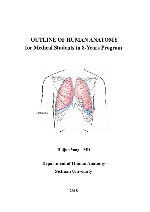
OUTLINE OF HUMAN ANATOMY for Medical Students in 8-Years ProgramHuijun Yang MDDepartment of Human AnatomySichuan University2018Unit 1 Introduction for Human AnatomyWhat is Human Anatomy?Human anatomy is one of the basic medical sciences that deals primarily with structure and function of the human body. It is importance for doctor to practice all specially areas of medicine. Anatomy, is closely associated with radiology and surgery, it forms an essential basis for all branches of medicine. Its importance to the surgeon has long been apparent but today such developments as new imaging techniques, biopsy procedures, and noninvasive therapeutic methods make an accurate knowledge of anatomy equally essential to the practice of all specially areas of medicine.Dissection is a technique to study the structure of the body. During dissection you will dissect, observe, palpate the parts of the body.It is good method of studying the body. Anatomy is the study of living human beingsThe student should remember that the purpose of such study is to allow him to visualize the living body in action so that he can appreciate the effects of injury or disease, and can recognize an abnormality from his knowledge of the normal. To achieve this kind of information there is no substitute for the personal process of looking at the body by dissection while thinking of the functions of its various parts and checking these points by observation and palpation.Anatomy is Basis of Medical LanguageAnatomy students learn a new language consists of at least 4,500 words. When you learn these words you will be able to speak the anatomical language fluently. You will feel ease talking to your clinical colleagues because the anatomical language constitutes most of the words making up the medical language.To describe the relationship of one structure to another, ANATOMICAL NOMENCLATURE should be used. To describe the body and to indicate the position of its parts and organs relative to each other, anatomists around the world have agreed to use several terms of position and direction and various planes of the body. Because the clinicians also use these terms, it is important for student to take time at the beginning of your professional career to learn them well. Practise, using them, so that it will be clear what you mean, when you describe parts of the body in patients’histories or during discussion of patients with yourclinical colleagues.Systemic AnatomyThe organ is made up by four basic tissues The systems of the human body are usually grouped some organs, which have similar structures and function together and described as follows:1. The Skeletal SystemThis system consists of bones, certain cartilage and joints. Its supports the body and protects the organs, provides a system of levers and a point of attachment for muscles that enable the body to move, and manufactures red blood cells and some white blood cells in the bone marrow. Bone tissue also stores the body’s main supply of reserve calcium and phosphorus.Understand the concepts: the structure, function of the bone as living organs, the classification of the bone, the classification of the articulation, the basic constituents of the synovial joint, and their important structure, the terms of movements.2. The Muscular SystemThere are three types of muscles: skeletal, smooth and cardiac muscles. The muscular system consists of skeletal muscles. The muscle constitute two portions: muscular fibers and tendons, the fibrous cords of connective tissue that attach muscles to bones, and the motor nerves that stimulate muscles contractions. Muscle allows movement; help us to maintain a correct posture; and produce much of our body heat.Understand the concepts: the classification of the muscle; the shape, architecture of the skeletal muscle, motor unit of muscle. Remember the facts that the manner in which a muscle acts on a joint depends on its position relative to the joint, and muscles are often classified in group by principal action, which they have on particular joint.Understand the synovial bursa and sheath, which lie between the tendons and the bone or enclose the tendons of muscles.3. The Digestive SystemThe digestive system consists of digestive tubes and associated digestive gland. The digestive tube f rom the mouth to anus, includes the teeth, tongue esophagus stomach, small and large intestines. The digestive gland includes the salivary gland liver and pancreas.Understanding the concept: Which organs form the digestive tract? What is the basic function of each?List the major accessory organs and associated structures of the digestive system4.The Respiratory SystemThe Respiratory system is composed of the nose, pharynx, larynx, trachea, and lung. The function of the respiratory system is exchange the gases between blood and air the oxygen from the air moves into the blood; then carried into the tissues. In a reverse process, waste carbon dioxide from the blood is carried to the lungs, where it is eventually exhaled from the body into the air outside.Understand the concepts:what are the structures and functions of the each organs of the respiratory system?5. The Urinary SystemThe Urinary system consists of kidneys, ureters, urinary bladder and urethra. The functions of the urinary system are produce and eliminate urine. In doing so, it rids the body of waste helps regulate blood pressure and the composition and volume of blood, and helps to maintain the body’s acid -base and water-salt balance.Understand the concepts: What are the structures and functions of the each organs of the urinary system?6. The Reproductive SystemThe male and female genital system have reproductive organs (testes or ovaries) that secrete sex hormones and produce reproductive cells (sperms or eggs), and a set of ducts and accessory glands and organs such as the prostate gland, penis, uterus, and vagina. The function of the reproductive system is responsible for maintaining the human species through reproduction and heredity.Understand the concepts: What are the structures and functions of the each organs of the reproductive system? Trace the pathway of a sperm cell from the site of production until it leaves the body of the male.7. The Cardiovascular SystemThe cardiovascular system consists of the heart, blood vessels (artery, capillary and vein). The heart is a muscular pump. It pumps blood through a complex system of blood vessels.Understand the structural characteristics, and functions of the arteries, capillaries and veins. Pay attentions to the anastomoses exist among the arteries, veins, or between arteries and veins.Understand the blood traverses 2 separate circuits. Trace the pathway of blood, and list the major blood vessels of pulmonary circuit. Trace the pathway of blood, and list the major blood vessels of systemic circuit.8. The Lymphatic SystemUnderstand the lymph, a clear water, resembles blood plasma in chemical composition, come from the tissue fluid. List the lymph channels, including the lymph capillaries, vessels, trunks and ducts, which drain the lymph to veins.Understand lymph nodes interrupt the flow of lymph, vary in size, and act as filters for lymph and factories for lymphocytes. Describe the structures of a lymph node.Know the main routes of lymph drainage and particularly the positions of primary lymph nodes, which drain lymph from the various parts of the body. This information makes it possible for the clinician to determine the position in the body of a pathological condition, which cause enlargement of a particular group of primary lymph nodes and to gauge the extent of the spread of the disease by the involvement of secondary or tertiary lymph nodes.9. The Endocrine SystemIt consisting of ductless gland (e.g., the hypothesis cerebra or pituitary gland), which produce secretions called hormones that are carried by circulatory system to all parts of the body.Recognize the locations and functions of the major endocrine glands.10. The Nervous SystemThe nervous system is the master system that control and coordinates the activities of all other systems.Understand the structural characteristics of neurons which are functional units of this system, and the other kind of cells----neuroglia support, insulate, and nourish the neurons.According to their positions, this system could be divided into central nervous system (CNS) located in cranial cavity (brain) and spinal canal (spinal cord), and peripheral nervous system (PNS) outside those 2 cavities, and consists of 31 pairs of spinal nerves and 12 pairs of cranial nerves, which connect CNS with peripheral structures.Understand the concepts: Some parts of the CNS and PNS, which chiefly control the voluntary muscles of the body and are concerned with consciousness, make up somatic nervous system (SNS), or called voluntary nervous system. Some parts of the CNS and PNS, which concern chiefly with regulation of visceral activities, are referred to as the autonomic nervous system (ANS), or called involuntary or visceral nervous system. The ANS classically described to consist of the fibers that innervate smooth muscle, cardiac muscles and glands. And, the fibers, which transmit the visceral sensations, such as sudden distention and strong contractions of viscera, are called visceral afferent fibers.Understand the concepts: The ANS could be divided into sympathetic and parasympathetic systems. Both of these two systems innervate the same structures, have different (usually contrasting) but coordinated effects.Understand the concepts:The collection of cell bodies of the neuron forms ganglion in PNS, but the nucleus, cortex in CNS. The collection of the processes of the neuron forms nerves in PNS, but tract in CNS.Understand the distribution of a typical spinal nerve, and the concepts of the dermatome, and myotome.Surface AnatomyProvides surface landmarks of important anatomical structures, many of which are located some distance beneath the skin.The aim of surface anatomy is the visualization (in the “mind’s eye”) of structures, which lie beneath the skin and are hidden by it.The use of surface anatomy begins when the doctor, dentist, first examines a patient. To examine a patient without knowledge of surface anatomy is impossible. Palpation, or physical examination with the hands and fingers of a doctor is a clinical technique. For example, palpation of arterial pulses is part of every routine evaluation of the living body.Radiology and AnatomyAnatomy is essential for understanding radiology. When you begin to practice your profession, you will examine the anatomy of the body in radiographs nearly every day. You will see anatomical structures this way much more frequently than you will see them displayed at operation or autopsy. Familiarity with normal radiographs allows you to recognizeabnormalities, e. g., tumors or fractures.When faced with an injured patient, you must be able to visualize in “your mind’s eye” the injured part (structure) and its surroundings, which beneath the skin. When you examine a sick patient, you must be able to visualize the diseased organs and its associated structures. Knowledge of radiological anatomy helps you to do these things.Each image on normal radiographs should be studied and identified on a skeleton and in your dissection.The Anatomical PositionAll description in human anatomy is expressed in relation to the anatomical position this position is adopted worldwide for giving description and must be understood. By using the anatomical position, any part of the body can be related to any other part of it. A person in the anatomical position is standing erect (or lying supine as if erect) with the head, eyes, and toes directed forward, the upper limbs by the sides with the palms facing forward and the lower limbs together with the digits (toes) pointing forward. You must always visualized the anatomi cal position in your “mind’s eye” when describing patients (or cadavers) lying on their backs (the supine position), sides, or fronts (the prone position). Always describe the body as if it was in the anatomical position, otherwise confusion as to the meaning of your description may exist and serous consequences could result.The anatomical PlanesAnatomical description is also based on four imaginary planes (median, sagittal, coronal, and horizontal) that pass through the body in the anatomical position.Terms of RelationsVarious adjectives are used to describe the relationship of part of the body in the anatomical position, several pairs of terms are used, including, anterior and posterior, Superior and inferior, medial and lateral, proximal and distal, superficial and deep, external (outer) and internal (inner).Introduction to DissectionThere is no substitute for dissection in studying HUMAN ANATOMY, i.e., a three-dimensional approach to the structures of the human body. Observe and palpate the topohraphic relations of various structures to each other, feel the texture of blood vessels,nerves, and various tissues, text the rigidity of bones and the strength of ligaments. All of them are important for your study.Eight or nine students are assigned to a group. They will dissect a cadaver, In the LAB, one student is dissector (operator), another one is his or her partner, whose duty is to help the dissector dissected to expose and clean the structures, or to read the “ESSENTIAL ANATOMY DISSECTOR” or to fine out appropriate illustrations in the “ATLAS OF HUMAN ANATOMY”.During the whole anatomy courses, more emphasis will be placed on self-learning and problem solving. Dissecting is the best way of learning anatomy. In the Lab period, by observing, feeling, discussing, and summarizing briefly the structures, students can acquire most of the fundamental knowledge.The student must always remember that former living persons have donated their bodies for medical studies benevolently and in good faith. Therefore, the cadaver must be treated with respect and dignity.The students should read the “PLAN FOR DISSECTION”, so they can have a general view of this part of human body, and roughly know how to dissect the structures, which will be met during the Lab.Unit 2 Lower LimbBones of the Lower LimbRecognize the three portions of hip bone, the visible and palpable landmarks, such as, crest, tubercle, spine, on the three portions. Be familiar with the external feature of the femur, especially its head, neck, and its lower extremity.Recognize the characteristics of the tibia and fibula, the formation of the arches of the foot, and the factors, which maintain the arches.Superficial Structures of the Lower LimbUnderstand the origin, course, tributaries, and confluence of the great and small saphenous veins, and its relationships with bony landmarks. Understand the perforating veins connect the superficial veins and deep veins of the lower limb.Recognize the superficial inguinal lymph nodes lying along the upper portion of the great saphenous vein or inguinal ligament.ThighUnderstand the anatomical characteristics of the deep fascia of the thigh, and its thickened and weakened portions (the saphenous opening). Be familiar with the formation and subdivisions of the femoral sheath.Understand the origins, insertions, functions, and innervation of the muscles of the thigh.Be familiar with the boundaries, shapes of the femoral triangle, the structures in it, and the communications of it with other portion of the lower limb.Recognize the origin, courses of the femoral artery, and its main branches in the thigh, and the surface anatomy of the femoral artery, femoral vein and femoral nerve at the base of Femoral triangleGluteal Region and Posterior Region of ThighUnderstand the arrangement of the muscles on the gluteal region, and their origins, insertions functions and innervation, including small lateral rotators of the hip.Be familiar with the courses, distributions of the blood vessels and nerves, which emerge above or below the piriformis.Understand the origins, insertions, functions, and innervations of the muscles in the posterior region of the thigh, and the concept of the hamstring muscles.Understand the formation, courses, and distributions of the sciatic nerve, and its anatomy in the gluteal region.Popliteal FossaRecognize the shape, boundaries of the popliteal fossa, and the structures in it. Notice the courses, arrangement, and branches of the blood vessels and nerves in it.Leg and FootUnderstand the origins, insertions, functions, and innervations of the muscles, which act on the ankle joint and foot, the relationships of the long tendons of the muscles of the leg with the ankle joint.Understand the characteristics of the deep fascia of the leg, and its thickenings in the region of the ankle joint, i.e., retionacula.Be familiar with the courses of the tibial, common peroneal nerves, and the innervations of their branches.Understand the main artery of each compartment of the leg.Notice the characteristic of the skin covering the dorsum of the foot, and the dorsal venous arch forming in the superficial fascial .Joints of the Lower LimbUnderstand the bony components of the hip joint, the shapes of the head of the femur, and the acetabulum of the hip bone. Notice the other structures, which enhance the stability, the movements permitted of those joint, and the neurovascular supply of the joint.Understand the origins, insertions of the flexors, adductors, and extensors of the hop joint, and the relationships between their positions with the joint and their functions. Be familiar with the principle of nerve supply of the muscles groups.Understand the clinical significance of the relationships of the psoas.Be familiar with the feature characteristics of the bones which take part in the formation of the knee joint, the attachment of the capsule of the joint, the extent of the synovial sac (cavity) of the joint, and the structures which increase the stability or mobility of this joint, including the ligaments, menisci, etc.Understand the movements permitted on the knee joint, and the muscles producing movement of these joint.Understand the articular surfaces of the bones comprising the ankle joint, and the capsule, ligaments of the joint, and the movements of the ankle joint and foot, the invertion and evertion of the foot.Self-learning and Problem Solving1. Try to palpate the bony marks of the whole lower limb, and find out the saphenous opening and draw the surface projections of the long and small saphenous veins, femoral and sciatic nerves and the femoral artery by means of these bony marks.2. Observe the boundaries of the popliteal fossa on the legs of your classmates.Unit 3 Upper LimbBones of Upper LimbUnderstand the pectoral girdle (clavicle and scapula) are very mobile, and mainly attached to the ribs, sternum, and vertebrae by muscles. Identify the shapes, positions of the bones lyingin the upper limb, such as, clavicle, scapular, humerus, etc. The important landmarks on these bones could be found on yourself body.Recognize the differences between the bones of the upper limb with those in the lower limb.Superficial Structures of Upper LimbBe familiar with the origins, courses, communications, and confluences of the cephalic and basilic veins. Understand the distribution of the cutaneous nerves. (don’t want to waste time to look for them in lab.)Be familiar with the structure, shape, location of the mammary gland, and its blood supply and lymphatic drainage. Especially, pay attention to the location of the “axillary tail” and the characteristics of suspensory ligament of the breast. Remember that all the structures of the mammary gland are embedded on the subcutaneous tissue.Pectoral Region and AxillaBe familiar with the shape, location, inlet, and outlet to the axilla; the structures forming the of walls of axilla, and the structures located in it. Understand the origins, insertions, and functions of the muscles in the pectorial region and the muscles attached to the scapula, especially the muscles called rotator cuff. Be familiar with the blood supply and innervation of these muscles..Recognize the course of the axillary artery, and the distributions of its branches. Be familiar with the formation and subdivisions of the brachial plexus, and its relationship to brachial artery. Understand the groups of the axillary lymph nodes, and their location and draining area. Recognize the course of axillary vein in relation to axillary artery.Recognize the formation of the quadrangular and triangular spaces, and identify the nerves and blood vessels passing them.ArmUnderstand the arrangement of the flexors and extensors of the arm, their origins, insertions, nerve supplies.Understand the courses of the radial, ulnar, median, and musculocutaneous nerves; and the courses of the brachial vessels; and the relationships between the nerves and vessels.Cubital FossaUnderstand the boundaries of the cubital fossa and the arrangement of the structures in the fossa. After the dissection, you should find exact position of the important nerves and blood vessels on the both sides of the tendon of biceps brachii.ForearmRecognize the origins, insertions and innervation of the flexors and extensors of the forearm. Understand the functions of them.Recognize the origins and insertions of the muscles belonging to the supinators or pronators of the forearm. And, find the origins, insertions, innervations, and functions of the muscles, which pass through the anterior or posterior aspects of the wrist joint. Notice that they should be looked in groups.Recognize the courses of the two terminals of the brachial artery, and their accompanying veins; and the courses of the ulnar, radial and median nerves in the forearm.HandUnderstand the structural natures of the skin of the palm and dorsum of the hand, and the formation, extent, shape, and function of the palmar aponeurosis.Recognize the formations of the artery arches in hand by the radial and ulnar arteries, and the distributions of the branches given off by the arches.Be familiar with the course, and distributions of the radial, ulnar, and median nerves in the hand.Understand the lymphatic and venous drainage of the hand.Recognize the insertions of the long tendons of the extrinsic muscles of the hand, the origins, insertions of the intrinsic muscles of the hand.Understand the arrangement of the synovial sheeth enclosing the long tendons in front or behind the wrist.Recognize the formation of the carpal tunnel, and the structures passing though or over it.Understand the fasical spaces of hand and fibrous digital sheath, and its clinical significance.Joints of Upper LimbUnderstand the characteristics of the sternoclavicular joint, which is the only joint connect the upper limb with the trunk.Compare the structures which form the glenohumeral joint (Shoulder joint) with those structures which form hip joint, and understand by which ways, the extent of the movements of the shoulder joint is increased.Identify the accessory ligaments of the scapula around the shoulder joint, and under their functions.Recognize the structures, which form the elbow joint.Understand the radioulnar joints, especially pay attention to the interosseous membrane joining the radius and ulna.Understand the formation and movement of the wrist (radiocarpal) joint. And, especially pay attention to the 1st metacarpophalangeal joint.Understand the movements of the fingers.Self-learning and Problem Solving1. Try to analyze the paralysis and/or anesthesia result in damage to the nerves in upper limb, for example, in the level of middle arm2. Understand when an individual nerve is cut, the extent of the damage will depend upon the level at which the cut is made. For instance, if the radial nerve has been cut below the branches to the triceps, extension of the elbow joint is not impaired, but severance above these branches will result in impairment.Unit 4 ThoraxBones of the ThoraxRecognize a typical rib, and understand the concepts of true, false and floating ribs, costal margin. Recognize the features of the sternum, the vertebral level of the angle of Louis, and the clinical significance of the useful bony prominence.Understand the formation of the bony thoracic cage. Describe the inlet (superior aperture) and outlet (inferior aperture) of the cage. Understand the movements of the cage, and its physiological significance of the movements.Intercostal SpacesRecognize the muscles, blood vessels and nerves filling the intercostal spaces. Be familiar with the direction of the muscular fibers, and the courses of the blood vessels and nerves.Understand the disposition of the azygos system of vein, and their drainage.Recognize the formation of a typical spinal nerve, the four components of the a spinal nerve.The Lungs and PleuraUnderstand the structural characteristics of the pleura. Recognize the subdivisions, reflections of pleurae. Understand why the pleural sacs are looked as potential spaces. Notice the innervation of the pleura.Recognize the shapes, fissures, and subdivisions of the lungs, the hilum of the lung and the root of the lung.Understand the concepts of the main, lobe, and segmental bronchus, the differences between the two main bronchi, including the diameter, the angles forming with the trachea, and the relationships.Understand the concepts of the bronchial tree and the bronchiopulmaonary segments.Be familiar with the surface markings of the pleura and the lung. Notice the difference between the lower margins of the lung and pleura, and the concept of pleura recesses. Understand the bare area, which free from pleura located on the anterior chest wall.Be familiar with the location, drainage of each group of the thoracic lymph nodes, and their clinical significance.MediastinumUnderstand the definition of the mediastinum, and its subdivisions.Middle MediastinumRecognize the fibrous and serous pericardium, pericardial cavity. Understand the structures covered by the serous pericardium, and the spaces (and the sinuses) existing in the cavity.Recognize the shape, size, external feature, and position of the heart. The structure of the heart, and the cusps, valves, fossa, and the orifices, which could be seen in the four chambers of the heart, note their physiological significance for the direction of the blood flow.Understand the surface markings of the heart, and the cardiac valves.Be familiar with the origin, course, branches of the two coronary arteries. Understand theterritories of their main branches. Recognize the definite route of venous return of heart.Be familiar with the components of the conducting system of the heart, their positions, function and blood supply.Superior and Posterior MediastinumUnderstand the general principle of the disposition of the contents of the superior and posterior mediastinum, and the arrangement of the veins lying in the superior mediastinum, and their formation, and confluences.Recognize the subdivisions of the aorta, and the relationships of the ascending, arch and descending aorta in the thorax. Be familiar with the courses, relationships of the three large branches of the arch of the aorta. Understand the courses, relationships, and distributions of the branches of the descending aorta in the thorax.Recognize the position and important relationships of the pulmonary artery, and its branches and ligamentum arterosum.Be familiar with the position, structural characteristics, relationships of the trachea and two main bronchi. Understand the vertebral level of the bifurcation of the trachea.Recognize the length, position, structural characteristics of the esophagus. Be familiar with its relationships to other important structures, and the levels of its three narrowings.Be familiar with the origin, course, ending of the thoracic duct; and its content and draining area.Be familiar with the courses of the phrenic nerves in the thorax, and the structures supplied by it.Understand the courses of the two vagi nerves in the thorax; and the important branch of left one, the left recurrent laryngeal nerve, its course and relationships to the structures nearby the aortic arch.Understand the locations of the cardiopulmonary and esophageal plexuses, the nerves taking part in and arising from those plexuses.Autonomic Nervous System of ThoraxBe familiar with the formation of the sympathetic trunk, the course of the sympathetic trunks in the thorax, and ganglia on the trunk, and the white and gray rami, which connect the trunk and the spinal nerves. Understand the formation of the greater and lesser sphanchnic。

《人体显微形态学实验(一)》教学大纲(供临床医学专业七年制、五年制本科生用)重庆医科大学基础医学实验教学中心人体显微形态学实验室2012年9月修订前言1.学科性质、学科主要内容及特点:随着学校实验教学改革得深入,将《细胞生物学》与《组织学与胚胎学》实验课合并形成独立实验教学,称之为《人体显微形态学实验(一)》,它就是基础医学形态学科之一,其实践性非常强,主要观察学习:①正常人体得微细结构,了解其与功能得相互关系;②人体发生、发育基本过程,变化规律以及常见先天性畸形,③细胞生物学实验得基本原理、操作步骤及观察细胞内成分改变得能力。
该门实验课就是医学教学中不可缺少得环节。
实验课内容结合教材分为绪论、细胞与基本组织观察、器官系统观察、人体胚胎学实验、实验操作与细胞内成分得观察等五大部分。
其中三次细胞生物学实验均为动物及细胞得活体实验,要求正确掌握取材、制片、固定、染色技术,根据实验原理,恰当应用生化方法显示颜色,并通过显微镜观察到细胞内发生得变化,属于综合性实验。
该课程为七年制及五年制医学类专业学生开设。
通过观瞧教学录像、光镜下观察组织切片、多媒体示教讲解、电镜图片、胚胎标本与模型观察等直观教学手段,以及学生自已制片、固定染色及观察,帮助学生对《细胞生物学》、《组织学与胚胎学》得基本理论、基本知识加以验证、便于掌握与记忆,培养学生观察标本、独立思考以及分析问题得能力。
实验课中得综合性实验有利于培养学生综合分析与动手得能力。
先进得多媒体形态学互动教学系统,可促进学生与教师间得更多交流,学生可通过完成实验报告,提高对相关知识得掌握程度以及实验效率与水平,为后续相关学科打下良好得基础。
2.课程学习要求:通过教学,要求学生达到:①能正确识别各种细胞、组织与器官得光镜结构以及主要细胞、组织、器官得超微结构特点,了解结构与功能得关系。
②掌握人胚早期发育得基本过程以及胚胎附属结构得构造,熟悉人体各系统发生过程得概况,掌握主要器官得发生过程及常见畸形。

形态实验学教学大纲形态学实验是医学基础课程中的重要内容,研究正常人体结构功能及病理情况下所发生的改变。
形态学实验在培养学生严谨的科学态度,分析问题,解决问题能力方面具有重要的作用。
随着医学形态学科迅猛发展,交叉学科、边缘学科不断涌现,客观上要求当代大学生具备更广泛知识面,不仅要具有宽厚的普通基础知识及深厚的医学知识,还要具备一定的医学发展前沿知识及掌握相当的研究技能。
课程要紧紧围绕专业培养为目标,更新教育思想、教育观念,以开展素质教育为先导,着重对于学生创新能力、实践能力的培养。
因此创建适应于21世纪需要的人才培养模式转化的关键,其核心之处在于教学改革,而高等医学院校实验教学改革尤为重要。
我们以实验室体制改革为切入点,促使形态学实验教学的系统改革,创建了一门新颖的独立课程——形态实验学。
率先在临床医学七年制硕士班中试点,不断实践、不断修改、不断完善。
总纲形态实验学是一门重要的医学基础课,涉及原组织胚胎学、细胞生物学、病理学、法医学、微生物与免疫学及寄生虫学等学科的大部分实验内容。
本课程开设12个综合性实验,以器官或疾病为主线,内容由浅入深,密切联系功能与临床,打破原来课程的界限,突出交叉融合,促使学生创新能力及实践能力的培养。
并在实验中着重强调学生动手能力及分析综合能力。
综合实验并非几个学科的简单拼凑,而是真正体现各相关学科的内在联系,是一门新的实验课程。
本大纲分掌握、熟悉及了解三个层次。
掌握为主要内容,要牢固掌握,灵活应用,透彻理解。
熟悉内容要记住,理解主要内容及方法。
了解为次要内容,有一总体认识及理解。
一、学时分配总学时150学时,其中课程间融合性实验120学时,余下30学时为相关学科开设未融合的内容。
二、本课程基本要求第一章形态学实验基本方法与研究技能实验目的形态实验学是应用多种实验技术和染色方法及各种显微镜,对机体细胞、组织、器官、结构与功能关系进行深入研究,近年来随着科学技术的发展,研究方法在经典技术的基础上取得了巨大的进展,特别对细胞在功能活动中的各种酶活性和各种物质的含量变化,亦可进行精确的定位及定量。
人体显微形态学实验教学大纲(供临床医学等专业本科生用)重庆医科大学基础医学实验教学中心二零零八年三月前言人体显微形态学实验是医学基础教学的重要组成部分,相关课程涉及:细胞生物学、遗传学、组织学、胚胎学、病理学等。
人体显微形态学实验技术涉及光镜和电镜下观察人体正常细胞、组织微细结构和病理改变所用的多种研究方法,如组织切片制作、组织细胞化学、免疫细胞化学、原位杂交、组织细胞培养、显微摄像等,不仅临床医学专业和其它医学相关专业学生应该了解,更是基础医学专业学生的学习课程之一。
本课程的主要内容有:①常用仪器及基本使用方法:主要学习形态学常用仪器的基本结构、原理、特点和使用方法。
②经典验证性实验:为传统形态学实验部分,基本按原有经典实验的编写方式。
但在各节增加内容提要,将相关理论作简要概述,并加入适当的图片,增强形态学的可视性特点。
每张切片或者标本观察后,留出空位,让学生自己总结形态特征或者诊断依据。
不出理论复习思考题,而是在实验指导中增强培养学生的观察、分析能力。
③综合性实验:包括综合性形态学的研究方法和病案综合讨论等。
主要介绍研究方法的基本原理、实验步骤和应用方面。
病案综合讨论主要引导学生综合分析,培养学生科学思维能力。
④创新性实验:学生自己发现问题,设计研究自己观察、提出的问题。
或者由教师提出问题,由学生查阅文献,提出实验设计,并与老师共同探讨实验方案及方法。
实验完成后,进行数据分析、论文写作。
通过这些实验来培养学生创新思维能力和基本的医学科研能力。
人体显微形态学实验(上)Ⅰ实验一显微镜的使用及细胞形态观察(综合实验)一、目的要求:1、掌握显微镜的结构、熟练使用和维护方法,几种组织细胞的形态结构2、熟悉组织细胞的镜下特点3、了解生物制图的基本要求二、实验原理:一切有机体的生命活动都是在细胞内或由细胞与细胞协同完成的。
对细胞结构完整性的任何破坏,都会导致细胞生命活动有序性与自控性的失调,从而引起整个生物体的失常。
《人体形态学-组织学》课程教学大纲(护理专业)课程基本信息课程编号:BJ0107012课程类别:学科基础课(西医基础)课程性质:必修课学时/学分:总学时23,理论13学时,实验(见习)10学时一、课程简介组织学是专门研究人体正常微细结构及其相关功能的一门学科,主要介绍组成人体的基本组织以及各系统中各主要器官的组织结构;各器官内特异性的微细结构;主要细胞的大小、形态结构、微细结构、部分分子结构和功能;同时介绍这些组织结构、微细结构和细胞的微细结构、分子结构与该器官的功能关系。
就护理学专业而言,主要讲述组织学四大基本组织方面的基本知识,为后续课程教学及临床医疗工作奠定基础。
选课对象:护理学本科。
二、课程目标1.建立知识目标通过学习,要求掌握组织学四大基本组织基本内容:人体的基本组织以及各系统中各主要器官的组织结构;各器官内特异性的微细结构;主要细胞的大小、形态结构、微细结构、部分分子结构和功能。
了解组织学与胚胎学的发展简史和研究方法。
为继续学习生理学、生物化学、病理学、病理生理学和内、外、妇、儿等临床各科打下坚实的基础。
2.建立能力目标在授课时逐步培养学生形态与功能相联系、理论与实践相结合、基础与临床相联系的融会贯通的整体的思维能力,运用医学术语进行语言表达的能力、批判性思维能力、运用网络资源获取新知识和相关信息的能力、与人合作的能力培养,让学生逐渐具备自主学习和终身学习的能力。
3.建立态度目标对教学内容作适度调整,融入后续学科和临床医学,实行整合医学课程的教学模式,巩固学生的专业思想和对医学专业的热爱。
三、教学目的要求与内容第一章绪论【目的要求】1.了解组织学的含义和研究内容以及意义。
2.了解组织学的发展概况。
3.了解组织学的研究方法。
4.了解组织学的研究方法。
【教学内容】1.组织学的含义和研究内容及其在医学中的地位和作用(即意义)2.组织学发展概况(自学)。
3.组织学的学习方法。
4.组织学的研究方法。
《人体形态与功能》课程标准课程代码:000436适用专业:康复治疗技术学时:112学分:8开课学期:第一学期第一部分前言1.课程性质与地位人体形态与功能是基础医学的重要骨干学科之一,是护理专业、临床医学及其相关专业开设的一门主干课程。
它是研究、揭示人体正常形态、结构、发生、发育规律、生命活动规律及其发生、调节机制的科学,在医学的发展中,起着促进基础研究与临床应用之间相互转化的重要作用。
学生对人体形态与功能学知识的深入理解和掌握,将为病理学、药理学等的学习,以及毕业后继续教育,奠定坚实的基础。
按我校教学计划规定,《人体形态与功能》总学时112,理论学时92,实验学时28。
2.课程的设计思路人体形态与功能是一门理论与实验相结合的基础课程,教学中应以形态与功能相关、局部与整体统一、理论与实践相结合以及进化发展的观点来理解人体的形态、结构与功能;重视基本理论、基础知识和基本技能的培养,做到重点突出,抓住关键,使学生对所学内容深刻理解、牢固掌握;要加强智能型人才的培养,采用启发式教育,体现“以学生为中心”,基于智慧职教云课堂平台,将课本、微课、课件、文档、习题等多种资源综合运用,循序渐进,由浅入深,鼓励学生自学和讨论,充分发挥学生学习的主动性和创造性,通过课堂讲授、线上学习、标本模型切片观察,培养学生观察事物、发现问题、分析问题及解决问题的能力;适当联系临床,以培养学生初步的临床思维能力,提高学生学习的积极性和目的性,达到学以致用、活学活用的教学目的。
第二部分课程目标1.知识目标(1)了解机体生命活动的基本规律;(2)掌握本学科重要的基本理论、基本知识和基本实验技能;(3)熟悉人体结构与功能、人体与环境的关系等。
(4)了解本课程的研究方法、研究动态及新理论、新知识、新进展。
2.能力目标(1)能初步应用本课程的基础知识和基本理论解释人体、人体的各种生命现象,简单分析某些疾病的发病原因;(2)能正确使用实验的基本仪器设备,掌握基本操作技术、为学习相关课程和临床实践做准备;(3)具备一定的自学能力和分析问题、解决问题的能力,初步形成临床思维能力。

广东药学院人体形态学教学大纲供药科学、中药学、生命科学与生物制药等药学类专业使用人体解剖学教研室组织胚胎学教研室2012年8月修订前 言一、课程性质、目的和任务人体形态学属于形态学科范畴,包括人体解剖学、组织胚胎学和病理解剖学三部分,因病理解剖学单设一门课程,故本课程此处只涉及人体解剖学和组织胚胎学两部分。
人体解剖学是研究人体正常形态结构的科学,主要任务是根据培养目标的要求,阐明人体各器官的形态结构和主要功能,为学生后继课程(生理学、药理学等)的学习和将来进行药物的研发打下基础。
在人体解剖学的教学过程中,要从形态与功能相关、局部与整体统一,以及进化发展的观点来理解人体的形态结构,使学生在学习和掌握人体解剖学基本内容的过程中,步培养和树立辩证唯物主义的世界观。
要积极贯彻理论联系实际的原则,不仅要学好理论,更要注意加强实验课的训练。
组织胚胎学又包括组织学和胚胎学两部分。
组织学以微细结构的形态描述为基本内容,从微观水平阐述机体的结构与相关功能。
胚胎学则以生殖细胞发生、受精、胚胎发育、胚胎与母体的关系、先天性畸形等为基本内容,主要阐明从受精卵发育为新生个体的过程及机理。
组织学是药学类专业学生学习生理、生化、药理等后继课程和临床药物开发与应用所必备的基础。
为加大预防化学物质(包括药物)的致畸,本课程增加了胚胎早期发育和胎盘的组织结构等教学内容。
二、课程基本要求本课程的内容分为掌握、熟悉和了解三个层次。
掌握部分指最基本的概念和知识,要求学生理解透彻,重点掌握,并能灵活应用。
熟悉部分指课程中比较重要的内容,要求学生熟悉其主体内容。
掌握和熟悉的内容在考试中占考题容量的90%左右。
了解部分指对涉及的概念和内容有所了解,作为扩大知识面的内容,在考试中占考题容量的10%左右。
在教学过程中要培养学生严谨的科学态度、严格的科学作风和严密的科学方法,加强学生的观察能力、思维能力和自学能力的培养。
因学时有限,部分章节是以学生自学为主,主要用于培养学生的自学能力和拓展视野。
病理学实验Pathological Experiment(供五年制临床医学、预防、麻醉、影像医学专业用)一、课程性质和任务形态学实验包括解剖学、组织胚胎学和病理学三部分,解剖学实验包括系统解剖学和局部解剖学。
解剖学按人体器官功能系统及局部分区,阐述人体正常器官形态结构以及各区域内器官的形态、位置、毗邻和层次关系,组织学是阐述人体各种组织、器官的正常结构和组成成分。
病理学是医学科学中的重要基础学科,主要从形态学角度,用直观的方法观察和研究疾病的发生和发展规律,也是一门基础联系临床的桥梁课程。
实验采用录像、多媒体、实物投影和显微镜等多种手段,通过对大体标本、组织切片的观察,加深对理论知识的了解和认识。
通过临床病例讨论(Clinical and Pathological Conference ,CPC),进一步使基础理论与临床实践密切结合,培养学生认识疾病,为临床阶段的学习打下坚实的理论基础。
病理学实验是培养当代医学生的重要组成部分,通过形态学实验可使学生掌握机体各系统、各种组织器官的基本结构和组成成分,掌握各种常见病、多发病的病理变化,充分理解和分析疾病的临床表现与病理学改变之间的关系,同时也是学生早期接触临床、培养临床思维不可缺少的培训过程,是连接基础和临床的重要桥梁。
通过形态学实验可培养和提高学生的动手能力、创新能力和临床思维能力。
二、基本内容和要求1.尸体解剖、组织的损伤、适应与修复基本要求:掌握萎缩心脏、肝脂肪变性、肝浊肿、肝脓肿、脾梗死、肾盂积水、足干性坏疽、淋巴结干酪样坏死大体病变特点,肝脂肪变性、肾小管上皮细胞水肿显微镜下病变特点。
熟悉各器官的病理观察要点。
了解尸体解剖常用的几种术式,尸体解剖的基本程序、取材规则,各种组织器官的肉眼形态、颜色、质地以及各器官的重量和大小。
2.局部血液循环障碍肾贫血性梗死,肺出血性梗死,肠出血性梗死大体病变特点。
肝淤血,肺淤血,肺水肿,混合血栓,肾贫血性梗死显微镜下病变特点。
形体课教学大纲一、课程简介形体课是一门旨在提高学生身体姿势、姿态和形象的课程。
通过学习正确的站立、行走和坐姿等基本动作,培养学生的仪态美和形象塑造能力。
本课程旨在通过系统的学习和实践,使学生掌握形体美的基本要素,提高个人形象气质,增强自信心和自尊心。
二、课程目标1. 培养学生正确站立姿势和行走方式,建立良好的身体形象;2. 培养学生仪态端庄、自信的气质,提升个人魅力;3. 培养学生良好的身体协调性和默契度,提高团队合作能力;4. 培养学生对身体的认识和关注,养成良好的生活习惯。
三、课程内容1. 基本动作训练- 正确站立姿势的练习- 步伐和行走方式的培养- 坐姿和站姿的调节与训练2. 身体协调训练- 手眼身体的协调性训练- 身体各部位的协调训练- 团队合作中的身体协调能力培养3. 姿势和仪态修正- 姿势的观察和矫正- 仪态的培养和修正- 动作表达和形象展示的训练4. 健康生活习惯培养- 身体放松和休息- 均衡饮食和适量运动- 良好的睡眠质量保障四、教学方法1. 示范法:教师通过示范正确的动作和姿势,引导学生模仿并练习;2. 指导法:教师针对学生的个体差异,进行个别指导和辅导;3. 合作学习法:以小组为单位进行合作活动,培养学生的团队合作能力;4. 情景模拟法:通过情境模拟,让学生在真实场景中练习,提高实际应用能力。
五、教学评估1. 课堂表现评估:评价学生在课堂上的参与度、动作准确性和仪态表现等;2. 作业评估:评价学生完成家庭作业的质量和效果;3. 考核评估:通过期末考试或课程结业演示进行评估,考察学生对形体课内容的掌握情况。
六、教学资源和器材1. 投影仪和电脑:用于播放示范视频和教学课件;2. 镜子:学生能够观察自己和他人的动作姿态;3. 合适的教学服装和鞋子:方便学生进行各种动作。
七、课程注意事项1. 学生需穿着舒适、合适的服装和鞋子参加课程;2. 学生需自觉遵守课堂纪律,尊重他人,保持专注;3. 学生需参与课堂活动,并按时完成课后作业;4. 学生需遵守课程安排,按时参加考核评估。
《人体形态学实验Ⅰ》实验教学大纲课程名称:人体形态学实验Ⅰ英文名称:Human morphologyⅠ授课学时:56学时授课学分:3.5分课程性质与课程属性:独立设课,专业基础性实验课授课时间:第一学期授课对象:临床医学本科专业制定实验教学大纲的依据:临床医学本科专业人才培养计划课程简介:人体形态学实验Ⅰ是临床医学专业学生的专业基础必修课,該课程主要讲授人体各器官系统的正常形态结构特征、位置与毗邻、生长发育规律及其功能意义,通过观察尸体、标本和模型,结合临床实际、提升医学生综合分析,形象思维能力,使医学生正确、全面地认识人体系统结构,为学好后续课程打好必要的形态学基础。
课程考核方式与办法、评分标准:本课程属考试课程,采用平时考查与实物标本考核相结合的考核方法,平时考查综合评价占20%(包括课堂提问、作业、实验报告),实物标本考核综合评价占80%。
实验项目及内容提要:本课程共开设25个实验,其中必修实验23个;选修实验2个,其中基础型实验8个,占实验项目总数的32%,综合设计型实验15个,占实验项目总数的60%,研究探索型实验2个,占实验项目总数的8%。
研究探索型实验将在课外进行。
具体内容见附表:实验教材及参考书目:1.吴仲敏,郑景璋,陈永峰,等. 《基础医学实验系列教程》人体形态学分册,第一版,杭州:浙江大学出版社,2009.2.朱晞. 《人体解剖学实习指导》,第一版,杭州:浙江大学出版社,2005.3.柏树令. 《系统解剖学》,第5版,北京:人民卫生出版社,2006. 4.K.L.MOORE,A.M.R.AGUR,ESSENTIAL CLINICAL ANATOMY, 2002.5.郭光文. 《人体解剖彩色图谱》,北京:人民卫生出版社,2001.实验教学内容:实验一全身骨学【目的意义】理解全身骨的分布、骨的形态与功能关系,骨折及其相关因素分析。
【实验要求】掌握骨的形态分类和构造,椎骨的一般形态结构,颈椎、胸椎、腰椎以及骶骨的形态特征,肋骨、胸骨的形态结构,锁骨、肩胛骨、肱骨、尺骨及桡骨的位置及形态结构,手骨的构成,髋骨的构成及形态结构;股骨、胫骨、腓骨的位置及形态结构,足骨的构成,颅的分部、各部骨的数量及名称,蝶骨、颞骨、上颌骨、下颌骨、舌骨的形态结构,颅的前面观、颅底内面观、颅侧面观的结构特点。
《正常人体结构》课程教学大纲【课程编号】【课程名称】正常人体结构【课程性质】专业基础课【学时】84学时【实验/上机学时】16学时【考核方式】理论考【开课单位】护理学院【授课对象】本科护理学专业学生一、课程的性质、目的和任务《正常人体结构》是高等护理专业的专业基础课,正常人体结构的教学目的在于使学生理解和掌握正常人体形态结构的知识,为学习护理学专业的其他基础课程和临床课程奠定必要的形态学基础。
《正常人体结构》是医学、护理学最基本、最重要的基础理论课程之一,通过学习本课程知识,在于使学生具备描述人体各个局部和器官的形态结构和位置关系,解决人体形态异常所导致临床护理问题,从而能够胜任临床护理岗位。
只有掌握坚实的《正常人体结构》知识,才能正确理解人体的生理功能和病理变化,准确判断人体的正常与异常,区别生理和病理状态,为其他学科奠定基础,毕业后才能胜任以人为中心的护理工作。
二、教学内容、基本要求和学、课时分配绪论(2学时)基本要求:1、了解正常人体结构的概念和其在医学教育中的地位。
2、掌握人体器官的构成与系统的划分。
3、重点掌握正常人体结构的常用术语。
教学内容和课时分配:2学时1、正常人体结构的定义和教学地位。
(1学时)2、器官的组成和系统的划分。
(1学时)3、正常人体结构基本术语。
(1学时)4、学习正常人体结构的方法。
(1学时)重点:正常人体结构基本术语难点:器官的组成和系统的划分第一章:上皮组织(2学时)基本要求:1、掌握被覆上皮的结构特点、分类和分布2、熟悉上皮细胞的特殊结构;3、了解腺上皮和腺、内分泌和外分泌的概念。
教学内容和课时分配:2学时1、被覆上皮。
(1学时)2、腺上皮和腺。
(0.5学时)3、上皮细胞表明的特化结构(0.5学时)重点:上皮组织的特点难点:各种被覆上皮的结构特点、分布和功能第二章:结缔组织(4学时)基本要求:1、掌握结缔组织的特点和分类;疏松结缔组织各种成分的结构和功能;血细胞的结构、功能和正常值;软骨组织的结构;骨组织的结构。
人体形态学教学大纲一、课程概述人体形态学是医学教育的基础课程之一,旨在让学生了解人体的结构、功能及生长发育过程。
通过学习人体形态学,学生可以掌握人体各系统的组成、器官的位置与功能,以及人体在生长发育过程中的变化规律。
本教学大纲将涵盖人体形态学的基本内容,包括解剖学、组织学和胚胎学等。
二、课程目标1、掌握人体各器官系统的形态结构及功能;2、理解人体在生长发育过程中的变化规律;3、掌握人体形态学的相关实验技能;4、培养学生对人体形态学的兴趣及自主学习能力。
三、教学内容1、解剖学:包括骨骼、肌肉、内脏、神经系统等人体各器官系统的形态结构及功能;2、组织学:研究细胞、组织和器官的微观结构及功能,以及人体各系统的组成和功能;3、胚胎学:探讨人类胚胎的早期发育过程,包括受精、卵裂、囊胚形成、原肠胚形成等;4、实验技能:通过实验操作,让学生掌握人体形态学的实验技能,如解剖技术、显微镜观察等。
四、教学方法1、理论授课:通过课堂讲解、图片展示、多媒体教学等多种方式,让学生了解人体形态学的基本概念和知识;2、实验操作:通过实验课程,让学生掌握人体形态学的实验技能,如解剖技术、显微镜观察等;3、自主学习:鼓励学生通过阅读教材、文献等途径,自主探索和学习人体形态学知识;4、小组讨论:通过小组讨论,鼓励学生交流学习心得,提高学习效果。
五、考核方式1、课堂表现:根据学生在课堂上的表现,如回答问题、参与讨论等,进行评价;2、作业:布置相关作业,如论文、实验报告等,评价学生的学习效果;3、期末考试:通过笔试、口试等方式,评价学生对人体形态学知识的掌握程度。
六、教学时数本课程总计54学时,其中理论授课36学时,实验操作18学时。
具体分配如下:1、解剖学:18学时;2、组织学:12学时;3、胚胎学:6学时;4、实验技能:18学时。
七、教学评估1、通过学生的课堂表现、作业和期末考试成绩,对学生的学习效果进行评估;2、通过问卷调查等方式,收集学生对课程的反馈意见,以改进教学方法和内容。
形体课教学大纲第一部分:课程概述形体课是一门旨在培养学生身体素质,促进身心健康发展的课程。
通过练习身体姿势、体态调整以及形体表达等方式,学生可以提高身体协调性、柔韧性和力量,培养自信心和表现能力。
本课程旨在引导学生形成正确的站姿、坐姿和行走方式,了解和掌握基本的形体表达技巧,为日常生活和职业发展打下坚实的基础。
第二部分:课程目标1. 培养正确的身体姿势和行走方式。
通过教学,学生将学会正确站立、坐姿和行走姿势,树立良好的身体形象。
2. 提高身体协调性和柔韧性。
通过形体操练和拉伸训练,学生将增强身体的协调性和柔韧性,减少身体不适症状。
3. 培养表现力和自信心。
通过形体表现训练,学生将提高表达能力和自信心,增强与他人交流的能力。
4. 培养团队合作意识。
学生将通过集体形体操练和合作训练,培养合作精神和团队意识。
第三部分:课程内容1. 身体基本姿势训练- 正确站姿和坐姿的练习与调整- 肩背放松与腰腹收紧的训练- 颈椎、腰椎、膝关节的保护与调整2. 身体协调性和柔韧性训练- 手臂、腿部和躯干的拉伸训练- 平衡感和灵活性的练习- 体操动作的练习和表演3. 形体表达技巧训练- 姿势塑造与表情训练- 舞蹈动作的学习与表演- 声音和肢体语言的配合练习4. 团队合作训练- 集体形体操练- 合作舞蹈表演- 形体游戏和互动训练第四部分:考核与评价为了准确评估学生对形体课的掌握程度,将采用以下方式进行考核与评价:1. 课堂表现:学生积极参与课堂活动、完成作业和任务,遵守纪律等方面的表现。
2. 技能测试:对学生在课堂上学到的基本姿势、表达技巧和动作进行测试。
3. 个人表演:要求学生在期末展示课上进行个人或小组形体表演。
第五部分:教学资源为了更好地开展形体课教学,在教学过程中将利用以下资源:1. 教材:选用优质的形体课教材,以满足课程教学需要。
2. 器械:如拉力带、瑜伽垫等,用于辅助学生进行身体拉伸和锻炼。
3. 多媒体设备:使用投影仪等设备,展示形体操练视频和形体表演的范例。
《人体大体形态学实验(二)》教学大纲(供临床医学专业七年制、五年制本科生使用)重庆医科大学基础医学实验教学中心人体大体形态学实验室2012年7月修订前言随着科学技术的发展、研究技术和方法的不断更新及临床诊疗的需要,对人体形态结构深入认知的需求亦不断提高。
《人体大体形态学实验(二)》是配合局部解剖学(systematic anatomy)理论教学,着重观察学习人体的某一局部层次结构,毗邻关系的一门学科的实验课程,是学习其它基础医学和临床医学课程的必修课。
因此,我们将《人体大体形态学实验(二)》教学方式设计为以学生自己动手解剖为主,在解剖操作中强调层次概念,以器官为中心的血管、淋巴管、神经的局部位置与毗邻关系以及辨认和寻找这些结构的标志。
这样可以提高学生的动手能力。
老师辅以提示和小结,并在小结中增加一些和临床问题相关的一些讨论内容,以提高学生的学习兴趣。
本大纲根据《局部解剖学》规划教材和我校编写的《人体大体形态学实验》,结合我校五年制临床医学及相关专业的教学培养计划和学时安排而修订,供临床医学及相关专业本科学习使用。
《人体大体形态学实验(二)》学时安排共计65学时,共13次实验,下肢15学时(占27.1%), 上肢15学时(占27.1%),颈部10学时(占15.4%),胸部5学时(占7.7%),腹部20学时(占30.7%)。
在授课方式上,由教师首先布置本次课的内容和要求、操作注意事项,然后由学生在尸体上操作,期间由教师及教辅人员巡回指导,最后由老师小结本次课的重点内容。
另外,我们还充分利用多媒体教学,制作课件、动画、VCD等,来丰富教学手段。
在每章的“目的要求”项中规定了掌握、熟悉与了解的三级要求,并在“教学内容”项中分别注明教学重点、难点以及教学方法。
为提高学生的专业外语水平,在教学中逐渐试行双语教学,专业外语词汇占学生成绩的15%,要求的词汇范围见本校编《医学专业英语词汇》(基础部分)。
本实验课程所有实验项目均为综合型实验,即为结合临床相关学科知识的一些综合讨论性实验。
人体形态学实验B1一、课程说明课程编号:231302Z11课程名称:人体形态学实验B1/Humam Morphology Experiment B1课程类别:专业基础课程学时/学分:56/1.5先修课程:细胞生物学适用专业:检验技术专业、护理学专业、生物科学专业、生物信息学专业教材、教学参考书:罗学港主编. 人体解剖学(上册).北京:高等教育出版社.2010邹仲之, 李继承主编. 组织学与胚胎学(第八版).北京:人民卫生出版社.2013 高英茂主编.医学形态学实验.北京:科学出版社.2012二、课程设置的目的意义人体形态学实验涵盖人体解剖学、组织学与胚胎学两门实验课程。
人体解剖学是研究正常人体大体形态结构的科学,是一门重要的医学主干学科,是医学各学科的基础。
在医学科学、生命科学高度发展的今天,人体解剖学越来越显示出其重要性,如CT、MRI、介入医学等先进诊疗手段的发展无不依赖于解剖学的发展。
人体解剖学的发展与现代医学的进步密切相关。
同时,人体解剖学又是一门极具实验性、实践性的学科。
对人体构造认识的每一点、每一滴都是来自于尸体解剖观察和临床实践。
医学生在临床实践前必须熟悉人体解剖结构,这就决定了人体解剖学的教学必须具备独特的、良好的实践条件。
人体解剖学分按功能系统阐述人体各器官的形态结构及各系统之间的联系。
而组织学与胚胎学是二门相互关联的形态课程,组织学是研究显微镜下正常机体的形态结构与功能关系的科学。
胚胎学是研究人的个体发生和发育规律以及先天性畸形的科学,均是医学生必修的医学基础课。
学习的目的在于通过理论与实践相结合的教学法,使学生认识和掌握人体各系统和器官的形态结构和基本功能,为学习其它基础医学课程和临床医学课程奠定基础。
三、课程的基本要求知识:(1)人体的标准姿势、轴、面、方位术语和“标准姿势”的概念。
骨的形态、构造和功能。
四肢骨、肩带骨和躯干骨的名称、位置排列及主要结构。
颅的组成和功能。
颅底内、外面观和颅的前面、侧面、上面观的结构。
形体课程教学大纲一、课程简介形体训练是融体操、舞蹈、音乐于一体,通过徒手和垫上运动的以“美”为其特征的身体练习。
通过合理的、科学的身体练习,达到增强体质、增进健康和提高用肢体语言表达思想感情的能力。
形体训练课程的设置,本着循序渐进的原则,使学生能系统的学习和掌握形体训练的基础理论和训练方法,让身体充分享受自由、舒缓、伸屈的动作,修塑高贵、纤美的身体形态,提高体育文化素养和审美情趣。
二、教学目标及要求掌握形体训练的基本理论和相关学科的基本知识及塑造形体美的一般规律,掌握形体训练的基本技术,基本技能和各类动作的基本核心动作,提高身体灵活性和可塑性,提高鉴别和评价形体美、动作美、气质风度美及表现美的基本能力。
学习本课程要求学生掌握形体训练的基本理论知识;提高对不良姿态的矫正能力和对音乐的感知与理解能力;将体验和领悟到的形体知识在实践操作的练习中再现,在已有知识的基础上,遵循人体运动规律和形体运动特点合理创编一些简单的姿态操和形体组合,使其终身受益。
三、教学重点和难点重点: 1、形体运动概述2、形体练习手段与方法3、形体舞蹈组合的创编4、形体矫正的手段与方法难点: 1、是否能准确把握每一个动作的正确姿态2、形体练习与舞蹈艺术结合的训练3、如何理解音乐4、如何合理运用形体练习的方法与手段四、教学内容、形式及学时分配五、教学大纲内容初级班(第一学期)理论部分:形体运动的概念;形体运动的特点与作用;形体运动的基本内容与方法;人体美的标准、以及影响形体美的因素。
实践部分:基本姿态练习:方向术语、站立姿态、手型和基本手位、脚形和基本教位,把杆基本练习:立、蹲、小踢、划圆、,基本步伐练习:柔软步、足尖步、滚动步、波尔卡、变换步,成套动作:基本姿态组合、基本步伐组合,身体素质:柔韧、力量、耐力、协调、弹跳。
初级班(第二学期)理论部分:形体运动的发展情况,形体运动的练习原则,形体运动的健康常识及注意事项,四季形体练习的特点和方法实践部分:把杆压腿、踢腿、控腿,手臂和腿的绕环、摆动,波浪练习:手臂波浪和躯干波浪,基本步伐练习:华尔兹,成套动作:华尔兹组合、形体徒手组合练习,身体素质:柔韧、力量、耐力、协调、弹跳提高班第一学期理论部分:形体运动的发展情况,形体运动的基本内容、方法和原则,如何理解音乐,形体运动的健康常识及注意事项实践部分:波浪练习:全身波浪,躯干绕环,腿部弹动和移重心,步伐练习:不同形体的走、跑、跳,基本技巧练习:头部的基本位置、脸部表情和眼神的合理运用,成套动作:波浪组合、徒手组合动作提高班第二学期理论部分:各种不良姿态的矫正方法,健康减肥,形体练习与舞蹈艺术结合的训练,如何合理运用形体练习的方法与手段实践部分:转体和平衡练习,徒手形体舞,自编动作六、教学进度形体课初级班(第一学期)形体课初级班(第二学期)形体课提高班(第一学期)形体提高班第二学期七、考核内容、评分方法及评分标准(一)考核内容初级班(第一学期)理论部分:教学大纲理论课内容。