肾结核的影像诊断 PPT
- 格式:ppt
- 大小:5.01 MB
- 文档页数:32

第229课—肾结核(RenalTuberculosis)近年来泌尿系统结核发病率呈上升趋势,其中以肾结核最为多见,那这期我们就简单看看肾结核在影像中的表现是怎么样的。
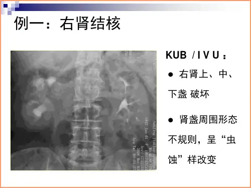
Incidence of urinary tract tuberculosis is on the rise in recent years, of which the most renal tuberculosis, that that's the simple we look at what's the renal tuberculosis's performance in the image.大多数无钙化的肾结核无异常X线表现,少数可见肾脏轮廓增大或缩小。
初期的肾结核病变,有时可出现肾实质内小斑点、片状钙化;后期可互相融合呈云絮状、环状钙化;终期肾脏可完全钙化,此时肾功能已经完全失代偿,称为肾自截;有时可见输尿管钙化及对侧肾区钙化灶。
Most abnormal X-ray findings without calcification of renal tuberculosis, a few visible kidney outline increased or reduced. The early renal tuberculosis, may sometimes small spots, patchy calcification in the renal parenchyma; Late to merging YunXuZhuang, annular calcification; End stage renal completely calcification, kidney function has been completely decompensation, called renal since section; Sometimes visible ureteral calcification and the contralateral renal calcifications.↑ 右肾结核(肾自截)腹部X线平片,可见右侧肾实质区域完全钙化,其密度不均,形态不整;右侧输尿管中段亦可见条形钙化影,左侧肾脏区域可见结节状钙化影Abdominal X-ray plain film, visible on the right side of renal parenchyma area completely calcification, its density, form whole; Middle ureter are visible on the right side bar calcification shadow, nodular calcification area visible within the left kidney↑左肾结核静脉肾盂X线造影,可见左侧肾的上组肾盏边缘不规则,似虫蚀样破坏Intravenous urography X-ray imaging and visible on the left side of the edge of renal calyces on group is irregular, like a worm corrosion damage↑ 右肾结核逆行性肾盂X线造影,显示右侧肾的肾盂、肾盏明显扩张,肾盏边缘不规则,肾实质内多发空洞形成Retrograde urography X-ray imaging, display the right renal pelvis and calyces obvious expansion, calyx margin irregular, renal parenchyma in multiple hole formation当结核病变位于肾实质内未与肾盏、肾盂相通时,可以表现为正常。