青光眼发病机制,治疗方式
- 格式:docx
- 大小:14.09 KB
- 文档页数:5
青光眼治疗指南疾病简介:青光眼(Glaucoma)是指眼内压力或间断或持续升高的一种眼病。
眼内压力升高可因其病因的不同而有各种不同的症状表现。
持续的高眼压可给眼球各部分组织和视功能带来损害,造成视力下降和视野缩小。
如不及时治疗,视野可全部丧失甚至失明。
故青光眼是致盲的主要病种之一。
发病原因劳累过度、睡眠不足、情绪波动、饮食不节或暴饮暴食等因素,可以影响血管神经调节中枢,使血管舒缩功能失调:一方面可使毛细血管扩张,血管通透性增加,造成睫状肌水肿、前移,堵塞前房角,使房水流出通道受阻;另一方面可使房水分泌过多,后房压力过高,周边虹膜受压向前移而使前房变浅,前房角变窄。
这些因素均可引起眼压的急剧升高,最终导致青光眼急性发作。
发病机制青光眼本身就是脏腑功能失调后引起全身病理变化过程中的一种眼部表现。
因此它不仅可由多种病变继发形成,且可加剧其它病变的演变、发展。
其中局部以外伤、屈光不正(近视、远视、散光)、晶体改变(白内障(Cataract)、老花眼、晶体脱位、人工晶体)、玻璃体改变(玻璃体混浊、液化)、视网膜病变(视网膜脱离、夜肓症(Night)、中浆、视网膜炎(Retinitis)、视网膜动静脉阻塞(Retinal vein occlusion)、出血)、眼部炎症。
全身以心脑血管(高血压、动脉硬化)、消化系统(胃溃疡(Gastric ulcer)、胃炎(Gastritis))、内分泌系统(糖尿病(Diabetes)、甲状腺疾病)、先天发育不良。
临床上因用药不当,引发青光眼并非鲜见,常见药物有:散瞳验光药物(如阿托品(Atropine)等),麻醉药物(利多卡因(Lidocaine)、普鲁卡因(Procaine)等),拟肾上腺素等药(肾上腺素(Adrenaline)、麻黄素(Ephedrine)等),扩血管药物,镇静安眠类(如安定),抗菌消炎类(磺胺)女性激素及避孕药。
疾病分类一、先天性青光眼(Congenital glaucoma):根据发病年龄又可为婴幼儿性青光眼及青少年性青光眼。
眼科晋升副主任医师专题报告(青光眼)眼科晋升副主任医师专题报告(青光眼)引言青光眼是一种常见的眼科疾病,严重影响了全球范围内的人眼健康。
本次专题报告旨在介绍青光眼的相关知识,并讨论晋升为副主任医师所需具备的要求。
青光眼概述青光眼是一种慢性进行性眼病,其特点是视野缺损和视神经损伤。
青光眼的发病机制通常与眼内压的升高以及视神经受损有关。
早期的青光眼可能没有明显的症状,但随着疾病进展,患者可能出现视力模糊、视野缩小以及眼睛疼痛等症状。
青光眼诊断和治疗青光眼的诊断通常需要综合考虑几个因素,包括眼压测量、视力检查、视野检查以及眼底检查等。
治疗方法主要包括药物治疗、激光治疗和手术治疗。
药物治疗常用于控制眼内压,以减缓疾病的进展。
激光治疗可以通过减少房水的产生或增加其排出来降低眼压。
手术治疗包括多种方法,如激光手术和过滤性手术等。
晋升副主任医师要求想要晋升为眼科副主任医师,需要具备以下要求:1. 专业知识与技术:具备扎实的眼科专业知识和熟练的临床技术,在青光眼的诊断和治疗上有较高的造诣。
2. 临床经验:积累丰富的青光眼临床经验,能够独立处理复杂病例,并能提供有效的治疗方案。
3. 学术研究能力:在眼科领域有一定的学术研究成果,如发表论文、参与学术会议等。
4. 团队合作精神:具备良好的团队合作精神,能够与其他医疗团队成员有效沟通和协作。
结论青光眼是一种常见的眼科疾病,其诊断和治疗需要眼科医生具备扎实的专业知识和技术。
想要晋升为副主任医师,除了具备丰富的临床经验和学术研究能力外,还需要具备团队合作精神。
希望本次专题报告对大家对青光眼及其晋升要求有所了解。
感谢大家的聆听。
什么是青光眼如何预防和治疗在我们的生活中,青光眼是一种较为常见但却可能导致严重后果的眼科疾病。
那么,究竟什么是青光眼?又该如何预防和治疗它呢?接下来,让我们一起深入了解一下。
青光眼,简单来说,是一组以视神经损害和视野缺损为共同特征的眼科疾病。
视神经就像一条“电缆”,负责将眼睛看到的图像信息传输到大脑。
而青光眼会损害这条“电缆”,导致信息传输出现问题,进而影响我们的视力。
青光眼的发病机制较为复杂。
眼压升高是青光眼最常见、也是最重要的危险因素。
正常情况下,我们眼睛内会不断产生和排出房水,以维持一定的眼压。
但如果房水的产生过多,或者排出通道受阻,眼压就会升高。
长期的高眼压会压迫视神经,使其逐渐受损。
此外,遗传因素、眼部结构异常、年龄增长、某些全身性疾病(如糖尿病、高血压)等也可能增加青光眼的发病风险。
青光眼的症状可能因类型和病情严重程度而有所不同。
开角型青光眼在早期通常没有明显症状,很多患者在不知不觉中视力逐渐下降,甚至到晚期出现严重的视野缺损时才察觉。
而闭角型青光眼在急性发作时,症状较为明显,可能会出现眼痛、头痛、恶心、呕吐、视力急剧下降等。
但需要注意的是,即使没有明显症状,也不意味着没有青光眼的风险。
那么,如何预防青光眼呢?首先,定期进行眼科检查是非常重要的。
建议 40 岁以上的人群,尤其是有青光眼家族史、高度近视、糖尿病、高血压等高危因素的人,每年至少进行一次全面的眼科检查,包括眼压测量、视神经检查、视野检查等。
这样可以早期发现青光眼,及时采取治疗措施,避免病情恶化。
其次,保持良好的生活习惯也有助于预防青光眼。
例如,保持规律的作息时间,避免长时间熬夜;避免过度用眼,尤其是长时间盯着电脑、手机等电子设备;保持心情舒畅,避免情绪波动过大,因为紧张、焦虑等不良情绪可能会导致眼压升高。
在饮食方面,要注意均衡饮食,多吃富含维生素 A、C、E 和抗氧化剂的食物,如胡萝卜、西红柿、蓝莓等,有助于保护眼睛。
同时,要控制饮酒和吸烟,因为酒精和尼古丁可能会影响眼部的血液循环,增加青光眼的发病风险。
青光眼小结青光眼是一种常见的眼病,也是导致全球盲目的重要原因之一。
青光眼的特点是眼球内压力增高,导致视神经损伤,进而引起视力丧失。
对于患者而言,及早发现和治疗青光眼至关重要。
在本文中,我将对青光眼的病因、诊断和治疗进行简要的总结。
青光眼的发病机制多种多样,目前还没有找到确切的原因。
但是,高眼压被认为是青光眼的主要危险因素之一。
眼球内的房水在产生和排出之间出现平衡失调,导致眼压升高。
长期高眼压会损害视神经,从而导致视力丧失。
此外,家族史、高度近视、年龄、种族和其他慢性眼部疾病也可能增加青光眼的风险。
青光眼的诊断主要包括眼压检查、视野检查和视神经检查。
眼压检查是最常用的诊断方法,通常使用非接触式眼压计或Applanation眼压计来进行检测。
正常眼压范围为10-20mmHg,高于此范围可能提示青光眼。
视野检查用于评估患者的视野损害程度,青光眼患者常出现边缘模糊和覆盖物感。
视神经检查可以通过扩瞳检查观察视神经盘的损伤情况,如视神经杯盘比值的改变和视神经纤维层缺损等。
青光眼的治疗主要包括药物治疗、激光治疗和手术治疗。
药物治疗是最常用的治疗方法,常用药物包括Beta受体阻滞剂、前列腺素类似物和碳酸酐酶抑制剂等。
这些药物通过减少房水产生或促进房水排出来降低眼压。
激光治疗主要通过激光切除小区域的组织,改善房水的排出,从而降低眼压。
手术治疗主要包括过滤性手术和植入性手术,通过创造人工排液通道或植入装置来降低眼压。
除了治疗,预防也是非常重要的。
定期眼科检查可以帮助早期发现青光眼,从而及早治疗,避免或延缓视力丧失。
避免眼部受伤,避免长时间用眼和控制体内疾病也可以减少青光眼的发生。
总之,青光眼是一种严重的眼病,对患者的视力影响很大。
早期发现和治疗青光眼非常重要。
通过眼压检查、视野检查和视神经检查可以确诊青光眼。
药物治疗、激光治疗和手术治疗是常用的治疗方法。
此外,预防也很重要,定期眼科检查和良好的生活习惯可以减少青光眼的发生。
青光眼的治疗方法有哪些青光眼能治好吗*导读:青光眼能好吗?青光眼如何治疗更有效?降眼压的治疗方式比较多,但是基本上都是使用青光眼药物,这里的药物主要就是我们通常用的眼药水。
……青光眼能好吗?警惕致盲发生!对于先天性青光眼的患者应该注意,这种情况是由于眼部组织发育异常引发的,一般婴儿的眼球会受到巨大压力影响,从而导致眼球不断增大,需要控制眼压!青光眼能好吗如果是孩子出生后被诊断为青光眼的话,属于先天性青光眼,需要选择最佳的手术时间为孩子进行减压手术治疗,以此来维持患者的视觉功能,如果眼部眼压持续升高的话,会对周围的视功能造成严重的损伤!如果患者属于急性发作的青光眼的话,不仅仅会出现局部的病症,还有可能会引发全身性的病症反应,例如恶心、呕吐、发烧等,有些患者还会出现虹视等情况,最终可能会导致患者失明。
青光眼的治疗方法降眼压的治疗方式比较多,但是基本上都是使用青光眼药物,这里的药物主要就是我们通常用的眼药水。
不过也有些药物是口服的、点滴注射的,不过后面这两种药物都只能在短期内使用,不适合长时间的使用。
目前的医学水平还无法完全治愈青光眼,只能是起到抑制作用。
使用药物的目的也只是降眼压。
药物治疗主要还是使用眼药水,比较适合长期实用。
降眼压还可以通过激光进行治疗,不过不同的青光眼类型会有不同的激光治疗方法,一些发展到晚期的青光眼同样都可以通过激光进行治疗。
另外也可以通过手术来进行治疗。
手术方法比较多。
有些手术手术是疏导眼睛里面的房水液体,使它灵活流动。
还有一种方法是引导眼睛内的水流出一部分。
针对晚期的青光眼一般是不能让水就出来的。
青光眼发展到的阶段不同,病情严重程度不同,采用的治疗方式都有所不同。
青光眼也可以使用的眼药水为1%葛根素滴眼液,患者可以每日使用2次,坚持使用眼药水。
这种眼药水主要功效是使收缩睫状突血管,缓解房水分泌而使得眼压变低。
患者还要注意的是,有一部分患者的血压出现稍微的降低,所以低血压病人要谨慎的使用。
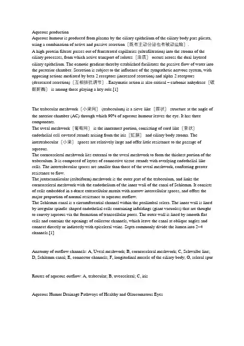
Aqueous productionAqueous humour is produced from plasma by the ciliary epithelium of the ciliary body pars plicata, using a combination of active and passive secretion〔既有主动分泌也有被动运输〕.A high-protein filtrate passes out of fenestrated capillaries (ultrafiltration) into the stroma of the ciliary processes, from which active transport of solutes〔溶质〕 occurs across the dual-layered ciliary epithelium. The osmotic gradient thereby established facilitates the passive flow of water into the posterior chamber. Secretion is subject to the influence of the sympathetic nervous system, with opposing actions mediated by beta-2 receptors (increased secretion) and alpha-2 receptors (decreased secretion)〔互相拮抗调节〕. Enzymatic action is also critical – carbonic anhydrase〔碳酸酐酶〕 is among those playing a key role.[1]The trabecular meshwork〔小梁网〕 (trabeculum) is a sieve-like〔筛状〕 structure at the angle of the anterior chamber (AC) through which 90% of aqueous humour leaves the eye. It has three components.The uveal meshwork〔葡萄网〕 is the innermost portion, consisting of cord-like〔索状〕endothelial cell-covered strands arising from the iris〔虹膜〕 and ciliary body stroma. The intertrabecular〔小梁〕 spaces are relatively large and offer little resistance to the passage of aqueous.The corneoscleral meshwork lies external to the uveal meshwork to form the thickest portion of the trabeculum. It is composed of layers of connective tissue strands with overlying endothelial-like cells. The intertrabecular spaces are smaller than those of the uveal meshwork, conferring greater resistance to flow.The juxtacanalicular (cribriform) meshwork is the outer part of the trabeculum, and links the corneoscleral meshwork with the endothelium of the inner wall of the canal of Schlemm. It consists of cells embedded in a dense extracellular matrix with narrow intercellular spaces, and offers the major proportion of normal resistance to aqueous outflow.The Schlemm canal is a circumferential channel within the perilimbal sclera. The inner wall is lined by irregular spindle-shaped endothelial cells containing infoldings (giant vacuoles) that are thought to convey aqueous via the formation of transcellular pores. The outer wall is lined by smooth flat cells and contains the openings of collector channels, which leave the canal at oblique angles and connect directly or indirectly with episcleral veins. Septa commonly divide the lumen into 2–4 channels.[1]Anatomy of outflow channels: A, Uveal meshwork; B, corneoscleral meshwork; C, Schwalbe line; D, Schlemm canal; E, connector channels; F, longitudinal muscle of the ciliary body; G, scleral spur Routes of aqueous outflow: A, trabecular; B, uveoscleral; C, irisAqueous Humor Drainage Pathways of Healthy and Glaucomatous EyesSimple intraocular pressure:High intraocular pressure has direct damage to the optic nerve. [2]High intraocular pressure directly oppresses optic nerve fibers, blocks axoplasmic transport, thereby damaging retinal ganglion cells. Increased intraocular pressure (IOP) causes stretching of the laminar beams and damage to retinal ganglion cell axons..[3, 4]Trans-lamina cribrosa pressure difference[5]Mitochondrial DNA mechanismAbnormally elevated intraocular pressure can directly lead to mtDNA damage and mutation, leading to mitochondrial dysfunction, thereby mtDNA further damages and mutates, forming a vicious circle, causing RGC progressive apoptosis.The vascular theory of glaucoma considers GON as a consequence of insufficient blood supply due to either increased IOP or other risk factors reducing ocular blood flow (OBF).Vascular dysregulation, rather than an atherosclerosis, leads to both low perfusion pressure and insufficient autoregulation. This in turn may lead to unstable ocular perfusion and thereby to ischemia and reperfusion damage.[6]High concentrations of glutamate causing RGC excitotoxic damage is the main cause of death in glaucoma patients.[7]Increased oxidative stress and increased ROS production play an important role in the development of glaucoma.[8]In recent years, it has been found that the immune system plays an important role in the development of GON.药物治疗的局限性:[9]1.很多患者仅用药物治疗不能降至理想的目标眼压。
急性青光眼怎么治患病有哪些要注意的急性青光眼通常发病是比较急的,一般在感染了两三天内都会发病的,在刚刚发病的时候,很多人都以为只是普通的一些眼部不适的症状,并没有马上发觉自己有可双月是患有青光眼的。
下面为大家介绍一下急性青光眼的治疗方式有哪些?急性青光眼的治疗方式有:第一、治疗的原则,主要是以手术为主,明确是急性青光眼以后要及早的进行手术治疗,并且要先用药物对病情进行控制,使房角达到开放的状态,一定要使患者的眼压达到下降的时候,炎症出现消退的情况以后才可以对患者进行手术治疗。
第二、常规治疗。
如果患者的病期是前驱期和间歇期,应该进行虹膜的切除手术,通过这种手术可以起到很好的根治效果,因为通过这种手术治疗可以使患者打破瞳孔的阻碍,房水可以通过这种切除六道前房,使得患者的前后房压得到很好的平衡状态,不至于发生关闭的情况。
第三、急性发作的青光眼应该积极的抢救,尽快的使患者的房角得到开放,避免患者的房角出现粘连的情况,如果患者的眼压非常高的时候会导致很多的并发症的出现,这样治疗的效果是比较差的,应该控制好眼压,达到正常的水平以后在手术治疗。
第四、手术治疗。
因为急性的青光眼患者发病是比较急并且是比较严重,通过药物和平常的治疗方法可以效果比较差,这个时候就必需要进行手术治疗,并且要做好手术治疗的前期准备。
急性青光眼患者要注意事项有:想要更好更快的治疗好青光眼的话,在平时的生活当中,青光眼患者还需要认真的去做好相关的预防工作,比方说养成良好的用眼习惯。
时间充裕的话,青光眼患者应该多参加一些户外云运动,提高自己预防疾病的能力。
现在对于急性青光眼治疗方法,主要就是通过减低眼睛里面的一些压力,或者是把眼睛里面的一些水引流出来,还有就是可以缩小瞳孔,或者是抑制抑制房水产生。
青光眼的主要诱发因素就是长期不良精神刺激,脾气暴躁、抑郁、忧虑。
所以保持心情舒畅,避免情绪过度波动对预防青光眼至关重要。
急性青光眼在刚刚开始的时候治疗是效果会比较好的,而且这也是对于眼睛的损伤会相对较小的,患者在治疗期间也需要注意生活中的各种事项的。
青光眼的治疗方法青光眼是一种常见的眼部疾病,如果不及时治疗,可能会导致视力下降甚至失明。
因此,了解青光眼的治疗方法对于患者来说至关重要。
接下来,我们将介绍一些常见的青光眼治疗方法,希望能给患者们带来帮助。
首先,药物治疗是治疗青光眼最常见的方法之一。
目前,市面上有许多种类的眼药水和口服药物可以用于治疗青光眼。
这些药物可以通过降低眼压的方式来减缓病情的发展。
常见的药物包括β受体阻滞剂、前列腺素类药物、碳酸酐抑制剂等。
患者在使用药物治疗时,应按照医生的建议进行规范使用,定期复查眼压,以确保疗效。
其次,手术治疗是一些青光眼患者在药物治疗无效或不适用的情况下的选择。
常见的手术包括激光治疗和手术手段。
激光治疗主要包括激光周围虹膜切除术、激光三角切除术等,通过激光作用于眼部组织,减少房水的产生或增加房水的排出,从而降低眼压。
手术治疗主要包括瞬时小梁切除术、非穿孔小梁切除术等,这些手术能够通过改变房水的排出通道来降低眼压。
患者在进行手术治疗前,应进行全面的眼部检查,以评估手术的适用性和风险。
此外,患者在日常生活中也可以通过一些辅助治疗方法来帮助控制青光眼。
例如,定期进行眼部按摩可以促进眼部血液循环,减轻眼部疲劳;保持良好的生活习惯,如充足的睡眠、合理的饮食等,有助于维持眼部健康;避免剧烈运动和提重物,以减少眼压的升高。
总之,青光眼的治疗方法多种多样,患者可以根据自身的情况选择合适的治疗方式。
在接受治疗的过程中,患者应密切配合医生的治疗方案,定期复查眼部情况,及时调整治疗方案。
希望通过治疗,患者能够控制病情,减轻症状,保持良好的视力。
最终,希望所有患者都能够早日康复,重获健康的双眼。
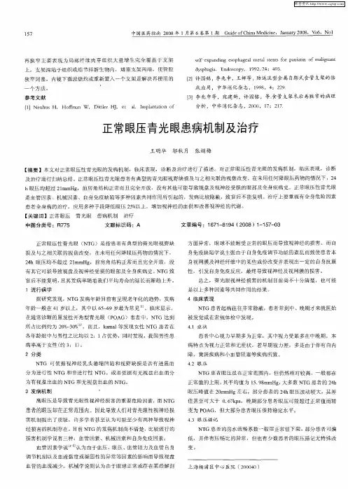
青光眼的发病机制及早期症状的再认识作者:石继来来源:《中国保健营养·中旬刊》2013年第11期【中图分类号】R775 【文献标识码】A 【文章编号】1004—7484(2013)11—0111—02青光眼是一种常见的严重危害人们健康、影响人们生产和生活的致盲眼病,可发生于各种年龄的人。
青光眼是一种发病迅速、危害性大、随时导致失明的常见疑难眼病。
特征就是眼内压间断或持续性升高的水平超过眼球所能耐受的程度而给眼球各部分组织和视功能带来损害,导致视神经萎缩、视野缩小、视力减退,最终导致失明,在急性发作期24-48小时即可完全失明。
青光眼属双眼性病变,可双眼同时发病,或一眼起病,继发双眼失明,一旦患上青光眼,就必须按双眼病变对待,不能盲目的认为单一眼患青光眼失明,还有另一只眼睛代偿。
因此了解青光眼的发病特点,及对其早期症状早发现和及时治疗是非常必要的。
临床上很多病人就诊时出现眼压升高,就其因劳累过度、睡眠不足、情绪波动、饮食不节或暴饮暴食等因素而影响血管神经调节中枢,使血管舒缩功能失调:一方面可使毛细血管扩张,血管通透性增加,造成睫状肌水肿、前移,堵塞前房角,使房水流出通道受阻;另一方面可使房水分泌过多,后房压力过高,周边虹膜受压向前移而使前房变浅,前房角变窄。
这些因素均可引起眼压的急剧升高,最终导致青光眼急性发作。
1 青光眼的发病机制青光眼本身就是脏腑功能失调后引起全身病理变化过程中的一种眼部表现。
因此它不仅可由多种病变继发形成,且可加剧其它病变的演变、发展。
其中局部以外伤、屈光不正(近视、远视、散光)、晶体改变(白内障、老花、晶体脱位、人工晶体)、玻璃体改变(玻璃体混浊、液化)、视网膜病变(视网膜脱离、夜肓、中浆、视网膜炎、视网膜动静脉阻塞、出血)、眼部炎症。
全身以心脑血管(高血压、动脉硬化)、消化系统(胃溃疡、胃炎)、内分泌系统(糖尿病、甲状腺疾病)、先天发育不良。
临床上因用药不当,引发青光眼并非鲜见,常见药物有:散瞳验光药物(如阿托品等),麻醉药物(利多卡、普鲁卡因等),拟肾上腺素等药(肾上腺素、麻黄素等),扩血管药物,镇静安眠类(如安定),抗菌消炎类(磺胺)女性激素及避孕药。
中医治疗青光眼最佳治疗方法
青光眼是一种常见的眼部疾病,如果不及时治疗,会给患者的视力和生活带来很大的影响。
中医作为我国传统的治疗方法,对于青光眼的治疗也有着独特的优势。
下面我们就来详细了解一下中医治疗青光眼的最佳方法。
首先,中医治疗青光眼的核心是调理气血。
中医认为,青光眼的发生与气血运行不畅有着密切的关系。
因此,通过中药调理和针灸疗法,可以有效地调理患者的气血,改善眼部的血液循环,从而达到治疗青光眼的目的。
其次,中医治疗青光眼还注重于调整体内的阴阳平衡。
在中医理论中,阴阳平衡是维持人体健康的关键。
通过中药调理和穴位按摩,可以帮助患者调整体内的阴阳平衡,从而减缓青光眼的发展,甚至逆转病情。
此外,中医还强调了对患者的情志调摄。
情志因素是导致青光眼发作的重要原因之一。
通过中医的心理疏导和情志调摄,可以帮助患者保持心情舒畅,减轻病情的发作频率,对青光眼的治疗有着积极的作用。
除了中医内部的治疗方法外,中医还强调了对患者生活习惯的调整。
比如,要避免长时间盯着电脑和手机屏幕,要保持充足的睡眠,要避免熬夜等不良习惯。
这些生活习惯的调整,对于青光眼的治疗和预防同样至关重要。
总的来说,中医治疗青光眼的最佳方法是综合运用中药调理、针灸疗法、情志调摄和生活习惯的调整。
通过综合治疗,可以达到治疗青光眼的最佳效果,帮助患者减轻病情,改善视力,提高生活质量。
希望通过本文的介绍,能够让更多的患者了解到中医治疗青光眼的优势和方法,选择适合自己的治疗方式,早日康复。
新生血管性青光眼的病因治疗与预防尽管一些早期临床报告指出,新生血管青光眼(NVG)患眼虹膜直到20世纪才有新血管。
NVG知识是基于完美的解剖学。
一、病因新生血管性青光眼的病因有40多种不同的疾病,几乎都广泛涉及眼后节缺氧或局部眼前节缺氧,主要包括视网膜中央静脉阻塞、糖尿病视网膜病变等疾病,约占1/3。
视网膜中央静脉阻塞取决于视网膜缺血分缺血型(占255%(75)和非缺血型(%)自然病程中无一例非缺血型发展为新生血管性青光眼,而缺血型中有18种%~60%多发生在静脉阻塞后2~3个月,80%病例发生在6个月内。
视网膜毛细血管的非灌注区主要通过眼底荧光血管造影来判断缺血。
注意非缺血型也可以转化为缺血型。
糖尿病是一个危险因素,糖尿病也是视网膜中央静脉阻塞的危险致病因素。
原发性开角青光眼与视网膜中央静脉阻塞有关,认为是机械压力引起的。
因此,视网膜中央静脉阻塞被视为原发性开角青光眼的危险因素。
此外,80%静脉阻塞患者的眼压低于对侧眼,认为这是代谢性酸中毒抑制房水形成的原因。
增殖性糖尿病性视网膜病变约22%新生血管性青光眼,糖尿病1型占15%多伴增殖性视网膜病变,2型占80%并伴有黄斑病变。
成人双眼新血管青光眼或虹膜新血管化几乎是由糖尿病视网膜病变引起的,但视网膜病变与虹膜新血管或青光眼的时间间隔不清楚。
白内障手术和玻璃体视网膜手术后更容易发生新的血管青光眼,主要与原糖尿病视网膜病变和视网膜缺氧有关。
伴有新生血管性青光眼的其他常见眼病有:视网膜中央动脉阻塞(1)%~17%),恶性黑色素瘤(0)等眼部肿瘤。
%~15%)视网膜母细胞瘤虹膜新生血管化可达30%~72%,玻璃体视网膜手术后虹膜再生血管化也达到23%~32%。
此外,它还出现在眼内血管疾病中Coats疾病、静脉周围炎、镰状血细胞变、虹膜异色症、剥脱综合征、巩膜炎、眼内炎、交感性眼炎、视神经纤维瘤病、原发性虹膜萎缩、网状组织细胞肉瘤、转移性癌、眼外伤Sturge-Weber综合征合并脉络膜血管瘤,甚至白内障切除术后。
青光眼的治疗方法有哪些
青光眼的治疗方法主要包括以下几种:
1. 药物治疗:使用眼压调节药物,如β受体阻滞剂、卡着通等,来减轻眼压和预防视神经损伤。
2. 激光治疗:利用激光减小眼房角的阻力,降低眼压,如激光三周治疗、玻璃体切割术。
3. 手术治疗:包括三种主要手术:过滤手术、引流手术和角膜瓣置换手术。
4. 中草药治疗:中草药治疗青光眼主要通过凉血降压、益肝明目、通络散寒、滋阴润燥等方式对症治疗。
5. 其他治疗方式:如针灸、磁疗、穴位按摩、氧疗等,可以起到辅助治疗的作用。
青光眼的成因是什么引言青光眼是一种慢性眼病,其特点是视神经损伤和视野缺损。
青光眼发病率逐渐增加,对患者的视力和生活质量造成了巨大的影响。
了解青光眼的成因对于预防和治疗本病至关重要。
本文将探讨青光眼的成因,并介绍相关的风险因素、遗传因素以及其他可导致青光眼发生的因素。
风险因素青光眼的发生与多种风险因素相关。
以下是一些常见的青光眼风险因素:1.高眼压:高眼压是青光眼最主要的危险因素。
眼压升高会给视神经带来压力,最终导致视神经损伤和视野缺损。
高眼压可能是由于眼房水循环紊乱引起的。
2.年龄:随着年龄的增长,患青光眼的风险也会增加。
中老年人更容易患上此病。
3.家族史:青光眼具有明显的遗传倾向。
如果家族中有亲人患有青光眼,那么罹患本病的风险将大幅提高。
4.种族:非洲人、亚洲人和拉美人患青光眼的风险较高。
5.眼球解剖结构异常:具有异常眼球结构,如眼角过尖或眼角开放角度狭窄等,容易导致房角堵塞,引起青光眼。
6.高度近视:近视度数较高的人患青光眼的风险较高。
7.眼外伤:曾经遭受过眼外伤的人患青光眼的风险也会增加。
遗传因素青光眼的发生与遗传因素有着密切的关联。
一些青光眼患者具有特定的基因变异,这些基因变异可能会导致眼房水循环紊乱,进而引发眼压升高和视神经损伤。
已经鉴定出一些与青光眼有关的遗传基因,例如: - CYP1B1基因:该基因突变常见于小角青光眼患者中,是一种罕见的遗传性青光眼类型。
- MYOC基因:该基因在开角青光眼患者中突变率较高,突变后可能会导致房角堵塞。
遗传因素对于青光眼的发病机制和病程的了解仍在不断深入研究中。
随着研究的进展,我们可以更好地预测和干预青光眼的发生。
其他因素除了高眼压和遗传因素外,青光眼的发生还与其他一些因素有关:1.使用类固醇药物:长期使用类固醇药物可能增加患青光眼的风险。
类固醇可导致眼房水排出减少,从而引发眼压升高。
2.糖尿病:糖尿病患者更容易患青光眼。
糖尿病引发的眼部病变可能导致房角堵塞和眼压升高。
Aqueous productionAqueous humour is produced from plasma by the ciliary epithelium of the ciliary body pars plicata, using a combination of active and passive secretion(既有主动分泌也有被动运输).A high-protein filtrate passes out of fenestrated capillaries (ultrafiltration) into the stroma of the ciliary processes, from which active transport of solutes(溶质) occurs across the dual-layered ciliary epithelium. The osmotic gradient thereby established facilitates the passive flow of water into the posterior chamber. Secretion is subject to the influence of the sympathetic nervous system, with opposing actions mediated by beta-2 receptors (increased secretion) and alpha-2 receptors (decreased secretion)(互相拮抗调节). Enzymatic action is also critical – carbonic anhydrase(碳酸酐酶) is among those playing a key role.[1]The trabecular meshwork(小梁网) (trabeculum) is a sieve-like(筛状) structure at the angle of the anterior chamber (AC) through which 90% of aqueous humour leaves the eye. It has three components.The uveal meshwork(葡萄网) is the innermost portion, consisting of cord-like(索状)endothelial cell-covered strands arising from the iris(虹膜) and ciliary body stroma. The intertrabecular(小梁) spaces are relatively large and offer little resistance to the passage of aqueous.The corneoscleral meshwork lies external to the uveal meshwork to form the thickest portion of the trabeculum. It is composed of layers of connective tissue strands with overlying endothelial-like cells. The intertrabecular spaces are smaller than those of the uveal meshwork, conferring greater resistance to flow.The juxtacanalicular (cribriform) meshwork is the outer part of the trabeculum, and links the corneoscleral meshwork with the endothelium of the inner wall of the canal of Schlemm. It consists of cells embedded in a dense extracellular matrix with narrow intercellular spaces, and offers the major proportion of normal resistance to aqueous outflow.The Schlemm canal is a circumferential channel within the perilimbal sclera. The inner wall is lined by irregular spindle-shaped endothelial cells containing infoldings (giant vacuoles) that are thought to convey aqueous via the formation of transcellular pores. The outer wall is lined by smooth flat cells and contains the openings of collector channels, which leave the canal at oblique angles and connect directly or indirectly with episcleral veins. Septa commonly divide the lumen into 2–4 channels.[1]Anatomy of outflow channels: A, Uveal meshwork; B, corneoscleral meshwork; C, Schwalbe line; D, Schlemm canal; E, connector channels; F, longitudinal muscle of the ciliary body; G, scleral spur Routes of aqueous outflow: A, trabecular; B, uveoscleral; C, irisAqueous Humor Drainage Pathways of Healthy and Glaucomatous EyesSimple intraocular pressure:High intraocular pressure has direct damage to the optic nerve. [2]High intraocular pressure directly oppresses optic nerve fibers, blocks axoplasmic transport, thereby damaging retinal ganglion cells. Increased intraocular pressure (IOP) causes stretching of the laminar beams and damage to retinal ganglion cell axons..[3, 4]Trans-lamina cribrosa pressure difference[5]Mitochondrial DNA mechanismAbnormally elevated intraocular pressure can directly lead to mtDNA damage and mutation, leading to mitochondrial dysfunction, thereby mtDNA further damages and mutates, forming a vicious circle, causing RGC progressive apoptosis.The vascular theory of glaucoma considers GON as a consequence of insufficient blood supply due to either increased IOP or other risk factors reducing ocular blood flow (OBF).Vascular dysregulation, rather than an atherosclerosis, leads to both low perfusion pressure and insufficient autoregulation. This in turn may lead to unstable ocular perfusion and thereby to ischemia and reperfusion damage.[6]High concentrations of glutamate causing RGC excitotoxic damage is the main cause of death in glaucoma patients.[7]Increased oxidative stress and increased ROS production play an important role in the development of glaucoma.[8]In recent years, it has been found that the immune system plays an important role in the development of GON.药物治疗的局限性:[9]1.很多患者仅用药物治疗不能降至理想的目标眼压。
2.药物副作用3.昂贵的药费4.长时间用药可能会影响今后的手术效果5.随着用药时间的延长,降眼压作用减弱6.可能与全身药物产生交叉反应7.药物产生的副作用干扰患者生活8.患者长期用药的依从性差Table 1 Summary of drugs used to treat glaucoma[10]LASER TREATMENT OF GLAUCOMALaser trabeculoplastySelective laser trabeculoplasty (SLT)Argon laser trabeculoplasty (ALT)Micropulse laser trabeculoplasty (MLT)Laser iridotomyDiode laser cycloablationLaser iridoplastyALT is effective as medical therapy in lowering IOP.The most common adverse eventwas a transient increase in IOP. The incidence of this event was 12% for an increase in IOP of .10 mm Hg and 34% for an increase in IOPof.5 mmHg. Other adverse events included a low-grade iritis.Complications• Bleeding occurs in around 50% but is usually mild and stops after only a few seconds; persiste nt bleeding can be terminated by increasing contact lens pressure.• IOP elevation. Usually early and transient but occasionally persistent.• Iritis. Especially if excessive laser is applied or post-laser steroid therapy is inadequate, or in darker irides (including those due to prostaglandin derivative treatment).• Corneal burns may occur if a contact lens is not used or if the AC is shallo w; these usually heal very rapidly without sequelae.• Cataract. Localized lens opacities occasionally develop at the treatment site; age-related cataract formation may be accelerated by iridotomy.TRABECULECTOMYNON-PENETRATING GLAUCOMA SURGERYDRAINAGE SHUNTS青光眼引流器的应用能减少结膜下和滤过道的瘢痕形成,从而大大提高手术的成功率。