
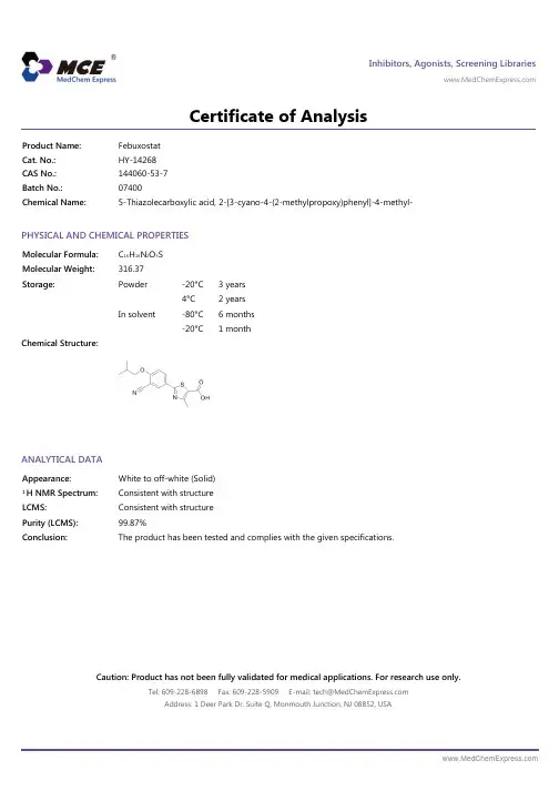
Febuxostat_COA_07400_MedChemExpress
- 格式:pdf
- 大小:91.63 KB
- 文档页数:1

ReviewStudy on the pharmacological activities and chemicalstructures of Viburnum dilatatumZhiheng Gao, Yufei Xi, Man Wang, Xiaoxiao Huang*, Shaojiang Song*Key Laboratory of Computational Chemistry-Based Natural Antitumor Drug Research &Development, Liaoning Province, School of Traditional Chinese Materia Medica, ShenyangPharmaceutical University, Shenyang 110016, ChinaAbstractViburnum dilatatum (jiami in Chinese), belonging to the Caprifollaceae family, is widely distributed in Japan and China. Phytochemical investigations of Viburnum dilatatum (V. dilatatum) have resulted in the isolation of triterpenoids, phenolic glycosides essential oil, norisoprenoids, etc. Research results have shown that the chemical constituents of V. dilatatum possess various pharmacological activities, including antihyperglycemic, antioxidant activity and antiulcer effects. This study reviewed the chemical constituents and pharmacological activities of V. dilatatum to provide practical and useful information for further research and development of this plant.Keywords: Viburnum dilatatum; pharmacological activity; chemical structures1 IntroductionViburnum dilatatum (called jiami in Chinese, gamazumi in Japanese and snowball tree in English), beloinging to family Caprifoliaceae, is a deciduous low tree distributed widely in the hills of northern China and Japan [1]. There are many types of chemical constituents in Viburnum dilatatum (V. dilatatum), including triterpenoids, * Author to whom correspondence should be addressed. Address:School of Traditional Chinese Materia Medica, Shenyang Pharmaceutical University, 103 Wenhua Rd., Shenyang 110016, China; Tel.: +86-24-43520793 (Xiaoxiao Huang); +86-24-43520707 (ShaojiangSong);E-mail:*******************(XiaoxiaoHuang); ****************(ShaojiangSong).Received: 2021-04-16 Accepted: 2022-08-28phenolic glycosides and norisoprenoids [2-4]. The leaves have been utilized as a traditional Chinese medicine, and phenolic compounds have been reported as the main active chemical component of the leaves. Many researchers have analyzed the functions of these medicinal components and found that these components have good antioxidant antihyperglycemic and antiulcer effects. For example, the gamazumi crude extract obtained from the squeezed juice of the fruit prevented oxidative injury in rats [5]. This review described the chemical structures and pharmacological activities of V. dilatatum, so as to help readers understand comprehensively the research progress of V. dilatatum and provide help for the development of V. dilatatum.2 Chemical constituents and structuresPrevious reports have indicated that the main chemical constituents of V. dilatatum are phenolic glycosides and triterpenoids.2.1 Phenolic glycosidesThirteen phenolic glycosides were isolated and identified from V. dilatatum by extensive spectroscopic methods, namely p -hydroxyphenyl-6-O -trans-caffeoyl-β-D -glucoside (1) [6], p -hydroxyphenyl-6-O -trans-caffeoyl-β-D -alloside (2) [6], 4-allyl-2-methoxyphenyl-6-O -β-D -apiosyl(1→6)-β-D -glucoside (3) [6], 1-(4’-hydroxy-3’-methoxypheny1)-2-[2’’-hydroxy-4’’-(3’’’-hydroxypropyl)]-1,3-propanediol-l-O -β-D -glucopyranoside (erythro isomer) (4-7) [7], neochlorogenic acid methyl ester (8-9) [7], cryptochlorogenic acid methyl ester (10-11) [7], cyanidin-3-sambubioside (Cy-3-sam) (12) [8], cyanidin-3-glucoside (Cy-3-glc) (13) [8], 5-O -caffeoyl-4-methoxyl quinic acid (4-MeO-5-CQA) (14) [8], chlorogenic acid (5-CQA) (15) [8], quercetin (16) [8], 2-(glucopyranosyloxy)-benzyl-3-(glucopyranosyloxy)-benzoate (17) [9] and jiamizioside E (18) [10]. These structures are shown in Fig. 1.Fig. 1 Phenolic glycosides isolated from V . dilatatumContinued fig. 12.2 TriterpenoidsThere were about seventeen triterpenoids isolated and characterized from V. dilatatum , such as viburnols A (19) [11], viburnols B (20) [11], viburnols C (21) [11], viburnols D (22) [11], viburnols E (23) [11], viburnols F (24) [12], viburnols G (25) [12], viburnols H (26) [12], viburnols I (27) [12], viburnols J (28) [12],viburnols K (29) [12], viburnudienone B 2methyl ester (30) [13], viburnenone H 2 (31) [13],v i b u r n e n o n e B 2 m e t h y l e s t e r (32) [13], viburnudienone B 1 methyl ester (33) [13], viburnenone H 1 (34) [13], and viburnenone B 2 methyl ester (35) [13]. The structures are shown in Fig. 2.Continued fig. 23 Pharmacological activities3.1 Antioxidant activityOxidative stress caused by free radicals and their derivatives leads to disturbances in redox homeostasis. Reactive oxygen species (ROS) are not only endogenously produced during intracellular metabolic processes but also generated by exogenous stimuli such as UV radiation, pollutants, smoke and drugs. The cell triggers its defense systems or undergoes apoptosis when intracellular oxidative status increases. It influences numerous cellular processes including core signaling pathways, which are associated with development of systematic and chronic disorders, such as aging and cancer. Therefore, it is critical to remove cellular oxidants and restore redox balance.solution of V. dilatatum (GSS) had strong antioxidant activity in vivo and prevent stress-induced oxidative damage by the XYZ-dish method and the澳electron spin resonance (ESR) method [14]. The experimental result showed that the concentrations of lipid peroxide in plasma, liver and stomach in the GSS group were reduced. Furthermore, the activities of plasma lactic dehydrogenase, amylase and creatine phosphokinase are ordinarily increased by stress. However, these activities in the GSS group decreased to that in the control group. It was concluded that gastric ulcer formation, increase of lipid peroxidation in plasma and tissues and elevation of plasma enzymatic activities were confirmed in rats with water immersion restraint stress. It was also found that intake of GSS could protect the stomach and other tissues from oxidative damage.Kim et al. identified and isolated two major anthocyanins by NMR and LC-ESI-MS/MS, namely, cyanidin 3-sambubioside (I) and kuromanin (II) [15]. By the electron spin resonance method, the superoxide anion radical scavenging activities of I and II were evaluated with the IC 50 values of 17.3 and 69.6 µM, and their activities on hydroxyl radicals were evaluated with the IC 50 values of 4.3 and 53.2 mM. As the positive control, the IC 50 values of ascorbic acid were 74.2 µM on superoxide anion radicals and 3.0 mM on hydroxyl radicals, respectively. The above results suggested that these anthocyanins with radical scavenging properties might be the key compounds contributing to the antioxidant activity and physiological effects of V . dilatatum fruits.Woo et al. determined the free radical scavenging capacity of VD (the leaves of V. dilatatum ) [16]. Anti-oxidant activity of the extracts was assessed by the ability to scavenge 2,2-diphenyl-1-picrylhydrazyl (DPPH) or 3-ethylbenzothiazoline-6-sulfonic acid (ABTS) radicals. Butylated hydroxytoluene (BHT), a synthetic antioxidant, or α-tocopherol, was used as the positive control in these assays. The experimental result showed that VD inducedincrease in radical scavenging activity. In addition, lipid peroxidation inhibitory activity was determined via measurement of MDA (Malondialdehyde) levels using mouse liver tissue homogenate treated with various concentrations of the extracts. The concentration-dependent decrease in MDA levels observed was consistent with radical scavenging activities of the extracts. To examine whether VD extracts could protect mam-malian cells from oxidative stress, cultures of a human mammary gland-derived epithelial cell line MCF-7 were treated with each extract prior to challenging them with tBHP. The intracellular ROS (Reactive oxygen species) production was determined with the relative intensity of dichlorofluorescein fluorescence. While intracellular ROS formation was significantly promoted by tBHP treatment, the augmented ROS level was significantly reduced after the treatment with VD extracts.3.2 Antihyperglycemic effectIwai et al. used an oral glucose tolerance test on the diabetic rats [17]. They found that the elevation of plasma glucose level after oral administration of 2 g/kg glucose was suppressed by the repeated administration of the freeze-dried powder of V. dilatatum fruit juice (CEV). The α-glucosidase inhibitory activities of isolated compounds from CEV were also measured. Cyanidin 3-sambubioside and 5-caffeoyl quinic acid A showed inhibitory activity. These results suggested that V. dilatatum fruit had the antihyperglycemic effects.4 ConclusionV. dilatatum is distributed widely in the hills of northern China and Japan. Currently, the studies on V. dilatatum have been conducted at home and abroad, but few studies focus on its chemical components and pharmacological activities. Previousphytochemical investigations showed that the constituents of V. dilatatum included triterpenoids, phenolic glycosides, norisoprenoids and other compounds. This study describes thirteen phenolic glycosides and seventeen triterpenoids and their different degrees of antihyperglycemic, antioxidant activity and antiulcer effects, aiming to provide a reference for further studies on V. dilatatum and pharmaceutical development.References[1] Jeffrey B, Harborne A. Colour atlas of medicinal plantsof Japan. Phytochemistry, 1981, 20: 1467.[2] Miyazawa M, Hashidume S, Takahashi T, et al. Aromaevaluation of gamazumi (Viburnum dilatatum) by aroma extract dilution analysis and odour activity value.Phytochem Anal, 2012, 23: 208-213.[3] Kurihara T, Kikuchi M. Studies on the constituentsof flowers. IV. On the components of the flower of Viburnum dilatatum Thunb. J Health Sci, 1975, 95: 1098-1102.[4] Machida K, Kikuchi M. Norisoprenoids from Viburnumdilatatum. Phytochemistry, 1996, 41: 1333-1336. [5] Iwai K, Onodera A, Matsue H. Mechanism of preventiveaction of Viburnum dilatatum Thunb (gamazumi) crude extract on oxidative damage in rats subjected to stress. J Sci Food Agric, 2010, 83: 1593-1599.[6] Machida K, Nakano Y, Kikuchi M. Phenolic glycosidesfrom Viburnum dilatatum. Phytochemistry, 1991, 30: 2013-2014.[7] Machida K, Kikuchi M. Phenolic compounds fromViburnum dilatatum. Phytochemistry, 1992, 31: 3654-3656.[8] Kim MY, Iwai K, Matsue H. Phenolic compositions ofViburnum dilatatum Thunb. fruits and their antiradical properties. J Food Compos Anal, 2005, 18: 789-802. [9] Lu D, Yao S. Phenolic glycoside from the roots ofViburnum dilatatum. Nat Prod Commun, 2009, 4: 945-946.[10] Wu B, Zeng X, Zhang Y. New metabolite fromViburnum dilatatum. Nat Prod Commun, 2010, 5: 1097-1098.[11] Machida K, Kikuchi M. Viburnols: Novel triterpenoidswith a rearranged dammarane skeleton from Viburnum dilatatum. Tetrahedron Lett, 1996, 37: 4157-4160. [12] Machida K, Kikuchi M. Viburnols: Six noveltriterpenoids from Viburnum dilatatum. Tetrahedron Lett, 1997, 38: 571-574.[13] Machida K, Kikuchi M. Studies on the Constituents ofViburnum Species. XIX. Six New Triterpenoids from Viburnum dilatatum Thunb. Chem Pharm Bull, 1999, 47: 692-694.[14] Iwai K, Onodera A, Matsue H, et al. Antioxidant activityand inhibitory effect of Gamazumi (Viburnum dilatatum THUNB.) on oxidative damage induced by water immersion restraint stress in rats. Int J. Food Sci Nutr, 2001, 52: 443-451.[15] Kim MY, Iwai K, Onodera A, et al. Identification andAntiradical Properties of Anthocyanins in Fruits of Viburnum dilatatum Thunb. J Agric Food Chem, 2003, 51: 6173-6177.[16] Woo YJ, Lee HJ, Jeong YS, et al. Antioxidant Potentialof Selected Korean Edible Plant Extracts. Bio Med Res Int, 2017, 2017: 1-9.[17] Iwai K, Kim MY, Akio O, et al. Alpha-glucosidaseinhibitory and antihyperglycemic effects of polyphenols in the fruit of Viburnum dilatatum Thunb. J Agric Food Chem, 2006, 54: 4588-4592.。

A new iridoid derivative from the roots ofScrophularia buergerianaWU Xi-min 1†, ZHANG Liu-qiang 1†, CHEN Xiao-chong 1, FENG Li 1,XING Wang-xing 2*, LI Yi-ming 1*(1. School of Pharmacy, Shanghai University of Traditional Chinese Medicine, Shanghai 201203, China;2. School of Medicine, Hangzhou Normal University, Hangzhou 310036, China )Abstract : Phytochemical investigation of the roots of Scrophularia buergeriana Miq. (Scrophulariaceae), resulted in the isolation of a new iridoid derivative named as buergerinin (1). Its structure was elucidated as rel-(1R , 5R , 6R )-(2-oxa-bicyclo[3.3.0]oct-7-en-6, 7-diyl)dimethoxypropane based mainly on MS and 1D and 2D NMR spectroscopic analyses.Key words : Scrophularia buergeriana ; iridoid derivative; buergerinin CLC number : R284 Document code : A Article ID : 0513-4870 (2014) 07-1019-03北玄参根中的一个新环烯醚萜衍生物吴喜民1†, 张刘强1†, 陈小冲1, 冯 丽1, 邢旺兴2*, 李医明1*(1. 上海中医药大学中药学院, 上海 201203; 2. 杭州师范大学医学院, 浙江 杭州 310036)摘要: 从北玄参块根中发现了一个新的环烯醚萜衍生物, 命名为buergerinin (1)。
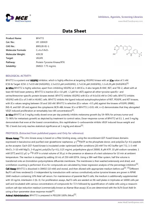
Inhibitors, Agonists, Screening Libraries Data SheetBIOLOGICAL ACTIVITY:BFH772 is a potent oral VEGFR2 inhibitor, which is highly effective at targeting VEGFR2 kinase with an IC 50 value of 3 nM.IC50 & Target: IC50: 2.7±0.9 nM (hVEGFR2), 1.5±0.53 μM (mVEGFR2), 1.7±0.36 μM (hVEGFR1), 1.1±0.29 μM (hVEGFR3)[1]In Vitro: BFH772 is highly selective; apart from inhibiting VEGFR2 at 3 nM IC 50, it also targets B–RAF, RET, and TIE–2, albeit with atleast 40–fold lower potency. BFH772 is inactive (IC 50>10 μM; >2 μM for cKIT) against all other tyrosine specific– andserine/threonine–specific protein kinases tested. BFH772 inhibits VEGFR2 with IC 50 of 4.6±0.6 nM in CHO cells. BFH772 inhibits VEGFR2 with IC 50 of 3 nM in HUVEC cells. BFH772 inhibits the ligand induced autophosphorylation of RET, PDGFR, and KIT kinases,with IC 50 values ranging between 30 and 160 nM. BFH772 is selective (IC 50 values >0.5 μM) against the kinases of EGFR, ERBB2,INS–R, and IGF–1R and against the cytoplasmic BCR–ABL kinase. IC 50 of BFH772 (<0.01 nM, n=2) demonstrates that they abrogated VEGF induced proliferation at remarkably low nM concentrations [1].In Vivo: BFH772 at 3 mg/kg orally dosed once per day potently inhibits melanoma growth (by 54–90% for primary tumor and71–96% for metastasis growth) as depicted by treatment to control ratios. Dose–response curves of BFH772 at 0.3, 1, and 3 mg/kg demonstrate that even at the lowest concentrations, this naphthalene–1–carboxamide inhibits VEGF induced tissue weight and TIE–2 levels but only reaches statistical significance at 1 mg/kg and above [1].PROTOCOL (Extracted from published papers and Only for reference)Kinase Assay:[1]In vitro kinase assay is based on a filter binding assay, using the recombinant GST–fused kinase domainsexpressed in baculovirus and purified over glutathione–sepharose, γ–[33P]ATP as the phosphate donor, and poly(Glu:Tyr 4:1) peptide as the acceptor. Each GST–fused kinase is incubated under optimized buffer conditions [20 mM Tris–HCl buffer (pH 7.5), 1–3 mM MnCl 2, 3–10 mM MgCl 2, 3–8 μg/mL poly(Glu:Tyr 4:1), 0.25 mg/mL polyethylene glycol 20000, 8 μM ATP, 10 μM sodium vanadate, 1mM DTT] and 0.2 μCi γ–33P ATP in a total volume of 30 μL in the presence or absence of a test substance for 10 min at ambient temperature. The reaction is stopped by adding 10 mL of 250 mM EDTA. Using a 384–well filter system, half the volume istransferred onto an Immobilon–polyvinylidene difluoride membrane. The membrane is then washed extensively and dried, and scintillation counting is performed. IC 50s for compounds are calculated by linear regression analysis of the percentage inhibition [1].Cell Assay: BFH772 is dissolved in DMSO (10 mM) and stored, and then diluted with appropriate medium before use [1]. [1]DifferentBa/F3 cell lines rendered IL–3 independent by transduction with various constitutively active tyrosine kinases are grown in RPMI 1640 medium containing 10% fetal calf serum. For maintenance of parental Ba/F3 cells, the medium is additionally supplemented with 10 ng/mL interleukin–3 (IL–3). For proliferation assays, Ba/F3 cells are seeded on 96–well plates in triplicates at 10000 cells per well and incubated with various concentrations of compounds for 72 h followed by quantification of viable cells using a resazurin sodium salt dye reduction readout (commercially known as Alamar Blue assay). IC 50s are determined with the XLFit Excel Add–In using a four–parameter dose response model [1].Animal Administration: BFH772 is prepared in PEG200 100% (Mice)[1].Product Name:BFH772Cat. No.:HY-100419CAS No.:890128-81-1Molecular Formula:C 23H 16F 3N 3O 3Molecular Weight:439.39Target:VEGFR Pathway:Protein Tyrosine Kinase/RTK Solubility:DMSO: 7.75 mg/mLBFH772 is dissolved in N–methyl pyrrolidone/polyethylene glycol200 (30:70, v/v) (Rat)[1].[1]Mice[1]Female FVB mice weighing between 18 and 20 g are housed in groups of six. Porous chambers containing VEGF (2 μg/mL) in 0.5 mL of 0.8% w/v agar (containing heparin, 20 U/mL) are implanted subcutaneously in the flank of the mice (n=6 per group). VEGF induces the growth of vascularized tissue around the chamber. This response is dose–dependent and can be quantified by measuring the weight and TIE–2 levels of the tissue. Mice are treated either orally once daily with compounds or vehicle (PEG200 100%, 5 mL/kg) starting4–6 h before implantation of the chambers and continuing for 4 days. The animals are sacrificed for measurement of the vascularized tissues 24 h after the last dose. Tissue weight is taken and then a lysate prepared for TIE–2 ELISA analysis .Rat[1]Catheters are implanted into the femoral artery and vein of na?ve female rats strain OFA for BFH772, and BAW2881, or in the jugular vein and femoral artery in female Sprague–Dawley rats for compounds 4, 9, and 10. Animals are allowed to recover for 96 h and are housed in single cages with free access to food and water throughout the experiment. Female OFA rats received 2.5 mg/kg ofBAW2881 dissolved in ethanol/dimethylisosorbide/polyethylene glycol400/D5W (10/15/35/40 v/v) or 1 mg/kg of BFH772 dissolved in N–methyl pyrrolidone/polyethylene glycol200 (30:70, v/v) via injection into the femoral vein. D5W is glucose 5%/water (v/v). Oral administration: BAW2881 and BFH772 are formulated as a micronized suspension (dissolved/suspended in 0.5% carboxymethyl cellulose in distilled water) and administered by gavage to female OFA rats to deliver a dose of 25 mg/kg for BAW2881 or 3 mg/kg BFH772 (n=4 rats per group). For compounds 4, 9, and 10, female Sprague–Dawley rats at 8 weeks of age received an intraveno References:[1]. Bold G, et al. A Novel Potent Oral Series of VEGFR2 Inhibitors Abrogate Tumor Growth by Inhibiting Angiogenesis. J Med Chem. 2016 Jan 14;59(1):132–46.Caution: Product has not been fully validated for medical applications. For research use only.Tel: 609-228-6898 Fax: 609-228-5909 E-mail: tech@Address: 1 Deer Park Dr, Suite Q, Monmouth Junction, NJ 08852, USA。

=====================================================================Acq. Operator : Li Shan(LCMS-02) Seq. Line : 50Acq. Instrument : HY-LCMS-02 Location : P1-C-01Injection Date : 5/15/2015 12:20:58 PM Inj : 1Inj Volume : 3.000 µlAcq. Method : D:\AGLIENT 1260\DATA\20150515\20150515 2015-05-15 08-47-06\100-1000MS+3MIN-1.5_(0.02%FA).MLast changed : 5/15/2015 8:47:06 AM by Li Shan(LCMS-02)Analysis Method : D:\AGLIENT 1260\METHOD\BL30-90A,210NM(RP-HPLC).MLast changed : 5/15/2015 3:12:48 PM by Li Shan(LCMS-02)(modified after loading)M ethod Info : HPLC Catalog No : HY-13202 Batch#04465 A-RP-132Additional Info : Peak(s) manually integratedmin0.51 1.52 2.53mAU20040060080010001200DAD1 C, Sig=254,4 Ref=off (D:\AGLIENT...0\DATA\20150515\20150515 2015-05-15 08-47-06\CPK2015-515-04465.D)1.362 1.462 1.8372.110=====================================================================Area Percent Report=====================================================================Sorted By : SignalMultiplier : 1.0000Dilution : 1.0000Do not use Multiplier & Dilution Factor with ISTDsSignal 1: DAD1 C, Sig=254,4 Ref=offPeak RetTime Type Width Area Height Area# [min] [min] [mAU*s] [mAU] %----|-------|----|-------|----------|----------|--------|1 1.362 MM 0.0528 1.80688 5.70622e-1 0.03642 1.462 MM 0.0554 4955.89258 1490.00452 99.88903 1.837 MM 0.0677 2.64214 6.50008e-1 0.05334 2.110 MM 0.0641 1.05738 2.74959e-1 0.0213Totals : 4961.39899 1491.50011=====================================================================*** End of Report ***===========================================================Acq. Operator : Li Shan(LCMS-02) Seq. Line : 50Acq. Instrument : HY-LCMS-02 Location : P1-C-01Injection Date : 5/15/2015 12:20:58 PM Inj : 1Inj Volume : 3.000 µlAcq. Method : D:\AGLIENT 1260\DATA\20150515\20150515 2015-05-15 08-47-06\100-1000MS+3MIN-1.5_(0.02%FA).MLast changed : 5/15/2015 8:47:06 AM by Li Shan(LCMS-02)Analysis Method : D:\AGLIENT 1260\METHOD\BL30-90A,210NM(RP-HPLC).MLast changed : 5/15/2015 3:14:15 PM by Li Shan(LCMS-02)(modified after loading)M ethod Info : HPLC Catalog No : HY-13202 Batch#04465 A-RP-132Additional Info : Peak(s) manually integratedmin 0.51 1.52 2.530100000200000300000400000500000600000700000800000MSD1 TIC, MS File (D:\AGLIENT 1260\DATA\20150515\20150515 2015-05-15 08-47-06\CPK2015-515-04465.D) ES-API, Pos, Sc1.466MS Signal: MSD1 TIC, MS File, ES-API, Pos, Scan, Frag: 50 Spectra averaged over upper half of peaks. Noise Cutoff: 1000 counts. Reportable Ion Abundance: > 10%.Retention Mol. WeightTime (MS) MS Area or Ion1.466 4473754 280.00 I279.00 I278.00 Im/z 100200300400500600020406080100*MSD1 SPC, time=1.434:1.507 of D:\AGLIENT 1260\DATA\20150515\20150515 2015-05-15 08-47-06\CPK2015-515-04465.D ES-API Max: 432781279.0 278.0 *** End of Report ***。