
【大学 笔译精品】4.2_amplification_and_omission(1)
- 格式:pptx
- 大小:626.78 KB
- 文档页数:4



英汉互译4-amplificationUnit 4 AmplificationAmplification, also called addition, means supplying necessary words in our translation on the basis of accurate comprehension of the original. As a matter of principle, a translator is not supposed to add any meaning or subtract any meaning from the original. However, this does not mean that a translator should refrain from supplying necessary words to make his/her version both accurate in meaning and in keeping with the idiomatic use of the language to be translated into. In fact, it is precisely for the purpose of “faithful representation” of the author that we often resort to “amplification”.I. Amplification in English-Chinese Translation1.1. Adding Words Omitted in the Original"If so grand, why so poor? If so poor, why so grand?"“既然那么阔,干吗要叫穷?既然那么穷,干吗摆阔气?”Matter can be changed into energy, and energy into matter.物质可以转化为能,能也可以转化为物质。
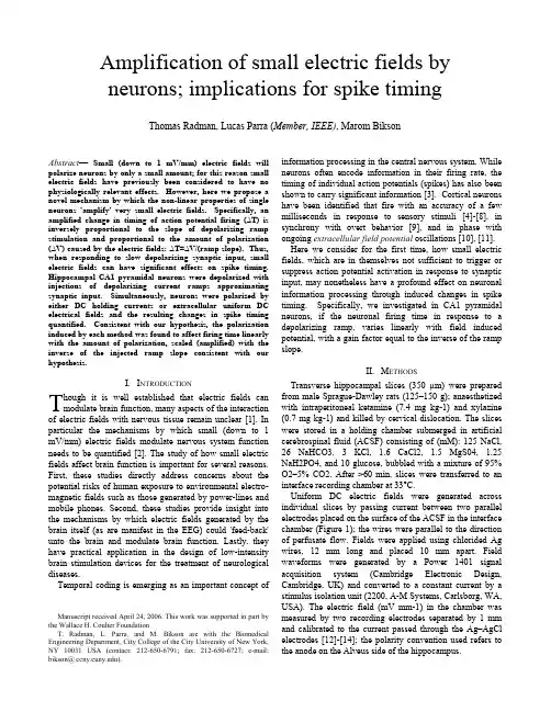
Amplification of small electric fields by neurons; implications for spike timing Thomas Radman, Lucas Parra (Member, IEEE), Marom BiksonAbstract— Small (down to 1 mV/mm) electric fields will polarize neurons by only a small amount; for this reason small electric fields have previously been considered to have no physiologically relevant effects. However, here we propose a novel mechanism by which the non-linear properties of single neurons 'amplify' very small electric fields. Specifically, an amplified change in timing of action potential firing (∆T) is inversely proportional to the slope of depolarizing ramp stimulation and proportional to the amount of polarization (∆V) caused by the electric fields: ∆T=∆V/(ramp slope). Thus, when responding to slow depolarizing synaptic input, small electric fields can have significant effects on spike timing. Hippocampal CA1 pyramidal neurons were depolarized with injections of depolarizing current ramps approximating synaptic input. Simultaneously, neurons were polarized by either DC holding currents or extracellular uniform DC electrical fields and the resulting changes in spike timing quantified. Consistent with our hypothesis, the polarization induced by each method was found to affect firing time linearly with the amount of polarization, scaled (amplified) with the inverse of the injected ramp slope consistent with our hypothesis.I.I NTRODUCTIONhough it is well established that electric fields can modulate brain function, many aspects of the interaction of electric fields with nervous tissue remain unclear [1]. In particular the mechanisms by which small (down to 1 mV/mm) electric fields modulate nervous system function needs to be quantified [2]. The study of how small electric fields affect brain function is important for several reasons. First, these studies directly address concerns about the potential risks of human exposure to environmental electro-magnetic fields such as those generated by power-lines and mobile phones. Second, these studies provide insight into the mechanisms by which electric fields generated by the brain itself (as are manifest in the EEG) could 'feed-back' unto the brain and modulate brain function. Lastly, they have practical application in the design of low-intensity brain stimulation devices for the treatment of neurological diseases.Temporal coding is emerging as an important concept of information processing in the central nervous system. While neurons often encode information in their firing rate, the timing of individual action potentials (spikes) has also been shown to carry significant information [3]. Cortical neurons have been identified that fire with an accuracy of a few milliseconds in response to sensory stimuli [4]-[8], in synchrony with overt behavior [9], and in phase with ongoing extracellular field potential oscillations [10], [11].Manuscript received April 24, 2006. This work was supported in part by the Wallace H. Coulter FoundationT. Radman, L. Parra, and M. Bikson are with the Biomedical Engineering Department, City College of the City University of New York, NY 10031 USA (contact: 212-650-6791; fax: 212-650-6727; e-mail: bikson@ ).Here we consider for the first time, how small electric fields, which are in themselves not sufficient to trigger or suppress action potential activation in response to synaptic input, may nonetheless have a profound effect on neuronal information processing through induced changes in spike timing. Specifically, we investigated in CA1 pyramidal neurons, if the neuronal firing time in response to a depolarizing ramp, varies linearly with field induced potential, with a gain factor equal to the inverse of the ramp slope.II.M ETHODSTransverse hippocampal slices (350 µm) were prepared from male Sprague-Dawley rats (125–150 g); anaesthetized with intraperitoneal ketamine (7.4 mg kg-1) and xylazine (0.7 mg kg-1) and killed by cervical dislocation. The slices were stored in a holding chamber submerged in artificial cerebrospinal fluid (ACSF) consisting of (mM): 125 NaCl, 26 NaHCO3, 3 KCl, 1.6 CaCl2, 1.5 MgS04, 1.25 NaH2PO4, and 10 glucose, bubbled with a mixture of 95% O2–5% CO2. After >60 min, slices were transferred to an interface recording chamber at 33°C.Uniform DC electric fields were generated across individual slices by passing current between two parallel electrodes placed on the surface of the ACSF in the interface chamber (Figure 1); the wires were parallel to the direction of perfusate flow. Fields were applied using chlorided Ag wires, 12 mm long and placed 10 mm apart. Field waveforms were generated by a Power 1401 signal acquisition system (Cambridge Electronic Design, Cambridge, UK) and converted to a constant current by a stimulus isolation unit (2200, A-M Systems, Carlsborg, WA, USA). The electric field (mV mm-1) in the chamber was measured by two recording electrodes separated by 1 mm and calibrated to the current passed through the Ag–AgCl electrodes [12]-[14]; the polarity convention used refers to the anode on the Alveus side of the hippocampus.T-+Ag/AgCl uniform field stimulation electrode+-18141062Electric fields were generated across tissue and the resulting potential of a single neuron was monitored. Application of a constant uniform field induced a membrane polarization. As explained, this polarization linearly affected the time to action potential in response to a depolarizing current ramp. The inverse of the ramp slope determined the efficacy of the field to induce this timing change. The polarization of the neuron was dependent on the direction of the field. Consistent with previous reports [14], the magnitude of the polarization was a linear function of field strength (Figure 4A). The steady-state sensitivity (coupling strength) of neurons ranged from 0.06 to 0.22 mV per 1 mV/mm applied uniform field (n =24).Fig. 1. A) Schematic of method for generating uniform extracellular fields across the CA1 region of the hippocampus and method for measuring artifact free transmembrane potential. B) Measured field strength distribution in mV/mm in relation to field generating electrodes.Conventional recording technniques were used to measure activity from the CA1 pyramidal cell region. Intracellular electrodes (70–120 M Ω, pulled on a P-97; Sutter Instruments, Novato, CA, USA) were filled with 3M potassium chloride. The voltage recorded by a field electrode (placed within 50 µm of the impaled neuron) was subtracted from the intracellular potential to obtain the transmembrane voltage and remove the exogenous potential artefact. Depolarizing intracellular current ramps were generated and triggered to halt upon action potential detection. The field waveforms were applied 400 ms before activation of depolarizing intracellular current ramps. The current ramps used (.1-.6nA/S) were selected to ensure a quasi-linear voltage response prior to sodium channel activation. Signals were subtracted, amplified, and low-pass filtered (1–10 kHz) with an Axoclamp-2B (Axon Instruments, Union City, CA, USA) and FLA-01 amplifiers (Cygnus Technology, Delaware Water Gap, PA, USA); digitized and processed using a Power 1401 and Signal software (CED).III. R ESULTSThe novel single neuron amplification mechanism was validated in hippocampal slices. CA1 Pyramidal neurons were slightly polarized by varied levels of DC intracellular holding current and their firing time in response to depolarizing ramps were measured. For each ramp, we determined the change in firing time resulting from a change in DC holding current. In separate experiments, the linear effects of DC electrical field polarization of a single neuron were then quantified and verified to be linear. Finally, current ramps were incorporated into electric field experiments to verify DC holding current timing amplification mechanisms held true for analogous extracellular DC electric field polarizations. A. Validation of Novel Amplification Mechanism Hyper-polarization of the neuron with an increasingly negative DC holding current, incrementally delayed action potential firing time in response to an intracellular ramp (n=10). The change in timing increased with the holding current, and inversely with ramp slope (Figure 2,3). These results show that the membrane dynamics of real CA1pyramidal neurons support the novel single neuron amplification mechanism proposed.B. Effects of Uniform DC Electric FieldsApplied fields could significantly modulate the firing latency of single neurons to intracellular ramp current injection. Fields inducing membrane hyperpolarization delayed action potential initiation while fields inducing membrane depolarization had the opposite effect (Figure 4B). The relationship between AP timing and depolarizing ramp slope, demonstrated for changes in intracellular holding current (Figure 3), was shown to be valid for polarization by extracellular fields (Figure 4).Using a ramp slope of .4 nA/s and 80 repetitions per field magnitude, we observed a significant (p<.05) change in timing under +/- 1 mV/mm field strengths (n=3). It is expected further experimental optimization can be incorporated in future studies of single neurons to resolve the effects of smaller fields on timing.IV. D ISCUSSIONHere we show the effects of electric fields, when coupled with a depolarizing input, affect the timing to firingthreshold. This process results from the non-linear properties of brain cells, and depends on the following: the threshold voltage is not a function of the baseline membrane potential100ms5mVAP Threshold∆T∆T = ∆V / Ramp Slope∆V∆TA) B)Fig. 2. Intracellular recordings of CA1 pyramidal cell response to depolarizing current ramps. A) Response to a .6nA/S intracellular current ramp. B) Response to a .4nA/S intracellular current ramp. In both cases hyperpolarization (∆V) delayed the AP firing time. The change (∆T), is inversely proportional to depolarizing ramp slope, ∆T A < ∆T B ). Action potentials clipped.or the depolarizing ramp slope. This holds if an injected current ramp to a neuron relates linearly to an induced voltage ramp [15], and that a uniform DC electric field has a linear relationship to neuronal polarization. Our previous studies have shown that hippocampal neuron polarization is a quasi-linear function of field magnitude, indicating that even very small extracellular fields will polarize neurons [14].We found that a 1 mV/mm uniform field induced a transmembrane potential change up to ~0.2 mV. Compared to the scale of depolarization necessary to bring a neuron from rest to threshold (~15 mV), these fields were previously considered insignificant with respect to actionpotential initiation. Previous action potential threshold studies identified changes due to electric fields less than 5 mV/mm [16]. Rather than spike generation, here we examined changes in timing, with respect to our novel amplification mechanism. Our results provide a mechanism for the effects on network spike timing previously demonstrated with exogenous uniform fields as low as 0.1 mV/mm [17], [18].In summary, these results demonstrate our hypothesis for magnification of the effect of small electric fields through changes in the timing of action potentials. These results are particularly relevant for situations in which small electric fields are manifest in the brain.A CKNOWLEDGMENTMadeline Lindvall; Chris Leonard of New York Medical College; Denis Paré of Rutgers University; Jack Belgum, Adair Osterle, and Rick Ayer of Sutter Instruments; Tim Bergel and Simon Gray of Cambridge Electronic Design (CED).-10-8-6-4-200.00.20.40.60.8 1.0Slope of Depolarizing Ramp (nA/s)E f f e c t o f P o l a r i z a t i o n o n T i m i n g (s /n A )Fig. 3. The inverse relationship between the ramp slope and the sensitivity to membrane polarization due to negative DC holding current can be summarized in a single plot. Experimental results (“x”) fit directly to the theoretical prediction of a 1/ramp slope timing change per transmembrane polarization. Error bars represent standard error of the slope of a regression line fitting holding current (nA) versus AP timing (s) data.Electric Field Strength (mV/mm)-20A)B)Polarization (mV)42-2-4-5-4-3-2-101234540200-20-40P o l a r i z a t i o n (m V )0.00.20.40.60.81.01.21.41.61.82.0403020100-10-30-40Electric Field Strength (mV/mm)T i m e t o A c t i o n P o t e n t i a l (s e c )Fig. 4. A) Application of a uniform field caused the polarization of a CA1 pyramidal neuron; the magnitude of the polarization was linearly related to the amplitude of the electric field (reported: mean +/- SD). B) Relationship between change in firing time and initial membrane polarization (induced by applied field) for three different ramp slopes. Note that for each ramp slope, the relationship between firing time (∆T) and polarization (∆V) is linear; moreover, the slope of this relationship is inversely related to the ramp slope. The injected current ramp (.4, .5, .6 nA/S) reported as voltage (6.8, 8.5, 10.2 mV/S respectively, scaled by this particular cell resistance of 17.5M Ω). The change in timing for any given membrane polarization increases (is ‘amplified’) as the slope of the ramp is decreased .R EFERENCES[1] Faber DS, Korn H. Field effects trigger post-anodal rebound excitationin vertebrate CNS. Nature . 1983 Oct 27-Nov 2;305(5937):802-4 [2] Parra LC, Bikson M. Model of the effect of extracellular fields onspike time coherence, 26th Annual International Conference of the IEEE Engineering in Medicine and Biology Society 1 - 4 September 2004[3] de Ruyter van Steveninck RR, Lewen GD, Strong SP, Koberle R,Bialek W. Reproducibility and variability in neural spike trains. Science . 1997 Mar 21; 275 (5307): 1805-8.[4] Mainen ZF, Sejnowski TJ. Influence of dendritic structure on firingpattern in model neocortical neurons . Nature . 1996 Jul 25;382(6589):363-6.[5] Trussell LO. S ynaptic mechanisms for coding timing in auditoryneurons. Annu Rev Physiol . 1999;61:477-96.[6]Reinagel P, Reid RC. Temporal coding of visual information in thethalamus. J Neurosci. 2000 Jul 15;20(14):5392-400.[7]Kara P, Reinagel P, Reid RC. Low response variability insimultaneously recorded retinal, thalamic, and cortical neurons.Neuron. 2000 Sep;27(3):635-46.[8]DeWeese MR, Wehr M, Zador AM.Binary spiking in auditory cortex.J Neurosci. 2003 Aug 27;23(21):7940-9.[9]Riehle A, Grammont F, Diesmann M, Grun S. Dynamical changesand temporal precision of synchronized spiking activity in monkeymotor cortex during movement preparation. J Physiol Paris. 2000Sep-Dec;94(5-6):569-82.[10]Harris KD, Csicsvari J, Hirase H, Dragoi G, Buzsaki G. Organizationof cell assemblies in the hippocampus. Nature. 2003 Jul31;424(6948):552-6.[11]Kashiwadani H, Sasaki YF, Uchida N, Mori K. Synchronizedoscillatory discharges of mitral/tufted cells with different molecularreceptive ranges in the rabbit olfactory bulb. J Neurophysiol. 1999Oct;82(4):1786-92.[12]Ghai RS, Bikson M & Durand DM (2000). Effects of applied electricfields on low-calcium epileptiform activity in the CA1 region of rathippocampal slices. J Neurophysiol 84, 274–280.[13]Durand DM & Bikson M (2001).Suppression and control ofepileptiform activity by electrical stimulation: a review. Proc IEEE 89, 1065–1082[14]Bikson M, Inoue M, Akiyama H, Deans JK, Fox JE, Miyakawa H,Jefferys JG. Effects of uniform extracellular DC electric fields onexcitability in rat hippocampal slices in vitro. J Physiol. 2004 May15;557(Pt 1):175-90. Epub 2004 Feb 20.[15]Fricker D, Verheugen J, Miles R (1999).Cell-attached measurementsof the firing threshold of rat hippocampal neurones. J Physiol 517.3,791-804[16]Jefferys JG. Influence of electric fields on the excitability of granulecells in guinea-pig hippocampal slices. J Physiol. 1981;319:143-52. [17]Deans JK, Bikson M, Fox JE & Jefferys JGR (2003). Effects of ACfields at powerline frequencies on gamma oscillations in vitro.Program No. 258.1 2003 Abstract Viewer/Itinerary Planner.Washington DC: Society for Neuroscience On-line[18]Francis JT, Gluckman BJ, Schiff SJ Journal of Neuroscience, 23(19)pp. 7255-7261, 2003。