PLGA文献讲座 ppt课件
- 格式:ppt
- 大小:254.50 KB
- 文档页数:20
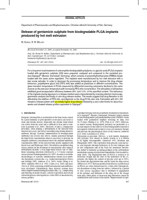
Fig.1:Effect of the admixture of PEG400on the glass transition temperature of PLGA and on the extrusion temperaturemization of the release profiles have been achieved inter alia by changes of the size of the dosage forms(Siepmann et al. 2004),by addition of monomers of the polymer(Yoo et al. 2004),by variation of the molecular weight(Kanellakopoulou et al.1999),by combining PLGA with other biodegradable polymers(Schmidt et al.1995)and of course by modify-ing the drug content(Cevher et al.2007).In this study,a plasticizer(PEG400)is added in different concentration to reduce the lag-phase and to gain a prolonged constant drug release.Gentamicin sulphate is an aminoglycoside broadspectrum antibiotic,which works by binding the50S subunit of the bacte-rial ribosome and hence interrupting the protein synthesis.The antibiotic is very potent and has a high temperature stability (Wang,Liu et al.2004).It is therefore often incorporated in polymer matrices via a melting process for extended drug release (Neut et al.2001).The production of the implants was performed by hot melt extru-sion(HME),an old manufacturing processfirst used in the plastics industry.Pharmacists have increasing interests in the HME technique as an alternative production method(Crowley et al.2007).The major advantages of this technique are that no solvents are involved in the process,only a few processing steps are needed,it is a continuous and well reproducible process, there are no requirements on the compressibility of the drug and the bioavaibility of the active ingredient can be increased enor-mously if it is molecularly dispersed in the polymer(McGinity et al.2007).Furthermore,hot melt extrusion allows produc-ing the favoured release behaviour by varying the composition of the formulations or by different geometry of the formed dosage form.The main disadvantage of HME is the thermal stress the active ingredient and the polymer are exposed to dur-ing processing(Breitenbach2002).In this study,biodegradable gentamicin sulphate loaded PLGA implants were produced via HME and PEG400was admixed as plasticizer to decrease the processing temperature and to affect the drug release behaviour beneficially.2.Investigations and results2.1.Plasticizer influence on glass transitionand extrusion temperatureGentamicin sulphate has no decreasing effect on the glass tran-sition temperature of PLGA(Fig.1).Therefore PEG400was added as plasticizer to produce the implants at lower extru-sion temperatures.With increasing PEG400-concentration up Fig.2:Drug content of the batches of all formulationsFig.3:Cumulative drug release of the20%GS batches with increasing PEG400 concentration compared to Septopal®to10%the glass transition temperature decreases linearly from 43◦C to about12◦C.Accordingly,it was possible to reduce the extrusion temperature from110◦C to85◦C and consequently minimize the temperature impact on the active ingredient.Nev-ertheless,the intense decrease of the glass transition temperature up to12◦C can eventually lead to stability problems during storage.2.2.Drug content of the extrudatesAs shown in Fig.2the measured drug contents reach well the specified contents with95%confidence intervals below 1.3%and each single extrudate exhibited an encapsulation effi-ciency of85%to115%.The p-values between the batches of the formulations exceeded0.1which indicates that there were no significant differences between the batches.Two exceptions were between0.05(>0.05indicates a significant difference)and 0.1,due to small confidence intervals.Thus,it can be stated that the manufacturing process led to reproducible implants.2.3.Release of gentamicin sulphate from melt extruded implantsFig.3shows the cumulative drug release of the20%gentam-icin sulphate batches with increasing concentration of PEG400 versus one Septopal®-sphere.After a burst effect,where about 20%of the drug amount is released due to the dissolving ofFig.4:Cumulative drug release of the25%GS batches with increasing PEG400 concentration compared to Septopal®gentamicin sulphate from the outer surface,water penetrates slowly into the extrudates and lets them swell.The drug that is further dissolved by intruding water diffuses into the release medium and thus generates pores which in turn accelerate the process of water penetration and drug liberation.In addition, the steady degradation of the polymer enhances drug release. PLGA hydrolytically decomposed into different products with chain lengths with free carboxyl groups at the end,which can-not penetrate out of the extrudate due to their size.Therefore the mass changes are only attributed to the loss in drug mass. The oligomers create an acidic microclimate inside the extru-date and hence accelerate the autocatalytic degradation process of PLGA(Shenderova et al.1999).In contrary the emerged frag-ments can diffuse away from the surface,so that the acidity here is neutralized by the buffer and the degradation of the surface is not autocatalysed.So,the outer layer is stable for a longer time.When the pores on the surface reach a size,where they allow an evacuation of the polymer fragments from the inside or when the oligomers degrade to dimers and monomers so that they can penetrate outside,a critical threshold is passed and the mass loss and the erosion of the extrudates starts(McGinity and O’Donnell1997).At this point the surface of the extru-dates can collapse and release an enlarged drug amount as can be seen at the20%gentamicin sulphate batch without plasti-cizer after approximately20days.By the end of the gentamicin sulphate release the liberation slows down until the drug is released almost completely after four tofive weeks.The increas-ing polyethylene glycol concentration does not significantly affect the release behaviour of the implants.In comparison to the release profile of the Septopal®-sphere,which releases gen-tamicin sulphate by diffusion from a non-biodegradable matrix system(polymethylmethacrylate)with release kinetics accord-ing to Higuchi the20%drug batches show more sigmoidal release curves.Although these batches release well above the minimal inhibitory concentration of Staphylococcus aureus,the main pathogen of osteomyelitis(Brady et al.2006),even over thefirst two weeks(data not shown),a higher drug release especially at the beginning of the antibiotic therapy would be preferable.The batches with25%gentamicin sulphate and increasing polyethylene glycol concentration,shown in Fig.4,exhibit a higher initial rate of release.After seven days about50%of the theoretical drug amount is liberated caused by the higher drug concentration yielding more pores.However,it is followed by a constant release over approximately four weeks.During this steady drug release the swelling of the extrudate and the liber-ation of the active ingredient equilibrate resulting in zero order Fig.5:Cumulative drug release of the30%GS batches with increasing PEG400 concentration compared to Septopal®kinetics with a coefficient of determination of0.99(formulation 4and6).The25%gentamicin sulphate batches,which again do not differ significantly with increasing plasticizer content, have release profiles equivalent to the Septopal®-sphere.Three extrudates with25%gentamicin sulphate are pharmaceutically equivalent to one Septopal®-sphere(containing7.5mg GS)and bioequivalence is anticipated by these release studies although this has to be verified in vivo.The release behaviour from30%drug content batches is shown in Fig.5.Basically,gentamicin sulphate is liberated quite fast, about55%,70%and80%drug respectively is set free depending on the plasticizer content after four days.In consideration of the fact that polyethylene glycol had no impact on the release rates of the batches with lower drug content,it can be assumed that the ebbing retarding effect of the batches is caused by the decreased PLGA-concentration.Although the30%gentamicin sulphate formulations combined with0%and5%plasticizer have the same PLGA-concentration as the25%drug batches combined with5%and10%PEG400,they show a less retarded drug release rate.This can be explained by the higher content of gentamicin sulphate,generating more pores and therefore triggering faster drug liberation.2.4.Water uptake and mass lossThe water uptake of all batches,ranked according to the PLGA-concentration,is illustrated in Fig.6.It becomes apparent that the water uptake corresponds mainly to the PLGA-content indepen-dent of further formulation ingredients.With a lower polymer Fig.6:Effect of the PLGA concentration on the water uptake of the extrudatesFig.7:Influence of the drug concentration and PEG 400concentration on the massloss of the extrudatescontent a less tighter matrix system can be build up and water can more easily infiltrate the extrudate,whereby it is unimpor-tant if gentamicin sulphate or polyethylene glycol are embedded between the polymer chains and spacing them apart.The smaller the PLGA-concentration is,the faster and the more water is taken up.While the water uptake was observed to be dependent on the polymer weight fraction,the mass loss is dependent on the drug amount (Fig.7).The 20%gentamicin sulphate batches initiallyshow a mass increase,probably generated by not completely freeze dried samples.Apart from that the other formulations exhibit a mass loss according to the released amount of drug.For example the 25%drug batches have a mass loss of approx-imately 16.5%after 16days which correlates properly to the drug amount released (approx.62%of 25%drug amount equals 15.5%mass loss).The mass loss data display the dependency of the 30%batches on the plasticizer content or rather on the remaining PLGA-concentration as well as the release curves.According to the faster drug liberation a higher mass loss is vis-ible with decreasing PLGA-concentration.The erosion of the polymer starts slowly after 16days and intensifies after approx.20days,which can also be observed in the increasing drug release of the 20%gentamicin sulphate batches.2.5.SEM-pictures of the extrudatesThe morphological changes during drug release are illustrated in the scanning electron pictures.The cutting zones of the extrudates,shown in Fig.8,reveal the included spherical gen-tamicin sulphate particles in a nonporous and homogeneous matrix leaving little holes after exposure to the release medium (Figs.9and 10).In Fig.10the more structural roughness of the surface compared to Fig.9denote the incorporated polyethylene glycol in the polymer.The surface of the extrudates changes over time to less smooth and more porous.This results from the degradation and after 21days erosion of the polymer becomesvisible.Fig.8:SEM-pictures of the cutting zones of a pure PLGA-extrudate (left)and a gentamicin sulphate loaded extrudate(right)Fig.9:SEM-pictures of the surfaces of extrudates with 25%gentamicin sulphate and 0%PEG 400after 1,16and 21days (left to right)exposure to phosphate buffer pH 7.4Fig.10:SEM-pictures of the surfaces of extrudates with25%gentamicin sulphate and10%PEG400after1,16and21days(left to right)exposure to phosphate buffer pH7.4Table:Composition of the formulationsBatch no.GS(%)PEG400(%)PLGA(%) 120080 220575 3201070 425075 525570 6251065 730070 830070 9301060 3.DiscussionAlthough the admixture of plasticizer decreases the glass tran-sition temperature of PLGA with increasing concentration and therefore the extrusion temperature of the blends,drug liberation is not affected up to a specific remaining PLGA-content,which is necessary to provoke a prolonged drug release.The release pro-files of the extrudates differ depending on the drug amount from sigmoidal to exponential release curves,whereby25%gentam-icin sulphate amount lead to an initial high drug release followed by zero order kinetics over about four weeks.These formula-tions are consequently a good alternative to Septopal®without the disadvantage of a second surgery,although further in vivo investigations have to be done.4.Experimental4.1.MaterialsAs biodegradable polymer Resomer®RG503H(Boehringer Ingelheim, Ingelheim,Germany)was used.Resomer®RG503H is a poly(D,L-lactic-co-glycolic acid)with a ratio of50:50,an intrinsic viscosity of 0.32–0.44dL/g and a free carboxylic acid as end group.The antibiotic gentamicin sulphate purchased from Caelo(Hilden,Germany)was used as active ingredient.The plasticizer polyethylene glycol400was obtained by BASF(Ludwigshafen,Germany).O-Phthaldialdehyde thioglycolic acid (Merck,Darmstadt,Germany)and sodium1-heptane-sulfonate(Sigma Aldrich,Taufkirchen,Germany)were used for the analytical methods. 4.2.Methods4.2.1.Manufacture of the implantsThe manufacture of the antibiotic loaded implants was performed by hot melt extrusion.Nine formulations(Table)with a drug content of20,25and30% gentamicin sulphate and plasticizer content of0,5and10%were produced. The polymer and the active ingredient were mixed with a turbula-blender (Willy A.Bachofen AG,Basel,Switzerland)for15min and subsequently manually granulated with PEG400for at least10min.These blends were hot melt extruded by a twin screw extruder(Minilab II,Thermo Fisher Sci-entific,Karlsruhe,Germany)with a die diameter of2mm.The temperature was set as low as possible between85◦C and110◦C depending on the plasticizer content.After cooling down,extrudates of9.8to10.2mg weight were produced by cutting.4.2.2.Drug contentTo determine the encapsulation efficiency,the extrudates were individually dissolved in dichloromethane.After addition of4mL water the samples were mixed to let the active ingredient migrate to the aqueous phase for at least one hour.The gentamicin sulphate content of the aqueous phase was analysed by HPLC.For determination of gentamicin sulphate by HPLC the drug has to be derivatisated as it does not possess UV absorbing chromophores.As derivatisation agent for pre-column derivatisation with UV detection o-phthaldialdehyde(OPA)was used.The o-phthaldialdehyd reagent was prepared as described in the USP XXXI.For derivatisation1.0mL of the sample was mixed with0.4mL of o-phthaldialdehyd solution and1.1mL of isopropanol,incubated at60◦C for15min and in afinal step cooled down to room temperature.The analysis of the drug content of the prepared samples was carried out using reversed phase HPLC with a HPLC system by the Agilent Series1100(Agilent Technologies,Santa Clara,USA).As station-ary phase a Merck LiChroCart125–4mm column,filled with LiChrospher 100RP18,5m and a pre column LiChroChart4–4,LiChroSpher100, 5mm(all Merck KGaA,Darmstadt,Germany)were used.The mobile phase was composed of700mL methanol,25mL water,50mL glacial acetic acid (all obtained from Merck KGaA,Darmstadt,Germany)and5g sodium 1-heptane-sulfonate.Theflow rate wasfixed at1.0mL per min,the column temperature was25◦C,the injection volume was50L and the detection wavelength was set to330nm.4.2.3.Differential scanning calorimetryThe thermal properties of the loaded implants were analysed with a differen-tial scanning calorimeter from Perkin Elmer,Waltham,Massachusetts,USA (DSC7).Samples of approximately7mg were weighed into aluminium pans,sealed hermetically and analysed under a nitrogen atmosphere with a heating procedure from10◦C to80◦C at a scan rate of10◦C/min related to an empty reference pan.4.2.4.Scanning electron microscopyTo analyse surface morphology SEM pictures were made with a Philips XL20(Philips B.V.,Eindhoven,The Netherlands)scanning electron micro-scope.The samples werefixed on a carbonfibrefilm and sputtered with gold in an argon atmosphere at a sputter current of50mA for180s using a SCD005Sputter coater(BalTec,Balzers,Liechtenstein).4.2.5.In vitro releaseFive gentamicin sulphate loaded extrudates of approximately10mg weight of each batch or one Septopal®-sphere(containing7.5mg(w/w)genta-micin sulphate)were placed into3mL phosphate buffer pH7.4(containing 250.0mL0.2M monobasic potassium phosphate solution with393.4mL 0.1M sodium hydroxide solution)as dissolution medium.The extrudates were incubated at37◦C and horizontally shaken.At a time interval of72h starting at day one the whole dissolution medium was collected for analyzing via HPLC and replaced by fresh buffer.This procedure was repeated until no more gentamicin sulphate could be detected.4.2.6.Water uptake and mass lossTo determine the swelling and the erosion of the extrudates water uptake und mass loss were analysed.Therefore implants were weighed exactly and placed in release medium over specific time periods.The implants were weighed wet after removing the water from the surface using soft wipes.After freeze drying(freeze dryer Alpha1-4LDC-1M,Martin Christ Gefriertrocknungsanlagen GmbH,Osterode,Germany)over two days the extrudates were weighed again to investigate the mass loss.The measure-ment of the water uptake und mass loss was only possible as long as the extrudates stayed in shape.ReferencesAmbrose CG,Clyburn TA,Louden K,Joseph J,Wright J,Gulati P, Gogola GR,Mikos AG(2004)Effective treatment of osteomyelitis with biodegradable microspheres in a rabbit model.Clin Orthop Relat Res421: 293–299.Ambrose CG,Gogola GR,Clyburn TA,Raymond AK,Peng AS,Mikos AG(2003)Antibiotic microspheres:preliminary testing for potential treatment of osteomyelitis.Clin Orthop Relat Res415:279–285.Böstman O,Pihlajamäki H(2000)Clinical biocompatibility of biodegrad-able orthopaedic implants for internalfixation:a review.Biomaterials21: 2615–2621.Brady RA,Leid JG,Costerton JW,Shirtliss ME(2006)Osteomyelitis:Clin-ical overview and mechanisms of infection persistence.Clin Microbiol Newsl28:65–71.Breitenbach J(2002)Melt extrusion:from process to drug delivery technol-ogy.Eur J Pharm Biopharm54:107–117.Castro C,Evora C,Baro M,Soriano I,Sanchez E(2005)Two-month ciprofloxacin implants for multibacterial bone infections.Eur J Pharm Biopharm60:401–406.Cevher E,Orhan Z,Sensoy D,Ahiskali A,Kann PL,Sagirli O,Mulazimoglu L(2007)Sodium fusidate-poly(d,l-lactide-co-glycolide)microspheres: preparation,characterisation and in vivo evaluation of their effectiveness in the treatment of chronic osteomyelitis.J Microencapsul24:577–595. Crowley MM,Zhang F,Repka MA,Thumma S,Upadhye SB,Battu SK, McGinity JW,Martin C(2007)Pharmaceutical applications of hot-melt extrusion:part I.Drug Dev Ind Pharm33:909–926.Flick AB,Herbert JC,Goodell J,Kristiansen T(1987)Noncommer-cial fabrication of antibiotic-impregnated polymethylmethacrylate beads. Technical note.Clin Orthop Relat Res223:282–286.Friess W,Schlapp M(2002)Release mechanisms from gentamicin loaded poly(lactic-co-glycolic acid)(PLGA)microparticles.J Pharm Sci91: 845–855.Garvin K,Feschuk C(2005)Polylactide-polyglycolide antibiotic implants. Clin Orthop Relat Res437:105–110.Jain RA(2000)The manufacturing techniques of various drug loaded biodegradable poly(lactide-co-glycolide)(PLGA)devices.Biomaterials 21:2475–2490.Kanellakopoulou K,Kolia M,Anastassiadis A,Korakis T,Giamarellos-Bourboulis EJ,Andreopoulos A,Dounis E,Giamarellou H(1999)Lactic acid polymers as biodegradable carriers offluoroquinolones:an in vitro study.Antimicrob Agents Chemother43:714–716.Lazzarini L,Mader JT,Calhoun JH(2004)Osteomyelitis in long bones.J Bone Joint Surg Am86-A:2305–2318.Lew DP,Waldvogel FA(2004)ncet364:369–379. McGinity JW,O’Donnell PB(1997)Preparation of microspheres by the solvent evaporation technique.Adv.Drug Deliv Rev28:25–42. McGinity JW,Repka MA,Koleng JJ,Zhang F(2007)Hot-Melt Extrusion Technology.In:Swarbrick J(ed)Encyclopedia of phar-maceutical technology.New York,Informa Healthcare USA.3: 2004–2020.Miller RA,Brady JM,Cutright DE(1977)Degradation rates of oral resorbable implants(polylactates and polyglycolates):rate modification with changes in PLA/PGA copolymer ratios.J Biomed Mater Res11: 711–719.Neut D,van de Belt H,Stokroos I,van Horn JR,van der Mei HC,Busscher JH(2001)Biomaterial-associated infection of gentamicin-loaded PMMA beads in orthopaedic revision surgery.J Antimicrob Chemother47:885–891.Schmidt C,Wenz R,Nies B,Moll F(1995)Antibiotic in vivo/in vitro release,histocompatibility and biodegradation of gentamicin implants based on lactic acid polymers and copolymers.J Control Release37: 83–94.Shenderova A,Burke TG,Schwendeman SP(1999)The acidic microclimate in poly(lactide-co-glycolide)microspheres stabilizes camptothecins. Pharm Res16:241–248.Shive MS,Anderson JM(1997)Biodegradation and biocompatibil-ity of PLA and PLGA microspheres.Adv Drug Deliv Rev28: 5–24.Siepmann J,Faisant N,Akiki J,Richard J,Benoit JP(2004)Effect of the size of biodegradable microparticles on drug release:experiment and theory. J Control Release96:123–134.Soundrapandian C,Datta S,Sa B(2007)Drug-eluting implants for osteomyelitis.Crit Rev Ther Drug Carrier Syst24:493–545.Wahlig H,Dingeldein E,Bergmann R,Reuss K(1978)The release of gentamicin from polymethylmethacrylate beads.An experimental and pharmacokinetic study.J.Bone Joint Surg.Br.60-B:270–275.Wang G,Liu SJ,Ueng SW,Chan EC(2004)The release of cefazolin and gentamicin from biodegradable PLA/PGA beads.Int J Pharm273:203–212.Wei G,Kotoura Y,Oka M,Yamamuro T,Wada R,Hyon SH,Ikada Y(1991) A bioabsorbable delivery system for antibiotic treatment of osteomyelitis. The use of lactic acid oligomer as a carrier.J Bone Joint Surg Br73: 246–252.Yoo JY,Kim JM,Khang G,Kim MS,Cho SH,Lee HB,Kim YS(2004) Effect of lactide/glycolide monomers on release behaviors of gentamicin sulfate-loaded PLGA discs.Int J Pharm276:1–9.。


动物医学进展,2020,41(12):96 101ProgressinVeterinaryMedicinePLGA纳米粒作为药物载体的靶向作用研究进展 收稿日期:2020 06 06 基金项目:国家自然科学基金项目(31872511) 作者简介:胡 馨(1997-),女,重庆人,硕士研究生,主要从事兽药学研究。
通讯作者胡 馨,支 慧,杨 艳,杨 杰,柴东坤,林 浪,刘云杰,宋振辉 ,封海波(西南大学动物科学学院,重庆402460) 摘 要:纳米科技在现代医学及药学的应用方面广泛发展,纳米药物载体在实现靶向性给药、缓释药物、降低药物的毒副作用等方面有重大优势。
聚乳酸 羟基乙酸聚合物(PLGA)是一种高分子有机化合物,具有生物相容性及生物可降解性,当前聚乳酸 羟基乙酸聚合物纳米粒(PLGANPs)被广泛地作为药物载体进行靶向治疗。
论文归纳总结了近年来国内外的相关文献报道,概述了PLGANPs的特点、制备方法与表征以及靶向作用的研究进展,着重讨论了PLGANPs作为药物载体在肿瘤组织、心脑血管、骨组织、免疫和基因类疾病中靶向作用的研究进展,并对未来发展前景进行了展望,为相关的科研提供参考。
关键词:聚乳酸 羟基乙酸聚合物;纳米粒;药物载体;靶向作用中图分类号:S854.53;S859.797文献标识码:A文章编号:1007 5038(2020)12 0096 06 靶向制剂是指通过局部给药的方式将药物输送至特定的组织、器官、细胞内,以提高药物的疗效和生物利用度,并减少毒副作用带来的危害。
聚乳酸 羟基乙酸聚合物[poly(lactic co glycolicacid),PLGA]是由乳酸和羟基乙酸的单体聚合而成的可降解的高分子有机化合物。
纳米粒(nanoparticles,NPs)是大小介于1nm~1000nm之间的一种固态胶体颗粒,可作为药物靶向传递的载体。
PLGA是乳酸(lacticacid,LA)与羟基乙酸(glycolicacid,GA)共聚合而成,当PLGA进入体内,通过酯键水解生成相应的单体酸、乳酸和羟基乙酸,然后经过三羧酸循环后转变成二氧化碳和水,因此该聚合物对人体无刺激性,无毒且拥有良好的生物相容性和降解性[1];PLGANPs极易于被吞噬细胞摄取,因此通过在纳米颗粒偶联吸附相应的配体可定位到需要的组织和器官。

PLGA纳米微球制备条件的优化聚乳酸-羟基乙酸共聚物(PLGA)为载体,二氯甲烷为有机溶剂,聚乙烯醇为乳化剂,采用水/油/水型(W1/O/W2)复乳化溶剂挥发制备载有原花青素的微球并对微球相关性质进行检测。
结果所得微球呈圆球状,微球平均粒径为10um,Zeta电位为-22.2mV,通过该方法制备的微球粒径分布较均匀,操作方便,为进一部优化包被原花青素的PLGA纳米微球提供了一定依据。
标签:原花青素;聚乳酸-羟基乙酸共聚物(PLGA);微球;乳化溶剂挥发法纳米材料作为一种可行的给药载体系统已成为可能。
聚乳酸-羟基乙酸共聚物(PLGA)作为生物可降解载体,因其具有良好的生物相容性、可生物降解性和安全性目前已被用作药物载体和生物支架,制备成纳米微球后,还可以缓慢释放包载的药物。
鉴于肿瘤发生率逐年上升,食源性药物能克服和减少化学性药物引起骨髓抑制、消化道反应、神经系统毒性等不良反应,原花青素属于黄烷-3-醇类化合物,具有抗氧化、抗炎、调节信号分子的表达、促进肿瘤细胞凋亡等抗肿瘤作用,但其生物利用度低,尤其是大分子成分(三聚体以上)吸收性差,为了提高其利用率与稳定性,本实验采用生物降解材料聚乳酸--羟基乙酸共聚物(PLGA)包裹原花青素,制备原花青素缓释微球。
尽可能保证颗粒的均一性、高包封率、高载药量、缓释性能。
1 材料与方法1.1 试剂PLGA(0%、5%、10%、20%PEG),葡萄籽原花青素,二氯甲烷,聚乙烯醇(PV A),三蒸水。
1.2 实验仪器移液枪,电子天平,超声混匀仪,磁力搅拌器,高速离心机,测微尺,激光粒度分析仪1.3 PLGA-原花青素纳米微球制备方法称取一定量葡萄籽原花青素溶于适量水中作为内水相,混匀,称取定量(PEG-)PLGA溶于二氯甲烷里作为有机相,同时配置一定浓度的PV A水溶液作为外水相,(需控制其完全溶解)将有机相和无机相混合,在超声条件下将内水相和油相混匀形成初乳,在恒温磁力搅拌器上将初乳加入到外水相中形成复乳后继续搅拌5h,然后在10000r/min下离心30min,蒸馏水洗三次,每次五分钟,之后冷冻保存。