BubR1 insufficiency causes early onset of aging-associated
- 格式:pdf
- 大小:1.89 MB
- 文档页数:6

DOI:10.16662/ki.1674-0742.2021.33.089艾司西酞普兰与喹硫平联合治疗首发抑郁症的效果分析杨晓江泉州市第三医院精神科,福建泉州362121[摘要]目的探讨艾司西酞普兰与喹硫平联合治疗首发抑郁症的效果。
方法方便选取2018年1月—2020年12月在该院就诊的116例首发抑郁症患者为研究对象,以随机数表法将其划分为两组。
对照组58例患者应用艾司西酞普兰治疗,研究组58例在此基础上联合喹硫平治疗。
比较两组患者的临床疗效、治疗前后的神经因子水平[髓鞘间隙蛋白(MBP)与神经营养因子(BDNF)],治疗前、治疗4周与8周时的认知功能[以重复性成套神经心理状态测验(RBANS)评价]以及不良反应情况。
结果研究组治疗的总有效率为94.83%较对照组79.31%高,差异有统计学意义(χ2=6.202,P<0.05)。
治疗后,研究组MBP(4.52±0.85)ng/mL较对照组(5.79±0.69)ng/mL低,BDNF(27.52±4.03)ng/mL较对照组(24.33±5.00)ng/mL高,差异有统计学意义(t=8.834、3.783,P<0.05)。
治疗4周与8周时,研究组RBANS评分(73.52±10.00)分、(78.62±10.40)分较对照组高(68.03±8.46)分、(71.32±9.05)分,差异有统计学意义(t=3.192,4.033,P<0.05)。
研究组不良反应发生率为10.34%,对照组不良反应发生率为8.62%,差异无统计学意义(χ2=0.100,P>0.05)。
结论首发抑郁症患者应用艾司西酞普兰与喹硫平联合治疗能够提高临床疗效,改善神经因子水平与认知功能,安全性佳,具有临床推广价值。
[关键词]艾司西酞普兰;喹硫平;首发抑郁症[中图分类号]R5[文献标识码]A[文章编号]1674-0742(2021)11(c)-0089-04. All Rights Reserved.Analysis of the Effect of Escitalopram and Quetiapine in the Treatment ofFirst-episode DepressionYANG XiaojiangDepartment of Psychiatry,Quanzhou Third Hospital,Quanzhou,Fujian Province,362121China[Abstract]Objective To explore the effect of escitalopram and quetiapine in the treatment of first-episode depression.Methods Conveniently selected the116first-episode depression patients who visited the hospital from January2018toDecember2020for research.They were divided into two groups using a random number table.58patients in the controlgroup were treated with escitalopram,and58patients in the study group were treated with quetiapine on this basis.Compared the clinical efficacy of the two groups of patients,the levels of neurological factors[myelin interstitial protein(MBP)and neurotrophic factor(BDNF)]before and after treatment,and the cognitive function[in a repetitive set ofneuropsychological status test(RBANS)evaluation]before and after treatment,4weeks and8weeks of treatment,andadverse reactions.Results The total effective rate of treatment in the study group was94.83%higher than79.31%in thecontrol group,the difference was statistically significant(χ2=6.202,P<0.05).After treatment,MBP(4.52±0.85)ng/mL in thestudy group was lower than that in the control group(5.79±0.69)ng/mL,and BDNF(27.52±4.03)ng/mL was higher thanthat in the control group(24.33±5.00)ng/mL,the difference was statistically significant(t=8.834,3.783,P<0.05).In thetreatment of4weeks and8weeks,the RBANS score of the study group was(73.52±10.00)points and(78.62±10.40)pointshigher than that of the control group(68.03±8.46)points and(71.32±9.05)points,the difference was statistically significant(t=3.192,4.033,P<0.05).The incidence of adverse reactions in the study group was10.34%,and the incidence of adversereactions in the control group was8.62%,the difference was not statistically significant(χ2=0.100,P>0.05).Conclusion Thecombined treatment of escitalopram and quetiapine in patients with first-episode depression can improve the clinicalefficacy,improve the level of neurological factors and cognitive function,with good safety and clinical promotion value.[Key words]Escitalopram;Quetiapine;First episode depression[作者简介]杨晓江(1978-),男,本科,主治医师,研究方向为精神病。

ʌ述评ɔ系统生物学方法在骨质疏松症中医证候研究中的应用❋章轶立1,2,齐保玉1,魏㊀戌1ә,戴建业3,王㊀旭1,申㊀浩4,谢雁鸣5(1.中国中医科学院望京医院,北京㊀100102;2.北京中医药大学中医学院,北京㊀100029;3.兰州大学药学院,兰州㊀730020;4.北京市丰台区长辛店社区卫生服务中心,北京㊀100072;5.中国中医科学院中医临床基础医学研究所,北京㊀100700)㊀㊀摘要:随着现代科学技术的进步及证候学研究的逐步深入,借助系统生物学的方法研究骨质疏松症中医证候以阐释证候科学内涵的研究逐年增多㊂本研究发现,目前骨质疏松症相关证候的基因组学与蛋白质组学研究仍停留在单个基因层面,主要包括骨钙素基因㊁雌激素受体基因以及Smad ㊁β-catenin 等蛋白,并未从整体的角度探究证候发生㊁发展的本质问题㊂代谢组学方面,虽然通过对骨代谢指标相关产物的检测,发现了与证候相关的代谢指标,但对于骨代谢终端产物的组学研究较少㊂未来研究仍需进一步运用组学技术手段,全方位㊁多层次㊁宽视角的探讨骨质疏松症中医证候的发生发展规律㊂㊀㊀关键词:骨质疏松症;证候;系统生物学;组学㊀㊀中图分类号:R2-03㊀㊀文献标识码:A㊀㊀文章编号:1006-3250(2021)04-0703-04Application of Systematic Biology Method in Traditional Chinese Medicine SyndromesResearch of OsteoporosisZHANG Yi-li 1,2,QI Bao-yu 1,WEI Xu 1ә,DAI Jian-ye 3,WANG Xu 1,SHEN Hao 4,XIE Yan-ming 5(1.Wangjing Hospital,China Academy of Chinese Medical Sciences,Beijing 100102,China;2.Beijing University of Chinese Medicine,Beijing 100029,China;3.School of pharmacy,Lanzhou university,Lanzhou 730020,China;4.Changxindian Community Health Service Center of Fengtai District,Beijing 100072,China;5.Institute of BasicResearch and Clinical Medicine,China Academy of Chinese Medical Sciences,Beijing 100700,China)㊀㊀Abstract :With the progress of science and technology and the gradual deepening of syndrome research ,the research on TCM syndromes of osteoporosis with the help of the method of systematic biology is increasing year by year.This study found that at present ,the genomic and proteomic studies of osteoporosis-related syndromes are still at the level of a single gene ,mainly including osteocalcin gene ,estrogen receptor gene ,Smad ,β-catenin and other proteins ,but fail to explore the nature of the occurrence and development of osteoporosis syndrome as a whole.In the aspect of metabolomics ,although the metabolites related to TCM syndrome have been found ,there are few studies on the end products of bone metabolism.Future research still needs to further use multi-omics technology to further explore the occurrence and development of TCM syndromes of osteoporosis.㊀㊀Key words :Osteoporosis ;Traditional Chinese medicine syndrome ;Systematic biology ;Omics❋基金项目:国家中医临床研究基地项目第二批科研专项(JDZX2015076)-中医综合干预方案预防原发性骨质疏松症骨折的前瞻性队列研究;中国中医科学院优秀青年科技人才(创新类)培养专项(ZZ13-YQ-039)-中医药防治脊柱退行性疾病的临床与基础研究;中国中医科学院循证能力提升建设项目(ZZ13-024-7)-骨伤科疾病中医药优先主题设置及循证研究实施方案设计;中国博士后科学基金(2019M662284)-基于多组学技术探究补肾中药治疗骨质疏松症的共有机制;中华中医药学会(2017 2019年度)青年人才托举工程项目(CACM-2017-QNRC2-A03)作者简介:章轶立(1991-),男,安徽芜湖人,在读博士研究生,从事老年病中医证候及中医药临床评价方法研究㊂ә通讯作者:魏㊀戌(1985-),男,四川绵阳人,研究员,博士研究生,博士研究生导师,从事骨关节退变与骨代谢疾病的临床与基础研究,Tel :134****6557,E-mail :weixu.007@ ㊂㊀㊀骨质疏松症(osteoporosis ,OP )主要表现为骨代谢异常,以全身性骨痛和易发生脆性骨折为特征性表现,与增龄关系密切,发病率呈逐年递增趋势[1]㊂中医药在提高OP 患者骨密度㊁改善临床症状㊁促进骨质疏松性骨折愈合等方面具有一定的优势[2-3]㊂证候作为中医临床诊治的重要依据,是中医学研究中的核心要素与学理支点[4]㊂系统生物学是研究生物系统中所有组分(如基因㊁蛋白质㊁代谢产物等)构成,以及在特定条件下(如遗传㊁环境变化等)各组分之间相互关系的学科,以多种组学技术为代表,为阐释中医证候生物学基础与辨证论治的科学内涵提供了重要的方法学支撑[5-7]㊂现就近年来系统生物学方法在OP 中医证候研究中的应用进行述评㊂1㊀基因组学研究基因组学关注微观的㊁相对稳定的生物基因精确结构㊁相互关系及表达调控,强调基因表达的差异是造成个体差异的主要原因㊂证候基因组学是在中医证候学理论指导下,运用基因组学的方法探讨OP 中医证候的科学内涵,特别是研究同病异证或异病3072021年4月第27卷第4期April 2021Vol.27.No.4㊀㊀㊀㊀㊀㊀中国中医基础医学杂志Journal of Basic Chinese Medicine同证时基因的差异表达情况,揭示与证候形成相关的基因及其功能[8-9]㊂肾藏精,主骨生髓,肾虚证与OP发病关系密切㊂郑洪新等[10]通过实验证实,OP肾虚证病理机制与转化生长因子β1(transforming growth factor-β1,TGF-β1)㊁TGFβ诱导早期应答基因1(TGF-βinducible early gene,TIEG1)mRNA等表达异常有关,并运用补肾益髓中药发挥对下丘脑-肾-骨反馈机制的调控作用以预防OP的发生发展㊂尚德阳等[11]发现,OP的发生可能与骨㊁肾㊁下丘脑组织中Smad泛素化调节因子1(Smad ubiquitination regulatory factor1,Smurf1)和Smad泛素化调节因子2(Smad ubiquitination regulatory factor2,Smurf2)的mRNA表达的异常变化有关,补肾中药可能通过调控上述因子表达发挥防治OP的作用㊂王爱坚等[12]研究提示,载脂蛋白E等位基因ε4频率升高与绝经后妇女肾虚证发生关系密切,肾虚证与载脂蛋白E基因多态性存在联系㊂在绝经后OP易感基因与基因多态性研究方面,国内葛继荣教授团队前期研究结果表明,OP证候与遗传特征可能存在关联性,在维生素D受体基因bb型中,绝经后OP肾阴阳两虚证腰椎骨密度明显低于肾阴虚证患者[13]㊂另有研究证实[14-15],绝经后OP肾阳虚证与骨钙素基因多态性㊁雌激素受体基因多态性存在关联,而肾阴虚证的发生与lncRNA uc431+的表达下调㊁富亮氨酸2糖蛋白1(leucine-rich-alpha-2-glycoprotein1,LRG1)的mRNA表达升高有关[16-18]㊂研究还发现,LINC00334等8条lncRNAs可能通过调控Janus激酶/信号传导与转录激活子(janus kinase/signal transducer and activator of transcription,JAK/STAT)信号通路㊁丝裂原活化蛋白激酶(mitogen-activated protein kinases,MAPKs)等信号通路参与绝经后OP肾阴虚证的发生发展过程[19]㊂此外,李颖等[20]研究发现,绝经后OP中医证候与线粒体DNA拷贝数㊁DNA氧化损伤的产物8 -羟基脱氧鸟苷酸含量存在相关性,其中肝肾阴虚证与线粒体DNA拷贝数相关性高,脾肾阳虚证与8 -羟基脱氧鸟苷酸关系密切㊂李生强等已完成对原发性骨质疏松症肾阴虚证㊁肾阳虚证骨组织基因表达谱的测定,不同肾虚证候相关基因均与免疫调节相关,肾阴虚证基因还与激素合成㊁组氨酸代谢㊁矿物质吸收等通路相关,而肾阳虚证基因还与TGF-β㊁细胞周期等信号通路相关[21-22]㊂基于现有文献,目前针对OP证候的基因组学研究主要停留在个别基因对OP证候的关联,并未从整体的角度探究OP证候发生发展的本质问题,未来研究仍需构建OP非肾虚证候相关的基因差异表达谱,筛选出与之有关的基因,并从功能基因组学的角度对其调控网络进行分析㊂同时,从 同病异证 和 同证异病 的角度比较基因表达谱的差异,寻找OP证候的同一性和差异性,进而揭示OP证候的科学内涵,并为其客观化诊断提供依据㊂2㊀蛋白质组学研究蛋白质组学是对基因组学的继承与发展,可系统分析细胞内动态变化的蛋白质组成㊁表达水平和修饰状态,了解蛋白质之间存在的相互关系,揭示蛋白质功能与细胞生命活动规律[23-24]㊂证候蛋白质组学研究有助于获得疾病证候的生物学实质与生物标志物,进一步使证候研究走向客观化与标准化[25]㊂国内学者已初步发现绝经后OP肾阳虚证与LTBP1蛋白表达下调相关联,而肾阴虚证与CLCFI 蛋白下调存在关联[26-28]㊂王蕾等[29]研究发现,破骨细胞相关因子蛋白表达水平与肾阳虚证㊁脾胃虚弱证㊁肝肾阴虚证㊁气滞血瘀证具有相关性㊂其中,巨噬细胞集落刺激因子㊁核因子κB受体活化因子可作为区别肾阳虚证与其他证候的生物标志物㊂邓洋洋等[30]发现,肾虚OP模型大鼠Smurf1蛋白在股骨㊁肾中表达降低,在下丘脑中表达水平升高,而补肾中药可能通过调控股骨㊁肾㊁下丘脑中Smurf1表达发挥防治原发性OP的作用㊂章建华等[31-32]运用左归丸与右归丸含药血清,对去卵巢大鼠肾阴虚证与肾阳虚证动物模型进行干预,结果证实两方均能促进成骨细胞增殖与碱性磷酸酶表达水平,并对细胞外调节蛋白激酶(extracellular regulated protein kinases,ERK)与β-catenin的蛋白表达具有一定调控作用㊂此外,对比不同动物模型中蛋白表达水平发现, 左归丸滋肾阴 作用更强,而 右归丸温肾阳 作用更强,与中医理论相符㊂伍超等[33]结合网络药理学与实验验证,发现肾精亏虚证可能与促红细胞生成素(erythropoietin,EPO)信号通路中低氧诱导因子-1(hypoxia inducible factor-1,HIF-1)㊁生长因子受体结合蛋白2(growth factor receptor-bound protein2,GRB2)㊁MAPK3等蛋白密切相关,而补肾益精中药通过调控EPO信号通路中靶点蛋白水平的降低,可能是治疗肾精亏虚证的作用机制之一㊂另1项研究表明,电针命门穴可促进骨形态发生蛋白(bone morphogenetic protein-2,BMP-2)及其信号传导蛋白Smad1/5表达水平,为电针治疗绝经后OP提供了基础研究证据[34]㊂与证候基因组学的研究现状相似,OP证候层面的蛋白质组学研究仍然集中于对疗效机制的科学阐释,多数聚焦于补肾类中药可能作用的靶点㊂而从证候衍变规律角度出发,探索证候动态变化及其生物学内涵的相关研究还不够深入,尚未形成完整的OP证候蛋白质组学证据链㊂3㊀代谢组学研究代谢组学是对生物体内的代谢物进行定量分析,试图寻找代谢物与生理病理变化相对关系的研407中国中医基础医学杂志Journal of Basic Chinese Medicine㊀㊀㊀㊀㊀㊀2021年4月第27卷第4期April2021Vol.27.No.4究,也是系统生物学的重要技术方法之一[35-36]㊂OP 属于全身代谢性疾病,代谢组学为OP临床诊断和治疗提供了一种整体的方法,对深入理解OP病理机制及中药等干预机制具有重要作用[37]㊂证候代谢组学通过测定不同证候间代谢产物的差异,为证候生物学基础研究开辟了新途径㊂徐琬梨等[38]应用核磁共振氢谱技术测定绝经后OP常见实证(肾虚对照组㊁肾虚血瘀组㊁肾虚痰湿组㊁肾虚气滞组)血清代谢产物,结果表明各组间存在差异代谢物,主要生物学功能涉及体内能量代谢㊁氨基酸代谢㊁蛋白代谢等方面,证实不同中医证候与代谢产物密切相关㊂张波等[39]通过OP肾虚血瘀证与骨质疏松症常规检测指标相关性研究证实,OP肾虚血瘀证可能与血清Ⅰ型胶原C末端肽㊁骨密度㊁25羟维生素D㊁雌二醇等具有相关性㊂帅波等[40]研究证实,骨转换标志物β-骨原交联㊁血清白细胞介素-6㊁肿瘤坏死因子含量与中医 本痿标痹 证候评分存在正相关,建议骨代谢与炎症指标可作为OP进展评价及疗效判定的依据㊂当前多数研究虽然通过对骨代谢指标相关产物检测,发现与OP证候相关的部分代谢指标,但针对骨代谢终端产物的代谢组学研究依然较少㊂此外,在中医药干预OP的作用机制研究中,虽然应用了代谢组学方法,但因与证候关联性较弱,因此未引用相关原始研究文献㊂除应用基因组学㊁蛋白质组学㊁代谢组学方法研究OP证候外,还有学者运用表观遗传学方法研究中医证候,主要表观遗传机理是miRNA㊁DNA甲基化,但相关研究仍处于探索阶段[41]㊂4 讨论随着生命科学大数据时代的到来,组学技术广泛运用于中医中药研究,各种组学方法已在证候生物学基础研究方面取得了积极进展[43]㊂系统生物学因其强调整体性㊁时效性的特点与中医学整体观念㊁辨证论治的思想较为吻合[44],借助系统生物学的方法与思路,不仅丰富了OP证候的理论内涵,拓宽了研究思路,也有助于搭建微观研究(基因㊁蛋白㊁代谢组学研究)与整体研究(证候研究)的桥梁,为OP证候客观化提供科学依据[45]㊂尽管目前运用系统生物学进行OP证候本质研究尚处于探索㊁发展阶段,但基于系统生物学特点㊁研究思路㊁技术方法为OP证候本质的研究带来了新的方向[46-47]㊂因此,将系统生物学运用于OP证候研究,建立多方向㊁多层次的组学技术平台,并通过计算生物学等数学语言定量描述生物体功能㊁表型及行为,对揭示OP证候本质具有重要意义,进而促进中医药现代化研究进程㊂参考文献:[1]㊀QASEEM A,FORCIEA MA,MCLEAN RM,et al.Treatment ofLow Bone Density or Osteoporosis to Prevent Fractures in Menand Women:A Clinical Practice Guideline Update From theAmerican College of Physicians[J].Ann Intern Med,2017,166(11):818-839.[2]㊀LIU Y,LIU JP,XIA Y.Chinese herbal medicines for treatingosteoporosis[J].Cochrane Database Syst Rev,2014,(3):CD005467.[3]㊀LIAO HH,YEH CC,LIN CC,et al.Prescription patterns ofChinese herbal products for patients with fractures in Taiwan:Anationwide population-based study[J].J Ethnopharmacol,2015,173:11-19.[4]㊀刘进娜,谢鸣,赵静,等.基于系统生物学和病证结合模型对中医证候表征的研究[J].中国科学㊃生命科学,2016,46(8):913-928.[5]㊀WANG X,ZHANG A,SUN H,et al.Systems biologytechnologies enable personalized traditional Chinese medicine:asystematic review[J].Am J Chin Med,2012,40(6):1109-1122.[6]㊀孙安会,袁肇凯,夏世靖,等.中医证候系统生物学研究的现状和展望[J].中华中医药杂志,2016,31(1):200-204. [7]㊀BURIANI A,GARCIA-BERMEJO ML,BOSISIO E,et al.Omictechniques in systems biology approaches to traditional Chinesemedicine research:present and future[J].J Ethnopharmacol,2012,140(3):535-544.[8]㊀GU P,CHEN H.Modern bioinformatics meets traditionalChinese medicine[J].Briefings in Bioinformatics,2014,15(6):984-1003.[9]㊀何清湖,周兴.从中西医学的异同探讨中医证候基因组学[J].湖南中医药大学学报,2012,32(3):3-5.[10]㊀郑洪新,燕燕,王思程,等. 肾藏精生髓主骨 藏象理论研究 肾虚骨质疏松症大鼠转化生长因子相关基因及蛋白表达的异常[J].世界科学技术 中医药现代化,2010,12(1):57-64.[11]㊀尚德阳,邓洋洋,孙鑫,等.补肾中药对肾虚骨质疏松症大鼠骨㊁肾㊁下丘脑组织中Smurf1/Smurf2的mRNA表达影响[J].中华中医药杂志,2015,30(10):3629-3633.[12]㊀王爱坚,王大健,裴云,等.绝经后肾虚证与骨代谢㊁雌激素及ApoE基因多态性的相关性研究[J].辽宁中医杂志,2011,38(10):1948-1950.[13]㊀葛继荣,李生强,朱小香,等.不同中医证型及维生素D受体基因BsmⅠ多态性与绝经后骨质疏松症患者骨密度的关系[J].中国组织工程研究,2006,10(15):42-44.[14]㊀李生强,谢冰颖,谢丽华,等.绝经后骨质疏松症肾虚证与基因多态性的相关性研究[J].福建中医药大学学报,2012,22(6):1-3.[15]㊀许惠娟,谢丽华,李生强,等.绝经后骨质疏松症肾阳虚证的关联基因LTBP1mRNA的表达研究[J].中国骨质疏松杂志,2014,20(5):476-480.[16]㊀李生强,许惠娟,陈娟,等.绝经后骨质疏松症肾阴虚证关联LincRNA uc431+的表达研究[J].中国骨质疏松杂志,2016,22(8):966-971.[17]㊀许惠娟,李生强,谢丽华,等.绝经后妇女骨质疏松症肾阴虚证与免疫关联基因LRG1㊁SRC mRNA表达的相关性[J].中华中医药杂志,2017,32(3):1347-1350.[18]㊀陈娟,谢丽华,李生强,等.绝经后骨质疏松症肾阴虚证关联基因CLCF1mRNA的表达研究[J].中国骨质疏松杂志,2014,20(6):618-622.[19]㊀陈娟,谢丽华,李生强,等.lncRNA在绝经后骨质疏松症肾阴虚证中的表达特征及调控网络分析[J].中国骨质疏松杂志,2015,21(5):553-559.[20]㊀李颖,黄宏兴,吴伙燕,等.线粒体DNA相关因子与骨质疏松症中医证型的关系研究[J].广州中医药大学学报,2015,32(4):656-660.[21]㊀李生强,冯尔宥,张怡元,等.原发性骨质疏松症肾阴虚证骨组织基因表达谱研究[J].中国骨质疏松杂志,2013,19(12):1215-1218.[22]㊀李生强,冯尔宥,谢冰颖,等.原发性骨质疏松症肾阳虚证骨组织全基因表达谱研究[J].中国骨质疏松杂志,2017,23(7):843-850.[23]㊀SUO T,WANG H,LI Z.Application of Proteomics in Researchon Traditional Chinese Medicine[J].Expert Rev Proteomics,2016,13(9):871-873.[24]㊀顾炜峰.蛋白质组学技术及其临床应用研究[J].中国医药导报,2009,6(16):5-8.[25]㊀宋明,陈家旭,刘玥芸,等.论蛋白质组学与中医证候研究[J].中华中医药杂志,2017,32(11):4804-4807. [26]㊀许惠娟,陈娟,谢丽华,等.绝经后妇女骨质疏松症肾阳虚证的关联蛋白LTBP1的表达及其cDNA测序的研究[J].中国骨质疏松杂志,2015,21(8):905-909.[27]㊀谢丽华,陈娟,许惠娟,等.绝经后骨质疏松症肾阴虚证差异表达基因CLCF1蛋白表达研究[J].中国骨质疏松杂志,2015,21(12):1425-1428.[28]㊀许惠娟,陈娟,李生强,等.绝经后妇女骨质疏松症肾阴虚证的免疫蛋白相关研究[J].中国骨质疏松杂志,2016,22(12):1509-1512.[29]㊀王蕾,谢智惠,袁春生,等.骨质疏松症患者破骨细胞相关细胞因子与中医证型的关系[J].南京中医药大学学报,2017,33(2):122-124.[30]㊀邓洋洋,孙鑫,李佳,等.去卵巢骨质疏松症模型大鼠股骨㊁肾㊁下丘脑中Smurf1信号转导蛋白的活性变化研究[J].中华中医药杂志,2014,29(2):574-578.[31]㊀章建华,邢婧,范连霞,等.骨质疏松肾阳虚和肾阴虚证型下左归丸含药血清干预成骨细胞ERK1/2,Wnt/β-catenin信号通路的研究[J].中国中药杂志,2017,42(20):3983-3989.[32]㊀章建华,邢婧,范连霞,等.骨质疏松肾阳虚㊁肾阴虚证型下右归丸含药血清对大鼠成骨细胞ERK1/2㊁Wnt/β-catenin信号通路的研究[J].中华中医药杂志,2018,33(7):3018-3022.[33]㊀伍超,韦佳慧,陈涵,等.补肾益精中药治疗肾精亏虚证相关疾病的生物学物质基础及作用机制的预测与验证[J].药学学报,2020,55(3):463-472.[34]㊀秦玮,纪峰,林莺,等.电针命门穴对去卵巢骨质疏松大鼠下丘脑骨形成蛋白BMP-2及其信号转导蛋白Smad1/5表达的影响[J].时珍国医国药,2016,27(6):1530-1532. [35]㊀BEGER RD,SUN J,SCHNACKENBERG LK.Metabolomicsapproaches for discovering biomarkers of drug-inducedhepatotoxicity and nephrotoxicity[J].Toxicology&AppliedPharmacology,2010,243(2):154-166.[36]㊀王娟,谢世平.中医证候的代谢组学研究现状[J].中医学报,2013,28(8):1148-1150.[37]㊀LV H,JIANG F,GUAN D,et al.Metabolomics and ItsApplication in the Development of Discovering Biomarkers forOsteoporosis Research[J].Int J Mol Sci,2016,17(12):2018.[38]㊀徐琬梨,李肖飞,田琪.基于代谢组学的绝经后骨质疏松症实性证素研究[J].辽宁中医杂志,2020,47(3):97-101. [39]㊀张波,杨传东,史耀勋,等.骨质疏松症(肾虚血瘀证)与骨吸收标志物的相关性研究[J].中国医药指南,2013,11(7):279-280.[40]㊀帅波,沈霖,杨艳萍,等.原发性骨质疏松症 本痿标痹 的核心病机研究[J].中国中医骨伤科杂志,2015,23(5):9-12.[41]㊀姜俊杰,刘玉庆,于洋,等.基于文献计量方法的表观遗传学在中医证候本质研究中的应用[J].中医药导报,2020,26(5):83-87.[42]㊀WANG P,CHEN Z.Traditional Chinese medicine ZHENG andOMICS convergence:a systems approach to post-genomicsmedicine in a global world[J].Journal of Integrative Biology,2013,17(9):451-459.[43]㊀MA T,TAN C,ZHANG H,et al.Bridging the gap betweentraditional Chinese medicine and systems biology:the connectionof Cold Syndrome and NEI network[J].Molecular Biosystems,2010,6(4):613-619.[44]㊀孙安会,袁肇凯,夏世靖,等.中医证候系统生物学研究的现状和展望[J].中华中医药杂志,2016,31(1):200-204. [45]㊀潘志强,方肇勤.中医证候本质研究现状及引入系统生物学技术新趋势[J].中国中医药信息杂志,2009,16(1):104-107.[46]㊀翟兴,韩爱庆,张文婷,等.我国中医药系统生物学研究文献计量学分析[J].中国中医药信息杂志,2014,21(4):13-16 [47]㊀WANG X,ZHANG A,SUN H,et al.Systems biologytechnologies enable personalized traditional Chinese medicine:asystematic review[J].American Journal of Chinese Medicine,2012,40(6):1109-1122.收稿日期:2020-06-142021年‘中国中医基础医学杂志“征订启事㊀㊀‘中国中医基础医学杂志“是由国家中医药管理局主管,中国中医科学院中医基础理论研究所主办的学术性期刊㊂本刊于1995年元月创刊㊂本刊为中文核心期刊㊃中国医学类核心期刊㊂已为中国科学引文数据库㊁中国学术期刊光盘版㊁中国生物学文摘和文献㊁中文科技期刊等数据库收录㊂从2020年1期开始本刊发表的论文已被中国知网㊁超星㊁维普网㊁万方数据 数字化期刊群全文收录㊂本刊设有理论探讨㊁实验研究㊁临床基础㊁针刺研究㊁方药研究㊁中医多学科研究㊁综述等栏目,适于中医及中西医结合科研㊁临床㊁教学人员阅读㊂本刊官网㊂若想获得更多信息,可通过微信公众号搜索 中国中医基础医学杂志 进行关注㊂国内刊号:CN11-3554/R;国际刊号:ISSN1006-3250㊂本刊为月刊,每月28日出版㊂版面大16开,正文144页㊂每册定价15元㊂国内各地邮局均可订阅,国内邮发代号为:80-330;国外邮发代号为:M-4690,中国国际图书贸易集团有限公司(北京399信箱)订阅㊂。

stochastic outlier selection -回复Stochastic Outlier Selection (SOS) is a powerful technique used to detect and remove outliers in large datasets. In this article, we will dive deep into the concept of SOS, explore its underlying principles and algorithms, and discuss its applications in various fields. By the end of this article, you will have a clear understanding of how SOS can be used to effectively identify outliers and improve data analysis outcomes.1. Introduction to Outliers:Outliers are data points that deviate significantly from the rest of the dataset. They can occur due to various reasons such as measurement errors, data entry mistakes, or true anomalous observations. Outliers can distort the overall analysis, affect statistical measures, and lead to incorrect conclusions. Therefore, it is essential to identify and handle outliers appropriately.2. Importance of Outlier Detection:Outlier detection plays a crucial role in many domains such as finance, healthcare, fraud detection, and anomaly detection in general. By accurately identifying outliers, organizations can take necessary actions to prevent potential risks, improvedecision-making processes, and enhance the overall performance of their systems.3. Introduction to SOS:Stochastic Outlier Selection (SOS) is a modern outlier detection method that combines the power of random sampling and outlier scoring. It was introduced by J. Papadimitriou in 2003. The main idea behind SOS is to sample subsets of observations from the dataset and score each observation based on its similarity to other sampled observations. This scoring process helps in identifying potential outliers.4. SOS Algorithm:The SOS algorithm involves the following steps:Step 1: Randomly select a subset of observations from the dataset. Step 2: Calculate the pairwise distances between the selected subset and the remaining observations.Step 3: Compute a similarity score for each observation based on its distance to the selected subset.Step 4: Rank the observations based on their similarity scores. Step 5: Identify the top-ranked observations as outliers.5. Advantages of SOS:SOS offers several advantages over traditional outlier detection methods:- Efficiency: SOS is scalable and efficient, making it suitable for large datasets.- Flexibility: SOS allows the user to adjust the sampling size and scoring parameters based on the specific requirements of the dataset.- Robustness: SOS can handle different types of data, including numerical, categorical, and mixed data.- Unsupervised Learning: SOS does not require any labeled data for training, making it suitable for unsupervised outlier detection tasks.6. Applications of SOS:SOS has been successfully applied in various domains:- Fraud Detection: SOS can help financial institutions detect fraudulent activities by identifying atypical transactions or patterns in financial data.- Anomaly Detection: SOS has been used in cybersecurity toidentify anomalous network behavior or intrusion attempts.- Image Analysis: SOS can be applied to image processing tasks to identify outliers in image datasets.- Geological Surveys: SOS is valuable in geology to identify anomalous mineral deposits or geological formations.7. Limitations and Future Work:While SOS is a powerful outlier detection technique, it does have certain limitations:- Threshold Selection: Setting an appropriate threshold for outlier detection can be challenging and may require domain knowledge for optimal results.- Interpretability: The scoring mechanism used in SOS does not provide direct information about the nature or cause of outliers.Future research can focus on refining the SOS algorithm, incorporating domain-specific knowledge into the scoring mechanism, and developing visualization techniques to interpret the identified outliers effectively.In conclusion, Stochastic Outlier Selection (SOS) is a robusttechnique for detecting outliers in large datasets. Its ability to combine random sampling and outlier scoring makes it a highly efficient and scalable approach. By leveraging SOS, organizations can improve their decision-making processes, identify potential risks, and enhance overall system performance. With ongoing research and development, SOS has the potential to become an even more valuable tool for outlier detection in various application domains.。
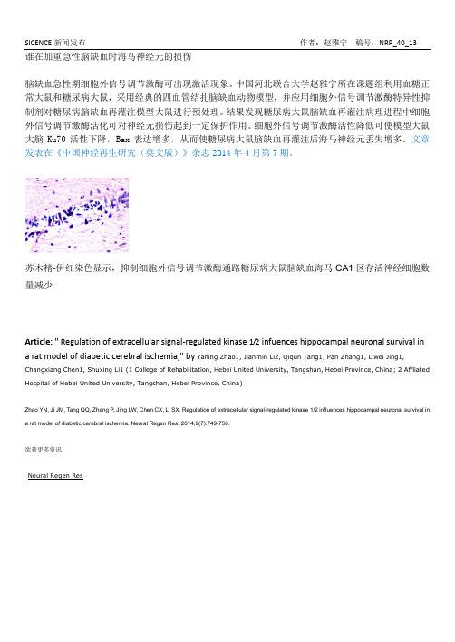
谁在加重急性脑缺血时海马神经元的损伤脑缺血急性期细胞外信号调节激酶可出现激活现象。
中国河北联合大学赵雅宁所在课题组利用血糖正常大鼠和糖尿病大鼠,采用经典的四血管结扎脑缺血动物模型,并应用细胞外信号调节激酶特异性抑制剂对糖尿病脑缺血再灌注模型大鼠进行预处理。
结果发现糖尿病大鼠脑缺血再灌注病理进程中细胞外信号调节激酶活化可对神经元损伤起到一定保护作用。
细胞外信号调节激酶活性降低可使模型大鼠大脑Ku70活性下降,Bax表达增多,从而使糖尿病大鼠脑缺血再灌注后海马神经元丢失增多。
文章发表在《中国神经再生研究(英文版)》杂志2014年4月第7期。
苏木精-伊红染色显示,抑制细胞外信号调节激酶通路糖尿病大鼠脑缺血海马CA1区存活神经细胞数量减少Article: " Regulation of extracellular signal-regulated kinase 1/2 infuences hippocampal neuronal survival in a rat model of diabetic cerebral ischemia," by Yaning Zhao1, Jianmin Li2, Qiqun Tang1, Pan Zhang1, Liwei Jing1, Changxiang Chen1, Shuxing Li1 (1 College of Rehabilitation, Hebei United University, Tangshan, Hebei Province, China; 2 Affliated Hospital of Hebei United University, Tangshan, Hebei Province, China)Zhao YN, Ji JM, Tang QQ, Zhang P, Jing LW, Chen CX, Li SX. Regulation of extracellular signal-regulated kinase 1/2 influences hippocampal neuronal survival in a rat model of diabetic cerebral ischemia. Neural Regen Res. 2014;9(7):749-756.欲获更多资讯:Neural Regen ResWhat aggravates hippocampal neuronal injury in acute cerebral ischemia?Activation of extracellular signal-regulated kinase 1/2 has been demonstrated in acute cerebral ischemia. Yaning Zhao and her colleagues, Hebei United University, China induced transientwhole-brain ischemia by four-vessel occlusion in normal and diabetic rats and intravenously injected diabetic rats with extracellular signal-regulated kinase 1/2 30 minutes before ischemia as a pretreatment. Results showed that during the pathological progression of cerebralischemia/reperfusion in rats, activation of extracellular signal-regulated kinase 1/2 exhibits protective effect on neuronal injury. Reduced extracellular signal-regulated kinase 1/2 decreases Ku70 activity, increases Bax expression and thereby increases the number of lost hippocampal neurons in diabetic rats after cerebral ischemia/reperfusion. These results were published in Neural Regeneration Research (Vol. 9, No. 7, 2014).Hematoxylin-eosin staining reveals that inhibition of extracellular signal-regulated kinase 1/2 activity aggravatesneuronal loss in the hippocampal CA1 region in a diabetic rat after cerebral ischemia/reperfusion.Article: " Regulation of extracellular signal-regulated kinase 1/2 infuences hippocampal neuronal survival in a rat model of diabetic cerebral ischemia," by Yaning Zhao1, Jianmin Li2, Qiqun Tang1, Pan Zhang1, Liwei Jing1, Changxiang Chen1, Shuxing Li1 (1 College of Rehabilitation, Hebei United University, Tangshan, Hebei Province, China; 2 Affliated Hospital of Hebei United University, Tangshan, Hebei Province, China)Zhao YN, Ji JM, Tang QQ, Zhang P, Jing LW, Chen CX, Li SX. Regulation of extracellular signal-regulated kinase 1/2 influences hippocampal neuronal survival in a rat model of diabetic cerebral ischemia. Neural Regen Res.2014;9(7):749-756.。

小鼠杏仁内侧核中白细胞介素1β的表达调控依赖于雌激素受体α(英文)张庆红;曹军;吕顺艳;黄艳红;胡玉珍;韦耿泽【期刊名称】《神经解剖学杂志》【年(卷),期】2004(20)3【摘要】研究雌激素受体 ( ER)敲除小鼠脑内,ERα和ERβ在介导内侧杏仁核中白细胞介素1β( IL -1β)表达的作用。
IL-1β表达有显著的性别差异 ,并且在 ER敲除小鼠含量减少。
细菌脂多糖 ( L PS)或卵巢切除能够促进野生型和ERβ敲除小鼠( BERKO) IL-1β表达 ,但对ERα敲除小鼠 ( ERKO)无作用。
相似的是 ,外源性雌激素能抑制野生型和 BERKO小鼠 IL -1β表达 ,后者时间稍有延搁 ,但对 ERKO IL-1β表达没有影响。
结果表明,ERα是内侧杏仁核 IL -1β表达调节的重要机制 ,提示【总页数】5页(P257-261)【关键词】小鼠;杏仁内侧核;白细胞介素1β;表达;雌激素受体;脑【作者】张庆红;曹军;吕顺艳;黄艳红;胡玉珍;韦耿泽【作者单位】第四军医大学基础医学部生理学教研室;第四军医大学口腔医院正畸科;第四军医大学西京医院妇产科【正文语种】中文【中图分类】R338.26【相关文献】1.大鼠下丘脑腹内侧核与杏仁皮质内侧核间电生理的年龄性变化 [J], 赵晓萍;滕国玺2.杏仁体内侧核损伤对小鼠社会行为的影响 [J], 王宇;李蕾;何志义3.小鼠双侧杏仁核去5-HT支配后的抑郁样行为学变化及杏仁核内FosB/ΔFosB的异常表达 [J], 陈菲菲;刘君涛;刘芳;宋亮;马传响;陈幽婷4.补肾中药对雄性大鼠杏仁核和皮质顶叶雌激素受体mRNA表达的影响 [J], 蔡晶;杜建;曹治云5.内侧杏仁核通过不同通路调控“趋向”和“回避”行为 [J], 刘峙皑;张瑛因版权原因,仅展示原文概要,查看原文内容请购买。
matlab 贝叶斯检测突变摘要:一、引言二、贝叶斯检测突变的原理三、MATLAB 在贝叶斯检测突变中的应用四、实例分析五、结论正文:一、引言在信号处理、图像处理以及数据挖掘等领域,突变检测是一项重要的研究内容。
在众多的突变检测方法中,贝叶斯检测突变凭借其强大的理论依据和实用性,成为了研究的热点之一。
而MATLAB 作为一款广泛应用于科学计算和可视化的软件,为贝叶斯检测突变提供了强大的支持。
本文将探讨贝叶斯检测突变的原理,并介绍如何利用MATLAB 实现贝叶斯检测突变。
二、贝叶斯检测突变的原理贝叶斯检测突变是基于贝叶斯公式和最小错误率原则的一种检测方法。
其基本思想是:对于给定的数据,计算各个特征在各个类别下的条件概率,找到特征与类别之间最匹配的组合,从而实现突变点的检测。
贝叶斯检测突变具有较强的理论依据,可以有效解决误检和漏检问题。
三、MATLAB 在贝叶斯检测突变中的应用MATLAB 提供了丰富的函数和工具箱,可以方便地实现贝叶斯检测突变。
以下是一个简单的MATLAB 实现贝叶斯检测突变的示例:1.读取数据:使用MATLAB 的读取函数读取数据,如使用`readtable`或`readmatrix`函数。
2.数据预处理:根据实际需求对数据进行预处理,如去除噪声、归一化等。
3.特征选择:选取与突变点相关的特征,如数据的一阶差分、二阶差分等。
4.计算条件概率:使用MATLAB 的统计函数,如`pdf`、`cdf`等,计算各个特征在各个类别下的条件概率。
5.最小错误率判别:根据最小错误率原则,找到使错误率最小的分类规则。
6.检测突变点:根据判别结果,对数据进行分类,找出突变点。
四、实例分析假设我们有两组信号数据,分别为正常信号和异常信号。
我们希望通过贝叶斯检测突变的方法,找出两组信号中的突变点。
具体操作如下:1.读取数据:使用`readtable`函数读取数据。
2.数据预处理:对数据进行归一化处理,以消除数据量纲的影响。
[键入文字]Cell:DBR1基因突变导致一些人更容易遭受病毒性大脑感染Cell:DBR1基因突变导致一些人更容易遭受病毒性大脑感染日期:2018-03-03 09:53:29 来源: 生物谷点击: 次对于之前健康的儿童来说,脑部感染是罕见的。
但每10000名接触过常见病毒(1型单纯疱疹病毒或流感病毒)的人中大约有1人会患上一种潜在致命性的疾病---脑炎。
在一项新的研究中,来自美国洛克菲勒大学等研究机构的Jean-Laurent Casanova及其同事们鉴定出就脑炎而言,单个基因发生的突变可能解释着脑干(大脑中控制着心率和呼吸等许多基本功能的部分)发生何种偏差。
Casanova实验室的临床调查助理教授Shen-Ying Zhang研究了7名来自无亲缘关系的家庭的儿童患者,这些儿童接触过一种常见病毒(1型单纯疱疹病毒,流感病毒或诺如病毒)并且遭受着威胁生命的或致命性的脑干感染。
相关研究结果发表在2018年2月22日的Cell期刊上,论文标题为Inborn Errors of RNA Lariat Metabolism in Humans with Brainstem Viral Infection 。
图片来自Cell,doi:10.1016/j.cell.2018.02.019。
这些研究人员发现一个被称作DBR1的基因发生突变,该基因产生的一种蛋白在mRNA剪接期间有助于处理mRNA中形成的环状结构。
没有这种蛋白,对病毒的免疫力会在脑干中受到选择性削弱。
Casanova的实验表明DBR1几乎完全丧失是这7名儿童患者的脑干遭受病毒入侵的罪魁祸首。
这些发现也揭示出mRNA加工机制与大脑特定区域的保护性免疫之间存在着意外之外的关联性。
这项研究是Casanova实验室正在开展的旨在鉴定出导致其他方面都健康的人患上传染病的突变的一个新的例子。
之前的研究已发现导致人体更容易遭受葡萄球菌感染、流感病毒感染和真菌感染的遗传因素。
炎症—老年痴呆,就差一个细胞因子炎症,是机体对刺激做出的一种防御反应,在生物体内起着重要的作用。
当受到刺激因子(如病毒、细菌、机械损伤、强酸强碱等)的刺激时,会诱发炎症反应,从而修复受损组织,抵抗损伤。
此外,炎症也会对机体产生不良影响,导致免疫系统紊乱。
近年来,有越来越多的研究报道了炎症与疾病的相关性,比如癌症、心血管疾病等。
而在神经退行性疾病方面,炎症也起着重要作用,但具体的分子机制,即哪些基因参与炎症调控从而影响疾病的发生发展,却是未知的。
一.现象在神经退行性疾病中,人们经常能观察到中枢神经免疫激活的现象。
β干扰素,一类免疫调节因子,在抗病毒和抗炎症反应中起到至关重要的作用。
研究者发现,缺少IFN-β,会促发一系列机制导致神经退行性疾病。
研究人员敲除了小鼠ifnb基因,ifnb-/-小鼠随着年龄的增长,行动能力、抓力和学习能力出现明显的退化。
海马组织中神经元的凋亡率上升以及数目下降。
此外,神经元的树突变少变短,黑质纹状体区域酪氨酸羟化酶(Th)活性下降,同时伴随着多巴胺含量的减少。
而帕金森病(PD)的主要特点是海马黑质区多巴胺含量减少,会影响抑制性乙酰胆碱的作用,从而导致震颤麻痹。
而Ifnb-/-小鼠的种种表型与帕金森病类似,因此该种小鼠可以作为帕金森病样老年痴呆病的动物模型用以研究帕金森病的发病机制以及治疗手段。
二.机制探索科研工作者探索了Ifnb-/-小鼠出现帕金森病老年痴呆病症状的调控机制。
通过大规模的全基因组测序,IFN-β信号通路和HD、PD、AD以及朊病毒疾病信号通路相关研究发现,Ifnb的缺失,会导致海马区α-synuclein 的积累,促进Lewy小体的形成,同时也会影响到自噬。
在自噬阻断剂及注射α-synuclein表达质粒实验中,发现自噬阻断后可以引起α-synuclein的积累,而α-synuclein表达质粒却不会影响到自噬。
因此Ifnb的缺失会影响自噬,促进α-synuclein的积累。
abdie f ghc©2004 N a t u r e P u b l i s h i n g G r o u p h t t p ://w w w .n a t u r e .c o m /n a t u r e g e n e t i c s(Supplementary Fig. 2 online). Decreased expression of BubR1 was confirmed by western-blot analysis (Fig. 1d ). BubR1 signals from Bub1b +/H , Bub1b +/–, Bub1b H/H and Bub1b –/H mouse embryonic fibroblasts (MEFs) were 42% (±15%), 29% (±9%), 11% (±3%) and 4% (±2%), respectively, of those from Bub1b +/+MEFs.We monitored 50 Bub1b +/+, 108 Bub1b +/H , 43 Bub1b +/–and 230Bub1b H/H mice to 15–16 months of age. One Bub1b H/H mouse devel-oped a life-threatening tumor. Six of 116 moribund or deceased Bub1b H/H mice had solitary tumors at autopsy. Bub1b H/H mice had a normal appearance until 2–3 months of age (Fig. 1f ; compare with wild-type mouse in Fig. 1e ) but typically developed cachexia and lor-dokyphosis at 3–6 months of age (Fig. 1h and Supplementary Fig. 2online; compare with wild-type mouse in Fig. 1g ). The median lifespan of Bub1b H/H mice was ∼6 months, compared with more than 15 monthsabcd fefor Bub1b +/–, Bub1b +/H and Bub1b +/+mice (Fig. 1i ). At age 2 months and older, Bub1b H/H mice developed progressive bilateral cataracts with features reminiscent of age-related human cataracts (Fig . 2a ). No cataracts were observed in Bub1b +/–, Bub1b +/H or Bub1b +/+mice.Histological analysis of skin showed that the dermis and subcutaneous fat cell layers were significantly thinner in 12-month-old Bub1b H/H mice than in control mice (Fig. 2b ,c and Supplementary Fig. 2online).Skinning of Bub1b H/H mice confirmed substantial loss of subdermal adipose tissue and highlighted their severe spinal kyphosis and muscle atrophy (Fig. 2d ). Dual-energy X-ray absorptiometry measurements confirmed that total body fat of Bub1b H/H mice declined prematurely (Fig. 2e ). Bub1b H/H mice also had less ability to repair wounds at an early age (Fig. 2f ). Taken together, these results (Supplementary Table 1online) suggest that multiple aging-associated phenotypes develop early in mice expressing BubR1 below a threshold level.To investigate whether the progeroid phenotypes could be due to a defective spindle assembly checkpoint, we measured the ability of Bub1b -mutant MEFs to induce a sustained preanaphase arrest in the presence of nocodazole. Approximately 11–12% of Bub1b +/+,Bub1b +/H and Bub1b +/–MEFs were arrested by 12 h (Fig. 3a ). InFigure 2 Aging phenotypes in Bub1b H/H mice. (a ) Cross-sections of lenses from Bub1b H/H and Bub1b +/+mice at various ages stained with hematoxylin and eosin. Bub1b H/H lenses were normal at day 10 but showed Morgagnian globules at day 60 (arrowheads), overt cataracts at day 120 and advanced stage nuclear cataracts at day 270. (b ) Dermal layer thickness of Bub1b H/H and Bub1b +/+mice at indicated ages (n =3 mice per group).(c ) Subcutaneous adipose layer thickness of Bub1b H/H and Bub1b +/+mice at indicated ages (n =3 mice per group). (d )Skinned 9-month-old Bub1b +/+(top) and Bub1b H/H (bottom) females. The Bub1b H/H mouse had little subcutaneous fat (arrows) and pronounced kyphosis (arrowheads).(e )Analysis of total body fat of Bub1b +/+and Bub1b H/H females at indicated ages (n =3 mice per genotype and age group). (f ) Closure of 3-mm punch biopsy wounds in Bub1b +/+and Bub1b H/H mice at 2 months (left panel) and 12 months (right panel) of age.abc de fFigure 3 Bub1b H/H cells have mitotic checkpoint defects. (a ) Mitotic index (defined as the percentage of mitotic cells) of nocodazole-treated MEF lines of indicated genotypes (n =3 for each genotype). The growth rates of these MEF lines were similar (growth curves not shown). (b ) Cyclin B–associated Cdc2 kinase activity of synchronized MEF cells at indicated time points after release into medium with high serum and nocodazole. The asterisk marks an aspecific spot on the autoradiogram.(c ) Spectral karyotype of a numerically normal Bub1b H/H metaphase with a gain of chromosome 3 and a loss of chromosome 14. (d ) Spectral image of a Bub1b H/H metaphase (MEF) that shows PMSCS. (e )Bub1b H/H MEF cell with a lagging chromosome in anaphase. DNA was stained with Hoechst. The arrowhead highlights alagging chromosome. (f )The average percentage of anaphases with lagging chromosomes per genotype.©2004 N a t u r e P u b l i s h i n g G r o u p h t t p ://w w w .n a t u r e .c o m /n a t u r e g e n e t i c scontrast, only 2.5% of Bub1b H/H MEFs were arrested by 12 h, sug-gesting that spindle assembly checkpoint function was severely compromised. We confirmed this result by flow cytometry (Supplementary Fig. 3 online). Next, we synchronized MEFs in G1and analyzed their cyclin B–associated Cdc2 kinase activity after releasing them into medium with nocodazole. Bub1b H/H cells had high Cdc2 kinase activity by 24–30 h after release, similar to Bub1b +/+cells (Fig. 3b ). Bub1b +/+cells sustained high levels of Cdc2activity until 42 h after restimulation, but Bub1b H/H cells did not maintain activity after 30 h, consistent with a spindle assembly checkpoint defect 8,9.The average percentage of aneuploid metaphases was much higher in passage 5 (P5) Bub1b H/H MEFs than in P5 Bub1b +/–, Bub1b +/H and Bub1b +/+MEFs (Table 1a ). We observed even more profound aneu-ploidy in P5 Bub1b –/H MEFs. We also carried out spectral karyotype analysis on metaphase spreads from Bub1b +/+and Bub1b H/H MEFs.Bub1b +/+metaphases (n =9) were karyotypically normal. In contrast,five of ten Bub1b H/H metaphases had numerical abnormalities.Furthermore, two numerically normal Bub1b H/H metaphases showed loss of one chromosome and gain of another (Fig. 3c ). We observed premature sister chromatid separation (PMSCS), a hallmark of a defective spindle assembly checkpoint 8,9, in 38% and 15% of mitotic figures from Bub1b –/H and Bub1b H/H MEFs, respectively, but in only 1–3% of Bub1b +/–, Bub1b +/H and Bub1b +/+MEFs (Table 1a and Fig.3d ). As expected, we observed more anaphase figures with lagging chromosomes in Bub1b –/H and Bub1b H/H cells than in Bub1b +/–,Bub1b +/H or Bub1b +/+cells (Fig. 3e ,f ). Together, these data suggest that the accuracy of chromosome segregation is greatly affected when BubR1 levels drop below a certain level.Bub1b –/H pups showed substantial aneuploidy at birth, but Bub1b H/H mice did not (Table 1b ). At age 2 months, however,Bub1b H/H mice had developed mild aneuploidy that increased in both degree and severity as mice aged further. Bub1b +/–and Bub1b +/H mice did not have detectable aneuploidy. We observed no genome mainte-nance defects other than chromosome number instability in Bub1b H/H and Bub1b –/H cells (Table 1a and Supplementary Fig. 4 online). The strong correlation between the onset and progression of the aging-associated phenotypes and the degree and the severity of the chromo-some number instability in Bub1b H/H mice are suggestive of a role for aneuploidy stemming from BubR1 dysfunction in the development of progeroid features.We next determined whether BubR1 deficiency triggers apoptosis or cellular senescence, two responses that have been linked to aging 10,11. We found similar numbers of apoptotic cells in kidney,liver and lung sections from 1-year-old Bub1b H/H and Bub1b +/+mice,and in Bub1b H/H and Bub1b +/+MEF cultures (data not shown).Senescence-associated β-galactosidase activity was high in kidney sections from 5-month-old Bub1b H/H mice, but not in those from age-matched Bub1b +/+mice and 2-month-old Bub1b H/H mice (Fig.4a ). P3 Bub1b H/H MEFs had comparable growth rates (Fig. 4d ) and numbers of cells positive for senescence-associated β-galactosidase activity to those of Bub1b +/+MEFs (Fig . 4b ). At P7, however,©2004 N a t u r e P u b l i s h i n g G r o u p h t t p ://w w w .n a t u r e .c o m /n a t u r e g e n e t i c sBub1b H/H MEF cultures had substantially slower growth rates and more cells positive for senescence-associated β-galactosidase activity than Bub1b +/+cultures (Fig. 4b ,d ). Bub1b –/H MEFs had even more profound growth inhibition and senescence-associated β-galactosi-dase activity (Fig. 4b ,d ), suggesting that the rate of senescence corre-lates with the degree of aneuploidy. Bub1b H/H MEFs senesced quickly in both 20% and 3% oxygen (Fig. 4c ), indicating that oxidative DNA damage 12is an unlikely cause of the senescence phenotype. Bub1b H/H MEFs showed early accumulation of the senescence markers p53, p21,p16 and p19 (Fig. 4e ). Taken together, these results suggest that aneu-ploidy in cells from BubR1-insufficient mice might elicit signals that drive them into a senescent state and cause early aging-related pheno-types as they accumulate.Bub1b H/H mice did not produce any pregnancies. Testicular weight of Bub1b H/H males (0.088 g ±0.009; n =8 testes) was slightly below that of Bub1b +/+males (0.106 g ±0.006; n =8 testes). Testes of 4-month-old Bub1b H/H mice were histologically normal (data not shown), but sperm counts of Bub1b H/H mice (9.3 ×10⁶±2.6 ×10⁶; n =3) were about four times lower than those of Bub1b +/+mice (37.1×10⁶±8 ×10⁶; n =3). Bub1b H/H spermatozoa had normal motility and morphology (data not shown) and were able to attach to and fertilize Bub1b +/+eggs in vitro , but they produced 2-cell-stage embryos at 13 times less frequently than Bub1b +/+spermatozoa (Fig. 5a ). Chromosome counts on metaphases of spermatocytes in meiosis II showed that 5% of Bub1b H/H karyotypes had abnormal chromosome numbers compared with 0% of Bub1b +/+karyotypes (Fig. 5b ). Thus, reduced expression of BubR1 seems to affect male fertility at the levels of meiotic chromosome segregation, sperm number and fertilization.Ovaries from 4-month-old Bub1b H/H mice appeared histologically normal (data not shown) and were capable of producing mature eggs that arrested at metaphase of meiosis II. But we observed highly abnormal metaphase II configurations in 9 of 13 arrested Bub1b H/H oocytes (Fig. 5c ). Oocytes from age-matched Bub1b +/+mice (n =14)yielded normal metaphase II configurations. We conclude that infer-tility in female mice expressing low levels of BubR1 is caused, at least in part, by meiotic chromosome segregation defects.T o determine whether reduced expression of BubR1 might have a role in natural aging, we measured BubR1 levels in wild-type mice of various ages. BubR1 was high in testes of 4-month-old mice but gradually decreased to very low levels as mice aged up to 32 months (Fig. 5d ). In contrast, the spindle assembly checkpoint proteins Bub3 and Rae1remained highly expressed as mice aged (Fig. 5d ). Reductions in BubR1expression were also seen in ovaries and spleens of 22-month-old mice,but not in lungs (Fig. 5e ,f and data not shown). Overall, these data are consistent with the idea that BubR1 might have a role in normal aging.The finding that very few Bub1b H/H mice had detectable tumors when they died, despite substantial chromosome number instabil-ity, is unexpected because aneuploidy is a hallmark of most human cancers 3,13,14, and because Bub1b is mutated or expressed at low levels in a subset of colorectal carcinomas with chromosomal insta-bility 15,16. Instead, BubR1-deficient mice have early onset of several aging-associated phenotypes and have severely shortened lifespans.This, together with the demonstration that BubR1 expression declines in several tissues as wild-type mice age, suggests that this checkpoint protein may be a key regulator of normal aging.Reproductive aging in female mammals occurs relatively early in life and is characterized by the production of increasing numbers of aneuploid oocytes. In humans, this leads to increased abortions and birth defects, such as Down syndrome 1. Chromosomal segre-gation defects associated with reproductive aging are reminiscent of those seen in oocytes of mutant mice expressing low levels of BubR1. Given this age-dependent decline in ovarian BubR1, we propose that downregulation of BubR1 might be a mechanism that contributes to age-related female infertility and certain birth defects. Paternal fertility also declines with advancing age 17,18, and a regulatory role for BubR1 is certainly conceivable, given the decline in BubR1 levels in testes of aging normal mice and the neg-ative impact of BubR1-deficiency on fertility of male mutant mice.METHODSGeneration of BubR1 mutant mice. We used a previously reported gene target-ing strategy to create a hypomorphic Bub1b allele in mouse embryonic stem cells 19(for details, see Supplementary Methods and Supplementary Fig. 1abcd eFigure 4 BubR1 insufficiency causes early onset of senescence. (a ) Senescence-associated β-galactosidase activity in kidney sections of 2- and 5-month-old Bub1b H/H and Bub1b +/+mice, and in P8 Bub1b H/H and Bub1b +/+MEFs.(b ) Percentages of Bub1b –/H , Bub1b H/H andBub1b +/+MEF cells cultured in 20% oxygen that were positive for senescence-associated β-galactosidase activity. Passages at which measurements were made are indicated.(c ) Percentages of MEF cells of indicated genotypes cultured in 3% oxygen that were positive for senescence-associated β-galactosidase activity. (d ) Growth curves of P3(left panel) and P7 (right panel) MEF cells of indicated genotypes. (e ) Expression ofsenescence-associated markers in Bub1b H/H and Bub1b +/+MEF cells at P2–P5.©2004 N a t u r e P u b l i s h i n g G r o u p h t t p ://w w w .n a t u r e .c o m /n a t u r e g e n e t i c sonline). Bub1b H/H mice obtained from four independently targeted embryonic stem cell clones had identical phenotypes. We monitored body weight in ten females of each genotype for 70 d. All mice were housed in a pathogen-free bar-rier environment for the duration of the study. Experimental procedures involving laboratory mice were reviewed and approved by the Institutional Animal Care and Use Committee of the Mayo Clinic.Western-blot analysis. We carried out western-blot analyses as previously described 20. We used affinity-purified rabbit antibody against mouse BubR1(382–420) to detect BubR1. We quantified BubR1 signals (n =6per genotype)by the use of Bio-Rad Quantity One Software (version 4.1.0) and normalized them to β-actin (AC-151, Sigma; 1:40,000 dilution). We also probed some blots with α-tubulin (T-9026, Sigma; 1:2,000 dilution) as a loading control. We used antibody against human BubR1 (1–350) to exclude production of truncated BubR1 products by Bub1b H and Bub1b –alleles. To quantify senescence-associ-ated proteins in various MEF lysates, we used the following antibodies at 1:200dilution (purchased from Santa Cruz unless noted otherwise): p16 (M-156; sc-1207), p19 (NB200-106; Novus Biologicals), p21 (M-19; sc-471), p53 (Fl-393-G, sc-6243-G). We detected Bub3 and Rae1 as previously described 8.Histopathology. We screened all major organs for overt tumors using a dissec-tion microscope and processed the collected tumors routinely for histopatho-logical confirmation. We fixed dissected tissues for histology in 10% formalin,processed them and embedded them in paraffin. We cut 5-µm sections of all tis-sues and stained them with hematoxylin and eosin using standard procedures.We stained dorsal skin sections and determined the thickness of the dermal and adipose layers by taking 40 random measurements of each mouse for each geno-type and age group (n =3). Calculations were done using a calibrated computer program (Spot Advanced by BioSpot). We stained ovary sections with affinity-purified antibody against BubR1 (382-420) as previously described 21.Bone and fat analyses by dual-energ y X-ray absorptiometry. We analyzed bone mineral content, bone mineral density and total body adipose using a LUNAR PIXI-mus small animal densitometer (LUNAR Corporate Headquarters) as described 22in three anesthetized mice (with avertin) of each genotype and age.Wound healing analysis. We analyzed the ability of mice to repair wounds as previously described 23. We introduced 3-mm punch biopsy wounds into dorsalskin of anesthetized mice. For a period of 6 d, we measured wound diameters using a digital caliper. Bub1b H/H mice did not survive at a standard anesthetic dose of 375 µg of avertin per g body weight but did survive with half that dose.Generation and culture of MEFs. We intercrossed Bub1b +/H mice to derive Bub1b +/+, Bub1b +/H and Bub1b H/H MEFs from individual 13.5-d-old fetuses.We intercrossed Bub1b +/–and Bub1b +/+mice to generate Bub1b +/+and Bub1b +/–MEFs, and Bub1b +/–and Bub1b +/H mice to generate Bub1b –/H MEFs.They were cultured in 20% oxygen, frozen at P2 and P3 and used for experi-mentation at indicated passages. For the studies described, we examined at least three Bub1b H/H , Bub1b +/+and Bub1b +/–clones. We generated growth curves using P3 and P7 MEFs. At day 0, we plated 1.5 ×10⁵MEFs per 35-mm dish and counted duplicate cultures at 24-h intervals thereafter (n =3 MEF lines per genotype). In certain experiments, we wanted to limit senescence due to oxida-tive stress 12, and so we used MEFs that were generated and cultured in 3% oxy-gen. MEFs were synchronized as previously described 8. We washed confluent cultures three times with phosphate-buffered saline and then cultured them in Dulbecco’s modified Eagle medium containing 0.1% fetal bovine serum and 0.04% bovine serum albumin for 18 h. We treated quiescent MEFs with trypsin and reseeded them in Dulbecco’s modified Eagle medium with 10% fetal bovine serum to allow their reentry into the cell cycle.Spindle assembly checkpoint analyses. We measured mitotic index (n =3MEF lines per genotype) and carried out FACS-based analysis of spindle assem-bly checkpoint activity as previously described 8. We carried out Cdc2-kinase assays as previously described 24.Karyotype analyses. We prepared metaphase spreads from MEFs and spleno-cytes and analyzed them for aneuploidy and PMSCS as previously described 8.For chromosome number analysis of spermatocytes, we minced testes between two microscope slides and instantly prepared metaphase spreads from the resulting cell suspensions. We carried out spectral karyotypic analysis of MEFs using the protocol, reagents, instrumentation and software from Applied Spectral Imaging.Analysis of DNA repair functions. We carried out DNA damage MEF sur-vival experiments as described 25,26. For mitomycin C (Sigma) and paraquat (Sigma) survival experiments, we seeded 104P2 MEFs in triplicate in a 96-well flat-bottom tissue culture dish. The next day, we replaced the mediuma cbd efFigure 5 Analysis of BubR1 in testis and ovary of mutant and normal mice. (a ) In vitro fertilization experiments with eggs from Bub1b +/+females and sperm from Bub1b +/+or Bub1b H/H males.(b ) Karyotype analysis of Bub1b +/+and Bub1b H/H spermatocytes at prometaphase of meiotic division II. Abnormal Bub1b H/H karyotypes showed either a gain or loss of one duplicated chromosome. (c ) Oocytes from Bub1b H/H mice arrested at metaphase II stained with α-tubulin and Hoechst.Normal metaphase configuration (left panel), metaphase with misalignedchromosomes (middle panel) and metaphase with an extra spindle (arrow) and misaligned chromosomes (arrowheads; right panel) areshown. (d ) Western-blot analyses of BubR1 levels in testis of young and old Bub1b +/+mice.(e ) Western-blot analyses of BubR1 levels in ovary of young and old Bub1b +/+mice. (f ) Cross-sections through ovary from young and oldBub1b +/+mice immunostained for BubR1 (green)and DNA (blue). Representative corpora lutea are shown.©2004 N a t u r e P u b l i s h i n g G r o u p h t t p ://w w w .n a t u r e .c o m /n a t u r e g e n e t i c swith drug-containing medium and allowed cells to grow for 72 h. For ultravi-olet-B and γ-irradiation experiments, we exposed 2 ×10⁴P2 MEFs to various doses of ultraviolet-B or ionizing irradiation, seeded them in triplicate and cultured them for 72 h. We measured cell survival by the use of the MTS assay (Promega). Data represent three independent MEF lines of each genotype.For γ-irradiation colony forming assays, we seeded 4 ×10⁴MEF cells in dupli-cate in 10-cm tissue culture dishes and exposed them to various doses of ion-izing radiation at P2. We grew cells in 3% oxygen for 14 d and counted colonies. In the colony-forming assay after treatment with mitomycin C, we seeded 4 ×10⁴P2 MEF cells in duplicate in 10-cm tissue culture dishes and allowed them to grow for 24 h. The indicated concentration of medium con-taining mitomycin C was added and cells were allowed to grow for 14 d in 3%oxygen. For the highest doses of γ-irradiation and mitomycin C, we used five times as many cells. We counted colonies containing >20 cells after staining with Coomassie blue.Senescence-associated β-g alactosidase staining. We stained cryosections of mouse kidney for senescence-associated β-galactosidase activity accord-ing to manufacturer’s protocol (Cell Signaling Technology). To quantify MEFs stained for senescence-associated β-galactosidase activity, we coun-terstained cells with Hoechst to visualize nuclei. The percentage of senes-cent cells was the total number of senescent cells divided by the total number of cells counted using immunofluorescence (n =3 MEF lines per genotype).Collection and analysis of metaphase II oocytes and in vitro fertilization. We collected ovulated oocytes and instantly fixed them as previously described 27.We stained fixed oocytes with α-tubulin (1:1,000 dilution of T-9026, Sigma)and Hoechst and analyzed them by confocal microscopy. We carried out in vitro fertilization experiments as previously described 28. We obtained aged testes from the aged rodent tissue bank maintained by the National Institute of Aging (Bethesda).Note: Supplementary information is available on the Nature Genetics website.ACKNOWLEDGMENTSWe thank J. Harden, R. Babu, M. Gazi, S. Guyse, M. Pittelkow, R. Goorha,J. Davenport, D. Pearson, K. Hafner (Cytogenetics Shared Resource), J. van Ree and S. Kaufman for supporting this project; R. Bram for critically reviewing the manuscript and for discussions; and T. Yen for providing BubR1 antibody. This work was supported by a grant from the US National Institutes of PETING INTERESTS STATEMENTThe authors declare that they have no competing financial interests.Received 30 March; accepted 7 May 2004Published online at /naturegenetics/1.Hassold, T. & Hunt, P . To err (meiotically) is human: the genesis of human aneu-ploidy. Nat. Rev. Genet.2, 280–291 (2001).2.Thomas, N.S. et al . Maternal sex chromosome non-disjunction: evidence for X chro-mosome-specific risk factors. Hum. Mol. Genet.10, 243–250 (2001).3.Jallepalli, P .V. & Lengauer, C. Chromosome segregation and cancer: cutting throughthe mystery. Nat. Rev. Cancer 1, 109–117 (2001).4.Wassmann, K. & Benezra, R. Mitotic checkpoints: from yeast to cancer. Curr. Opin.Genet. Dev.11, 83–90 (2001).5.Yu, H. Regulation of APC-Cdc20 by the spindle checkpoint. Curr. Opin. Cell Biol.14,706–714 (2002).6.Cleveland, D.W., Mao, Y. & Sullivan, K.F . Centromeres and kinetochores: from epige-netics to mitotic checkpoint signaling. Cell 112, 407–421 (2003).7.Wang, Q. et al . BUBR1 deficiency results in abnormal megakaryopoiesis. Blood 103,1278–1285 (2004).8.Babu, J.R. et al . Rae1 is an essential mitotic checkpoint regulator that cooperateswith Bub3 to prevent chromosome missegregation. J. Cell Biol.160, 341–353(2003).9.Michel, L.S. et al . MAD2 haplo-insufficiency causes premature anaphase and chro-mosome instability in mammalian cells. Nature 409, 355–359 (2001).10.Hasty, P ., Campisi, J., Hoeijmakers, J., van Steeg, H. & Vijg, J. Aging and genomemaintenance: lessons from the mouse? Science 299, 1355–1359 (2003).11.Campisi, J. Cancer and ageing: rival demons? Nat. Rev. Cancer 3, 339–349 (2003).12.Parrinello, S. et al . Oxygen sensitivity severely limits the replicative lifespan of murinefibroblasts. Nat. Cell Biol.5, 741–747 (2003).13.Lengauer, C., Kinzler, K.W. & Vogelstein, B. Genetic instabilities in human cancers.Nature 396, 643–649 (1998).14.Li, R., Sonik, A., Stindl, R., Rasnick, D. & Duesberg, P . Aneuploidy vs. gene mutationhypothesis of cancer: recent study claims mutation but is found to support aneu-ploidy. Proc. Natl. Acad. Sci. USA 97, 3236–3241 (2000).15.Cahill, D.P . et al . Mutations of mitotic checkpoint genes in human cancers. Nature392, 300–303 (1998).16.Shichiri, M., Yoshinaga, K., Hisatomi, H., Sugihara, K. & Hirata, Y. Genetic and epi-genetic inactivation of mitotic checkpoint genes hBUB1 and hBUBR1 and their rela-tionship to survival. Cancer Res.62, 13–17 (2002).17.Ford, W.C. et al . Increasing paternal age is associated with delayed conception in alarge population of fertile couples: evidence for declining fecundity in older men. The ALSPAC Study Team (Avon Longitudinal Study of Pregnancy and Childhood). Hum.Reprod.15, 1703–1708 (2000).18.Eskenazi, B. et al . The association of age and semen quality in healthy men. Hum.Reprod.18, 447–454 (2003).19.Meyers, E.N., Lewandoski, M. & Martin, G.R. An Fgf8 mutant allelic series generatedby Cre- and Flp-mediated recombination. Nat. Genet.18, 136–141 (1998).20.Kasper, L.H. et al . CREB binding protein interacts with nucleoporin-specific FGrepeats that activate transcription and mediate NUP98-HOXA9 oncogenicity. Mol.Cell. Biol.19, 764–776 (1999).21.Feldmann, K.A., Pittelkow, M.R., Roche, P .C., Kumar, R. & Grande, J.P . Expression ofan immediate early gene, IEX-1, in human tissues. His tochem. Cell Biol.115,489–497 (2001).22.Nagy, T.R. & Clair, A.L. Precision and accuracy of dual-energy X-ray absorptiometryfor determining in vivo body composition of mice. Obes. Res.8, 392–398 (2000).23.Tyner, S.D. et al . p53 mutant mice that display early ageing-associated phenotypes.Nature 415, 45–53 (2002).24.Wassmann, K. & Benezra, R. Mad2 transiently associates with an APC/p55Cdc com-plex during mitosis. Proc. Natl. Acad. Sci. USA 95, 11193–11198 (1998).25.Koomen, M. et al . Reduced fertility and hypersensitivity to mitomycin C characterizeFancg/Xrcc9 null mice. Hum. Mol. Genet.11, 273–281 (2002).26.de Boer, J. et al . Premature aging in mice deficient in DNA repair and transcription.Science 296, 1276–1279 (2002).27.Woods, L.M. et al . Chromosomal influence on meiotic spindle assembly: abnormalmeiosis I in female Mlh1 mutant mice. J. Cell Biol.145, 1395–1406 (1999).28.Kang-Decker, N., Mantchev, G.T., Juneja, S.C., McNiven, M.A. & van Deursen, J.M.Lack of acrosome formation in Hrb-deficient mice. Science 294, 1531–1533 (2001).©2004 N a t u r e P u b l i s h i n g G r o u p h t t p ://w w w .n a t u r e .c o m /n a t u r e g e n e t i c s。