caspase1抗体说明书
- 格式:pdf
- 大小:84.73 KB
- 文档页数:1
Caspase活性检测(基于p NA标记底物的比色法)在caspase 的底物多肽上标记发色团如:p NA,可用于检测凋亡细胞中的caspase活性及筛选caspases的激活剂或抑制剂。
本操作步骤主要针对使用酶标仪进行读数而设计。
其中使用的细胞抽提物、底物、抑制剂及p NA的用量为每个样本140ul。
若您希望设计针对分光光度计读数的实验,建议对每种溶液体积进行进一步优化。
所需溶液、试剂及设备• Caspase底物• Caspase抑制剂• 细胞裂解缓冲液:50mM HEPES, pH7.4, 100mM NaCl, 0.1%CHAPS, 1mM DTT, 100uM EDTA • 检测缓冲液:50mM HEPES, pH7.4, 100mM NaCl, 0.1%CHAPS, 10mM DTT, 100uM EDTA, 10%Glycerol• PBS:将8g NaCl、200mg KCl和1.44g Na2HPO4溶于800ml蒸馏水;用HCl调至pH7.4;定容至1L• 酶标板和酶标仪其它试剂(推荐)• 纯化的Caspase(阳性对照)• p NA细胞抽提物的制备以适合的凋亡诱导剂对细胞进行诱导(参考前述操作方法),同时做阴性对照,包括未处理细胞、用诱导剂的无诱导活性类似物处理细胞(若可得到),或一个凋亡诱导处理“零时间”样品。
应保证有足够细胞进行重复实验,包含或不含抑制剂的对照以检测蛋白浓度。
一般来说,我们下面推荐的裂解前细胞数量可以产生足够蛋白浓度供酶标仪检测;然而不同大小、体积的细胞及蛋白浓度可能需要增加铺板密度。
当裂解2×10⁷/ml细胞是,产生的蛋白浓度约为1-3mg/ml,体积10ul,含约10-30ug蛋白。
依据其细胞系,最少应有106个细胞,体积50ul的裂解缓冲液进行处理。
• 细胞计数,离心收集细胞。
以PBS缓冲液洗涤一次。
若细胞已被caspase抑制剂处理过,建议多洗几次以防止后继实验中可能造成的不利影响。

Caspase 1活性检测试剂盒(比色法)简介:Caspase(Cysteine-requiring Aspartate Protease)家族在介导细胞凋亡过程的起着极其重要的作用,其成员包括Caspase1~11等,均属于蛋白酶家族。
Caspase 1又称Interkeukin 1 conberting enzyme (ICE)或caspase-1,是唯一可以剪切IL-1前体蛋白或IL-18前体产生相应成熟细胞因子的Caspase 家族成员。
Caspase 1通过剪切Bcl-XL 来调节细胞凋亡,并通过对一些细胞因子前体的剪切来调控相关免疫反应。
Caspase 1活性检测试剂盒(比色法) (Caspase 1 Colorimetric Assay Kit) 的检测原理是利用Caspase 1催化底物 Ac-YVAD-p NA)产生黄色的游离硝基苯胺p NA(p -nitroaniline),通过测定分光光度比色法测定p NA 在400~410nm 处吸光值,从而间接获得caspase 1的活性。
组成:操作步骤(仅供参考):1、 制备标准曲线:① 配制适量的标准品稀释液。
② 用标准品稀释液稀释p NA(10mM),使p NA 分别达到200μM 、100μM 、50μM 、25μM 、12.5μM 、6.25μM ,另外设置一管不加p NA 仅含标准品稀释液作为零管,把以上系列浓度物质作为标准品。
③ 取p NA 浓度分别为200μM 、100μM 、50μM 、25μM 、12.5μM 、6.25μM 、0的标准品各100 μl 加入至96孔板或取适量体积加入至容量不超过100μl 的比色杯,测定405nm 处吸光值即A 405。
④ 用每一个标准品的A 405减去不含p NA 的空白对照的A 405,计算出实际的吸光值。
以每个浓度的标准品A 405为x 轴,以对应的p NA 浓度为y 轴,用Excel 制作p NA 浓度对A 405值的标准曲线。
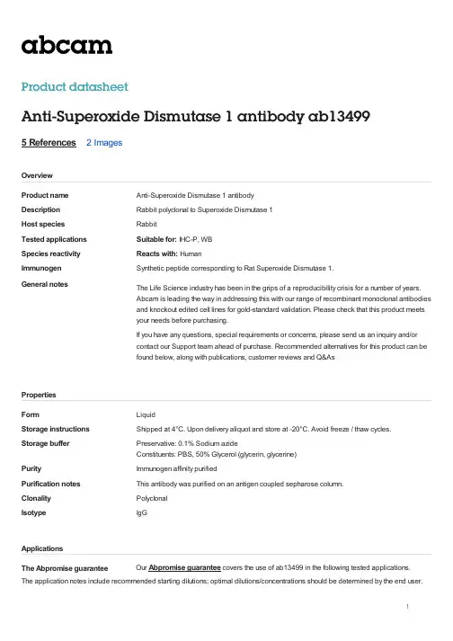
Product nameAnti-Superoxide Dismutase 1 antibody DescriptionRabbit polyclonal to Superoxide Dismutase 1Host speciesRabbit Tested applicationsSuitable for: IHC-P, WB Species reactivityReacts with: Human ImmunogenSynthetic peptide corresponding to Rat Superoxide Dismutase 1.General notes The Life Science industry has been in the grips of a reproducibility crisis for a number of years.Abcam is leading the way in addressing this with our range of recombinant monoclonal antibodiesand knockout edited cell lines for gold-standard validation. Please check that this product meetsyour needs before purchasing.If you have any questions, special requirements or concerns, please send us an inquiry and/orcontact our Support team ahead of purchase. Recommended alternatives for this product can befound below, along with publications, customer reviews and Q&AsFormLiquid Storage instructionsShipped at 4°C. Upon delivery aliquot and store at -20°C. Avoid freeze / thaw cycles.Storage bufferPreservative: 0.1% Sodium azide Constituents: PBS, 50% Glycerol (glycerin, glycerine)PurityImmunogen affinity purified Purification notesThis antibody was purified on an antigen coupled sepharose column.ClonalityPolyclonal Isotype IgGThe Abpromise guarantee Product datasheetAnti-Superoxide Dismutase 1 antibody ab134995 References 2 ImagesOverviewPropertiesApplicationsOur Abpromise guarantee covers the use of ab13499 in the following tested applications.The application notes include recommended starting dilutions; optimal dilutions/concentrations should be determined by the end user.Function Destroys radicals which are normally produced within the cells and which are toxic to biologicalsystems.Involvement in disease Defects in SOD1 are the cause of amyotrophic lateral sclerosis type 1 (ALS1) [MIM:105400].ALS1 is a familial form of amyotrophic lateral sclerosis, a neurodegenerative disorder affectingupper and lower motor neurons and resulting in fatal paralysis. Sensory abnormalities are absent.Death usually occurs within 2 to 5 years. The etiology of amyotrophic lateral sclerosis is likely tobe multifactorial, involving both genetic and environmental factors. The disease is inherited in 5-10% of cases leading to familial forms.Sequence similarities Belongs to the Cu-Zn superoxide dismutase family.Post-translational modifications Unlike wild-type protein, the pathogenic variants ALS1 Arg-38, Arg-47, Arg-86 and Ala-94 are polyubiquitinated by RNF19A leading to their proteasomal degradation. The pathogenic variants ALS1 Arg-86 and Ala-94 are ubiquitinated by MARCH5 leading to their proteasomal degradation.The ditryptophan cross-link at Trp-33 is reponsible for the non-disulfide-linked homodimerization. Such modification might only occur in extreme conditions and additional experimental evidence is required.Cellular localization Cytoplasm. The pathogenic variants ALS1 Arg-86 and Ala-94 gradually aggregates andaccumulates in mitochondria.Western blot - Anti-Superoxide Dismutase 1 antibody (ab13499)Anti-Superoxide Dismutase 1 antibody (ab13499) at 1 µg/ml + Cell lysates prepared from mixed human cell lines (A431, A549,HCT116, HeLa, HEK293, HepG2, HL-60, HUVEC, Jurkat, MCF7, PC3 and T98G)Predicted band size: 18 kDaApplication Abreviews NotesIHC-P Use a concentration of 2 µg/ml. Perform heat mediated antigenretrieval before commencing with IHC staining protocol.WB Use a concentration of 1 µg/ml. Detects a band of approximately19, 23 kDa (predicted molecular weight: 18 kDa).TargetImagesImmunohistochemistry (Formalin/PFA-fixed paraffin-embedded sections) - Anti-Superoxide Dismutase 1 antibody (ab13499)Ab13499 staining Human normal colon. Staining is localized to the nucleus.Left panel: with primary antibody at 2 ug/ml. Right panel: isotype control.Sections were stained using an automated system DAKO Autostainer Plus , at room temperature. Sections were rehydrated and antigen retrieved with the Dako 3-in-1 AR buffer citrate pH 6.0 in a DAKO PT Link. Slides were peroxidase blocked in 3% H2O2 in methanol for 10 minutes. They were then blocked with Dako Protein block for 10 minutes (containing casein 0.25% in PBS) then incubated with primary antibody for 20 minutes and detected with Dako Envision Flex amplification kit for 30 minutes. Colorimetric detection was completed with Diaminobenzidine for 5 minutes. Slides were counterstained with Haematoxylin and coverslipped under DePeX. Please note that for manual staining we recommend to optimize the primary antibody concentration and incubation time (overnight incubation), and amplification may be required.Please note: A ll products are "FOR RESEA RCH USE ONLY. NOT FOR USE IN DIA GNOSTIC PROCEDURES"Our Abpromise to you: Quality guaranteed and expert technical supportReplacement or refund for products not performing as stated on the datasheetValid for 12 months from date of deliveryResponse to your inquiry within 24 hoursWe provide support in Chinese, English, French, German, Japanese and SpanishExtensive multi-media technical resources to help youWe investigate all quality concerns to ensure our products perform to the highest standardsIf the product does not perform as described on this datasheet, we will offer a refund or replacement. For full details of the Abpromise, please visit https:///abpromise or contact our technical team.Terms and conditionsGuarantee only valid for products bought direct from Abcam or one of our authorized distributors。

caspass1通路调控机制
Caspase 1 是一种重要的炎症相关蛋白酶,参与调节炎症反应和免疫应答。
Caspase 1的活性受到多种机制的调控。
1. 转录调控:Caspase 1基因的表达受到多个转录因子的调控,如NF-κB、AP-1等,它们能够结合在Caspase 1基因的启动子区域上,促进或抑制Caspase 1的转录。
2. 翻译后修饰:Caspase 1的活性可以通过翻译后修饰来调控。
例如,Caspase 1的自我断裂(autocleavage)是其激活的重要步骤之一,这一过程受到其他蛋白酶的调控,如Caspase 8、Caspase 11等。
3. 组蛋白修饰:组蛋白修饰也参与了Caspase 1的调控。
研究表明,组蛋白去乙酰化修饰酶(HDACs)和组蛋白甲基转移酶(HMTs)可以调控Caspase 1特定位点上的甲基化、乙酰化等修饰,从而影响Caspase 1的活性和功能。
4.反式调控:一些信号通路分子可以与Caspase 1发生反式调控,调节Caspase 1的活性与功能。
例如,NLRP3炎症小体信号通路被认为是Caspase 1激活的一个重要启动机制。
总的来说,Caspase 1的活性和表达受到多种机制的调控,包括转录调控、翻译后修饰、组蛋白修饰和反式调控等。
这些调控机制的研究有助于深入了解炎症反应和免疫应答的调控机制,并有望为相关疾病的治疗提供新的治疗策略。
Caspase-1调控单核—巨噬细胞分化的效应及机制巨噬细胞是单核吞噬细胞系统中的重要成员,在天然免疫和获得性免疫中都发挥着重要作用。
体内巨噬细胞的来源包括单核细胞起源的巨噬细胞、原位增殖而生成的巨噬细胞以及卵黄囊巨噬细胞来源的巨噬细胞,其中单核细胞起源的巨噬细胞是组织巨噬细胞的主要来源。
在机体稳态或发生炎症的时候,单核细胞都能在微环境中一些因子的调控下向巨噬细胞分化。
单核-巨噬细胞分化过程的紊乱与多种疾病的发生发展密切相关,如自身免疫疾病、自身炎症性疾病、代谢性疾病以及肿瘤等。
因此,对于单核-巨噬细胞分化过程相关调控及分子机制的研究一直以来都是免疫学领域的热点。
Caspase-1是caspases家族中与炎症密切相关的一个成员,对于巨噬细胞的功能具有关键的调控作用。
Caspase-1缺失的巨噬细胞释放炎症因子IL-1p和IL-18的能力显著下降;同时caspase-1还能调控巨噬细胞由M2型到M1型的极化。
此外,caspase-1也能参与细胞分化程序的控制。
Caspase-1能够通过调控IL-1p的加工、释放而控制脂肪细胞和Th17细胞的分化。
但是,对于caspase-1在单核-巨噬细胞分化中的功能我们还知之甚少。
基于caspase-1广泛的生物学功能及其在单核细胞、巨噬细胞的核心地位,caspase-1在单核-巨噬细胞的分化过程中也具有重要的调控功能。
在本研究中,我们通过构建体外诱导人单核细胞白血病细胞株(THP-1、U937)或人外周血单核细胞(PBMCs)向巨噬细胞分化的模型,深入探讨了 caspase-1对于单核-巨噬细胞分化过程的调控效应以及所涉及的分子机理,主要研究内容和结果如下:1.我们运用PMA成功诱导THP-1和U937细胞向巨噬细胞分化,并通过分析此过程中细胞形态、表面标志分子(markers)及吞噬功能的变化情况而将单核-巨噬细胞的分化过程分为早期和晚期两个阶段。
2.我们发现caspase-1在THP-1和U937细胞中呈组成型的高表达;然而随着分化的进行其活性不断升高,并伴随有IL-1β和IL-18的释放。
caspase-1影响乳腺癌的生长及其对髓源性抑制细胞发育的调控作用陈勇军;郑薇;牛志远;吴珍;沈萍萍【期刊名称】《中国免疫学杂志》【年(卷),期】2013(029)011【摘要】目的:caspase-1能够调控炎症反应和肿瘤生长,但是关于caspase-1对体内髓源性抑制细胞(Myeloid-derived suppressor cells,MDSCs)发育的调控作用尚不清楚.本研究分析了caspase-1活性对于乳腺癌荷瘤小鼠体内外周血、脾脏以及肿瘤等部位MDSCs细胞数量比例的影响.方法:运用注射4T1细胞到BALB/c 雌性小鼠乳腺脂肪垫部位的方法构建原位乳腺癌种植模型,并在此模型基础上使用caspase-1特异性抑制剂Ac-YVAD-CMK(YVAD,5 mg/kg)对荷瘤小鼠进行腹腔注射给药,分别在第10天、15天和20天时分析脾脏和肿瘤的重量;并分离出外周血、脾脏、骨髓以及肿瘤部位的原代细胞,运用流式细胞仪分析CD11b+Gr-1+细胞(即MDSCs)的比例.同时,还使用H&E染色和免疫组化检测了肿瘤组织中肿瘤相关巨噬细胞(Tumor-associated macrophages,TAMs)的浸润情况.结果:YVAD给药能增加肿瘤重量并加剧脾肿大的程度;YVAD给药能显著增加除了骨髓以外的其他部位的多形核髓源性抑制细胞(PMN-MDSCs)的比例;YVAD不能改变肿瘤部位TAMs(CD11b+F4/80+)的数量,而在体外YVAD对于骨髓起源巨噬细胞的分化也没有调控作用.结论:caspase-1能够通过调控外周组织(外周血、脾脏)和肿瘤微环境中MDSCs细胞的发育而抑制肿瘤的发展,提示caspase-1可作为肿瘤治疗中的潜在靶点.【总页数】7页(P1128-1134)【作者】陈勇军;郑薇;牛志远;吴珍;沈萍萍【作者单位】南京大学生命科学学院,医药生物技术国家重点实验室,南京,210093;南京大学生命科学学院,医药生物技术国家重点实验室,南京,210093;南京大学生命科学学院,医药生物技术国家重点实验室,南京,210093;南京大学生命科学学院,医药生物技术国家重点实验室,南京,210093;南京大学生命科学学院,医药生物技术国家重点实验室,南京,210093【正文语种】中文【中图分类】R392.3【相关文献】1.胰岛素样生长因子和胰岛素样生长因子结合蛋白在软骨细胞生长和发育过程中的调控作用研究进展 [J], 刘瑾春;刘霞2.栽培因子对大豆生长发育及群体产量的影响Ⅱ. 肥水、生长调控措施对产量的影响 [J], 赵双进;张孟臣;杨春燕;王文秀3.髓源性抑制细胞在乳腺癌发生中的作用及调控机制 [J], 苑龙;杨雅妮4.生长轴对半滑舌鳎早期生长发育的调控作用 [J], 张雅星;王滨;柳学周;徐永江;史宝;姜燕;崔爱君5.白藜芦醇对大鼠脑组织缺血再灌注过程中细胞焦亡的调控作用及对小胶质细胞NLRP3炎症小体、Caspase-1及ZO-1的影响 [J], 孙玉洁; 张楠楠; 赵萌因版权原因,仅展示原文概要,查看原文内容请购买。
caspase家族蛋白酶的结构与功能按照结构同源性的大小,可以将caspase蛋白酶分为三个组,分别以caspase-1、caspase-2和caspase-3为代表。
其中最重要的是caspase-1、caspase-3 和caspase-8。
(一)caspase-1(ICE)1. ICE的结构与生物学作用ICE即IL-1b 转化酶(IL-1b converting enzyme),是单核细胞合成的一种蛋白酶,可以将34kD的IL-1b 前体(pro-IL-1b )剪切为17kD的成熟IL-1b ,这种剪切对于IL-1b 活性的发挥是必须的。
不表达ICE的细胞系转化IL-1b 基因后可以产生pro-IL-1b ,但不能分泌有活性的成熟IL-1b ;ICE特异性抑制剂可以阻断金黄色葡萄球菌刺激引起的IL-1b 的分泌。
ICE属于半胱氨酸蛋白酶,活性中心有高活性的巯基,对氧化剂很敏感,但对丝氨酸蛋白酶、金属蛋白酶或天冬氨酸蛋白酶的抑制剂不敏感。
(1)ICE活化时的剪切过程:ICE基因定位于11q13-23,编码的ICE前体(pro-ICE)全长404aa,约45kD,蛋白酶活性中心是位于283~287位置的Gln-Ala-Cys-Arg-Gly(QACRG)五肽序列,其中Cys285是发挥酶切活性的关键残基。
在活化过程中pro-ICE在4个位点Asp103~Ser104、Asp119~Asn120、Asp297~Ser298和Asp316~Ala317进行自我催化剪切形成两个片段P20和P10,P20和P10首先形成异源二聚体,然后两个P20/P10异源二聚体再通过P10小亚基多聚化形成同源二聚体,所以ICE的活性形式是(P20/P10)2。
pro-ICE活化过程中的剪切并不是在四个酶切位点同时进行的,最先被剪切的是第三个位点(Asp297~Ser298),形成P35和P12两个片段,P35的酶切活性比pro-ICE要高,它既可以进一步在第三个酶切位点剪切其它pro-ICE,还可以剪切其它三个酶切位点,形成P20和P10两个片段,再由P20和P10形成四聚体。
什么情况下Caspase-1启动细胞凋亡而不是细胞焦亡?大多数不必要或危险的细胞,如病毒感染细胞,会通过激活内置的自杀程序自杀。
这种细胞死亡被称为“程序性细胞死亡”。
在2000年左右,人们将这种细胞死亡方式定义为细胞凋亡,至少在哺乳动物中,这几乎被认为是程序性细胞死亡的唯一模式。
近年来,根据细胞的分子机制和形态学特征,人们又定义了其他类型的“非凋亡”程序性细胞死亡,如细胞焦亡和坏死。
在细胞凋亡过程中,一组叫做半胱天冬酶(caspases)的蛋白酶起“自杀酶”的作用,人体约有10种。
caspase-1是在哺乳动物中发现的第一种caspase,已被报道在脑梗塞、阿尔茨海默病和肌萎缩侧索硬化(ALS)等神经疾病的动物模型中诱导神经元凋亡。
随后有研究表明,在被细菌感染的巨噬细胞中,caspase-1诱导坏死样和炎症性程序性细胞死亡,后来这被称为细胞焦亡。
说明caspase-1可以在不同的情况下诱导细胞焦亡或凋亡(上图),这其中的机制未被揭示。
最近,有研究表明,细胞焦亡是由于一种叫做Gastermin D (GSDMD)的蛋白质被caspase-1水解引起的。
GSDMD基因被破坏的巨噬细胞在caspase-1激活后表现出细胞死亡,但比野生型巨噬细胞慢。
因此,caspase-1介导的细胞死亡机制似乎与GSDMD无关。
然而,这种细胞死亡的形式和分子机制仍不清楚。
结果由神奈川大学、北海道大学、东京大学和日本国家遗传学研究所的科学家组成的研究小组,研究了caspase-1介导的巨噬细胞程序性细胞死亡。
首先,他们发现缺乏GMDSD的巨噬细胞在感染沙门氏菌时会发生凋亡,野生型巨噬细胞在同样条件下也会发生凋亡。
但caspase-1缺陷的巨噬细胞在相同条件下几乎没有细胞死亡,很明显caspase-1介导了细胞焦亡和细胞凋亡这两种程序性细胞死亡。
接下来,研究小组调查了caspase-1介导的GSDMD缺陷细胞凋亡所需的caspase家族成员。
中 文 说 明 书适用产品目录号:G9951、G9952和G99532020 版 CTM456原英文技术手册TM456Caspase-Glo ®1Inflammasome AssayG9711, G9712 and G9713普洛麦格(北京)生物技术有限公司Promega (Beijing) Biotech Co., Ltd 地址:北京市东城区北三环东路36号环球贸易中心B座907-909电话:************网址:技术支持电话:800 810 8133(座机拨打),400 810 8133(手机拨打)技术支持邮箱:*************************TM4562020制作1Caspase-Glo® 1 Inflammasome Assay所有技术文献的英文原版均可在/ protocols 获得。
请访问该网址以确定您使用的说明书是否为最新版本。
如果您在使用该试剂盒时有任何问题,请与Promega 北京技术服务部联系。
电子邮箱:*************************1.产品描述 (2)2.产品组分和储存条件 (6)3.试剂制备和储存 (7)4.Caspase-Glo® 1 Inflammasome Assay操作步骤 (8)4.A 在培养细胞中测量Caspase-1活性的操作步骤 (8)4.B 在细胞培养基中测量释放的Caspase-1活性的操作步骤 (11)4.C 使用纯化的Caspase测量Caspase-1活性的操作步骤 (13)5.代表性数据 (15)6.一般注意事项 (20)6.A 炎症小体活化的动力学和Caspase Glo® 1 Inflammasome Assay (20)6.B Caspase-1的特异性 (22)6.C 检测灵敏度 (24)6.D 温度 (25)6.E 化学品 (25)6.F 混匀 (25)6.G 发光检测仪 (25)7.参考文献 (26)8.缓冲液和溶液的成分 (27)9.相关产品 (28)10.内容变更总结 (30)Promega (Beijing) Biotech Co., Ltd电话:************网址: 技术支持邮箱:*************************TM4562020制作21. 产品描述Caspase Glo ® 1 Inflammasome Assay (a,b )是一种均质的基于生物发光反应的检测方法,可用于选择性地测量半胱天冬-1(caspase-1)活性。
Caspase1介导细胞焦亡与炎症反应细胞焦亡是一种程序性细胞死亡形式,与细胞凋亡和自噬不同,细胞焦亡会导致细胞肿胀、细胞膜破裂和细胞内容物的释放,从而引发强烈的炎症反应。
Caspase1作为一种关键的炎症小体成分,在细胞焦亡和炎症反应中发挥着至关重要的作用。
Caspase1是一种半胱氨酸蛋白酶,主要存在于细胞质中。
在受到病原体感染、损伤或内源性刺激时,Caspase1会被激活,进而切割并激活炎症小体。
炎症小体是一种多蛋白复合物,由NLRP3、ASC和Caspase1组成。
当炎症小体被激活后,Caspase1会被招募到炎症小体中,并被切割成有活性的形式。
活化的Caspase1可以切割并激活多种炎症因子,如IL1β和IL18,这些炎症因子在细胞焦亡和炎症反应中发挥着重要作用。
IL1β和IL18能够诱导炎症细胞的募集和活化,促进炎症介质的释放,从而加剧炎症反应。
Caspase1还可以切割并激活Gasdermin D,Gasdermin D是一种细胞膜穿孔蛋白,能够导致细胞膜破裂和细胞内容物的释放,从而引发细胞焦亡。
除了直接参与细胞焦亡和炎症反应,Caspase1还可以通过调节其他细胞死亡途径来影响细胞焦亡和炎症反应。
例如,Caspase1可以切割并抑制细胞凋亡相关的蛋白,如Bcl2,从而促进细胞焦亡的发生。
Caspase1还可以切割并激活自噬相关的蛋白,如Beclin1,从而抑制自噬的发生。
Caspase1在细胞焦亡和炎症反应中发挥着至关重要的作用。
通过切割并激活炎症小体、炎症因子和细胞膜穿孔蛋白,Caspase1能够诱导细胞焦亡和炎症反应的发生。
Caspase1还可以通过调节其他细胞死亡途径来影响细胞焦亡和炎症反应。
因此,Caspase1在炎症性疾病和免疫性疾病中具有潜在的治疗价值。
Caspase1介导细胞焦亡与炎症反应细胞焦亡,作为一种程序性细胞死亡形式,在细胞免疫应答和炎症反应中扮演着重要角色。
SANTA CRUZ BIOTECHNOLOGY,INC.caspase-1(14F468):sc-56036Santa Cruz Biotechnology,Inc. 1.800.457.3801831.457.3800fax 831.457.3801Europe +BACKGROUNDCaspase-1,originally designated ICE (for IL-1converting enzyme),is a member of the group of caspases with large prodomains.Caspase-1promotes matura-tion of interleukin IL-1βand interleukin18(IL-18)by proteolytic cleavage of precursor forms into biologically active pro-inflamatory cytokines.Active caspase-1,a (p20/p10)2tetramer,is necessary and sufficient for cleavage of precursor IL-1as well as for induction of apoptosis in some cell lines.The highly conserved family of caspases mediate many of the morphological and biochemical features of apoptosis,including structural dismantling of cell bodies and nuclei,fragmentation of genomic DNA,destruction of regulatory proteins and propagation of other pro-apoptotic molecules.The human cas-pase-1gene maps to chromosome 2q14and encodes a cytoplasmic protein expressed in liver,heart,skeletal muscle kidney and testis.caspase-1is implicated in inflammation,septic shock,and other situations such as wound healing and the growth of certain leukemias.REFERENCES1.Alnemri,E.S.,et al.1995.Cloning and expression of four novel isoforms of human interleukin-1βconverting enzyme with different apoptotic activities.J.Biol.Chem.270:4312-4317.2.Gu,Y.,et al.1995.Interleukin-1βconverting enzyme requires oligomer-ization for activity of processed forms in vivo .Embo J.14:1923-1931.3.Tatsuta,T.,et al.2000.The prodomain of caspase-1enhances Fas-mediated apoptosis through facilitation of caspase-8activation.J.Biol.Chem.275:14248-14254.4.Wang,J.,et al.2000.Role of caspases in apoptosis,development,and cytokine maturation revealed by homozygous gene deficiencies.J.Cell.Sci.113:753-757.5.Eldadah,B.A.and Faden,A.I.2000.Caspase pathways,neuonal apopto-sis,and CNS injury.J.Neurotrauma 10:811-829.6.SWISS-PROT/TrEMBL (P29466).World Wide Web URL:http://www.expasy.ch/sprot/sprot-top.htmlCHROMOSOMAL LOCATIONGenetic locus:CASP1(human)mapping to 11q23;Casp1(mouse)mapping to 9A1.SOURCEcaspase-1(14F468)is a mouse monoclonal antibody raised against amino acids 371-390of caspase-1of human origin .PRODUCTEach vial contains 100µg IgG 1in 1.0ml of PBS with <0.1%sodium azide and 0.1%gelatin.STORAGEStore at 4°C,**DO NOT FREEZE**.Stable for one year from the date of shipment.Non-hazardous.No MSDS required.APPLICATIONScaspase-1(14F468)is recommended for detection of caspase-1of mouse,rat and human origin by Western Blotting (starting dilution 1:200,dilution range 1:100-1:1000),immunoprecipitation [1-2µg per 100-500µg of total protein (1ml of cell lysate)],immunofluorescence (starting dilution 1:50,dilution range 1:50-1:500)and immunohistochemistry (including paraffin-embedded sec-tions)(starting dilution 1:50,dilution range 1:50-1:500).Suitable for use as control antibody for caspase-1siRNA (h):sc-29235,caspase-1siRNA (m):sc-29922,caspase-1siRNA (r):sc-61878,caspase-1shRNA Plasmid (h):sc-29235-SH,caspase-1shRNA Plasmid (m):sc-29922-SH,caspase-1shRNA Plasmid (r):sc-61878-SH,caspase-1shRNA (h)Lentiviral Particles:sc-29235-V,caspase-1shRNA (m)Lentiviral Particles:sc-29922-V and caspase-1shRNA (r)Lentiviral Particles:sc-61878-V.Molecular Weight of caspase-1:45kDa.Positive Controls:HL-60whole cell lysate:sc-2209,HL-60+LPS cell lysate:sc-24704or Jurkat whole cell lysate:sc-2204.RECOMMENDED SECONDARY REAGENTSTo ensure optimal results,the following support (secondary)reagents are recommended:1)Western Blotting:use goat anti-mouse IgG-HRP:sc-2005(dilution range:1:2000-1:32,000)or Cruz Marker™compatible goat anti-mouse IgG-HRP:sc-2031(dilution range:1:2000-1:5000),Cruz Marker™Molecular Weight Standards:sc-2035,TBS Blotto A Blocking Reagent:sc-2333and Western Blotting Luminol Reagent:sc-2048.2)Immunopre-cipitation:use Protein A/G PLUS-Agarose:sc-2003(0.5ml agarose/2.0ml).3)Immunofluorescence:use goat anti-mouse IgG-FITC:sc-2010(dilution range:1:100-1:400)or goat anti-mouse IgG-TR:sc-2781(dilution range:1:100-1:400)with UltraCruz™Mounting Medium:sc-24941.4)Immuno-histochemistry:use ImmunoCruz™:sc-2050or ABC:sc-2017mouse IgG Staining Systems.DATARESEARCH USEFor research use only,not for use in diagnostic procedures.caspase-1(14F468):sc-56036.Western blot analysis of caspase-1expression in HL-60(A ),LPS-treated HL-60(B )and THP-1(C )whole cell lysates.----ABCWestern blot analysis of caspase-1cleavage in untreated (A )and PMA (sc-3576)and LPS (sc-3535)treated (B )THP-1whole cell lysates.Antibody tested is caspase-1(14F468):sc-56036(A ,B ).Note cleavage of caspase-1in lane B .––––AB<。