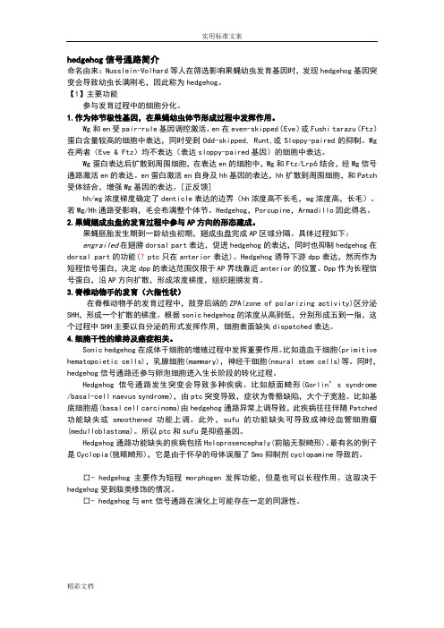
hedgehog 信号通路模式图
- 格式:ppt
- 大小:122.50 KB
- 文档页数:2

Hedghog信号通路与肿瘤发生【关键词】 Hedgehog Signaling Pathway Patched Smoothened Cubitus interruptus Gli 0 引言 Hh是由英文“刺猬”(hedgehog)简写而来的。
这类基因最早是在果蝇里发现,果蝇和其他动物一样身体分成多个节段,幼虫的每个节段内一部分有毛、一部分无毛,Hh基因突变使无毛部分变成有毛部分,所以被戏称为“刺猬”基因。
果蝇Hh基因是美国霍普金斯大学毕淇实验室在90年代初克隆的,在果蝇只有一个Hh基因,以后多个实验室在高等动物发现有三个Hh基因。
Hedgehog通路不仅在胚胎正常发育中起着重要作用,通路的异常还可引发畸形和肿瘤。
本文就Hedgehog通路的构成、途径及在胚胎发育和肿瘤形成中的作用、肿瘤治疗的进展进行综述。
1 Hedgehog通路的基本构成 1.1 Hedgehog蛋白家族果蝇只有一个hedgehog基因,脊椎动物有3种hedgehog基因,包括:Desert hedgehog(Dhh), Indian hedgehog(Ihh), Sonic hedgehog(SHh)。
Dhh与果蝇的Hedgehog基因的关系最近;Ihh和SHh之间的关系较近。
Hedgehog蛋白是一种分泌蛋白,必须经过自身的修饰才能获得活性。
Hh蛋白包含一个N端信号结构域,和一个C端催化结构域。
C端催化结构域可以共价结合胆固醇,并使其结合到N端信号结构域,再将N端信号结构域一个半胱氨酸棕榈酰化,这个过程需要Skinny hedgehog酰基转移酶。
从鸡的sonic hedgehog (SHh)蛋白出发,用BLAST法找到其在人、小鼠、大鼠等脊椎动物的同源蛋白共16个,组成Hedgehog蛋白家族。
1.2 Patched(Ptch)蛋白 Ptch蛋白是细胞表面接受Hh信号蛋白的受体,具有二种功能,一是与Hh结合,二是抑制Smoothened(Smo)。
Controlled cell proliferation is a predominant theme in normal embryonic and post-embryonic development, and, in many instances, cell-type specification and cell proliferation are intimately coupled. Several secreted intercellular signaling proteins that behave as morphogens during pattern formation are also implicated in the regulation of the cell cycle. Hedgehogs (Hhs) are one such class of morphogens that regulate an enormous variety of developmental events in the fly and vertebrate embryo and plays a central role in several cancers.The vertebrate Hh family is represented by at least three members: Dhh (Desert Hh), Ihh (Indian Hh) and Shh (Sonic Hh), two Patched homologs, Ptc1 (Patched-1) and Ptc2 (Patched-2); and three homologs of Ci (Cubitus interruptus, a 155 kDa cytoplasmic zinc finger protein), Gli1, Gli2 and Gli3 (Ref.1). Shh is the most extensively characterized vertebrate homolog, and is involved in morphogenesis of several organs including the eye, hair and lungs. It acts as both a short-range, contact-dependent factor and as along-range, diffusible morphogen. Shh genes are highly conserved and have been identified within a variety of species, including human, mouse, frog, fish, and chicken. In the human embryo, Shh is expressed in the notochord, the floorplate of the neural tube, the gut, and in the developing limbs. Dhh and Ihh play more restricted roles: Dhh acts in the regulation of spermatogenesis and organization of the perineurium, which ensheaths peripheral nerves, and Ihh in coordinating proliferation and maturation of chondrocytes during development of the endochondral skeleton. Hh signals act as morphogens to induce distinct cell fates at specific concentration thresholds. In Drosophila, Hh patterns the segment, wing, leg, eye, and regions of the fly brain either directly, or through the recruitment of other signaling factors such as Dpp (Decapentaplegic) and Wg (Wingless) (Ref.2).The Hh-signaling pathway comprises three main components: the Hh ligand; a transmembrane receptor circuit composed of the negative regulator Ptc plus an activator, Smo (Smoothened) a GPCR (G-Protein Coupled Receptor); and finally a cytoplasmic complex that regulates the Ci or Gli family of transcriptional effectors. Additional pathway components are thought to modulate the activity or subcellular distribution of these molecules. There is positive and negative feedback at the transcriptional level as the Gli1 and Ptc1 genes are direct transcriptional targets of activation of the pathway (Ref.3). Ptc, a twelve-pass membrane protein binds Hh ligand, and in the absence of ligand, Ptc interacts with and inhibits Smo, a seven-pass membrane protein. This repression culminates in a transcription factor, Ci (Ci75) in Drosophila and Gli in vertebrates acting as a transcriptional repressor. When Hh binds Ptc, its interactions with Smo are altered such that Smo is no longer inhibited. This leads to Ci/Gli protein entering the nucleus and acting as a transcriptional activator for the same genes it represses when Ptc is free to interact with and inhibit Smo. The determination of diverse cell fates by Shh signaling occurs by regulating the combination of Gli genes expressed in a cell. The transcriptional effects of Hh signaling are directed to particular target genes by the specificity of the Ci zinc fingers in DNA sequence recognition (Ref.4). The processing and nuclear import of Ci is regulated via a complex of Ci with the cytoplasmic members of the Hh signaling pathway, Cos2 (Costal-2; Cos-FlyBase), Fused (Fu) and SUFU (Suppressor of Fused). Cos2 tethers the Ci-containing complex to the microtubules. On Hh signaling, the complex is released from microtubules and full-length Ci enters the nucleus (Ref.5). Kinases including GSK3Beta (Glycogen Synthase Kinase-3Beta), Slimb and PKA (Protein Kinase-A) oppose activation of the Shh pathway by regulating the stability of intermediate signaling transcription factors of Hh pathway. SUFU interacts directly with Ci proteins, repressing Hh signaling. In the absence of Hh signal, Cos2 and SUFU binding to Ci prevent Ci activation and retain it in the cytoplasm. Most of Ci is available for cleavage in a process which is dependent upon its phosphorylation by the PKA and which involves Cos2and Slimb. Uncleaved, full-length Ci is actively exported from the nucleus. Upon Hh reception, Fu is activated and acts on Cos2 and SUFU, alleviating their negative effect on Ci. As a result, Ci cleavage is reduced, Ci155 nuclear import overcomes its export and Ci is activated. Ci activation requires Cos2 and Fu to antagonize SUFU negative effect. Activated nuclear Ci interacts with the CBP (CREB Binding Protein) to fully activate the transcription of Hh target genes (Ref.6).Since their isolation, members of the Hh family of intercellular signaling proteins have been recognized as key mediators of many fundamental processes in embryonic development. Their activities are central to the growth, patterning, and morphogenesis of many different regions within the body plans of vertebrates and insects, and most likely other invertebrates (Ref.7). Inactivation of Shh or components in its signal transduction pathway, such as Gli2 and Gli3, gives rise to various degrees of lung and foregut malformations, with fusion of lung lobes, hypoplasia and esophageal atresia or stenosis (Ref.1). Further, misregulation of Hh signaling in humans is associated with congenital malformations of the CNS (Central Nervous System, spina bifida, holoprosencephaly type 3, hpe3), head (cleft palate), and limb (syn- and polydactyly) and with a predisposition for developing a variety of tumors of the skin (basal cell carcinoma) and CNS (medulloblastoma, glioblastoma) (Ref.8). Hh signal transduction has been the focus of intense research over the past decade due to the central role it plays in development and its emerging biomedical relevance in areas ranging from regenerative medicine to oncology (Ref.3).。
Hedgehog信号通路在哺乳动物生殖系统中的作用1. Hedgehog信号通路Nusslein-V olhard和Wieschaus在对果蝇进行影响幼虫表皮层图式形成的突变体筛选时发现了hedgehog 基因(hh),果蝇和其他动物一样身体分成多个节段,幼虫的每个节段内一部分有毛、一部分无毛,hh 基因突变使无毛部分变成有毛部分,所以被戏称为“刺猬”基因,随后Hedgehog 信号通路的组成成分和具体途径在果蝇中被确定。
果蝇Hedgehog 信号通路中的组成成分(主要包括hh、ptch 和Gli 家族转录因子ci)及其功能被高度保守和复杂化的存在于哺乳动物中。
果蝇只有一个hh 基因,哺乳动物中发现其同源基因有 3 个,分别为Sonic hedgehog(Shh)、Indian hedgehog (Ihh)和Desert hedgehog (Dhh),研究较多的是Shh,因其在哺乳动物中作用最为广泛[2]。
经典的哺乳动物Hedgehog 信号通路是由Hh 配体、跨膜蛋白质受体Patched(Ptch1 和Ptch2)和Smoothened(Smo)组成的受体复合物、下游转录因子Gli 蛋白(Gli-1、Gli-2、Gli-3)组成以及最近被克隆和阐述的丝氨酸/苏氨酸蛋白激酶Fuesd(Fu) 和Fu 抑制剂(SuFu)的脊椎动物同源物。
Hh蛋白家族成员是一类具有自我剪切功能的分泌性信号蛋白,均由氨基端(Hh-N)和羧基端(Hh-C)两个结构域组成,其中Hh-N具有Hh蛋白的信号活性,而Hh-C则具有自身蛋白水解酶活性和胆固醇转移酶功能。
Shh、Ihh和Dhh的共同点是由这三种基因编码而成的信号都激动同样一条信号级联放大通路。
Hh编码的前体蛋白合成后并无生物学活性,只有前体蛋白C末端的一部分氨基酸自身磷酸化切除了C末端后,剩下的N末端片段再经双重脂质修饰后才有活性,这可能与Hh蛋白在细胞内的极性分布有关,并可能影响到它与受体的结合。
doi:10.3969/j.issn.1000⁃484X.2019.15.022㊃专题综述㊃Hedgehog 信号传导通路及其生物学功能①闵 明 王婷婷 张继东(遵义医科大学免疫学教研室贵州省基因检测与治疗特色重点实验室,遵义563000) 中图分类号 R392.9 文献标志码 A 文章编号 1000⁃484X (2019)15⁃1903⁃05①本文为国家自然科学基金(31660338)㊁贵州省科技计划项目[黔科合基础(2016)1167]㊁贵州省普通高等学校科技拔尖人才支持计划[黔教合KY 字(2016)079]㊁遵义医学院博士科研启动项目(F⁃739)㊁贵州省留学人员科技创新项目[(2016)20]和遵义医科大学研究生创新计划(ZYK014)㊂作者简介:闵 明,女,在读硕士,主要从事生殖免疫研究,E⁃mail:mjdmm4550@㊂通讯作者及指导教师:张继东,男,博士,副教授,硕士生导师,主要从事生殖免疫研究,E⁃mail:choukeitou @㊂[摘 要] 从胚胎到成体的发育过程中,Wnt㊁TGF⁃β㊁Notch㊁Hedgehog(Hh)这4条信号通路是主要的信号调节通路㊂他们调控着胚胎早期发育㊁细胞的增殖分化以及维持机体的稳态㊂其中Hh 信号传导通路在多种生理调控中起着关键的作用㊂Hh 信号通路不仅参与细胞生长㊁分化,组织器官的发生和形成,在组织器官的损伤修复㊁免疫调节方面也发挥着重要作用㊂但是Hh 信号通路异常激活也会引起发育异常甚至引发肿瘤㊂[关键词] Hedgehog;Gli;生物学功能;初级纤毛Hedgehog signaling pathway and its function in biologyMIN Ming ,WANG Ting⁃Ting ,ZHANG Ji⁃Dong .Department of Immunology ,Zunyi Medical University ,Special Key Laboratory of Gene Detection &Therapy of Guizhou Province ,Zunyi 563000,China[Abstract ] Four well⁃known signal transduction pathways,Wnt,TGF⁃β,Notch and Hedgehog(Hh)play a vital role in diversebiological processes,such as embryonic development,cell proliferation,differentiation and homeostasis.Among them,Hh signaling is an indispensable part in physiological regulation.Not only does the Hh signaling participate in cell growth,differentiation and morphogenesis of organs,but also exerts great influence in immune regulation and regeneration after injury.However,disorder of Hhsignaling pathway may lead to abnormal development and tumor.[Key words ] Hedgehog;Gli;Function in biology;Primary cilia Hedgehog(Hh)信号传导通路最初是在果蝇身上发现的,是一条高度保守的信号通路途径[1]㊂果蝇的Hh 基因突变导致幼虫体表出现许多刺突,形似刺猬,故名Hedgehog㊂Hh 信号转导通路无论在胚胎期还是胚胎形成后,几乎对所有器官的发育都至关重要㊂机体内环境稳态的维持,需要Hh 信号通路在特定的时间㊁组织细胞中被正常激活,异常的激活会导致组织细胞异常增殖分化,引发发育异常或者肿瘤㊂本文将综述Hh 在生理和病理状态下的生物学功能,以进一步全面而深入地总结Hh 信号通路的最新研究进展,旨在为更好地理解和研究此信号通路在生物体内的调控作用提供参考㊂1 Hh 信号传导通路Hh 信号通路主要由配体㊁受体㊁转录因子3部分构成㊂配体一共有3种,包括Sonic hedgehog (Shh )㊁Desert hedgehog (Dhh )和Indian hedgehog (Ihh)㊂受体有Patched(Ptch)和Smoothened(Smo)两种,Ptch 受体又分为Ptch1和Ptch2㊂转录因子Gli (Glioma⁃associated oncogene )拥有3种类型:Gli1㊁Gli2和Gli3㊂Gli1参与激活目的基因转录,Gli2和Gli3既能激活又能抑制目的基因的转录㊂与其他调控生长发育信号通路不同的是,脊椎动物Hh 信号通路依赖于高度专业化的细胞器 初级纤毛㊂初级纤毛是突出于细胞表面的微管样结构的细胞器,广泛存在于各种类别细胞表面[2]㊂初级纤毛是具有感受能力的细胞器,能感受Hh 信号,调节信号通路[3]㊂在没有Hh 配体的情况下,Ptch ㊃3091㊃闵 明等 Hedgehog 信号传导通路及其生物学功能 第15期受体位于初级纤毛的底部,抑制Smo向初级纤毛内转移,从而抑制其活化;Gli则被依赖环磷酸腺苷(cyclic adenosine monophosphate,cAMP)的激酶A (Protein kinase A,PKA)磷酸化后分解为Gli抑制物(GLIR),GLIR抑制细胞核内目的基因转录㊂PKA 的调节亚基和催化亚基都位于纤毛基底部,感受cAMP促进GLIR的生成,调节Gli2不恰当地活化[4]㊂当纤毛感受到Hh信号,并且Hh与Ptch受体结合,解除对Smo的抑制作用,Smo即向初级纤毛内转移,到达纤毛顶端与Sufu(Suppressor of fused homolog)相互作用,使Gli活化转导入细胞核,与目的基因启动子结合,使目的基因转录㊂此通路为经典的Hh信号激活途径㊂经典途径的Hh信号传导高度依赖初级纤毛,要求纤毛在结构和功能上保持良好的完整性[5]㊂非经典的Hh信号传导则是不依赖受体,Gli直接被活化来调控目的基因的表达[6]㊂之前普遍认为只有初级纤毛参与Hh信号通路的调控,近来有研究报道在缺乏初级纤毛的肺部,Shh通过运动纤毛介导的非经典途径调节细胞间的cAMP,进而调节运动纤毛的运动频率和气道表面液体pH值[7]㊂2 Hh信号通路介导的生物学功能Hh也被称为成形素,调控祖细胞分化为特定成熟细胞,参与组织形成并发挥调控功能㊂但是Hh 信号通路调控紊乱,会引起发育异常和肿瘤生成㊂近年来,研究者在睾丸形成㊁神经系统发育㊁免疫调节㊁肿瘤形成方面进行了大量的研究㊂2.1 Hh信号通路参与T细胞的分化发育 胸腺是T细胞分化㊁发育㊁成熟的场所㊂T细胞在胸腺中需经历3个连续的发育阶段:CD4⁃CD8⁃双阴性(Double negative,DN)阶段㊁CD4+CD8+双阳性(Double posit⁃ive,DP)阶段㊁CD4+或CD8+单阳性(Single positive, SP)阶段㊂DN阶段根据CD44和CD25的表达情况依次分为CD44+CD25⁃(DN1)㊁CD44+CD25+(DN2)㊁CD44-CD25+(DN3)㊁CD44-CD25-(DN4)[8]㊂Hh配体参与T细胞发育每一阶段的调节,而且相同配体在细胞不同发育阶段的作用不尽相同㊂DN1向DN2分化时,Ihh㊁Shh㊁Gli2㊁Gli3能促进分化,使处于DN2时期细胞的数量增多㊂Dhh㊁Shh㊁Ihh阻碍DN3㊁DN4向DP分化㊂Shh-/-基因敲除会使DN3㊁DN4无法完成T细胞抗原受体β链(T cell receptorβ⁃chain,TCR⁃β)链重排,导致DN4无法进入阳性选择而发生凋亡㊂Gli3在此阶段能抑制Shh 作用,促使DN向DP分化[9]㊂在Dhh-/-基因敲除小鼠中,虽然胸腺细胞总数会增多,但是细胞多停留在DN3阶段,延迟进入阳性选择[10]㊂DP高表达Ihh,能反馈调节DN3,控制细胞分化进程,维持稳态㊂DP在Shh缺失的情况下,向SP分化的比例会增高㊂另外,Hh信号通路通过调控胸腺上皮细胞(Thymic epithelial cells,TECs)的分化,间接调控T 细胞的分化发育[11]㊂2.2 Hh信号通路参与炎性反应2.2.1 Hh信号通路参与脂肪炎性反应 脂肪组织的慢性低度炎性反应被认为是引起肥胖的关键因素㊂脂肪组织的炎性反应主要表现为巨噬细胞的浸润,尤以M1型巨噬细胞为主㊂炎性反应会使细胞损伤释放损伤相关物质,例如高迁移率族蛋白1 (High mobility group box⁃1protein,MGB1)㊁S100蛋白㊁氧化型低密度脂蛋白(Low density lipoprotein, LDL)等㊂其中由活化的巨噬细胞释放的S100A8作为巨噬细胞的趋化物,进一步促进巨噬细胞迁移[12]㊂巨噬细胞作为Hh的靶细胞,受Hh信号通路的调控㊂Hh信号通路能抑制脂肪组织炎性反应,加强糖代谢,减轻脂肪重量㊂在条件敲除骨髓细胞Smo基因而创建Lys⁃Smo-/-小鼠模型中,小鼠在高脂饮食条件下会出现脂肪组织重量增加㊁炎性反应加重和糖耐量受损等症状[13]㊂2.2.2 Hh信号通路参与胃肠道炎性反应 研究表明,胃肠道上皮细胞主要表达Shh㊁Ihh配体,相应的配体与基质细胞表面受体结合,激活Hh信号通路抑制胃肠道炎性反应㊂在幽门螺杆菌感染的胃部,壁细胞分泌的Shh参与免疫抑制[14]㊂在结肠中,Hh信号通路活化后诱导基质细胞分泌抗炎因子IL⁃10;促使调节性T细胞参与抗炎反应[15];抑制成纤维细胞分泌趋化因子配体趋化因子12(C⁃X⁃C motif chemokine 12,CXCL12),降低对炎性细胞的趋化作用,从而抑制炎性反应㊂反之,抑制Hh信号通路,会引起肠道性炎症,破坏肠道结构,影响肠道功能[16]㊂2.3 Hh信号通路参与神经系统调节 Hh信号通路对于神经系统的调控,从胚胎发育开始就显得极其重要㊂在胚胎期,外胚层细胞增殖㊁内陷,最终离开外胚层表面形成中空的神经管㊂神经底板和脊索分泌的Shh,在神经管内形成不同的浓度梯度[17]㊂神经祖细胞(Neural progenitor cells,NPCs)在不同浓度梯度的Hh信号调控下,不同区域的NPCs分化为不同类型的细胞[18,19]㊂神经管是中枢神经系统的原基,闭合的神经管前段发育为脑,后段发育为脊髓㊂Bay等[20]发现在胚胎期小脑发育过程中,浦肯野纤维细胞分泌的Shh通过Gli调控细胞周期,刺㊃4091㊃中国免疫学杂志2019年第35卷激小脑颗粒细胞前体细胞(Cerebellar granule neuron precursors,CGNPs)分裂增殖,形成小脑的内部颗粒层㊂药物如糖皮质激素或基因突变使Shh㊁Ptch㊁Smo减少,引起CGNPs增殖减慢,小脑发育不良,髓母细胞瘤形成㊂然而添加Shh激动剂(Sonic hedgehog agonist,SAG)后,浦肯野纤维细胞的数量恢复到正常水平,小脑发育状况有所改善[21]㊂由此猜想,Shh参与了神经的损伤修复㊂Angeloni等[22]建立大鼠海绵体神经(Cavernous nerve,CN)损伤的模型,探究Shh对CN的修复作用㊂正常情况下盆底神经节(Pelvic ganglia,PG)合成的Shh被转运到CN,用于维持CN的形态结构㊂当CN发生损伤断裂时,Shh转运障碍,CN中的Shh 降低,引起CN形态改变以及脱髓鞘和轴突变性㊂在较高浓度的Shh治疗下,由于胶质纤维酸性蛋白(Glial fibrillary acidic protein,GFAP)降低,从而更有效地减低CN损伤㊂在发生脱髓鞘反应后,胼胝体内脑室来源的少突胶质细胞在Shh信号调控下重新形成髓鞘[23]㊂研究者对面部神经损伤进行研究时,发现面部神经细胞中Shh表达上调,Shh作用于成纤维细胞,使成纤维细胞参与损伤神经的修复[24]㊂由此可见,Shh参与神经系统的发育,并作为神经保护剂参与神经修复,维持神经系统稳态㊂2.4 Hh信号通路在睾丸形成中的作用 睾丸中管周肌样细胞㊁内皮细胞㊁间质细胞(Leydig)㊁初级精母细胞㊁次级精母细胞㊁圆形精子中都有受体Ptch1的表达[25]㊂Dhh与Ptch1结合,参与生殖细胞㊁管周肌样细胞㊁Leydig细胞的分化以及睾丸索的形成[26]㊂Hh信号通路两个重要组分 Gli1㊁Sufu,在大鼠生殖细胞不同发育阶段,呈现不同的表达水平㊂Sufu从第9阶段(Ⅸ期)精细胞开始表达,在第10~ 13阶段(Ⅹ~ⅩⅢ期)表达最强,在第15~18阶段(Ⅰ~Ⅵ期)不表达㊂Gli的表达则不同,Gli表达于胞质,在第9~14阶段(Ⅸ~ⅩⅣ期)不表达,第16~ 18阶段(Ⅱ~Ⅵ期)高表达㊂这种差异表达说明Sufu使Hh信号通路在Ⅸ~ⅩⅣ期呈现关闭状态,使Gli停留在胞质中,抑制Hh信号通路的活化,调控生殖细胞的发育㊂如果加入Hh信号通路抑制剂环杷明,还会引起生殖细胞凋亡增加㊂所以,Hh信号通路参与生殖细胞的增殖㊁分化㊁凋亡[27]㊂除此以外,有研究表明Hh信号通路通过调节干细胞微环境间接调节干细胞的增殖㊁分化甚至凋亡[28]㊂Sertoli细胞是睾丸中唯一能合成Dhh的细胞, Dhh作用于间质细胞祖细胞,使其分化为Leydig细胞㊂Leydig细胞是睾丸中合成睾酮的细胞,但在胚胎期只有睾丸间质细胞干细胞(Stem Leydig cells,SLCs)不具有睾酮合成的能力㊂在Dhh㊁Wnt㊁Notch 等信号通路和促黄体素(Luteinizing hormone,LH)㊁卵泡刺激素(Follicle⁃stimulating hormone,FSH)的共同调控下,SLGs逐步分化为睾丸间质细胞祖细胞(Progenitor Leydig cells,PLCs)㊁未成熟的睾丸间质细胞(Immature Leydig cells,ILCs)和成熟的睾丸间质细胞(Adult Leydig cells,ALCs)[29]㊂在Dhh-/-基因敲除小鼠中,Leydig细胞分化障碍,不能有效地参与睾丸索的形成㊂不完整的睾丸索,不能起到很好的屏障作用,使少部分生殖细胞穿越睾丸索,零星分布在间质中㊂在间质中的生殖细胞不能进行减数分裂,导致生精障碍[30,31]㊂然而Hh信号通路的持续激活,会使苗勒管退化受阻,出现睾丸和苗勒管同时存在的现象[32]㊂2.5 Hh信号通路参与肿瘤形成 肿瘤之所以具有无限增殖能力,归因于肿瘤干细胞的不断自我更新的能力㊂研究表明Hh参与了肿瘤干细胞增殖和分化的调控,而且Hh信号通路持续异常活化,与肿瘤的发生具有很高的相关性[33,34]㊂Hh信号通路通过配体非依赖型㊁配体依赖的自分泌型㊁配体依赖的旁分泌型3种方式异常活化㊂针对Hh信号通路异常激活这一现象,应运而生了一系列针对各组分的阻断剂㊂5E1是针对Ptch 的单克隆抗体;XL⁃139㊁LEQ506是Smo抑制剂;Gli 的拮抗剂有GANT56㊁GANT61等[35]㊂Gli的拮抗剂GANT61被证实不仅可以抑制乳腺癌细胞增殖,而且能减少细胞活力,降低癌细胞侵袭性㊂在传统治疗方法达不到预期的情况下,有研究者将抑制剂与传统治疗方法相结合,以期达到更好的治疗效果㊂例如在进行放疗和顺铂化疗治疗宫颈癌的同时加入Smo抑制剂,发现不仅没有增加胃肠道毒理作用,而且使宫颈癌细胞增殖减慢,淋巴结转移减少[36]㊂但是随着一些抑制剂在临床上的广泛使用,部分患者出现了耐药现象,尤其是针对Ptch和Smo受体的阻断药㊂而且随着研究的深入,发现耐药机制不仅涉及编码Hh组分基因突变,也可由基因突变以外的原因引起㊂在使用维莫德吉治疗基底细胞瘤(Basal cell carcinomas,BCCs)出现耐药现象的患者中,就有大约50%的耐药患者不存在受体基因突变㊂Whitson等[37]针对这一耐药现象进行深入研究,揭示了其耐药机制,即通过RhoA⁃mDia(mouse Diaphanous)⁃actin⁃SRF⁃巨核细胞白血病1(Mega⁃karyoblastic leukemia1,MKL1)级联反应,激活非经㊃5091㊃闵 明等 Hedgehog信号传导通路及其生物学功能 第15期典Hh信号通路㊂RhoA在成蛋白家族成员mDia的作用下活化,促使细胞骨架中球状肌动蛋白(Globular actin,G⁃actin)转换为纤维状肌动蛋白(Fibros actin,F⁃actin)㊂G⁃actin的减少使MKL1蛋白释放增多,进而转入细胞核内㊂F⁃actin积聚增多,使MKL1活化㊂活化的MKL1作为转录因子血清反应因子(Transcription factor serum response factor,SRF)的辅助因子,和SRF共同结合到Hh信号通路靶基因旁边位点,和Gli1形成一种蛋白复合物,增强Gli1的活性,促进目的基因的转录㊂新的耐药机制的发现,也诠释了一个新的药物靶点 MKL1㊂他们的实验也证明了在Hh信号通路抑制剂维莫德吉耐药情况下使用MKL1抑制剂,可以更有效地降低Gli1的表达,抑制肿瘤的增长㊂在受体阻断剂耐药情况严重的背景下,Gli拮抗剂和新药物靶点的发现以及相关药物的开发被寄予很高的希望㊂3摇结语Hh信号通路作为一条经典的调控生长发育的信号通路,调控功能强大,调控范围几乎涉及所有的组织㊁器官㊂目前研究成果主要集中在调节T细胞分化发育㊁免疫调节㊁睾丸形成㊁神经系统发育㊁肿瘤形成方面㊂随着广泛而深入的研究,其他的生理学功能相继被发现,例如Hh参与B细胞的分化成熟㊁软骨成骨㊁血管形成㊁肝脏修复等[38⁃41]㊂不仅如此, Hh信号通路还和其他信号通路共同调节生命活动㊂然而不容忽视的是,Hh信号通路的异常活化会引发发育异常和肿瘤㊂针对不同的肿瘤,Hh信号通路具体的调控机制还需要继续深入研究㊂只有全面而深入地了解Hh信号通路在生理和病理状态下的生物学功能及其调控机制,才能为进一步的研究提供新的思路,更好地实现其临床应用价值㊂参考文献:[1] Zhao L,Wang L,Chi C,et al.The emerging roles of phosphatasesin Hedgehog pathway[J].Cell Commun Signal,2017,15(1):35.[2] Dores C,Alpaugh W,Su L,et al.Primary cilia on porcine testicularsomatic cells and their role in hedgehog signaling and tubular mor⁃phogenesis in vitro[J].Cell Tissue Res,2017,368(1):215⁃223.[3] Breslow DK,Hoogendoorn S,Kopp AR,et al.A CRISPR⁃basedscreen for Hedgehog signaling provides insights into ciliary function and ciliopathies[J].Nat Genet,2018,50(3):460⁃471. [4] Bangs F,Anderson KV.Primary cilia and mammalian Hedgehogsignaling[J].Cold Spring Harb Perspect Biol,2017,9(5):pii:a028175.[5] Eguether T,Cordelieres FP,Pazour GJ.Intraflagellar transport isdeeply integrated in hedgehog signaling[J].Mol Biol Cell,2018,29(10):1178⁃1189.[6] Teperino R,Aberger F,Esterbauer H,et al.Canonical and non⁃canonical Hedgehog signalling and the control of metabolism[J].Semin Cell Dev Biol,2014,33:81⁃92.[7] Mao S,Shah AS,Moninger TO,et al.Motile cilia of human airwayepithelia contain hedgehog signaling components that mediate non⁃canonical hedgehog signaling[J].Proc Natl Acad Sci U S A, 2018,115(6):1370⁃1375.[8] Bredenkamp N,Jin X,Liu D,et al.Construction of a functionalthymic microenvironment from pluripotent stem cells for theinduction of central tolerance[J].Regen Med,2015,10(3): 317⁃329.[9] Barbarulo A,Lau CI,Mengrelis K,et al.Hedgehog signalling in theembryonic mouse thymus[J].J Dev Biol,2016,4(3):22. [10] Kariuki SM.Desert hedgehog is a negative regulator of CD44⁃CD25+double negative T lymphocytes developmental stage inthymic differentiation[J].Int J Res Med Sci,2018,6(3):734⁃738.[11] Solanki A,Yanez DC,Ross S,et al.Gli3in fetal thymic epithelialcells promotes thymocyte positive selection and differentiation byrepression of Shh[J].Development,2018,145(3):pii:dev146910.[12] Sekimoto R,Fukuda S,Maeda N,et al.Visualized macrophagedynamics and significance of S100A8in obese fat[J].Proc NatlAcad Sci U S A,2015,112(16):E2058⁃E2066. [13] Braune J,Weyer U,Matz⁃Soja M,et al.Hedgehog signalling inmyeloid cells impacts on body weight,adipose tissue inflammationand glucose metabolism[J].Diabetologia,2017,60(5):889⁃899.[14] Razumilava N,Gumucio DL,Samuelson LC,et al.IndianHedgehog suppresses intestinal inflammation[J].Cell MolGastroenterol Hepatol.2017,5(1):63⁃64.[15] Lee JJ,Rothenberg ME,Seeley ES,et al.Control of inflammationby stromal Hedgehog pathway activation restrains colitis[J].ProcNatl Acad Sci U S A,2016,113(47):E7545⁃E7553. [16] Westendorp BF,Buller N,Karpus ON,et al.Indian Hedgehogsuppresses a stromal cell⁃driven intestinal immune response[J].Cell Mol Gastroenterol Hepatol,2018,5(1):67⁃82. [17] Demers CJ,Cox G,Collins SD,et al.Directing the spatialpatterning of motor neuron differentiation in engineered microenvi⁃ronments[J].Conf Proc IEEE Eng Med Biol Soc,2016:477⁃480.[18] Pusapati GV,Kong JH,Patel BB,et al.G protein⁃coupledreceptors control the sensitivity of cells to the morphogen SonicHedgehog[J].Sci Signal,2018,11(516):pii:eaao5749. [19] Li P,Markson JS,Wang S,et al.Morphogen gradientreconstitution reveals Hedgehog pathway design principles[J].Science,2018,360(6388):543⁃548.[20] Bay SN,Long AB,Caspary T.Disruption of the ciliary GTPaseArl13b suppresses Sonic hedgehog overactivation and inhibitsmedulloblastoma formation[J].Proc Natl Acad Sci U S A,2018,115(7):1570⁃1575.[21] Nguyen V,Sabeur K,Maltepe E,et al.Sonic Hedgehog agonistprotects against complex neonatal cerebellar injury[J].Cerebellum,2018,17(2):213⁃227.[22] Angeloni N,Bond CW,Harrington D,et al.Sonic hedgehog isneuroprotective in the cavernous nerve with crush injury[J].JSex Med,2013,10(5):1240⁃1250.[23] Sanchez MA,Armstrong RC.Postnatal Sonic hedgehog(Shh)responsive cells give rise to oligodendrocyte lineage cells duringmyelination and in adulthood contribute to remyelination[J].ExpNeurol,2018,299(Pt A):122⁃136.[24] Bobarnac Dogaru GL,Juneja SC,Shokrani A,et al.The role ofHedgehog⁃responsive fibroblasts in facial nerve regeneration[J].Exp Neurol,2018,303:72⁃79.(下转第1912页)People′s Medical Publishing House,2009:10⁃15. [38] Cui H,Xie N,Tan Z,et al.The human long noncoding RNA Lnc⁃IL7R regulates the inflammatory response[J].Eur J Immunol,2014,44:2085⁃2095.[39] Ye Z,Xu J,Li S,et al.Lnc IL7R promotes the growth of fibroblastlike synoviocytes through interaction with enhancer of zestehomolog2in rheumatoid arthritis[J].Mol Med Rep,2017,15(3):1412⁃1418.[40] Guttman M,Donaghey J,Carey BW,et al.lincRNAs act in thecircuitry controlling pluripotency and differentiation[J].Nature,2011,477(7364):295⁃300.[41] Cui HC,Xie N,Tan Z,et al.The human long noncoding RNA,lnc⁃IL7R,regulates inflammatory response[J].Eur J Immunol,2014,44(7):2085⁃2095.[42] Heward JA,Lindsay MA.Long non⁃coding RNAs in the regulationof the immune response[J].Trends Immunol,2014,35(9):408⁃419.[43] Adami G,Orsolini G,Adami S,et al.Effects of TNF inhibitors onparathyroid hormone and Wnt signaling antagonists in rheumatoidarthritis[J].Calcif Tissue Int,2016,99(4):360⁃364.[44] Nike Müller,Frank Döring,Maja Klapper,et al.Interleukin⁃6andTumour Necrosis Factor⁃a differentially regulate lincRNAtranscripts in cells of the innate immune system in vivo in humansubjects with rheumatoid arthritis[J].Cytokine,2014,68(1):65⁃68.[45] Lu MC,Yu HC,Yu CL,et al.Increased expression of longnoncoding RNAs LOC100652951and LOC100506036in T cellsfrom patients with rheumatoid arthritis facilitates the inflammatoryresponses[J].Immunol Res,2016,64(2):576⁃578. [46] Zhang HJ,Wei GF,Wang SJ,et al.LncRNA HOTAIR alleviatesrheumatoid arthritis by targeting miR⁃138and inactivating NF⁃κBpathway[J].Int Immunopharmacol,2017,50:283⁃290. [47] 夏 燕,冯 佳,陈安平,等.类风湿关节炎外周血LncRNA差异表达研究[J].中国免疫学杂志,2016,32(1):9⁃12.Xia Y,Feng J,Chen AP,et al.Differential expression of LncRNAin peripheral blood of rheumatoid arthritis[J].Chin J Immunol,2016,32(1):9⁃12.[收稿2018⁃08⁃10 修回2018⁃10⁃08](编辑 刘咸筠)(上接第1906页)[25] Liu C,Rodriguez K,Yao HH.Mapping lineage progression ofsomatic progenitor cells in the mouse fetal testis[J].Development,2016,143(20):3700⁃3710.[26] Yao HH,Whoriskey W,Capel B.Desert Hedgehog/Patched1signaling specifies fetal Leydig cell fate in testis organogenesis[J].Genes Dev,2002,16(11):1433⁃1440.[27] Makela JA,Saario V,Bourguiba⁃Hachemi S,et al.Hedgehogsignalling promotes germ cell survival in the rat testis[J].Repro⁃duction,2011,142(5):711⁃721.[28] Li S,Wang M,Chen Y,et al.Role of the hedgehog signalingpathway in regulating the behavior of germline stem cells[J].Stem Cells Int,2017,2017:1⁃9.[29] Chen H,Wang Y,Ge R,et al.Leydig cell stem cells:Identification,proliferation and differentiation[J].Mol CellEndocrinol,2017,445:65⁃73.[30] Yao HH,Capel B.Disruption of testis cords by cyclopamine orforskolin reveals independent cellular pathways in testisorganogenesis[J].Dev Biol,2002,246(2):356⁃365. [31] Clark AM,Garland KK,Russell LD.Desert hedgehog(Dhh)gene is required in the mouse testis for formation of adult⁃typeLeydig cells and normal development of peritubular cells and sem⁃iniferous tubules[J].Biol Reprod,2000,63(6):1825⁃1838.[32] Migone FF,Hung PH,Cowan RG,et al.Overactivation ofhedgehog signaling in the developing Mullerian duct interfereswith duct regression in males and causes subfertility[J].Reproduction,2017,153(4):481⁃492.[33] Yin Y,Liu L,Zhao Z,et al.Simvastatin inhibits sonic hedgehogsignaling and stemness features of pancreatic cancer[J].CancerLett,2018,426:14⁃24.[34] Peacock CD,Wang Q,Gesell GS,et al.Hedgehog signalingmaintains a tumor stem cell compartment in multiple myeloma[J].Proc Natl Acad Sci U S A,2007,104(10):4048⁃4053.[35] Riaz SK,Khan JS,Shah STA,et al.Involvement of hedgehogpathway in early onset,aggressive molecular subtypes andmetastatic potential of breast cancer[J].Cell Commun Signal,2018,16(1):3.[36] Chaudary N,Pintilie M,Hedley D,et al.Hedgehog inhibitionenhances efficacy of radiation and cisplatin in orthotopic cervicalcancer xenografts[J].Br J Cancer,2017,116(1):50⁃57. [37] Whitson RJ,Lee A,Urman NM,et al.Noncanonical hedgehogpathway activation through SRF⁃MKL1promotes drug resistancein basal cell carcinomas[J].Nat Med,2018,24(3):271⁃281.[38] Solanki A,Lau CI,Saldana JI,et al.The transcription factor Gli3promotes B cell development in fetal liver through repression ofShh[J].J Exp Med,2017,214(7):2041⁃2058. [39] Haraguchi R,Kitazawa R,Imai Y,et al.Growth plate⁃derivedhedgehog⁃signal⁃responsive cells provide skeletal tissuecomponents in growing bone[J].Histochem Cell Biol,2018,149(4):365⁃373.[40] Caradu C,Guy A,James C,et al.Endogenous Sonic Hedgehoglimits inflammation and angiogenesis in the ischaemic skeletalmuscle of mice[J].Cardiovasc Res,2018,114(5):759⁃770.[41] Machado MV,Diehl AM.Hedgehog signalling in liverpathophysiology[J].J Hepatol,2018,68(3):550⁃562.[收稿2018⁃09⁃30 修回2018⁃12⁃11](编辑 刘咸筠)。
hedgehog信号通路简介命名由来:Nusslein-Volhard等人在筛选影响果蝇幼虫发育基因时,发现hedgehog基因突变会导致幼虫长满刚毛,因此称为hedgehog。
【1】主要功能参与发育过程中的细胞分化。
1.作为体节极性基因,在果蝇幼虫体节形成过程中发挥作用。
Wg和en受pair-rule基因调控激活。
en在even-skipped(Eve)或Fushi tarazu(Ftz)蛋白含量较高的细胞中表达,同时受到Odd-skipped, Runt,或Sloppy-paired的抑制。
Wg 在两者(Eve & Ftz)均不表达(表达sloppy-paired基因)的细胞中表达。
Wg蛋白表达后扩散到周围细胞,在表达en的细胞中,Wg和Ftz/Lrp6结合,经Wg信号通路激活en的表达。
en蛋白激活en自身及hh基因的表达,hh扩散到周围细胞,和Patch 受体结合,增强Wg基因的表达。
[正反馈]hh/wg浓度梯度确定了denticle表达的边界(hh浓度高不长毛,wg浓度高,长毛)。
若Wg/Hh通路受影响,毛会布满整个体节。
Hedgehog,Porcupine,Armadillo因此得名。
2.果蝇翅成虫盘的发育过程中参与AP方向的形态建成。
果蝇胚胎发生期到一龄幼虫初期,翅成虫盘完成AP区域分隔。
具体过程如下:engrailed在翅膀dorsal part表达,促进hedgehog的表达,同时也抑制hedgehog在dorsal part的功能(?ptc只在anterior表达)。
Hedgehog诱导下游dpp表达,然而作为短程信号蛋白,决定dpp的表达范围仅限于AP界线靠近anterior的位置。
Dpp作为长程信号蛋白,沿AP方向扩散,形成浓度梯度,组织翅膀发育。
3.脊椎动物手的发育(六指性状)在脊椎动物手的发育过程中,肢芽后端的ZPA(zone of polarizing activity)区分泌SHH,形成一个扩散的梯度。
Hedgehog信号通路在哺乳动物生殖系统中的作用1. Hedgehog信号通路Nusslein-V olhard和Wieschaus在对果蝇进行影响幼虫表皮层图式形成的突变体筛选时发现了hedgehog 基因(hh),果蝇和其他动物一样身体分成多个节段,幼虫的每个节段内一部分有毛、一部分无毛,hh 基因突变使无毛部分变成有毛部分,所以被戏称为“刺猬”基因,随后Hedgehog 信号通路的组成成分和具体途径在果蝇中被确定。
果蝇Hedgehog 信号通路中的组成成分(主要包括hh、ptch 和Gli 家族转录因子ci)及其功能被高度保守和复杂化的存在于哺乳动物中。
果蝇只有一个hh 基因,哺乳动物中发现其同源基因有 3 个,分别为Sonic hedgehog(Shh)、Indian hedgehog (Ihh)和Desert hedgehog (Dhh),研究较多的是Shh,因其在哺乳动物中作用最为广泛[2]。
经典的哺乳动物Hedgehog 信号通路是由Hh 配体、跨膜蛋白质受体Patched(Ptch1 和Ptch2)和Smoothened(Smo)组成的受体复合物、下游转录因子Gli 蛋白(Gli-1、Gli-2、Gli-3)组成以及最近被克隆和阐述的丝氨酸/苏氨酸蛋白激酶Fuesd(Fu) 和Fu 抑制剂(SuFu)的脊椎动物同源物。
Hh蛋白家族成员是一类具有自我剪切功能的分泌性信号蛋白,均由氨基端(Hh-N)和羧基端(Hh-C)两个结构域组成,其中Hh-N具有Hh蛋白的信号活性,而Hh-C则具有自身蛋白水解酶活性和胆固醇转移酶功能。
Shh、Ihh和Dhh的共同点是由这三种基因编码而成的信号都激动同样一条信号级联放大通路。
Hh编码的前体蛋白合成后并无生物学活性,只有前体蛋白C末端的一部分氨基酸自身磷酸化切除了C末端后,剩下的N末端片段再经双重脂质修饰后才有活性,这可能与Hh蛋白在细胞内的极性分布有关,并可能影响到它与受体的结合。
Hedgehog信号通路与癌症|MedChemExpress癌症medptcHh 信号通路分子包括 Hedgehog 配体 (SHH、DHH 和 IHH)、Ptch 受体 (Ptch-1 和 Ptch-2,跨膜蛋白)、Smoothened (SMO)、驱动蛋白 Kif7、蛋白激酶 A (PKA)、3 种 Gli 转录因子 Gli1/2/3 (Gli1 仅具转录激活因子作用,Gli2 和 Gli3 同时具有激活因子和抑制因子作用) 以及 Sufu (融合抑制因子,Hh 信号传导的负调节因子)。
根据Hh 信号通路激活后是否依赖 Gli 蛋白发挥生物学效应,Hh 通路激活可以分为两种不同的途径:经典以及非典型信号途径。
经典信号通路激活在没有Hh 配体的情况下,Hh 受体如Ptch-1,定位于初级纤毛,可以阻止 SMO 积累且抑制 SMO 活性。
蛋白激酶,如 PKA、GSK3β 和CK1α,磷酸化 GLI2 和 GLI3,导致蛋白体介导全长 Gli 裂解为截短形式 Gli2R、Gli3R,并作为 Hh 靶基因表达的阻遏物。
此外,Sufu 通过与细胞质和细胞核中的 Gli 结合,充当该途径的另一个负调节因子,防止 Hh 靶基因的激活。
在存在 Hh 配体 (如Shh) 的情况下,Hh 配体会与 Ptch-1结合后,Ptch-1 被内化 (Endocytosis),解除对 Smo 的抑制,允许 SMO 的积累和激活,Hh 信号通过由 Kif7、Sufu 和全长 Gli 组成的细胞质蛋白复合物向 Smo 下游传递。
Smo 移动到初级纤毛的顶端并向 Sufu 发出信号以释放 Gli 激活剂 (GliA)。
然后 GliA 迁移到细胞核并激活靶基因的表达。
图 1. 经典信号通路激活[8]左:有 Hh 配体的通路激活 (ON-state);右:无配体的情况 (OFF-state)非典型信号通路激活非经典的 Hh 信号转导是指对 Hh 信号通路的一个或多个组成部分的信号反应,而不是上述 Hh-Ptch-Smo-Gli 经典通路。
Hedgehog信号通路Hedgehog信号通路在哺乳动物生殖系统中的作用1. Hedgehog信号通路Nusslein-V olhard和Wieschaus在对果蝇进行影响幼虫表皮层图式形成的突变体筛选时发现了hedgehog 基因(hh),果蝇和其他动物一样身体分成多个节段,幼虫的每个节段内一部分有毛、一部分无毛,hh 基因突变使无毛部分变成有毛部分,所以被戏称为“刺猬”基因,随后Hedgehog 信号通路的组成成分和具体途径在果蝇中被确定。
果蝇Hedgehog 信号通路中的组成成分(主要包括hh、ptch 和Gli 家族转录因子ci)及其功能被高度保守和复杂化的存在于哺乳动物中。
果蝇只有一个hh 基因,哺乳动物中发现其同源基因有3 个,分别为Sonic hedgehog(Shh)、Indian hedgehog (Ihh)和Desert hedgehog (Dhh),研究较多的是Shh,因其在哺乳动物中作用最为广泛[2]。
经典的哺乳动物Hedgehog 信号通路是由Hh 配体、跨膜蛋白质受体Patched(Ptch1 和Ptch2)和Smoothened(Smo)组成的受体复合物、下游转录因子Gli 蛋白(Gli-1、Gli-2、Gli-3)组成以及最近被克隆和阐述的丝氨酸/苏氨酸蛋白激酶Fuesd(Fu) 和Fu 抑制剂(SuFu)的脊椎动物同源物。
Hh蛋白家族成员是一类具有自我剪切功能的分泌性信号蛋白,均由氨基端(Hh-N)和羧基端(Hh-C)两个结构域组成,其中Hh-N具有Hh蛋白的信号活性,而Hh-C则具有自身蛋白水解酶活性和胆固醇转移酶功能。
Shh、Ihh和Dhh的共同点是由这三种基因编码而成的信号都激动同样一条信号级联放大通路。
Hh编码的前体蛋白合成后并无生物学活性,只有前体蛋白C末端的一部分氨基酸自身磷酸化切除了C末端后,剩下的N末端片段再经双重脂质修饰后才有活性,这可能与Hh蛋白在细胞内的极性分布有关,并可能影响到它与受体的结合。
hedgehog信号通路简介命名由来:Nusslein-Volhard等人在筛选影响果蝇幼虫发育基因时,发现hedgehog基因突变会导致幼虫长满刚毛,因此称为hedgehog。
【1】主要功能参与发育过程中的细胞分化。
1.作为体节极性基因,在果蝇幼虫体节形成过程中发挥作用。
Wg和en受pair-rule基因调控激活。
en在even-skipped(Eve)或Fushi tarazu(Ftz)蛋白含量较高的细胞中表达,同时受到Odd-skipped, Runt,或Sloppy-paired的抑制。
Wg在两者(Eve & Ftz)均不表达(表达sloppy-paired基因)的细胞中表达。
Wg蛋白表达后扩散到周围细胞,在表达en的细胞中,Wg和Ftz/Lrp6结合,经Wg信号通路激活en的表达。
en蛋白激活en自身及hh基因的表达,hh扩散到周围细胞,和Patch 受体结合,增强Wg基因的表达。
[正反馈]hh/wg浓度梯度确定了denticle表达的边界(hh浓度高不长毛,wg浓度高,长毛)。
若Wg/Hh通路受影响,毛会布满整个体节。
Hedgehog,Porcupine,Armadillo因此得名。
2.果蝇翅成虫盘的发育过程中参与AP方向的形态建成。
果蝇胚胎发生期到一龄幼虫初期,翅成虫盘完成AP区域分隔。
具体过程如下:engrailed在翅膀dorsal part表达,促进hedgehog的表达,同时也抑制hedgehog在dorsal part的功能(?ptc只在anterior表达)。
Hedgehog诱导下游dpp表达,然而作为短程信号蛋白,决定dpp的表达范围仅限于AP界线靠近anterior的位置。
Dpp作为长程信号蛋白,沿AP方向扩散,形成浓度梯度,组织翅膀发育。
3.脊椎动物手的发育(六指性状)在脊椎动物手的发育过程中,肢芽后端的ZPA(zone of polarizing activity)区分泌SHH,形成一个扩散的梯度。