DICOM CT 图像分割异常检测结果分析(IJEM-V9-N5-4)
- 格式:pdf
- 大小:265.23 KB
- 文档页数:10
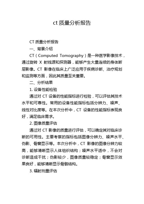
ct质量分析报告CT质量分析报告一、背景介绍CT(Computed Tomography)是一种医学影像技术,通过旋转X射线源和探测器,能够产生大量连续的身体断层影像。
CT影像在临床上广泛应用于疾病诊断、治疗规划和监测等方面,因此其质量至关重要。
二、分析结果1. 设备性能检验通过对CT设备的性能指标进行检验,可以评估其技术水平和可靠性。
常用的设备性能指标包括分辨力、噪声、线性对比度等。
在本次分析中,CT设备的性能指标表现良好,满足临床需求。
2. 图像质量评估通过对CT影像的质量进行评估,可以确定其对临床诊断的可用性。
主要考察的指标包括图像分辨力、噪声水平、伪影、骨窗显示等。
本次分析中,CT影像的图像分辨力较高,能够清晰显示人体组织结构;噪声水平适中,不会对诊断造成干扰;伪影较少,图像质量较稳定;骨窗显示效果良好,能够清晰显示骨骼结构。
3. 辐射剂量评估CT影像的质量不仅取决于图像本身,还与辐射剂量有着密切关系。
合理的辐射剂量可以保证影像质量的同时,尽量降低对人体的辐射损伤。
本次分析中,CT影像的辐射剂量处于合理范围内,既满足影像质量要求,又减少了对患者的辐射损伤。
4. 人员培训评估CT设备的操作人员对影像质量的掌控也是影响质量的重要因素。
通过对人员的培训评估,可以判断其操作技术是否熟练,是否能够合理调节设备参数来获取高质量的影像。
本次分析中,CT设备的操作人员技术熟练,能够根据需要进行合理的参数调节,保证了图像质量的可靠性。
三、改进建议1. 进一步提高技术水平尽管CT设备在本次分析中表现良好,但在不断发展的医学影像领域,技术水平仍需不断提高。
可以通过进一步学习和培训,学习国内外最新的CT技术和应用,提高设备的性能指标和图像质量。
2. 定期维护和保养CT设备作为精密的医学设备,需要定期进行维护和保养,以保证其运行的稳定性和可靠性。
建议制定严格的设备维护计划,并确保设备的仪器校准和故障排除及时有效。

医学图像分割算法的性能评价与改进方法一、引言医学图像分割是指将医学图像中感兴趣的目标物体从背景中准确地提取出来的过程。
医学图像分割在医疗诊断和疾病治疗领域具有极其重要的作用。
随着计算机技术的不断发展,各种医学图像分割算法得到了广泛的应用。
然而,由于医学图像本身具有复杂性、噪声干扰、边缘模糊等特点,如何准确地进行医学图像分割仍然是一个具有挑战性的问题。
本文将对医学图像分割算法的性能评价进行探讨,并介绍了一些常见的改进方法。
二、医学图像分割算法的性能评价指标准确地评价医学图像分割算法的性能是提高算法的关键。
以下是常用的医学图像分割算法的性能评价指标。
1. Jaccard指数(JI)JI指数是衡量分割结果与真实分割之间相似性的度量标准。
JI指数的计算公式为:JI = TP / (TP + FP + FN),其中,TP为真正例(算法分割正确),FP为假正例(算法分割错误),FN为假反例(算法未将目标分割出来)。
2. Dice系数(Dice)Dice系数是评估分割结果与真实分割之间相似性的指标。
Dice系数的计算公式为:Dice = 2 * TP / (2 * TP + FP + FN)。
3. 值得注意的是,除了JI和Dice系数,还有一些其他常用的性能评价指标,如准确率(Accuracy)、灵敏度(Sensitivity)、特异度(Specificity)等,选择适当的指标以评价医学图像分割算法的性能。
三、常见的医学图像分割算法改进方法为了提高医学图像分割算法的性能,研究者们提出了许多有效的改进方法。
以下是一些常见的改进方法。
1. 基于机器学习的改进方法机器学习在医学图像分割中被广泛应用。
基于机器学习的算法可以通过学习已有的医学图像数据集,提取出区分目标和背景的特征,并做出准确的分割预测。
常见的机器学习方法包括支持向量机(SVM)、决策树、随机森林等。
2. 基于深度学习的改进方法深度学习在医学图像分割中取得了很大的突破。

CT图像后处理质量控制报告分析CT(Computed Tomography)是一种医学影像技术,它通过使用X射线和计算机技术来生成人体的横断面图像,从而帮助医生更好地诊断和治疗疾病。
在CT图像后处理过程中,质量控制至关重要,因为图像质量直接影响到医生对病情的判断和诊断结果的准确性。
本文将从CT图像后处理的总体流程、质量控制指标的设定和实际应用情况等方面进行分析,以期为医疗机构提供合理的质量控制方案和改进措施。
一、CT图像后处理的总体流程CT图像后处理是对原始的CT图像进行进一步处理,以改善图像的质量、清晰度和对比度,使医生能够更准确地诊断病变。
通常的CT图像后处理流程包括以下几个步骤:1. 图像重建:将从患者身上获得的原始数据进行处理,生成横断面图像。
这个步骤需要确保重建参数的选择合理,以获得清晰的图像。
2. 骨干重建:对CT图像进行骨干窗显示,以便更清晰地显示骨骼结构。
3. 螺旋重建:通过将原始数据进行螺旋扫描重建得到更清晰的图像。
4. 后处理滤波:对图像进行滤波处理,以去除噪声和增强对比度。
5. 三维重建:将二维图像进行三维重建,以提供更全面的信息。
以上所述仅是CT图像后处理流程的一个概括,实际情况可能会因设备类型、应用场景等因素而有所差异。
二、质量控制指标的设定针对CT图像后处理的质量控制,需要明确一些指标用于评价图像的质量。
一般来说,可从以下几个方面进行评价:1. 空间分辨率:指图像能分辨出不同结构的能力,通常使用线对模块函数(MTF)来评价。
2. 对比度分辨率:指图像能够显示出不同物质的对比度程度,这对于显示病变非常重要。
3. 噪声水平:指图像中无用的杂乱信号,即噪声,应尽量降低。
4. 几何失真:指图像显示的物体与其实际形状的偏差程度。
5. 伪影:指图像中出现的不真实的结构,通常由于仪器故障或者处理算法的问题引起。
这些指标可以通过人工观察或者自动化算法来评估,以确保CT图像后处理的质量符合临床要求。

医学影像中的异常区域检测与分割算法研究随着医学影像技术的不断发展,大量的医学影像数据被产生和积累,这为医学诊断和治疗提供了宝贵的信息。
然而,在海量的医学影像数据中,寻找和提取异常区域成为医学专业人员面临的一项重要任务。
因此,研究和开发高效准确的医学影像异常区域检测与分割算法具有重大意义。
异常区域的检测与分割是医学影像处理领域的一个关键问题。
为了帮助医生准确诊断疾病,异常区域的精确定位和提取至关重要。
传统的医学影像异常区域检测与分割方法通常基于图像的灰度、纹理和形状等特征进行计算和分析,然后利用数学模型和算法进行图像处理和分割操作。
然而,由于医学影像的复杂性,传统方法在处理复杂疾病或特定情况下可能存在一定的局限性。
近年来,随着深度学习和计算机视觉的快速发展,基于深度学习的医学影像异常区域检测与分割方法得到了广泛关注和研究。
深度学习技术通过构建深度神经网络模型,可以自动学习和提取医学影像中的特征,以实现高效准确的异常区域检测与分割。
其中,卷积神经网络(CNN)被广泛应用于医学影像的异常区域检测和分割任务。
通过多层次的卷积和池化操作,CNN 可以捕获图像中的局部和全局特征,并自动学习特定疾病的表征。
在基于深度学习的医学影像异常区域检测与分割算法中,常用的方法包括U-Net、Faster R-CNN、Mask R-CNN等。
U-Net是一种经典的卷积神经网络模型,它通过构建编码器和解码器网络结构进行医学影像的分割操作。
编码器网络用于提取图像层次化的特征表示,而解码器网络用于将特征映射恢复到原始图像分辨率,从而得到医学影像的分割结果。
Faster R-CNN和Mask R-CNN是一种基于目标检测的异常区域检测和分割算法,通过引入候选框和区域建议网络,进一步提高了检测和分割的性能。
为了提高医学影像异常区域检测与分割算法的准确性和鲁棒性,研究者们不断改进和优化深度学习模型。
一些研究表明,在训练深度学习模型时,使用大规模的医学影像数据集和合适的数据增强技术可以提高算法的性能。

异常检测中的图像处理与分析技术导言:在现代社会中,图像处理与分析技术已经被广泛应用于各个领域,包括安全监控、医学影像、自动驾驶等。
其中,异常检测是图像处理与分析技术中的一个重要研究方向。
通过对图像进行处理与分析,可以有效地识别和定位异常事件,为人们的生活和工作提供了极大的便利。
本文将详细介绍异常检测中的图像处理与分析技术,包括基本概念、方法原理、应用案例等内容,旨在为读者提供全面的了解。
第一章:异常检测的基本概念1.1 异常检测的定义与分类异常检测是通过对数据进行分析与处理,识别出与正常模式不符的事件或对象。
根据异常检测的输入数据类型,可以将其分为基于图像的异常检测和基于视频的异常检测。
本章主要介绍基于图像的异常检测。
1.2 基本原理与方法基于图像的异常检测方法主要分为基于特征的方法和基于像素的方法两大类。
前者通过提取图像的局部或全局特征进行异常检测,后者则直接对图像像素进行统计分析。
第二章:基于特征的异常检测技术2.1 局部特征提取与描述局部特征提取是基于特征的异常检测方法中的核心环节。
该方法旨在寻找图像中的不规则特征点或线条,并对其进行描述,以便后续的异常检测工作。
2.2 特征匹配与配准特征匹配与配准是基于特征的异常检测方法中的关键步骤。
通过将待检测图像与已有数据库中的正常图像进行匹配和配准,可以找出异常区域并定位异常事件。
第三章:基于像素的异常检测技术3.1 像素分布统计分析基于像素的异常检测方法主要是通过对图像中的像素值进行统计分析,找出异常像素。
这种方法一般适用于背景相对固定的场景。
3.2 像素差分与阈值检测基于像素的差分与阈值检测方法主要是通过将当前图像与历史图像进行差分,并根据设定的阈值判断差异像素是否异常。
第四章:异常检测的应用案例4.1 安防监控领域在安防监控领域中,异常检测技术可以帮助监控人员实时发现异常事件,如入侵行为、人员聚集等,并及时采取相应措施。
4.2 医学影像领域异常检测技术在医学影像领域的应用十分广泛。

生物医学图像中的异常检测与分析方法研究引言生物医学图像是在医学研究和临床诊断中广泛应用的一种重要工具。
正常和异常的生物医学图像之间的差异可以提供有用的信息,帮助医生做出准确的诊断和治疗决策。
因此,异常检测与分析在生物医学图像领域中具有重要意义。
本文将介绍生物医学图像中常用的异常检测与分析方法,并讨论其应用和挑战。
一、异常检测方法1. 统计方法统计方法是一种常用的异常检测方法,通过对生物医学图像中的像素值进行统计分析,确定异常像素并标记出来。
常用的统计方法包括基于阈值、基于聚类和基于概率模型的方法。
其中,基于阈值的方法简单直接,但对噪声和图像变化敏感,可能会产生较多的误检率。
基于聚类的方法可以有效地区分正常和异常像素,但需要准确设置聚类参数。
基于概率模型的方法可以通过概率分布来描述图像像素的特性,具有较高的准确性和鲁棒性。
2. 基于机器学习的方法基于机器学习的异常检测方法通过训练模型来学习异常模式,并根据学习到的模型来进行异常检测。
常见的机器学习方法包括支持向量机、决策树和神经网络等。
这些方法可以通过特征提取和特征选择来构建生物医学图像的模型,并使用模型来预测新样本的异常程度。
基于机器学习的方法具有较高的准确性和灵活性,但需要大量的训练数据和特征工程。
二、异常分析方法1. 图像特征提取与描述图像特征提取与描述是生物医学图像分析中的关键步骤,旨在从图像中提取有用的信息和特征,以辅助异常检测和分析。
常见的图像特征包括形状、纹理、颜色和灰度等。
图像特征提取与描述可以通过局部二值模式、灰度共生矩阵和小波变换等技术来实现。
特征选择与降维是图像特征提取与描述中的重要问题,有助于去除冗余和不相关的特征,提高异常检测和分析的性能。
2. 异常定位与识别异常定位与识别是在生物医学图像中确定和标记异常区域的过程。
常见的异常定位与识别方法包括像素级别方法和区域级别方法。
像素级别方法通过像素分类或像素分割的方式来定位和识别异常,可以提供较高的定位精度,但对噪声和图像变化敏感。
CT图像后处理质量控制报告分析1. 引言1.1 研究背景CT图像后处理质量控制报告分析CT图像后处理质量控制报告分析是医学影像学中非常重要的一环,它通过对CT图像后处理过程中质量控制的实施与评估,保证了影像诊断结果的准确性和可靠性。
随着医疗技术的不断发展,CT图像后处理质量控制报告分析也越来越受到重视。
在过去的研究中发现,CT图像后处理质量控制报告分析对于提高影像诊断的准确性和精度起到了关键作用。
通过对CT图像后处理过程中可能存在的问题进行分析和评估,可以帮助医学影像学工作者及时发现和解决问题,从而保证影像诊断的质量。
CT图像后处理质量控制报告分析还可以为医院科室管理提供重要参考依据。
通过对CT图像后处理质量控制报告的分析,可以及时了解影像质量的相关情况,为科室管理提供决策支持。
研究背景CT图像后处理质量控制报告分析的重要性不容忽视,对于提高影像诊断水平、加强医疗质量管理具有重要意义。
1.2 研究目的CT图像后处理质量控制报告分析CT图像后处理质量控制报告分析的研究目的在于通过对CT图像后处理过程中产生的质量问题进行深入分析,探讨其原因和解决方法,为提高CT图像后处理质量提供有效的参考。
具体来说,研究目的包括以下几个方面:通过对CT图像后处理质量控制报告进行分析,可以揭示出当前存在的问题和不足之处,为改进和提升CT图像后处理质量提供依据。
研究目的还在于探讨CT图像后处理质量控制报告的编制标准和要求,以及相关的技术指标和评价方法,帮助实验室或医疗机构建立完善的质量控制体系。
通过研究CT图像后处理质量控制报告的分析,可以为医疗机构或科研单位提供参考,指导其在实际工作中如何针对质量问题进行系统性的监控和管理,从而保障CT图像后处理质量的稳定和可靠性。
1.3 研究意义CT图像后处理质量控制报告分析CT图像后处理质量控制对于医学影像领域具有重要意义。
随着医学影像技术的不断发展,CT图像后处理已经成为了医学影像诊断的重要环节。
医学图像分割技术在病灶检测中的应用案例分析1.引言医学图像分割技术在病灶检测中的应用具有重要意义,可以帮助医生精确识别和定位病灶区域,从而提高诊断准确性和效率。
本文将以几个应用案例为例,介绍医学图像分割技术在病灶检测中的优势和应用前景。
2.背景传统的医学图像分割方法主要基于手工设计的特征和规则,存在主观性和局限性。
而现代的医学图像分割技术利用计算机视觉和机器学习的方法,能够自动学习和提取图像特征,从而实现更准确的病灶检测和分割。
3.应用案例1:乳腺癌病灶检测乳腺癌是女性常见的恶性肿瘤之一。
利用医学图像分割技术,可以将乳腺磁共振图像中的肿瘤区域进行精确分割,帮助医生快速准确地检测和评估病变。
通过对乳腺癌病灶的分割,医生可以制定更精确的治疗方案,提高治疗效果。
4.应用案例2:脑部病变检测脑部病变是造成神经系统疾病的重要因素。
利用医学图像分割技术,可以将脑部核磁共振图像中的异常病灶进行自动分割,实现对脑部病变的定位和评估。
这对于神经外科手术的指导和疾病诊断有着重要的意义,能够减少手术风险,并提高疾病的早期诊断和治疗效果。
5.应用案例3:前列腺癌检测前列腺癌是男性常见的恶性肿瘤之一。
医学图像分割技术可以帮助医生在前列腺磁共振图像中自动分割出潜在的肿瘤区域。
这为医生提供了更准确的定位信息,有助于早期诊断和治疗。
此外,医学图像分割技术还可以辅助医生评估前列腺癌的分级和预测患者的预后,为制定个性化治疗方案提供参考。
6.应用案例4:皮肤癌检测皮肤癌是常见的恶性肿瘤之一。
医学图像分割技术可以将皮肤镜图像中的病变区域进行自动分割,帮助医生快速识别和定位皮肤癌病变。
这对于非侵入性早期诊断和治疗至关重要。
此外,医学图像分割技术还可以辅助医生跟踪病变的发展和治疗效果,为患者提供更精确的治疗方案。
7.技术挑战与发展趋势医学图像分割技术在病灶检测中的应用面临着一些挑战,包括不同病灶的形态和纹理差异、图像噪声和复杂的背景等。
为了解决这些挑战,研究人员不断提出新的分割方法和算法,如基于深度学习的图像分割方法。
CT图像后处理质量控制报告分析
CT(计算机断层扫描)图像后处理在临床医学中已经成为非常重要的工具。
通过 CT 图像后处理技术,我们可以从 CT 扫描获得高质量的立体图像,并发现肿瘤、血管、骨骼等内部器官的异常情况。
因此,对 CT 图像后处理的质量控制十分重要。
在CT图像后处理质量控制方面,主要涉及以下几点:
1.图像几何校准:当 CT 设备使用不当时,会导致图像几何姿态不正确。
因此需要校准仪器,消除不正确的几何形态,最终可以得到准确的三维图像数据。
2.图像质量评估:在 CT 图像处理之前,需要对 CT 扫描进行质量评估来确定扫描是否符合标准,以便进行图像后处理。
评估指标包括:分辨率、多普勒效应、斑点噪声和切片位置偏移等。
3.选择适当的滤波算法:图像滤波是一种处理图像中噪声的方法,因此选择适当的滤波算法对于减少噪声是至关重要的。
低通滤波器可以平滑图像,而高通滤波器可以增强CT 图像中的像素差异。
4.分割技术:图像分割通过将图像分成不同的部分,可以更好地区分不同的组织和结构。
分水岭算法、基于阈值的方法和基于形态学的方法等是常用的图像分割技术。
5.三维可视化:三维模型可以提供更详细的图像信息,基于三维模型的切面和立体呈现可以使医生更好地理解图像结果。
6.图像存档和管理:CT 图像后处理完整的过程生产了大量的图像数据,因此,存档和管理非常重要。
在存档和管理过程中,需要保证图像的安全及其可用性。
在 CT 图像后处理中,需要考虑机器/软件参数设置和操作的标准化。
通过对图像后处理的质量控制,可以更好地提高 CT 图像的质量,并且最终改善诊断结果。
CT图像后处理质量控制报告分析CT(Computed Tomography)是一种常用的医学影像检查方式,通过使用X射线和计算机技术来生成横断面图像,以帮助医生诊断和治疗疾病。
CT图像后处理质量控制是确保CT 图像质量和准确性的重要环节,对于提高诊断效果和保障患者安全具有重要意义。
下面我们将对CT图像后处理质量控制报告进行分析,以便更好地了解和改进CT图像后处理质量控制工作。
一、CT图像后处理质量控制报告的基本内容CT图像后处理质量控制报告主要包括以下内容:1.设备运行情况:包括CT设备的运行状态、图像采集参数设置等。
2.图像质量评估:对采集到的CT图像进行质量评估,检查图像的清晰度、对比度、噪声水平等。
3.后处理软件操作:对CT图像进行后处理的软件操作情况进行评估,包括后处理参数设置、图像重建等。
4.质量问题记录:记录在CT图像后处理过程中出现的质量问题和异常情况。
5.改进建议:针对CT图像后处理过程中存在的问题提出改进建议和措施。
二、CT图像后处理质量控制报告的分析方法对CT图像后处理质量控制报告进行分析,可以采用以下方法:1.对比历史数据:将当前的CT图像后处理质量控制报告与历史数据进行对比分析,了解质量水平的变化趋势。
2.定量分析:采用定量分析方法,对图像质量评估结果进行统计和分析,找出存在的问题和改进的方向。
3.问题分析:对质量问题记录进行深入分析,找出问题的原因和可能的解决方案。
4.建议整合:对改进建议进行整合和归纳,形成改进方案。
三、CT图像后处理质量控制报告的问题与改进在对CT图像后处理质量控制报告进行分析过程中,我们发现了一些常见的问题和改进的方向:1.设备运行情况记录不全:部分CT设备运行情况的记录不全,导致无法全面评估设备运行的稳定性和准确性。
建议对设备运行情况进行更详细和全面的记录。
2.图像质量评估方法不够科学:部分CT图像质量评估方法仍较为主观,缺乏科学性和客观性。
建议引入更加科学的图像质量评估方法,例如使用图像质量评估指标进行定量评估。
I.J. Engineering and Manufacturing, 2019, 5, 46-55Published Online September 2019 in MECS ()DOI: 10.5815/ijem.2019.05.04Available online at /ijemAnalysis of CT DICOM Image Segmentation for AbnormalityDetectionRashmi Kulkarni a, Bhavani K aa Department of Information Science and Engineering, Dayananda Sagar College of Engineering,Bengaluru-560078,India.Received: 11 June 2019; Accepted: 23 August 2019; Published: 08 September 2019AbstractThe cancer is a menacing disease. More care is required while diagnosing cancer disease. Mostly CT modality is used for Cancer therapy. Image processing techniques [1] can help doctors to diagnose easily and more accurately. Image pre-processing [2], segmentation methods [3] are used in extraction of cancerous nodules from CT images. Many researches have been done on segmentation of CT images with different algorithms, but they failed to reach 100% accuracy. This research work, proposes a model for analysis of CT image segmentation with filtered and without filtered images. And brings out the importance of pre-processing of CT images.Index Terms: Image processing, noise, filtration, image pre-processing, segmentation, nodule extraction.© 2019 Published by MECS Publisher. Selection and/or peer review under responsibility of the Research Association of Mode rn Education and Computer Science* Corresponding author.E-mail address: kulkarni.rashmi.25@Analysis of CT DICOM Image Segmentation for Abnormality Detection 47 1.IntroductionCancer is a life-threatening disease. The doctors should identify cancer nodules in the early stages. CT modality is used in cancer treatment. The medical images are acquired by scanning the patients with high x-ray beam. These scanned medical images are saved as DICOM format.The CT images may embedded with noises during image acquisition. Spekle noise, Gaussian noise, salt and pepper noise are common in medical images. Pre-processing of medical images is done to get more accurate results. In pre-processing of images, the filters like median, CLAHE [11], Wiener, Morphological, Gabor, Gaussian, Anisotropic Diffusion, Haar wavelet Filter are used to remove the noise and to enhance the images.PSNR is used to find the ratio of signal strength to the corrupted signal called noise. The more the PSNR value, the more the signal strength will be. An image with good PSNR is considered as good picture.Finding the best filters for medical images is very important. In this research work, different filters are applied on five organs like Lung, Pancrease, Bladder, Stomach and Thyriod. Analysis of images is done by taking PSNR into consideration and by visualising the images.Segmentation is the processes, in which the region of interest can be extracted [12] from the whole image. Segmentation algorithms like Watershed, Otsu, Thresholding algorithm and many more are used for image segmentation.Finding the cancer cells growth at the earlier stage is a good option to come out of this problem. We need to build a system which detects cancer cells at the earlier stages. By using different filtering techniques on DICOM images and accurate segmentation algorithm we need to improve the accuracy of system for finding the cancer cells. In this paper, we tried to improve PSNR value by using filters on images and to increase the accuracy of finding the cancer cells by applying appropriate segmentation technique.2.Literature ReviewHere, survey of some techniques used for pre-processing of CT images and its segmentation techniques is discussed [2].Suren Makaju et al. [4] proposed a model to detect cancer nodules in CT lung images. The median and Gaussian flitters are used to smooth the image and remove speckle noise from image. Watershed algorithm is used for segmentation. Feature extraction is done on the images. Used machine learning method called Support Vector Machine (SVM) as classifier to differentiate nodule as malignant or benign [4]. Proposed model detects the cancer with 92% accuracy [4]. Hasan Koyuncu et al. [5] proposed BFO model for image enhancement before abdominal organ & tumor segmentation. Block Matching and 3D Filtering (BM3D) algorithm, Fast Linking Spiking Cortical Model (FLSCM) [5] and Otsu algorithm are used. PSNR values are compared and found that BFO model gives good PSNR values. Jiayong Yan et al. [6] proposed a promising method to segment the liver metastases on contrast-enhanced sequential CT images. Marker-controlled watershed transform and Fuzzy connectedness algorithm [6] are used. Ashwani Kumar Yadav et al. [7] proposed a model for segmenting Brain MRI and CT Angiography images. The pre-processed images are segmented by thresholding method. The results are calculated by two parameters. That is completeness and other one is correctness [7]. M.Jayanthi et al. [8] compared the results of segmentation algorithms for liver CT images. Median filtering technique is used to remove unwanted noise[8] in images to get accurate results. Histogram Techniques, seeded region growing method, connected component algorithm, NS based thresholding algorithms are the segmentation algorithms [8] used for liver images. The dice similarity values are compared and found that the NS based thresholding algorithm works better for liver CT images than any other. P.Arjun et al. [9] proposed an improved region growing algorithm to enhance the segmentation of the48 Analysis of CT DICOM Image Segmentation for Abnormality Detectionliver from abdominal CT images[9]. Gaussian filter to remove Gaussian noise. Thresholding segmentation algorithms are applied. . K-means clustering algorithm[9] is used to classify the dataset. An improved region growing algorithm [9] partitioned the image from overlapped image to non-overlapped image. The results are compared with traditional region growing algorithm and K-means clustering. It is found that an improved region growing algorithm is having high accuracy of 97.04% where as traditional region growing is 86.03% and K-means clustering is 87.52% [9].Analysing this survey, the accuracy of current model are good but not satisfactory. The current researches used jpeg format of CT images. The accuracy can be achieved 100% if the pre-processing of images is done properly. In this work, proposed a model to analyse the filters for filtration of CT images and to analyse the results of segmentation with filter and segmentation without filter. DICOM format images are used as input dataset.3.Proposed ModelAccording to survey, it is found that pre-processing for CT image is very necessary to get the more accurate results. In proposed system, before going for segmentation of CT images, pre-processing of images[2] is done by filtering techniques. The proposed system consists of three phases. In first phase, analysis of filters on CT images is done by filtering the images with 8 different filters, and found out the best filter to remove noise in CT images. In second phase, analysis of segmentation is done on filtered CT image and on non-filtered CT image. Nodules are extracted. In third phase, the results of filtered segmentation and non-filtered segmentation are compared and analyzed.3.1 Type conversionFirstly, the CT DICOM images are converted from matrix scale to gray scale images. It converts the images with intensity between 0 to 1. That is from black (0) to white (1).3.2. Image pre-processingIn CT images, the noises are implanted while acquiring the images. The noises like speckle noise, Gaussian noise, salt and pepper noise are common. In this work , 8 different filters like Median Filter, CLAHE filter[10], Wiener Filter, Morphological Filter, Gabor filter, Gaussian Filter, Anisotropic Diffusion filter , Haar wavelet Filter are applied on Lung , Pancrease, Bladder, Stomach and Thyroid CT DICOM images. The filtered images are compared by considering their PSNR values and by visualising the images. The filter which results into more clarity image with good PSNR values is selected for CT image pre-processing. Here, the median, Gaussian and CLAHE filter gives the better results. So these filters are considered for pre-processing of CT images.3.3. SegmentationThis process locates objects or boundaries which help in acquiring the region of interest in the image [12]. It helps to segment the particular part of the organ in which we are interested in. In this work, Otsu segmentation algorithm [13] is applied on the filtered and non-filtered images. Otsu algorithm classifies the images using threshold values.Analysis of CT DICOM Image Segmentation for Abnormality Detection 49 3.4. Nodule extractionFinally, the cancer nodules are extracted [14] from the organ. The nodule counts of filtered segmented image and nodule counts of non-filtered segmented image are compared.Fig. 1. Architecture of proposed system4.ImplementationFor implementation, the patient’s real CT scan images are taken from LIDC (Lung Image Database Consortium image collection)[16] and TCIA(The Cancer Imaging Archive)[17]. LIDC is a web-accessible international resource for development, training, and evaluation of computer-assisted diagnostic (CAD) methods for lung cancer detection and diagnosis [16]. In this work the images are used are in DICOM format. The DICOM images will be of size 512*512. DICOM format contains the metadata. It contains patient’s details like patient’s ID, age, sex, acquisition value, date of scan etc.The proposed model is implemented in MATLAB R2014a. MATLAB is one of the tools for research development and analysis [18]. The DICOM viewer is used for analyzing the CT images. The patient details are seen through DICOM viewer.In this model various filters are used on DICOM images and tested with combination of median, Gaussian and CLAHE filter. This combination is resulting good PSNR value. After that segmentation algorithm is applied on images. The accuracy of model in finding cancer cells is found out.50 Analysis of CT DICOM Image Segmentation for Abnormality Detection5.Results and AnalysisIn the first phase of this work, the different filters are applied on Lung, Pancrease, Bladder, Stomach and Thyroid. The median filter is used to remove salt and pepper noise. Gaussian filter is used to remove Gaussian noise. And CLAHE filter [11] is used for image enhancement. The results are analyzed and found out that the median, Gaussian and CLAHE filter can be used for image pre-processing.In second phase of this work, the segmentation is done on filtered image and on non-filtered image by using Otsu segmentation algorithm [15]. Cancer nodules are extracted.In the above, Fig2, 3, 4 shows the original image of a lung, segmented image of lung without filter and extraction of cancerous nodules respectively. Without filtration the nodule count resulted as 23.Fig. 2. Original ImageFig. 3. Segmented without filterAnalysis of CT DICOM Image Segmentation for Abnormality Detection 51Fig. 4. Extracted Nodules with count=23And Fig.5, 6, 7 represents filtered image of original lung image, segmentation with filter and nodule extraction. With filtration, the image clarity is improved and the extracted nodule count resulted as 14.Which matched with nodule count given by LIDC data set.Fig. 5. Filtered ImageFig. 6. Segmented with filter52 Analysis of CT DICOM Image Segmentation for Abnormality DetectionFig. 7. Extracted Nodules with count=14Here, 24 Lung images are tested and found out the accuracy. The test results are tabulated below. Table 1. Analysis of CT ImagesImages of Lung ExpectednodulecountNodulecountwithoutfiltrationNodulecount withfiltrationRemarkimage1.dcm 4 1 4 Trueimage2.dcm 12 3 12 Trueimage3.dcm 4 0 4 Trueimage4.dcm 4 0 4 Trueimage5.dcm 9 18 9 Trueimage6.dcm 14 23 14 TrueImage7.dcm 2 0 2 Trueimage 8.dcm 4 0 4 Trueimage9.dcm 11 28 11 Trueimage10.dcm 6 0 6 Trueimage11.dcm 17 24 17 Trueimage12.dcm 16 23 16 Trueimage13.dcm 34 60 32 Falseimage14.dcm 3 0 3 Trueimage15.dcm 2 16 2 Trueimage16.dcm 14 52 14 Trueimage17.dcm 1 7 1 Trueimage18.dcm 6 18 5 Falseimage19.dcm 5 23 5 Trueimage20.dcm 4 10 4 Trueimage21.dcm 4 18 4 Trueimage22.dcm 4 18 4 Trueimage23.dcm 2 0 2 Trueimage24.dcm 5 24 5 TrueTrue Nodules Detected images (TN) = 22 (1) False Nodules Detected images (FN) = 2 (2) Total number of images tested = 24 (3)Analysis of CT DICOM Image Segmentation for Abnormality Detection 53220.91691.6%24TN Accuracy TN FN ====+(4)The above results depicts that, the proposed system achieved the accuracy of 91.6% in detecting cancer nodules in patients which is higher than other models [9]. 6. ConclusionThough many current systems are developed for cancer detection by using segmentation, the results are not satisfactory. Therefore, proposed a system for analysis of CT image segmentation. From proposed model, it is found that, the combination of Median, Gaussian and CLAHE filters give the better results for CT images. The PSNR value is more that means more noise is reduced by using combination of 3 filters. The comparison of filtered image segmentation and non-filtered image segmentation results. Found that segmentation with filter gives the better result than the segmentation without filter, referring to the values of table.1. So pre-processing of CT DICOM image is very necessary to get accurate results of finding cancer nodules at the earlier stages. The proposed system achieved the accuracy of 91.6%. Only 8.4% of test cases failed to find accurate numbers of cancer nodules in the images. 7. Future ScopeAlthough the proposed model is having accuracy of 91.6% in finding cancer cells, it is also having some limitations. The accuracy of system can be improved by using improved segmentation algorithms like watershed algorithm. And proposed system doesn’t classify the stages of cancer like I, II, III, IV. So this can be improved by using supervised and unsupervised classification techniques of machine learning. References[1] Rafael C.Gonzalez, Richard E.Woods , “Digital Image processing”, 3rd edition ,Pearson publication,2013. [2] Rashmi Kulkarni, Bhavani K, “A Survey on Noise Reduction and Segmentation Techniques in CTDICOM Images”, International Research Journal of Engineering and Technology (IRJET), Volume: 05 Issue: 05, May 2018.[3] M.Jayanthi, “Comparative Study of Different Techniques Used for medical image Segmentation of Liver from Abdominal CT Scan”, IEEE WiSPNET conference,2016.[4]Suren Makaju, P.W.C. Prasad, Abeer Alsadoon, A. K. Singh, A. Elchouemi, “Lung Cancer Detection using CT Scan Images”, 6th International Conference on Smart Computing and Communications, ICSCC 2017, 7-8 December 2017.[5]Hasan Koyuncu, Rahime Ceylan, “A Hybrid Tool on Denoising and Enhancement of Abdominal CT Images before Organ & Tumour segmentation”, IEEE 37th International Conference on Electronics and Nanotechnology (ELNANO),2017.[6]Jiayong Yan, John Q. Fang, “Segmentation of Liver Metastasis on CT Images Using the Marker-controlled Watershed and Fuzzy Connectedness Algorithms”, IEEE 2016.54 Analysis of CT DICOM Image Segmentation for Abnormality Detection[7]Ashwani Kumar Yadav, Ratnadeep Roy, Rajkumar, Vaishali, Devendra Somwanshi, “Thresholding andMorphological Based Segmentation Techniques for Medical Images”, IEEE International Conference on Recent Advances and Innovations in Engineering (ICRAIE-2016), December 23-25, 2016.[8]M.Jayanthi, “Comparative Study of Different Techniques Used for medical image Segmentation of Liverfrom Abdominal CT Scan”, IEEE WiSPNET 2016.[9]P.Arjun, M.K.Monisha, A.Mullai yarasi, G.Kavitha, “Analysis of the liver in CT images using animproved region growing technique”, 2015 International Conference on Industrial Instrumentation and Control (ICIC) Col/ege of Engineering Pune, India. May 28-30,2015.[10]Ravindra Pal Singh, Manish Dixit, “Histogram Equalization: A Strong Technique for ImageEnhancement”, International Journal of Signal Processing, Image Processing and Pattern Recognition Vol.8, No.8 (2015), pp.345352,2015.[11]Brij Bhan Singh, Shailendra Patel, “Efficient Medical Image Enhancement using CLAHE Enhancementand Wavelet Fusion”, International Journal of Computer Applications (0975 – 8887) Volume 167 – No.5, June 2017.[12]Khobragade, S., Tiwari, A., Patil, C., Narke, V. , “Automatic detection of major lung diseases usingChest Radiographs and classification by feed-forward artificial neural network.” IEEE International Conference on Power Electronics, Intelligent Control and Energy Systems (ICPEICES),2016.[13]Wuli Wang, Liming Duan, Yong Wang, “Fast Image Segmentation Using Two-Dimensional Otsu Basedon Estimation of Distribution Algorithm”, Journal of Electrical and Computer Engineering Volume 2017, Article ID 1735176,2017.[14]Smitha P, Shaji L, Dr. Mini M. G., “A Review of Medical Image Classification Techniques”, InternationalConference on VLSI, Communication & Instrumentation (ICVCI) , Proceedings published by International Journal of Computer Applications(IJCA),2011.[15]Hetal J. Vala, Astha Baxi, “A Review on Otsu Image Segmentation Algorithm”, International Journal ofAdvanced Research in Computer Engineering & Technology (IJARCET), ISSN: 2278 – 1323, Volume 2, Issue 2, February 2013.[16]LIDC CT DICOM image dataset retrieved from :https:///display/Public/LIDC-IDRI[17]TCIA CT DICOM image dataset of Lung, Pancrease, Bladder, Stomach, Thyroid retrieved from:https:///display/Public/Wiki[18]Miah, M.B.A., & Yousuf, M.A. (2015) “Detection of lung cancer from CT image using image processingand neural network.” International Conference on Electrical Engineering and Information Communication Technology (ICEEICT), 2015.Analysis of CT DICOM Image Segmentation for Abnormality Detection 55Authors’ ProfilesMrs Rashmi Kulkarni received Bachelor of Engineering in Computer Science &Engineering in2016 and Master Degree M.Tech in computer Science and Engineering in2018 from Visvesvaraya Technological University, Karnataka. Currently she is workingas Technical Mentor in private institute. Her areas of interests are Medical ImageProcessing, Machine Learning, and Artificial Intelligence.Mrs Bhavani K received Bachelor of Engineering in Computer Science & Engineeringin 2006 and Master Degree M.Tech in Computer Science and Engineering in 2009. Sheis currently pursuing Ph.D from Visvesvaraya Technological University, Karnataka. Herarea of research include Medical image processing, Computer Vision, AI.How to cite this paper:Rashmi Kulkarni, Bhavani K. "Analysis of CT DICOM Image Segmentation for Abnormality Detection", International Journal of Engineering and Manufacturing(IJEM), Vol.9, No.5, pp.46-55, 2019. DOI: 10.5815/ijem.2019.05.04。