UltraVIEW VoX 活细胞共聚焦
- 格式:pdf
- 大小:2.17 MB
- 文档页数:16

乳酸对粪肠球菌的抑菌作用及作用机制王凤婷;靳盼盼;刘芳;孙芝兰;吴海虹;王道营;许晓曦;徐为民【摘要】目前乳酸常被用来控制食品中腐败菌的生长,但是有关其对食品优势腐败菌抑菌机制方面的研究较少.本试验以一株从肉制品中分离得到的腐败菌粪肠球菌R612-Z1为目标菌株,研究乳酸对其的杀菌效果及作用机制.结果表明,各浓度(0.25%、0.50%、1.00%)的乳酸均有杀菌效果而且作用明显,通过激光共聚焦显微镜及流式细胞仪检测发现,处理后细胞膜通透性增强.扫描电镜及腺嘌呤核苷三磷酸(ATP)浓度的测定结果说明,胞内物质流出聚集在细胞外侧,胞外ATP浓度和核酸类物质浓度明显上升,膜电势快速升高.因此,乳酸通过破坏粪肠球菌的细胞壁,增加细胞膜通透性,改变细胞内外电势,导致内容物流出,从而达到杀菌效果.%Lactic acid(LA) is widely used to control the growth of contaminating bacteria in food,but there are few reports about the antibacterial mechanism of it. The antimicrobial activity of LA against a dominated spoilage bacterium, Enterococcus faecalis R612-Z1,isolated from water-boiled salted duck was studied. The results showed that all the selected concentrations of LA (0.25%,0.50%, 1.00%) had obvious bactericidal action. The permeability of cell membranes was increased for the bacterial cells treated with 1.00% LA, which was detected using the laser scanning confocal microscope and flow cytometry. The intracellular substances were leaked according to the results of scanning electron micrographs and the detection of extracellular ATP and UV-absorbing materials. The membrane potentials were found to be increased. There-fore,the antibacterial effect of LA on E. faecalis was mainly completed by the massive leakage of intracellular component,which was caused by damaging the cell membrane and membrane potential.【期刊名称】《江苏农业学报》【年(卷),期】2018(034)001【总页数】7页(P200-206)【关键词】粪肠球菌;乳酸;抑菌机理【作者】王凤婷;靳盼盼;刘芳;孙芝兰;吴海虹;王道营;许晓曦;徐为民【作者单位】江苏省农业科学院农产品加工研究所,江苏南京210014;东北农业大学食品学院,黑龙江哈尔滨150030;江苏省农业科学院农产品加工研究所,江苏南京210014;东北农业大学食品学院,黑龙江哈尔滨150030;江苏省农业科学院农产品加工研究所,江苏南京210014;江苏省农业科学院农产品加工研究所,江苏南京210014;江苏省农业科学院农产品加工研究所,江苏南京210014;江苏省农业科学院农产品加工研究所,江苏南京210014;东北农业大学食品学院,黑龙江哈尔滨150030;江苏省农业科学院农产品加工研究所,江苏南京210014【正文语种】中文【中图分类】TS201.3食品的安全、质量及营养价值是人们密切关注的问题[1]。
荧光共振能量转移(FRET)影像系统Olympus(北京)销售服务有限公司上海分公司PDF created with pdfFactory Pro trial version 荧光共振能量转移(FRET)影像系统一、研究目的随着生命科学研究的不断深入, 光学显微镜使我们理解了细胞结构和有关功能。
但是分子 生物学研究已经显示了分子事件,例如信号传导和基因翻译,需要蛋白质的装配成特殊的大 分子复合体等。
对各种生命现象发生的机制,特别是对细胞内蛋白质间相互作用的研究变得尤 为重要。
传统的生物物理或生物化学方法例如亲和色谱法或免疫沉淀反应法和近来的酵母双杂 交、磷酸化抗体、免疫荧光、放射性标记等方法等,都需要破碎细胞或对细胞造成损伤,无 法做到在活细胞生理条件下实时地对细胞内蛋白质-蛋白质间相互作用进行动态研究。
而基于强度的影像技术FRET方法,使得研究活细胞内的这些相互作用变得容易了,荧光 共振能量转移( FRET)是用于对生物大分子之间相互作用定性、定量检测的一种有效方法。
根 据所基于的荧光显微镜配置不同而有不同的应用侧重,可在多细胞,单细胞,细胞膜,细胞 器等不同层次对生物大分子间的相互作用距离,动力学特性等进行研究。
二、FRET的原理和实现方法FRET的原理和发生的基本条件:1. 2. 3. 4. 发色团之间的距离在10A到100A 。
供体D的荧光光谱和受体A的吸收光谱足够多的重叠。
供体D的量子产率和受体A的吸收系数足够大。
D和A的跃迁偶极矩有最佳的相对取向,或者两者之一有一定的快速旋转的自由度。
FRET的实现方法:1) 稳态方法(基于供体、受体的三通道计算校准) 供体荧光的减弱-主要的方法 受体荧光的增强 激发光谱和吸收光谱的比较 2) 3) 光漂白方法 (Pb-FRET) 时间分辨方法(TR-FRET) 供体荧光的衰减 受体荧光的增长PDF created with pdfFactory Pro trial version FRET 特点:1) 动态实验,采集速度快 / 高速Shutter、高速CCD 2) 3) 4) 维持活细胞活性-CO2培养箱、恒温培养箱、恒温板 尽量减少光毒性,减少光照时间 保证长时间观察奥林巴斯 FRET 系统组成:1、显微镜 2、光源、高速荧光激发光切换控制和电动光闸 3、电动 XY 载物台 4、环境控制 5、高灵敏度冷 CCD 6、多种部件同时工作的控制软件 7、图像分屏器——DualView三、Olympus FRET系统详细技术参数一)显微镜:Optics 光学性能Ø 光学系统(Optical System): 奥林巴斯 2005 年最新推出的 UIS2 无限 远光学系统(UIS2 Infinity optical system) (UIS2 光学系统具有的高光 透过率和全光谱范围的色差校正,及高信噪比的特点,非常适合荧光 方面的研究,可以说是目前最先进的光学系统之一) 光路设计: V型光路把反射时的光线损失减少到最小程度,保证最大光 通过量System Flexibility系统适应性Ø Ø Ø 光口: 双层多光口设计(奥林巴斯首创)保证了输入/输出灵活性,提 供 6 条射入/射出光路,最多可同时接 4 路采集原像的图像获取系统。

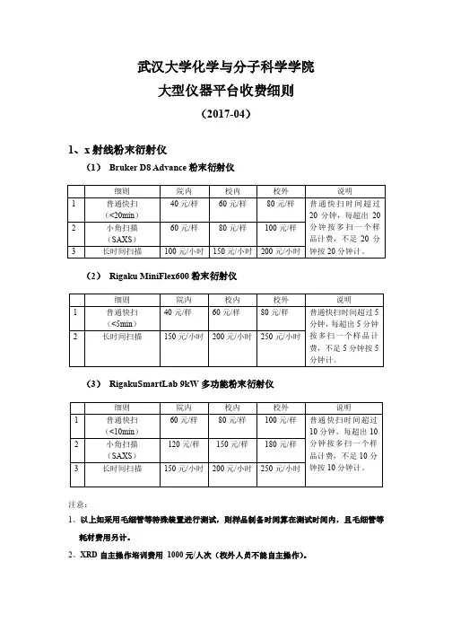
武汉大学化学与分子科学学院大型仪器平台收费细则(2017-04)1、x射线粉末衍射仪(1) Bruker D8 Advance粉末衍射仪细则院内校内校外说明1 普通快扫(<20min)40元/样60元/样80元/样普通快扫时间超过20分钟,每超出20分钟按多扫一个样品计费,不足20分钟按20分钟计。
2 小角扫描(SAXS)60元/样80元/样100元/样3 长时间扫描100元/小时150元/小时200元/小时(2) Rigaku MiniFlex600粉末衍射仪细则院内校内校外说明1 普通快扫(<5min)40元/样60元/样80元/样普通快扫时间超过5分钟,每超出5分钟按多扫一个样品计费,不足5分钟按5分钟计。
2 长时间扫描150元/小时200元/小时250元/小时(3) RigakuSmartLab 9kW多功能粉末衍射仪细则院内校内校外说明1 普通快扫(<10min)60元/样80元/样100元/样普通快扫时间超过10分钟,每超出10分钟按多扫一个样品计费,不足10分钟按10分钟计。
2 小角扫描(SAXS)120元/样150元/样180元/样3 长时间扫描150元/小时200元/小时250元/小时注意:1、以上如采用毛细管等特殊装置进行测试,则样品制备时间算在测试时间内,且毛细管等耗材费用另计。
2、XRD自主操作培训费用1000元/人次(校外人员不能自主操作)。
2、x射线单晶衍射仪细则院内校内校外1 扫单胞50元/样100元/样200元/样2 晶体结构数据采集(整套数据8h以内)400元/样600元/样800元/样3 晶体结构数据采集(整套数据超过8h)每小时加收100元/样每小时加收150元/样每小时加收200元/样4 2D衍射(4h以内)400元600元800元5 低温液氮费30元/小时30元/小时60元/小时注意:1、以上如采用毛细管等特殊装置进行测试,则样品制备时间算在测试时间内,且毛细管等耗材费用另计。
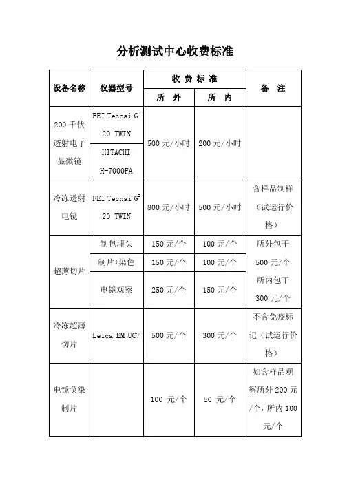
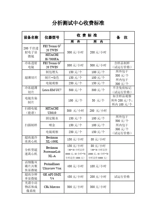
蔡司LSM900共聚焦显微镜的成
像原理
蔡司LSM900共聚焦显微镜的成像原理
共聚焦显微镜是指利用共聚焦显微成像技术(Confocal microscopy)作为技术基础,应用光学手段进行成像的一种光学显微镜。
共聚焦显微成像技术是一种利用逐点照明和空间针孔调制来去除样品非焦点平面的散射光的光学成像手段,相比于传统成像方法可以提高光学分辨率和视觉对比度。
共聚焦显微镜是一种利用逐点照明和空间针孔调至来去除样品非焦点平面的散射光成像技术的显微镜,是拥有高对比度和高分辨率的专业级显微镜。
常用语细胞及生物荧光样品观察分析;绿荧光蛋白分析;荧光复位杂交分析;光切片扫描以及
3D图像处理等应用中。
蔡司LSM900共聚焦显微镜的成像原理主要用于生物领域,共聚焦成像也被称为细胞CT。
能够获得焦平面细胞内一个层面的细胞图像,因无其它层面信号的干扰,能获得非常清晰的细胞图像。
共聚焦成像经过多年发张,如超分辨技术具有多种一样的实现方式相同,有多种方式能够实现共聚焦,不同的成像方式之间特点互补。
共焦成像是利用激光点扫描共焦成像的原理,即通过单个激光点激发样品中的荧光获得荧光信号,通过针孔屏蔽非焦平面信息,从而获得单片细胞图像。
在转盘可以聚焦的成像原理上,激光或普通激发光通过转盘变成多个激发点,转盘的旋转使激发光扫描整个样品,得到的荧光通过转盘的针孔去除非焦点细胞图像。
更多信息请咨询北京瑞科钟毅科技相关人员。
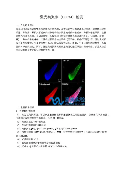
激光共聚焦(LSCM)检测一、实验技术简介激光扫描共聚焦显微镜是采用激光作为光源,在传统光学显微镜基础上采用共轭聚焦原理和装置,并利用计算机对所观察的对象进行数字图象处理的一套观察、分析和输出系统。
主要系统包括激光光源、自动显微镜、扫描模块(包括共聚焦光路通道和针孔、扫描镜、检测器)、数字信号处理器、计算机以及图象输出设备(显示器、彩色打印机)等。
通过激光扫描共聚焦显微镜,可以对观察样品进行断层扫描和成像。
因此,可以无损伤的观察和分析细胞的三维空间结构。
同时,通过激光扫描共聚焦显微镜也是活细胞的动态观察、多重免疫荧光标记和离子荧光标记观察的有力工具。
二、主要技术指标1、共聚焦扫描系统(1)独立双向扫描镜。
可以在正置显微镜和倒置显微镜之间迅速互换。
仪器永久不用校正;可调的计算机控制单共焦针孔、孔径20~600μm(2)光谱扫描区400~850nm(3)单线扫描频率≥2000线/秒(4)图扫描率≥3幅/秒512×512pixels;≥20幅/秒512×32pixels(5)扫描分辨率4096*4096扫描放大1~32X;多方位序列扫描方式;任意形状区域扫描-范围:≥22mm。
(6)光谱斜陡率≤1%(7)透射光检测器用于微分干涉相衬法检验(8)低噪音冷却型光电倍增管(PNT)控测器12bi2、激光装置:具有多通道AOTF(四通道,八谱线),连接四种不同波长的可见光激光可以自动选择激光谱线和功率,具等待功能。
光纤耦合连接低能量激光。
目前使用五激光,八谱线。
Ar激光发生器(458nm/5mw,476nm/5mw,488nm/20mw,514nm/20mw)-蓝;He/Ne激光发生器(543nm/1.2mw)-绿;He/Ne激光发生器(633nm/10mw,594nm/2mv)-红,紫外激光发生器(405nm/25mv)3、显微镜:DMIRE2倒置电动荧光显微镜,拥有10x,20x,40x干镜以及63x,100x油镜。
•优博专栏•荣筋拈痛方对白细胞介素-1 诱导大鼠退变软骨细胞增殖的影响黄艳峰\陈俊2,林洁\赵忠胜\叶锦霞u,李西海2,吴广文C福建中医药大学中西医结合研究院,福州350122;2福建省中西医结合老年性疾病重点实验室,福州350122)摘要:目的:探讨荣筋拈痛方对白细胞介素-IP(IL-ip)诱导退变软骨细胞增殖的作用机制。
方法:C C K8法筛选荣筋拈痛方对退变软骨细胞最佳干预条件后,将软骨细胞随机分为正常组、模型组、抑制剂组和荣筋拈痛方组,分组干预后采用激光共聚焦观察各组丨丨型胶原蛋白表达;流式细胞术检测各组周期分布情况;R T-P C R与W eS ten1B丨o t检测各组G1/S检测点相关调节因子pl6、CyclinDl、C D K4、pR B基因和蛋白表达。
结果:荣筋拈痛方lO O pg/m L干预退变软骨细胞48h可显著增强软骨细胞活力(P<0.01)。
与正常组比较,模型组和抑制剂组n型胶原蛋白表达量显著降低(P<0.01);与模遨组和抑制剂组比较,荣筋拈痛方组n型胶原蛋白表达量显著增加(P<0.01)。
与正常组比较,模型组和抑制剂组G o/G,期比例升高,G2/M期、S期比例显著降低(P<0.01);与模型组和抑制剂组比较,荣筋拈痛方组(VG,期比例显著降低,G2/M期、S期比例1著升高(P<0.01)。
与正常组比较,模型组和抑制剂组CyclinDl、C D K4、P丨《B基因和蛋白表达量显著降低,P16基因和蛋白表达量显著增加(P<0.01,P<0.05);与模型组和抑制剂组比较,荣筋拈痛方组CyclinDl、C D K4、P R B基因和蛋白表达量显著增加,pl6基因和蛋白表达量显著降低(P cO.Ol):结论:荣筋拈痛方可调控软骨细胞G,/S期的转换,促进n型胶原蛋白表达和软骨细胞增殖。
关键词:荣筋拈痛方;膝骨性关节炎;本痿标痹;A细胞介素-1P;退变软骨细胞;细胞周期;增殖;大鼠基金资助:国家自然科学基金面上项目(N o.82074465),福建省自然科学基金面上项目(N o.2020101750),陈可冀中西医结合发展基金(N〇.C K J2020005)Effects of Rongjin Niantong Formula on proliferation of rat degeneration chondrocytesinduced by interleukin-ipHUANG Yan-feng1,CHEN Jun2, LIN Jie丨,ZHAO Zhong-sheng1,YE Jin-xia12,L I Xi-hai2, WU Guang-wen1,2('Academy of Integrative Medicine, Fujian University of Traditional Chinese Medicine, Fuzhou 350122, China;2Fujian Key Laboratory of Integrative Medicine on Geriatrics, Fuzhou 350122, China )Abstract:Objective: To investigate the mechanism of Rongjin Niantong Formula on the proliferation of degenerative chondrocytes induced by interleukin-ip (IL-ip). Methods: After screening the best intervention conditions of Rongjin NiantongFormula on degenerative chondrocytes by CCK8 method, chondrocytes were randomly divided into normal group, modelgroup, inhibitor group and Rongjin Niantong Formula group. After group intervention, the expression of type I I collagen ineach group was observed by laser confocal method; The cycle distribution of each group was detected by flow cytometry; RT-PCR and Western Blot were used to detect the gene and protein expressions of regulatory factors p i6, Cyclin Dl, CDK4, andpRB at G,/S detection points. Results: Rongjin Niantong Formula 100|ig/mL for 48h could significantly enhance the activity ofdegenerative chondrocytes (尸<0.01). Compared with the normal group, the expression of type I I collagen in the model group通信作者:吴广文,福建ft、福州市闽侯上街邱阳路丨号福建中医药大学中西医结合研究院.邮编:35〇丨22.电话:*************E-mail: ********************and the inhibitor group was reduced (P<0.01); Compared with the model group and the inhibitor group, the expression of type I I collagen in the Rongjin Niantong Formula group increased (^O.Ol). Compared with the normal group, the proportion of G,/G, phase was increased in the model group and the inhibitor group, while the proportion of G:/M phase and S phase was decreased (P<0.01). Compared with model group and inhibitor group, the proportion of G,/G(phase in Rongjin Niantong Formula group was decreased, while the proportion of G2/M phase and S phase was increased (P<0.01). Compared with normal group, the expression levels of Cyclin Dl, CDK4, pRB gene and protein in model group and inhibitor group were decreased, while the expression levels of p i6 gene and protein were increased (P<0.01, P<0.()5). Compared with model group and inhibitor group, the expression levels of Cyclin Dl, CDK4, pRB gene and protein in Rongjin Niantong Formula group were increased, while the expression levels of pl6 gene and protein were decreased (P<0.01). Conclusion: Rongjin Niantong Formula can regulate the transition of chondrocytes in G,/S phase, promote the expression of type I I collagen and the proliferation of chondrocytes.K e y w o r d s;Rongjin Niantong Formula; Knee osteoarthritis; Deficiency in origin and excess in superficidity; Interleukin-ip : Degenerative chondrocytes; Cell cycle; Proliferation; RatsFunding! General Program of National Natural Science Foundation of China (No.82074465), General Program of Natural Science Foundation of Fujian Province (N〇.2020J01750), Integrative Medicine Developmental Foundation of CHEN Ke-ji (N〇.CKJ2020005)膝骨性关节炎(knee osteoar丨hiitis,K O A)是临 床所有骨关节炎(osteoarthritis,O A)中发病率最高 疾病之一,多见于中老年女性群体m。
活细胞工作站使用指南1. 开机顺序:1) 分为上下两列开关:先下列按照一、三、二、四、五的顺序打开,上列依顺序由下向上打开;2) 打开激光器开关;3) 打开电脑;4) 打开气瓶,顺时针缓慢拧开,压力阀上调一小刻度即可;5) 小室加水,要用超纯水(如小瓶没水,请到各自实验室填充);6) 选择合适的适配器(33mm 培养皿或玻片)后,将细胞放入小室,盖好小室上层小盖。
2. 打开软件。
桌面——双击Volocity ——出现 界面1) 新建文件夹(为了保证每次数据的安全性及有序性,要求每次试验都建立新的文件夹)方法:点击 Creat a new library 图标——选择新建文件夹路径(C:\Ultraview \ioz\当时月份的文件夹)——输入文件名(名称格式为 本人姓名+年月日)——点击 Creat ——进入软件界面2) 打开已有的文件夹进行数据处理点击 Open a existing library 图标——选择新建文件夹路径(C:\Ultraview \ioz\当时月份的文件夹\目标文件名)——点击 Open ——进入软件界面3. 软件界面及显微镜界面构成:2) 显微镜界面:图2:显微镜正面示意图(学生可操作的部分有两部分):图44. UltraVIEW VoX 图像采集简要操作流程1) 将样品放入小室,盖好小室上层小盖;2) 打开软件,单击图1 第三部分中的“video preview ”图标,点击图1 第六部分中的⑦打开明场光路;3) 选择合适的物镜后在显微镜下调整样品对焦;4) 点击图2.第一部分中的“L100”将显微镜光路切换至共聚焦成像模式;5) 选择合适的激光通路6) 依次调节各路激光的激光功率,敏感度(sensitivity )及曝光时间(exposure time );激光功率最大一般控制在40%左右,如果荧光较弱,敏感度可以拉的很大,但曝光时间一般最大控制在500ms 左右。
Stereo microscopesfor Materials and Life SciencesNexius ZoomThe new standard in microscopyEuromex NexiusZoom stereo microscopes enable you to observe your specimen with the highest precision in three dimensional imagingThese high quality stereo microscopes are perfect for analyzing all kinds of material surfaces and for preparing biological samplesNexiusZoom EVO The interpupillary distance is adjustable between 54 and 75 mm dditional lenses 0.3x, 0.4x, 0.5x, 0.75x, 1.5x and 2.0x are availableAll optics are anti-fungus treated and anti-reflection coated for maximum The newly optimized Greenough Optical System offers more depth of focus without compromising on low distortion and color aberrationESD SAFE MICROSCOPESElectrostatic protected microscopesfor all kinds of industry, inspection andNZ.1903-P-ESDNZ.1903-U-ESDNZ.1903-P-ESD The body and stand of the microscope are coated with a special electrostatic dissipative paint eliminating harmful electrostatic discharges, thus making the microscope suitable for all static-sensitive environmentsElectrostatic discharge (ESD) is the unwanted sudden flow of electricity between two electrically charged objects. ESD can cause a range of harmful effects as well as permanent damage to solid state electronicTo solve this Euromex has introduced electrostaticeliminating harmful electrostatic discharges makes environments. Ideal for all kinds of industry, assembly applicationsthe examination of your gems in perfectdarkfield conditionsfluorescenceLEDhalogenNZ.1702-GEMFThe ergonomic tilting stand allows easyadjustment between 0 and 45ºNZ.1902-GEMLSTANDErgonomic stand with 0 to 45° backwards tilting arm, including 76 mm headholder, suitable for darkfield and brightfield. Built-in 100-240 V power supplySTAGEFlat darkfield stage with clamp object holder, iris diaphragm and incidentillumination. Clamp object holder can be placed on left or right side1325Ergonomic flat pillar stand, including incident and transmitted 3 W LED illumination, Universal single-arm stand for free observation of (large) objects. Mounted on a heavy base for maximum stability. Supplied without illumination. However, a wide 100-240 V power supply, both illumination intensities can be adjusted separately. sustaining Goosenecks and one transmitted 3 W LED illumination. Built-in 100-240 V power supply. Maximum object height 148 mm.Including 76 mm head holderNZ.9018NexiusZoom standsThe NexiusZoom stereo microscopes are supplied with a large choice of objectives and stands. Long working distances enable working comfortably under the microscope. Ideal for education, laboratories and industryThe NexiusZoom stands can be combined with both NexiusZoom and NexiusZoom EVO binocular and trinocular heads. Choose your system from the pre-combined sets or combine a microscope head with the stand of your choice10 6879121311Universal double-arm stand for free observation of (large) objects.Mounted on a heavy base formaximum stability. Guided double arm that allows titling of the head. Suppliedwithout illumination. However, a wide range of illumination systems is available.Maximum object height 213 mm. NZ.9090 or NZ.9095 head holderAP stand8Articulated stand for free observation of (large) objects. For working comfortablyunder the microscope. Mounted on a heavy base for maximum stability. Flexiblejointed arm for maximum flexibility. Supplied without illumination. However, a widerange of illumination systems is available. Maximum object height 185 mm.NZ.9081 head holderNZ.9027A stand9Identical to the AP stand but with a table-clamp mount.NZ.9081 head holderNZ.902510Large 320 x 285 mm pillar stand including adjustable transmitted 3 W LED illuminationand 76 mm head holder. Built-in 100-240 V power supply. Including 76 mm head holderLarge 320 x 285 mm pillar stand without illumination. Including 76mm head holder.With stone holding tweezers and built-in Iris. Comes with additional 1 W LED incident illumination. Built-in12NexiusZoom illuminationGood illumination of an object is crucial for stereo microscopy. When the correct illumination is chosen the details of the observed object becomes more distinct. Comfort and desired light intensity are the factors that determine the choiceSegmentation of LED’s (LE.1973)LE.5211 and LE.5211-LEDLE.5212 combined with a NZ.9000 stand and head (NZ.5902)132AE.19601Positioning table allows easy and precise positioning of objects when using high magnifications. The round positioning table AE.1960, ø 20 cm is ideal for moving objects smoothly and precisely around a central point. Suitable for all free hanging standsAE.5168-NZ2Heating stage with X/Y mechanical stage. Temperature range: up to 50°C. LED display for set temperature (1 degree) and measured temperature (0.1 degree). For -S and -P stands*NZ.95053Mechanical 180 x 155 mm X-Y stage with 75 x 75 mm translation and transparent glass plate.For –S and –P stands*The stages are designed for -S and -P stands. For other models, send your request * Supplied with new microscopes onlyNexiusZoomMechanical stages & positioning tablesStages can be an ideal tool for stereo microscopes when delicate positioning or heating of a specimen is neededWD = working distance, OH = object height, FoV = Field of view, Total mag. = total magnificationNZ.8903 NZ.8904 NZ.8905 NZ.8907 NZ.8915 NZ.8920MODELSMax. frames (p/sec)Signal/Noise(db)Dynamic (db)Sensibility V/lux-secProduct numberCMEX-5 Pro143968.0 1.76DC.5000-PRO39100CMEX-10 Pro 835.563.50.31DC. 10000-PRO 25CMEX-18 Pro642651.3DC. 18000-PRO1832Digital Solutions for NexiusZoomCMEX 5, 10 & 18 PROThe Euromex digital solution products offer quick and accurate solutions for all industrial applications. Systems for computer and/or stand-alone use are available, all with calibrated measurement functions Check product quality, save images or videos, generate reports with great ease, high speed and accuracy This is a selection of the most popular cameras in the fieldDetailed information and all models are available The CMEX-5 Pro, -10 Pro and -18 Pro cameras are equipped with a 5.1, 10 or 18 MP CMOS sensor with 12 bits grayscale conversion and a 24 bits color rendering. These cameras are equipped with a USB-3 data interface enabling fast frame rate and are supplied with ImageFocus Alpha softwareWARE FEATURES (HDMI MODE)HD-Pro HDMI | VC.3038JEPG 1920 x1080 pixels ASF 1920x1080 pixels16 GB (max. 32 GB)0.036 ~ 8000 msUser defined gridlive and captured imagesThe Euromex HD cameras offer the perfect solution to modern microscopy computer, because the software is built into the camera and the image can be displayed on any HDMI screen. Save high resolution images and calibrated measurements directly from the camera VC.3038WF 10x/22 mm eyepiece only with crosshairs for NZ EVO micrometer anddditional 0.3x lens for NZ. Working distance 287 mm.dditional 0.4x lens for NZ. Working distance 220 mm.Only suitable for models with universal or boom standsdditional 0.5x lens for NZ. Working distance 183 mm.NZ.6210-CM NZ.9095HEADHOLDERSNZ.9040NZ.9520AE.1112A B365 nm6500 K C D395 nm420 nmv. 029281For your own safety it is highly recommended to wear protective orange glasses when using the 365 nm gooseneckEUROMEX MICROSCOPEN BV is a leading manufacturer of microscopes and other optical instruments. Founded in 1966, Euromex has become a world-class supplier of biological and stereo microscopesThe corporate office is based in Arnhem, The Netherlands. A facility with a 2.000 m2 conditioned logistics warehouse, an opto-mechanical workshop, an R&D department and a high-level quality control departmentAround the world, Euromex operates in more than 80 countries through distributors,resellers and agents. A wide variety of customers such as schools and educational institutes, clinical and research laboratories and a broad range of industrial customersare using Euromex microscopesThe Euromex Quality System is certified according to ISO9001:2015 and supports our pursuit of continuous improvement and our on-going commitment to provide our world-wide customers assurance of product qualityManagement SystemISO 9001:2015ID 0000037140G L O B A L H E A D Q UA R T E R SEuromex Microscopen bvPapenkamp 20 6836 BD Arnhem The NetherlandsTel: +31 (0) 26 323 22 11****************E U R O P E A N S A L E S OF F I C EEuromex Microscopen Spain sl Carretera de Barcelona 88, Entresuelo Esc. B - Local 908302 Mataró, Spain Tel: +34 (0) 937 415 609****************DISCLAIMERAll information and materials contained on this website have been prepared solely for the purpose of providing general information about the products offered by Euromex Microscopen bv. The content of and all information published on this website are provided in good faith as a convenience to you and may be used for information purposes only and at all times in accordance with these terms of accessEuromex Microscopen bv does not warrant the completeness or accuracy of information obtained from its website and does not undertake to update or correct its content or the information and/or materials contained on it on a regular basisEuromex Microscopen bv reserves the right to make improvements and/or changes to(including the removal of) the products described or referred to on the website and to delete and/or move any such information at any time and without notice ___Euromex is a registered trademark of Euromex Microscopen bv。
REALIZETHE POWER OF3D LIVE CELLIMAGINGUltra VIEW® VoX3D Live Cell Imaging SystemGET A MORE COMPLETE PICTURE OF LIVE CELL ACTIVITYMore information. Greater accuracy. Endless possibilities.When a vesicle disappears, where does it go? How does a membrane changeacross the whole cell? How many bacteria are invading a cell? You face biological mysteries that 2D imaging alone can’t unravel. But with the Ultra VIEW VoX 3D live cell imaging system, you can find all the answers you’re looking for.It’s the only 3D spinning disk system that offers acquisition to analysis. Not only will Ultra VIEW VoX tell you more about cellular structure and function than other systems, 3D analysis also provides greater context to cellular changes and more accurate results.The Ultra VIEW VoX features leading innovation, such as the advanced Yokogawa ® CSU-X1 spinning disk scanner, patented ProSync ® technology and award-winning Volocity ® software. With this system, you can achieve faster acquisition rates, greater sample protection and higher image data quality than you ever imagined. With the power of 3D live cell imaging, picture all that you can do with your research.PerkinElmer gives you the only 3D spinning disk solution from acquisition to analysis for multiple applications.Ultra VIEW VoX is a highly configurable system that gives you state-of-the-art technology. By integrating the latest software, hardware and computing advancements, it’s the solution youneed for your most challenging biological problems. Designed for high speed, maximum sample protection and unprecedented 3D results, we can help you configure the ideal system for your research.MANY BIOLOGICAL PROBLEMSONE 3DS OLUTIONThe features you want. The benefits you need.Yokogawa ® CSU-X1 spinning disk confocal scannerPatented ProSync ® technologyIntegrated acquisition- to-analysis systemFlexible range of hardwarePhotokinesis optionAuxiliary laser output supportCross-platform systemRegular software updatesUpgradeabilityExpert teamTechnical support deskLatest technology spinning disk for low toxicity confocal imaging of living cellsDelivers unparalleled speed, minimum photobleaching and superb image qualityCapture and analyze multichannel, multiposition, 2D or 3D time-lapse experiments on one systemWide range of lasers, cameras, upright and inverted microscope support, automated microscope control, hardware options (e.g., XY, Z, shutters, etc.) and environmental control options FRAP, FLIP, photoswitching and photoactivation applications Use Ultra VIEW VoX laser source to power other devices such as TIRF illuminatorsMacintosh ® and Windows ® 32-bit as well as Windows ® 64-bit systems to suit your preference and performance needsGet the latest computing advancements, processing technologies and software tools to meet your continually evolving application needs Highly flexible design to meet the multiple needs of your laboratory now or in the futureA specialist imaging sales team that understands your science and can tailor the system to your application needs; an experienced global service organization to support you in your lab; a global user forum sharing scientific and technical knowledge Dedicated team of imaging experts on a global help desk to support your applications, software and hardwareOnline FRAP AnalysisVOLOCITY:THE POWERBEHIND 3DView cells longer while minimizing damage. Observe cellularinteractions and dynamics in far more detail. Measure and interpret data with greater confidence. It’s all possible with Ultra VIEW VoX powered by our industry-leading Volocity software. With Volocity, you get a fully integrated software solution for 3D acquisition, visualization and analysis.When the success of your experiment depends on the accuracy of your results, there’s only one choice: Ultra VIEW VoX with high-performance Volocity.Volocity high-performance 3D imaging software gives you everything you need to gather and share more accurate insights about cellular activity. Perform 2D and 3D morphological analysis, measure fluorescence localization and colocalization, and study trends. Present and publish your images, measurements and statistical data.FRAPUltra VIEW VoX allows you to observe in 3D around the FRAP position.FRETChoice of published algorithms to guide you to accurate FRET analysis.ColocalizationGenerate colocalization channels and relate these back to your data for greater confidence in your results.Ratiometric ImagingAccurately detect and measure fluorescence intensity changes.RestorationApply proven algorithms to achieve even higher resolution images.Tracking and ChartingPerform more complex and dynamic experi-ments; track objects in the XY and Z planes.Volocity AcquisitionVolocity QuantitationVolocity Visualization3D Live Cell ImagingExperienced Specialist TeamGlobal Service OrganizationApplication FocusDedicated Technical Support DeskRegular Software Updates A Complete SolutionF or a complete listing of our global offices, visit /ContactUs Copyright ©2009, PerkinElmer, Inc. All rights reserved. PerkinElmer ® is a registered trademark of PerkinElmer, Inc. All other trademarks are the property of their respective owners.008842_01Printed in USAPerkinElmer, Inc.940 Winter StreetWaltham, MA 02451 USA P: (800) 762-4000 or (+1) 203-925-4602ULTRA VIEW VOX.THE POWER OF FLEXIBILITY TODAY AND TOMORROW.We can help you configure the 3D live cell imaging system that’s right for you. Please contact us at .Ultra VIEW VoX and Volocity software are part of a PerkinElmer Complete Solution that also includes dedicated technical support and regular software updates.Images on page 2, clockwise from upper left: Stem Cells, Neuron, Alzheimer’s Disease Plaque, Drosophila Embryo.Acknowledgments: Professor Elizabeth Head, Sanders-Brown Center on Aging, and Dr. T. Fauquier, National Institute of Medical Research.。
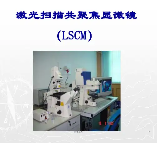
.b ž激光扫描共聚焦显微镜( LSCM) 是随着光学、视频、计算机等技术的迅速发展而诞生的一种高科技产品。
它是在荧光ž显微镜成像基础上加装了激光扫描装置, 利用计算机进行图像处理, 使用紫外或可见光激发荧光探针, 从而得到细胞或组织内部微细结构的荧光图像, 成为形态学﹑分子细胞生物学﹑神经科学﹑药理学﹑遗传学等领域新一代强有力的研究工具。
同时,激光扫描共聚焦显微镜也是活细胞的动态观察、多重免疫荧光标记和离子荧光标记观察的有力工具。
不仅可对活的或固定的细胞及组织进行无损伤的“光学切片”; 进行单标记或双标记细胞及组织标本的荧光定性定量分析; 还可用于活细胞生理信号, 离子含量的实时动态分析监测, 粘附细胞的分选, 细胞激光显微外科和光陷阱技术等。
可以无损伤的观察和分析细胞的三维空间结构。
- www 生物秀-专心做生物w ww .b b i o o .c o mIntroductionžLSCM 是一种高科技显微镜ž荧光显微镜成像为基础,加装了激光扫描装置, 计算机进行图像处理, 使用紫外或可见光激发荧光探针, 得到细胞或组织内部微细结构的荧光图像。
ž无损伤的“光学切片”ž细胞三维立体机构ž实时动态分析监测激光扫描共聚焦显微镜( LSCM) 是随着光学、视频、计算机等技术的迅速发展而诞生的一种高科技产品。
生物秀-专心做生物w w w .b b i o o .c o mž光学显微镜部分ž激光发射器ž扫描装置ž光检测器ž计算机系统( 包括数据采集, 处理, 转换, 应用软件)ž图像输出设备LSCM 的基本组成生物秀-专心做生物w w w .b b i o o .c o mLSCM 的原理激光光源:激光扫描束经照明针孔形成点光源, 普通显微镜采用的自然光或灯光是一种场光源, 标本上每一点的图像都会受到邻近点的衍射光或散射光的干扰。
而LSCM 以激光为光源, 激光具有单色性强﹑方向性好﹑高亮度﹑相干性好等优点, 可以避免普通显微镜的缺点。