多晶型药物指定晶型筛选方案设计
- 格式:pdf
- 大小:347.58 KB
- 文档页数:13
药品晶型研究及晶型质量控制指导原则当固体药品存在多晶型现象,且不同晶型状态对药品的有效性、安全性与药品质量产生影响时,应对药品固体制剂、半固体制剂、悬浮剂等中的药用晶型物质状态进行定性或定量控制, 药品的药用晶型应选择优势晶型,并保持制剂中晶型状态为优势晶型,以保证药品的有效性、安全性与质量可控。
优势晶型系指当药物存在有多种晶型状态时, 晶型物质状态的临床疗效佳、安全、稳定性高等, 且适合药品开发的晶型。
1 . 药物多晶型的基本概念用于描述固体化学药物物质状态, 由一组参量(晶胞参数、分子对称性、分析排列规律、分子作用力、分子构象、结晶水或结晶溶剂等) 组成。
当其中一种或几种参量发生变化而使其存在有两种或两种以上的不同固体物质状态时,称为多晶型现象(polymorphism) 或称同质异晶现象。
通常,难溶性药物易存在多晶型现象。
固体物质是由分子堆积而成。
由于分子堆积方式不同,在固体物质中包含有晶态物质状态(又称晶体) 和非晶态物质状态(又称无定型态、玻璃体)。
晶态物质中分子间堆积呈有序性、对称性与周期性; 非晶态物质中分子间堆积呈无序性。
晶型物质范畴涵盖了固体物质中的晶态物质状态(分子有序) 和无定型态物质状态(分子无序) 。
优势药物晶型物质状态可以是一种或多种, 故可选择一种晶型作为药用晶型物质, 亦可按一定比例选择两种或多种晶型物质的混合状态作为药用晶型物质使用。
2 . 晶型样品的制备采用化学或物理方法, 通过改变结晶条件参数可获得不同的固体晶型样品。
常用化学方法主要包括: 重结晶法、快速溶剂去除法、沉淀法、种晶法等; 常用物理方法主要包括: 熔融结晶法、晶格物理破坏法、物理转晶法等。
晶型样品制备方法可以采用直接方法或者间接方法。
各种方法影响晶型物质形成的重要技术参数包括溶剂(类型、组成、配比等) 、浓度、成核速率、生长速率、温度、湿度、光度、压力、粒度等。
鉴于每种药物的化学结构不同, 故形成各种晶型物质状态的技术参数条件亦不同, 需要根据样品自身性质合理选择晶型样品的制备方法和条件。
同⼀⼚家开发,最早报道晶型专利和原研化合物/制剂所有权⼈是否⼀致?同⼀⼚家开发,原研晶型专利⽐较容易确认。
如遇到不是同⼚家开发,请对不同不同所有权⼈之间的关系进⾏说明(收购?合作关系?授权使⽤?),以作为对原研晶型专利确认的依据。
晶型专利:专利号,所有权⼈,到期时间,是否有中国同族,是否已经授权?2)晶型⼀致性①此处主要⽐较⾃制与⽂献报道的区别,有条件可以⽐较⾃制⽚与原研⽚,可根据需要加⼊图表进⾏说明。
②晶型稳定性的初步研究:对湿、热、光照稳定性;空⽓中与密封条件下的稳定性数据的对⽐;粉碎前后的晶型变化等(制剂还要增加处⽅制备前后的晶型对⽐)。
3)多晶型根据⽂献调研情况来介绍可能的多晶型。
4)稳定性研究将测试的多批次稳定性数据进⾏⽐较,可根据需要加⼊图表进⾏说明。
4、申报资料中晶型部分的撰写呈现形式1)3.2.S.2:对于多晶型药物,申请⼈应在⽣产⼯艺开发阶段通过精制⼯艺的优化和筛选制备优势稳态晶型,保证原料药批间晶型⼀致性。
注:应包括结晶条件的考察,如:溶剂体系、降温速率、晶种加⼊考察、保温析晶温度及时间、搅拌⽅式等,提供说明⽬标晶型成为优势晶型的依据。
批间晶型的⼀致性:各阶段代表性批次的检测结果,如⼩中试,试⽣产,⼯艺验证批晶型的XRD,DSC,TG等晶型数据及对应图谱。
2)3.2.S.2、3、7:对药物制剂关键质量属性产⽣影响的多晶型药物,需研究证明批间晶型⼀致性(3.2.S.2)和晶型放置过程稳定性(3.2.S.7)。
共晶药物具有特殊的理化性质、确定的组分和化学计量⽐,可以通过X-射线单晶⾏射、X-射线粉末所射、固相核磁共振波谱、红外吸收光谱、差⽰扫描量热法和/成晶体形态等分析⽅法进⾏结构确证(不要求全部都做)。
3)3.2.S.4:如原料药的晶型和/或粒度分布对制剂质量产⽣影响,应被纳⼊原料药内控标准并制定专属的检测项⽬进⾏控制。
质量标准中晶型描述:⼀般仅对XRD的2θ⾓要求明确即可。
对于晶型质量研究的法规,⽬前为⽌,出台的不过以上⼏个,在吃透当前法规的要求,要结合⼯作⽇常的需求,领悟晶型研究换换相扣的逻辑关系,最后尘埃落地,落实到申报资料上,⼒求清晰完整,逻辑科学,交上⼀份满意的答卷。
药物多晶型的制备方法
药物多晶型可是个超重要的领域啊!它关系到药物的性质和效果呢!
药物多晶型的制备方法有好多呢!首先就是溶剂介导法啦,这可是常用的办法之一。
先把药物溶解在合适的溶剂里,形成过饱和溶液,然后通过控制温度、搅拌速度等条件,让药物结晶出来。
这里要注意哦,溶剂的选择可太关键啦,得选能溶解药物又不会引起不良反应的。
还有温度的控制也得精准,不然可就结不出想要的晶型啦!在这个过程中,安全性那是必须要保证的呀!不能让溶剂泄漏啥的,不然多危险呀!而且结晶过程要稳定,不能一会儿这样一会儿那样,那可不行!这种方法的稳定性还是比较可靠的,只要操作得当,一般都能得到不错的结果呢。
药物多晶型的制备过程中,安全性和稳定性就像两座大山一样重要呢!如果不安全,那不是开玩笑嘛,谁还敢用啊!稳定性不好,一会儿一个样,那怎么能行呢!就像建房子,根基得稳呀,不然不就塌了嘛!所以在制备过程中,每个环节都得小心翼翼,不能有丝毫马虎。
它的应用场景那可多啦去了!不同的晶型可能有不同的溶解性、稳定性、生物利用度等等。
这就好比不同口味的糖果,各有各的特点和优势呀!比如有些晶型溶解性好,那吸收就快,效果可能就更好;有些晶型更稳定,那储存就方便呀!这优势不就体现出来了嘛!这多晶型的研究可是能为药物研发带来很多新的可能性呢,能让药物更好地发挥作用,造福人类呀!
就拿那个很有名的阿司匹林来说吧,不同的晶型效果还真不一样呢!通过研究多晶型,找到了更适合的晶型,让阿司匹林的效果更好啦!这就是实际应用的效果呀,多厉害!这不就证明了药物多晶型的重要性嘛!
总之,药物多晶型的制备方法超级重要,我们一定要重视起来,好好研究,让药物发挥出更大的作用呀!。
药物研发晶型问题研究【摘要】随着药物研发的深入,难溶性药物在药物研发中的比例在不断的增大,晶型问题也日渐突出。
由于不同晶型的药物可能会影响其在体内的溶出、释放,进而可能在一定程度上影响药物的临床疗效和安全性。
因此,对于多晶型药物,在研制成固体口服制剂时,对晶型问题进行研究,有利于我们选择稳定可控的晶型用于临床。
【关键词】晶态物质;药品质量管理;制备工艺1.晶型现象固体物质可以由晶态物质与非晶态物质组成。
晶态物质是由于组成物质的分子、原子、离子在三维空间有序排列,具有周期性排列规律。
非晶态物质是指分子、原子、离子在三维空间无序堆积而成。
晶态物质又被称为晶体,当晶体结构测定发现样品分子中存在溶剂或水分子时,该样品晶体存在多晶型现象的可能性就会增大,因为样品分子容易与溶剂或水分子形成氢键,当被测样品的分子与不同的溶剂分子结合时,将会形成不同晶型的物质。
不含溶剂的晶体也可能由于分子的对称排列规律的不同而存在多晶型现象。
因此,晶型是化合物一个重要的理化性质,对于多晶型药物,因晶体结构不同,某些理化性质(如熔点、溶解度、稳定性)可能不同;而且在不同条件下,同一化合物的各晶型之间可能会发生相互转化。
例如,文献报道较多的抗结核药物利福平存在四种晶型:包括I型、II 型、SV型、及无定型四种。
研究发现利福平的理化性质和生物利用度与其晶型关系密切;其中I型和II型为有效晶型,两者溶解速率基本一致,但I型人体生物利用度方面优于II型。
2.原料药晶型研究应注意的几个方面难溶性固体药物,在作为固体口服制剂开发时,应对原料药的晶型进行研究。
因为同一化合物可能因其制备工艺的差异(如重结晶使用不同的溶剂或不同的结晶方法),而产生不同的晶型。
化合物晶型的改变除由其本身性质决定外,还受其它一些因素的影响。
例如,在不同温度、光照、湿度的条件下,化合物的晶型会随着失水或吸收水分而变化;不同的溶剂对化合物的晶型也会产生不同的影响。
对于全新的药物,首先应研究是否存在多晶型现象,考虑可能影响晶型的各种因素(温度、重结晶溶剂及重结晶条件),设计不同的重结晶方案。
药品研发中多晶型问题的考虑在药品研发过程中,多晶型问题一直是一个备受关注的话题。
多晶型是指一种化合物可以以不同的晶体形态存在,并且这些不同的晶型可以具有不同的物理性质、化学性质甚至药理学性质。
多晶型对药物的性质及其在临床应用中的表现有着重要的影响。
本文将从多晶型的形成机制、对药品研发的挑战以及解决多晶型问题的方法等方面对多晶型问题进行探讨。
一、多晶型的形成机制多晶型的形成机制是一个复杂的问题,受到多种因素的影响。
一般来说,多晶型的形成可以通过以下几种途径:1. 晶体结构的同质转变同一种物质在不同的条件下,如温度、压力等会形成不同的晶型结构。
这种同质转变通常是由于晶体结构中的原子、分子或离子位置的改变导致的。
2. 溶液结晶在溶液中,当溶液中物质的浓度、温度等条件发生变化时,会导致物质以不同的形式结晶。
当两种或多种不同的物质混合在一起时,由于它们之间的相互作用,会形成新的晶型结构。
二、多晶型对药品研发的挑战多晶型对药品研发会产生一系列的挑战,主要包括以下几个方面:1. 药效性和生物利用度的影响不同的晶型具有不同的生物利用度和药效性,因此不同的晶型会对药物的治疗效果产生影响。
2. 质量一致性和稳定性的考量不同的晶型具有不同的物理性质和化学性质,可能会引起药物的质量一致性和稳定性问题,从而导致生产过程中的变化和不良反应。
3. 制剂工艺的挑战不同的晶型在制剂过程中可能会产生不同的反应和性能,从而影响到制剂的工艺,增加了制剂的难度和成本。
4. 知识产权和市场竞争不同的晶型可能会对药品的知识产权保护和市场竞争产生影响,因此多晶型问题也是一个关乎商业利益的问题。
多晶型问题在药品研发中具有重要的意义,需要引起足够的重视和关注。
三、解决多晶型问题的方法针对多晶型问题,我们可以采取一系列的方法来解决:1. 原料选择在药品研发过程中,可以通过选择合适的原料来规避多晶型问题,如选择具有固定晶型结构的原料进行研发。
2. 制备工艺优化通过优化制备工艺,控制温度、压力等条件,可以尽量避免多晶型的形成,保证药品的一致性和稳定性。
药物晶型筛选溶剂设计-概述说明以及解释1.引言1.1 概述概述药物晶型筛选和溶剂设计是药物研发过程中非常重要的一环。
在药物研发中,晶型选择能够直接影响药物的物理和化学性质,进而对药物的稳定性、生物利用度和溶解性等方面产生影响。
而溶剂设计则是为了在晶型筛选过程中,找到适合药物晶型生长的溶剂,从而提高晶体的质量和高产率。
在药物晶型筛选中,晶型的选择不仅考虑到药物的物理化学性质,还需要兼顾制备上的可行性和工艺性。
因此,为了能够筛选到适合的晶型,需要进行一系列的实验和分析。
通过调整结晶条件和溶剂体系,能够优化药物晶体的形态、尺寸和晶型等特征。
而溶剂设计则是为了选择合适的溶剂,以提高晶体的质量和产率。
药物晶型筛选和溶剂设计对于药物研发具有重要的意义。
首先,晶型的选择能够直接影响药物的溶解性和稳定性,进而影响药物的生物利用度和药效。
因此,通过晶型筛选能够寻找到更适合的药物晶型,提高药物的疗效和药物的制剂性能。
其次,溶剂设计不仅可以提高晶体的质量,还可以提高晶体的产率,降低成本和提高效益。
因此,药物晶型筛选和溶剂设计在药物研发过程中具有非常重要的作用。
综上所述,药物晶型筛选和溶剂设计是药物研发过程中不可忽视的环节。
通过晶型筛选和溶剂设计,能够优化药物的药物性质和制剂性能,提高药物的疗效和生物利用度。
因此,加强对药物晶型筛选和溶剂设计的研究,对于促进药物研发的进展具有重要的意义。
1.2 文章结构文章结构部分的内容应该包括对整篇文章的组成和各个章节的内容进行简要介绍。
具体编写如下:文章结构分为引言、正文和结论三个主要部分。
引言部分主要包括概述、文章结构和目的。
首先概述了药物晶型筛选和溶剂设计的背景和意义。
然后介绍了文章的结构,包括主要章节和内容的安排。
最后明确了本文的目的,即通过对药物晶型筛选和溶剂设计相关知识的介绍和讨论,提高读者对该领域的了解和认识。
正文部分包括药物晶型筛选和溶剂设计两个小节。
其中,药物晶型筛选部分将介绍晶型的概念和分类、晶型筛选的方法和技术以及晶型对药物性质和稳定性的影响等内容。
药品研发中多晶型问题的考虑【摘要】药品研发中的多晶型问题是当前药物研究领域备受关注的一个重要议题。
本文将从影响药效的多晶型问题、多晶型的鉴定和表征、多晶型的稳定性研究、多晶型的制备方法以及多晶型的应用探索等方面展开论述。
多晶型问题对药品研发具有重要意义,其稳定性和制备方法对药物的质量和效果有着直接影响。
未来研究应该进一步深入探讨多晶型问题的重要性,探索新的方法和技术,以提高药物的研发效率和成果。
多晶型问题的重要性不容忽视,对于药品研发中的质量控制和疗效提升都具有重要意义。
【关键词】药品研发、多晶型、药效、鉴定、表征、稳定性、制备方法、应用探索、重要性、未来研究方向1. 引言1.1 药品研发中多晶型问题的考虑在药品研发过程中,多晶型问题是一个备受关注的重要议题。
多晶型指的是化学物质在结晶过程中形成的不同晶体结构,其存在对药效产生着重要影响。
药品的多晶型问题涉及到药物的物理性质、药效、药代动力学等方面,因此对于药品研发和制备具有重要意义。
在研发药品的过程中,研究药物的多晶型问题可以帮助科研人员更好地理解药物在溶解度、稳定性、生物利用度等方面的特性。
通过对多晶型的鉴定和表征,可以准确地确定药物的结构和性质,为制备高效、稳定的药物提供依据。
多晶型的稳定性研究可以帮助科研人员预测药物在不同条件下的稳定性和溶解度,为药品的制备和储存提供参考。
多晶型问题在药品研发中扮演着重要的角色,对于药品的研究、制备和应用具有重要意义。
随着科学技术的不断发展,我们对药物多晶型问题的理解将会更加深入,为药品研发带来更多的创新和进步。
2. 正文2.1 影响药效的多晶型问题多晶型是指一种药物分子在晶体结构内的排列方式不同,具有不同的晶体结构。
多晶型问题在药品研发中变得越来越重要,因为不同的多晶型可能导致药效、溶解性、稳定性等方面的差异。
1. 生物利用度:不同的多晶型可能导致药物在体内的吸收和分布特性发生改变,从而影响药效。
有些多晶型可能会被人体更容易吸收,增强药效;而有些多晶型则可能导致药物在体内难以吸收,降低药效。
晶型筛选方法《晶型筛选方法,包教包会啦!》嘿,朋友!今天我要跟你唠唠晶型筛选这个事儿,这可是个技术活,但别怕,听我给你细细道来,保证让你整得明明白白!首先呢,咱得搞清楚啥是晶型。
你就把它想象成一群调皮的小精灵,它们有不同的排列方式和姿态。
而我们的任务就是把这些不同姿态的小精灵给找出来。
第一步,准备工作要做好。
就像你要出门旅行,得先把行李收拾妥当一样。
咱得把要筛选的物质准备充足,别筛到一半发现不够了,那可就抓瞎啦!还有各种实验设备,得保证它们都能正常工作,别关键时刻掉链子。
这就好比战士上战场,枪可不能卡壳!第二步,选择合适的筛选方法。
这就像选衣服,得根据不同的场合选合适的。
常见的有溶剂结晶法、蒸发结晶法、冷却结晶法等等。
溶剂结晶法呢,就好比让小精灵们在溶剂里泡澡,泡舒服了它们就会以不同的姿势出现;蒸发结晶法呢,就像是把溶剂这个大游泳池的水慢慢抽干,逼得小精灵们不得不换姿势;冷却结晶法呢,就是给热热闹闹的小精灵们泼一盆冷水,让它们冷不丁地改变造型。
第三步,进行实验操作。
这可是关键步骤,得小心翼翼,就像走钢丝一样。
控制好温度、浓度、搅拌速度这些条件,一个不小心,可能就前功尽弃。
我跟你说,我有一次做实验,就因为搅拌速度没控制好,结果出来的晶型乱七八糟,那感觉,就像精心准备的生日派对被一场暴雨给搅黄了!第四步,分析筛选结果。
这就像在一堆水果里挑出最好的那个。
得用各种厉害的仪器,比如 X 射线衍射仪、热重分析等等,来看看这些晶型到底是啥样。
可别马虎,不然就像在一堆赝品里把宝贝给扔了。
第五步,重复实验验证。
这一步可不能偷懒,多做几次,确保结果的可靠性。
就像你考试做完题,得多检查几遍,不然一不小心丢了分,后悔都来不及。
最后,总结归纳。
把筛选出来的晶型特点、形成条件都整理清楚,这可是你的宝贵财富,以后再遇到类似的情况,就能轻松应对啦。
总之,晶型筛选就像是一场有趣的探险,每一步都充满了惊喜和挑战。
只要你按照这些步骤来,认真细心,再加上一点点运气,相信你一定能成功筛选出你想要的晶型!加油吧,朋友,期待你的好消息!。
・综 述・ [收稿日期]2001212224;[修回日期]2002206227[作者简介]王洪亮(1978-),男,天津宝坻人,河北医科大学药学院药剂学教研室在读硕士研究生,从事药剂学研究。
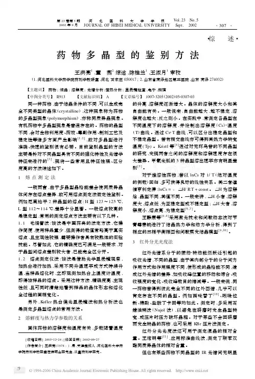
药物多晶型的鉴别方法王洪亮1,董 燕2综述,陈桂兰1,王淑月1审校(1.河北医科大学药学院药剂学教研室,河北石家庄050017;2.山东省菏泽地区第三医院,山东荷泽274032) 【主题词】 药物;结晶;溶解度;光谱分析;差热分析;显微镜检查,电子,扫描 【中图分类号】 R913 【文献标识码】 A 【文章编号】 100723205(2002)0520307203 同一种药物,由于结晶条件的不同,可以生成完全不同类型的晶体(crystalline ),这种现象称为药物的多晶型现象(polymorphism ),亦称同质异晶现象。
有机药物中多晶型现象是普遍存在的。
药物的晶型不同,会对生物利用度、药效、毒副作用、制剂工艺及稳定性等诸多方面产生影响[1,2],故对多晶型进行准确、快速的鉴别很有必要。
目前鉴别晶型的方法主要是针对不同晶型具有不同的理化特性及光谱学特征来进行的[3],现将一些常用且特征性强、区分度高的方法综述如下。
1 熔点测定法一般而言,由于多晶型晶格能差会使同质异晶体间存在熔点差异,故可用熔点测定法做定性鉴别。
例如尼莫地平2种晶型的熔点:H 型123~125℃,L 型112~114℃,差异十分显著。
一般熔点较高的是稳定型,常用的测定熔点方法主要有以下几种。
1.1 毛细管法:该法是中国药典的法定方法,它操作简便,使用样品量少,但测得的数值常略高于真实熔点,且主观性较强,需要操作者具有较熟练的实验技能。
尽管如此,它的精确度已可满足一般要求,对于晶型间熔点差别较大者,已能完全区分开。
1.2 熔点测定仪法:该法是借助光学显微镜观察,加热台进行加热,采用不同升温速率或方式持续升温,当样品熔化时,立即观测加热台上温度计温度,即得该样品的熔点。

药品研发中多晶型分析方法的研究进展综述方法:以“药物多晶型” “晶型多态性” “测定” “pharmaceutical poly morphs”“polymorphic transition”等为关键词,检索2006 年1 月-2016 年12 月在PubMed、Scifinder、中国知网、维普等数据库中收录的相关文献,然后进行归纳、总结。
药品研发中多晶型分析方法的研究进展摘要:固体药物晶型直接影响药物的安全有效性和质量可控性, 对固体药物晶型的研究已经成为研发和生产过程中必须注意的问题之一, 在国内外越来越受到重视。
通过查阅国内外有关文献资料, 本文简要介绍了药物多晶型及其对药物质量的相关影响,以及对多晶型药物的各种定量分析测定方法。
只有对药物的多晶型进行质量控制, 才能确保药品使用的安全性和有效性。
关键字:多晶型新药开发质量控制化学结构相同的药物, 可因结晶条件不同而得到不同的晶型,这种现象称为多晶型。
药物的晶型不同, 其物理性质如密度、熔点、溶解度和溶出速度均有不同。
多晶型药物根据其稳定性可分为稳定型、亚稳定型和不稳定型。
稳定型熔点高、化学稳定性较好,但溶出速率慢,溶解度小;不稳定型则溶出速率快、溶解度大,但化学稳定性相对较差;亚稳定型介于稳定型和不稳定型之间,储存一段时间会向稳定型转变。
在一定温度与压力下, 多晶型中只有一种是稳定型, 溶解度最小, 化学稳定性好, 而其他晶型则为亚稳定型, 它们最终可转变为稳定型。
但由于从亚稳定型转变为稳定型的过程通常比较缓慢, 因此许多结晶药物都存在多晶现象。
亚稳定型的溶解度大, 故溶出速度也较快。
一般来讲, 亚稳定型的生物利用度高, 为有效晶型, 而稳定晶型药物往往低效甚至无效。
因此, 药物多晶型的研究已经成为新药开发和审批, 药物的生产和质量控制以及新药剂型确定前设计所不可缺少的重要组成部分。
1 药物的多晶型及效应1.1药物的同质异晶晶体是由原子(或离子、分子)在空间周期地排列构成的固体物质,晶体中最小的立体单元叫做晶胞,按照晶胞三边之长及夹角的不同,可将晶体分为7个晶系,即立方(等轴)、六方、三方、四方、三斜、单斜、正交晶系。
药物多晶型佚名同一种元素或化合物在不同条件下生成结构、形态、物性完全不同的晶体的现象称为多晶现象。
McCrone在1965年提出的多晶型概念,强调了同一种分子在不同固态晶格中的不同填充、排列方式[1]。
近年来,其含义又扩大到互变异构多晶型、构象多晶型以及手性系统等[1]。
另外,关于蛋白质、核酸等生物大分子多晶现象的研究也日趋深入。
本文着重讨论有机药物领域中出现的多晶型。
有机药物的结晶基本上都属于分子晶格。
药物在分子中凭分子间引力结合。
如利福平分子中有-OH,-C=O,-NH等基团,可在利福平分子间、分子内、分子与溶媒间形成氢键,随着工艺条件的不同而产生不同的晶型。
晶癖是生长着的结晶因结晶条件(溶媒、杂质等)的影响,使分子不能均匀地达到各结晶面,从而产生不同的外形。
同一晶系的结晶,外观可呈现不同的形状(晶态),而多晶型则是由于结晶内部构造的分子排列不同而产生[2],二者有本质的不同。
另外,尽管溶媒化物与多晶型物也有区别,但同一药物的不同溶媒化物,也可表现出不同的熔点、溶解度,从而影响药物的生物利用度。
对于多晶型物的分析鉴定方法(热分析法、X-射线衍射法等)同样适用于溶媒化物的研究。
因此许多文献将溶媒化物称为“假多晶型物”[3,4]。
1研究多晶型的意义不同晶型的同一药物在溶解度、溶出速率、熔点、密度、硬度、外观以及生物有效性等方面有显著差异,从而影响药物的稳定性、生物利用度及疗效的发挥。
药物多晶型现象的研究已经成为日常控制药品生产及新药剂型确定前设计所不可缺少的重要组成部分。
了解药物的多晶现象及其性质,将有助于解决下列问题:保证在制备和贮存过程中药物的物理、化学稳定性;提高生物利用度,减少毒性,增进治疗效果;保证每批生产药物间的等效性;改善药物粉末的压片性能;防止药物在制备或贮存中产生不良晶型而影响外观质量。
2药物多晶型的研究手段[1]尽管不同晶型的药物其固态理化性质有绝对的差异,但由于仪器分辨率的限制,这些差异常常出现在分析范围的边缘,因此同时采用多种方法进行多晶型研究对于保证分析结果的可靠性具有重要意义。
药品研发中多晶型问题的考虑
多晶型问题在药品研发中是一个非常重要的考虑因素。
随着化学和物理学科技的快速发展,人们日益认识到多晶型的存在对于药物的研究开发、制备、储存和应用等方面产生的重要作用。
一个药物品种的多晶型是指在晶体结构上有不同的晶体态形态存在。
这些多晶态可以是单晶体或多晶体,它们的晶体结构可以有不同的因素影响,比如药物分子的构象,药物分子之间的作用力,晶格结构的排列方式等等。
药物的多晶型对于其药效进行影响。
由于多晶形的药物分子的结构差异性较大,因此它们的生物利用度,生物转化率和生物体内反应速率也会因此产生变化。
比如了解多晶型问题就可以在制备同一种药物的不同多晶型时确定其药物释放速率、生物利用度等特性,从而可以更好地评估其临床适应度。
另外,多晶型问题还能帮助研究者进行药物含量、质量和安全性的管理和控制,特别是在品牌公司的药品临床试验阶段。
对于药物的制备、储存和应用等方面也对多晶型问题进行了广泛探究。
在药物的制备过程中,多晶型选择是十分关键的,因为它会直接影响药物的质量和稳定性,同时在药物的储存过程中也需要考虑多晶型问题,以避免其在贮存过程中发生不必要的晶型变化。
另外,多晶型问题还可以提供重要的药物质量掌控数据,因此在药物制备、储存和应用等方面都有着不可忽视的作用。
通过对多晶型问题进行全面探究,可以帮助药品研发者更好地了解药物的药效变化和药品质量问题,从而能够更好地控制药品的研制水平、制备过程、储存过程和应用效果。
同时,对于多晶型问题的研究也可以为药物品种的研发提供重要的理论支持和应用方向,为药品的发展和推广提供依据。
药品研发中多晶型问题的考虑随着医药科技的不断发展,药品研发在不断探索各种新的方向和技术。
多晶型技术在药品研发中扮演着重要的角色。
多晶型是指同一种药物以不同的晶体形式存在。
在药物研发中,多晶型技术可以帮助我们获得更好的药效、更好的稳定性和更好的生物利用度。
药品研发中多晶型问题的考虑显得尤为重要。
本文将从多晶型的概念、多晶型的重要性以及多晶型问题的解决方案等方面进行深入探讨。
一、多晶型的概念多晶型是指同一种化合物在晶体结构上可以存在多种形式。
在化学中,很多化合物都能够以多晶型的形式存在,这些多晶型可能具有不同的物理、化学性质。
而在药物领域,多晶型也是一个非常重要的概念。
药物的多晶型可以影响药物的生物利用度、稳定性和制剂工艺等方面。
多晶型的研究对于药物的研发来说至关重要。
二、多晶型的重要性1.药效性能在药物研发中,药效性能是一个非常重要的指标。
不同的多晶型可能具有不同的物理、化学性质,这些性质对药物的药效性能有着直接的影响。
通过研究药物的多晶型,可以帮助我们找到更好的药效性能,从而提高药物的疗效。
2.稳定性3.制剂工艺在药物生产过程中,多晶型也会对制剂工艺产生影响。
不同的多晶型可能需要采用不同的工艺条件和方法进行制备,这就要求我们对药物的多晶型有深入的了解。
通过研究药物的多晶型,可以帮助我们设计出更合理的制剂工艺,提高药物的生产效率。
三、多晶型问题的解决方案在药物研发中,多晶型问题是一个具有挑战性的课题。
为了解决多晶型问题,我们需要采取一系列的研究和措施。
1.多晶型鉴定我们需要对药物的多晶型进行鉴定。
通过现代的实验手段和仪器设备,可以对药物的多晶型进行深入的研究和分析,确认不同多晶型的存在形式和性质,从而为后续的研究奠定基础。
2.多晶型筛选在确认了药物的多晶型后,我们需要对不同的多晶型进行筛选和评价。
通过实验研究,我们可以评估不同多晶型的药效性能、稳定性和制剂工艺等方面的性质,找出最具有潜力的多晶型。
药物制剂开发中对晶型的考虑随着药物开发研究的深入,难溶性药物在新药中的比例不断的增大,晶型问题越来越被重视。
不同晶型会影响药物在体内的溶出、吸收,进而影响药物的临床疗效和安全性,特别是一些难溶性口服固体和半固体制剂。
因此,对于多晶型药物,在研制成固体和半固体口服制剂时,对晶型进行研究有利于开发一种在临床治疗上有意义且稳定可控的晶型。
目前,国内对晶型问题也愈来愈重视,2015版药典附录新增“9105 多晶型药品的质量控制技术与方法指导原则”指出: 固体药物及其制剂中存在多晶型现象时,应使用“优势药物晶型物质状态”作为药物原料及其制剂晶型,以保证药品临床有效性、安全性与质量可控性。
下面就药物审评工作中遇到的晶型问题,以及研究开发这类多晶型药物所需注意的问题谈一些看法。
1、什么是药物的多晶型现象?固体物质根据其组成分子、原子、离子在三维空间的排列方式可以分为晶型(包括假晶型)和非晶体型。
晶体型的组成单元在三维空间排列固定有序,非晶型则相反。
所有晶型可以归纳为三斜晶系、单斜晶系、正交晶系、四方晶系、立方晶系、三方晶系、六角晶系共七个晶系14种晶格(如右图)。
有机药物的晶体大多为分子晶体,当药物分子中存在溶剂或分子时,因为药物分子易与溶剂或水分子形成氢键,药物分子与不同的溶剂分子结合,就会形成不同的晶型;不含溶剂的药物也可能由于分子的对称排列规律不同而存在的多晶现象,如药物结晶时的溶解、温度、湿度等,制剂过程中粉碎、混悬、压片等都会影响药物的晶型。
2、多晶现象会如何影响药物性质?众所周知,结构决定功能,同一药物的不同晶型在外观、溶解度、熔点、溶出度、生物有效性等方面都可能会有显著不同,从而影响了药物的稳定性、生物利用度及疗效,该种现象在口服固体制剂方面表现得尤为明显。
1)对药物理化性质及工业制剂的影响多晶型固体药物每个晶型有不同的表面自由能,而表面自由能大小是影响其溶出度的因素之一,像亚稳态的非极性表面自由能与稳态晶型基本相同,但极性表面自由能大于稳态的,因而总的表面自由能较大,更易被水润湿,在固体制剂崩解后形成的混悬液中,由于亚稳态粒子表面易水化,较厚的水化膜的反絮凝作用优于稳态晶型物,因而亚稳态的晶体粒子更易分散,顾有高的溶出度,如无味氯霉素共有A、B、C 3种晶型及无定型,我国1975年以前生产的无味氯霉素原料片剂、胶囊都为无效的A 型,后经改进生产工艺才生产出有生物活性的B型,并在质量标准中增加了非活性晶型的限度。
Apr.2009,Vol.6No.4药品研发中多晶型问题的考虑张玉琥(国家食品药品监督管理局药品审评中心,北京100038)作者简介:张玉琥,男,主任药师。
曾从事多年药物新剂型及新制剂研究工作,现从事化学药品技术审评工作。
E-mail:zhangyh@ 药品审评多晶型现象在固体化学药品研发过程中比较常见。
由于多晶型问题可能影响药品的安全有效性和质量可控性,应当针对不同情况采取相应的措施。
本文就药品研发中涉及的多晶型现象及相关问题的考虑作简要讨论,供药品研发和评价工作参考。
1晶型与多晶型现象固体药物有结晶型和非结晶型(无定形)之分。
构成药物结晶的基本单元为晶格,在晶格中药物分子以一定的规律排列。
而无定形是分子以无序的方式排列,不具有明确的晶格。
若药物结晶中包含结晶溶剂分子,就称为溶剂化物。
当该溶剂为水时(即含有结晶水),通常称为水合物。
药物的不同晶型是由分子在晶格中排列方式的不同所致。
需要注意晶型与结晶形态(晶癖)的区别。
前者由晶格中分子的排列来决定,后者是指形成的结晶的外观形状,如针状结晶、片状结晶等。
同一晶型的药物,可能具有不同的外观形状;反之,外观形状相同的结晶,其晶型也可能不同。
若固体药物可存在不同的晶型,或存在结晶型与无定形,或存在非溶剂化物与溶剂化物、不含或含有结晶水等现象,就称为该药物存在多晶型现象。
一些方法可用于研究和区分多晶型现象。
单晶X-射线衍射可对晶体结构提供直接的证据,是研究多晶型现象的可靠方法。
需要注意的是若获取单晶所采用的结晶条件与药物生产中实际采用的结晶条件不同,则单晶X-射线衍射得到的晶体特征并不代表药物实际的晶体特征。
粉末X-射线衍射是常用的研究和区分不同晶型的有效方法,该方法不仅可用于不同晶型的定性区分,在建立特征衍射峰与不同晶型含量之间的定量关系后,粉末X-射线衍射还可用于不同晶型比例的定量控制。
其他方法,包括显微观察、热分析(差示扫描量热、热重分析、热台显微镜等)、光谱法(红外光谱、拉曼光谱、固相核磁共振)等,亦有助于进一步研究多晶型现象。
药物晶型对药效的影响及其优化策略药物晶型指的是药物分子在固体状态下的排列方式和结构形态,包括多晶、单晶、无定形等形态。
药物晶型对药效具有重要影响,不仅能影响药物的物理性质,还能影响药物的药代动力学和药效学特性。
因此,对药物晶型的研究和优化显得尤为重要。
一、药物晶型对药效的影响1.药物晶型对药物的稳定性和溶解度有明显影响。
不同晶型的药物在溶液中的稳定性和溶解度可能有较大差异,从而影响药物的溶出速度和药效的表现。
2.药物的生物利用度也受到晶型的影响。
一些药物在特定晶型下吸收更好,因此对晶型进行优化可以提高药物的生物利用度。
3.药物晶型对药效的选择性和持续性也有影响。
不同晶型的药物可能表现出不同的选择性和持续性,有些晶型更适合特定类型的疾病治疗。
4.药物晶型还能影响药物的制备工艺和稳定性。
某些晶型可能在工艺条件下更容易制备,而且在储存和运输过程中更加稳定。
二、药物晶型的优化策略1.采用适当的晶型筛选方法来获取合适的药物晶型。
晶型筛选方法包括溶剂结晶法、熔融结晶法、混合溶剂结晶法等,可以通过这些方法获取理想的药物晶型。
2.利用晶型工程技术来优化药物晶型。
晶型工程技术包括共结晶、固溶体制备、超声共晶等方法,可以改变药物的晶型特性,并优化药物的药效。
3.采用合适的晶型控制剂来控制药物的晶型。
晶型控制剂可以通过干扰药物分子的晶型生长过程,来控制药物的晶型特性。
4.结合计算模拟方法来研究和优化药物的晶型。
计算模拟方法可以模拟药物分子在不同条件下的排列方式和结构形态,有助于预测和优化药物的晶型。
5.在药物研发和制备的过程中,重视药物晶型的影响和优化。
在药物研发的早期阶段,就应该考虑药物晶型对药效的影响,并在制备过程中合理选择和优化药物的晶型。
结语:是一个重要领域,通过对药物晶型的深入研究和优化,可以提高药物的药效和生物利用度,为药物研发和制备提供更多可能性。
因此,加强对药物晶型的研究和优化工作,对于提高药物的疗效和稳定性具有积极的意义。
Development of a Targeted Polymorph Screening Approach for a Complex Polymorphic and Highly Solvating APIANTHONY M.CAMPETA,BRIAN P.CHEKAL,YURIY A.ABRAMOV,PAUL A.MEENAN,MARK J.HENSON,BING SHI, ROBERT A.SINGER,KEITH R.HORSPOOLPharmaceutical Sciences,Pfizer Global Research&Development,Groton,Connecticut06340Received19February2010;revised21April2010;accepted22April2010Published online22June2010in Wiley InterScience().DOI10.1002/jps.22230ABSTRACT:Elucidation of the most stable form of an active pharmaceutical ingredient(API)isa critical step in the development process.Polymorph screening for an API with a complexpolymorphic profile can present a significant challenge.The presented case illustrates anextensively polymorphic compound with an additional propensity for forming stable solvates.In all,5anhydrous forms and66solvated forms have been discovered.After early polymorphscreening using common techniques yielded mostly solvates and failed to uncover several keyanhydrous forms,it became necessary to devise new approaches based on an advanced under-standing of crystal structure and conformational relationships between forms.With the aid ofthis analysis,two screening approaches were devised which targeted high-temperature deso-lvation as a means to increase conformational populations and enhance overall probability ofanhydrous form production.Application of these targeted approaches,comprising over100experiments,produced only the known anhydrous forms,without appearance of any new forms.The development of these screens was a critical and alternative approach to circumventsolvation issues associated with more conventional screening methods.The results providedconfidence that the current development form was the most stable polymorph,with a lowlikelihood for the existence of a more-stable anhydrous form.ß2010Wiley-Liss,Inc.and theAmerican Pharmacists Association J Pharm Sci99:3874–3886,2010Keywords:axitinib;polymorphism;polymorph screen;crystallization;desolvation;compu-tational chemistry;crystal structureINTRODUCTIONPolymorphism can be thought of as the state in which a solid chemical compound exists in more than one crystalline form1with only one polymorph being the thermodynamically most stable form at a specific temperature and pressure.This phenomenon is very common to most pharmaceutical APIs.It is well known that polymorphs of the same substance can have dramatic differences in pertinent pharmaceu-tical properties,such as solubility and stability that can often have a significant impact on bioavailability and overall drug product performance.A number of excellent texts on polymorphism and their influence on pharmaceutical development are available.2Thus,identifying the most appropriate solid form,typically the thermodynamically stable form,is a key element in the early developmental process for a new drug candidate.In regard to polymorph screening approaches,there are many common techniques employed that are designed to typically uncover all metastable and low-energy polymorphic forms.These include,for example,crystallizations through solvent evaporations,antisolvent crystallizations,slow and fast cooling of saturated API solutions to induce precipitation,and slurrying of solid API for extended periods of time.3A significant number of solvents and cosolvents of varying polarity and chemical composi-tion are usually employed,while variable tempera-tures are also incorporated in the design to assess enantiotropic behavior.These approaches have been incorporated into our practices for polymorph screen-ing and are typical throughout the pharmaceutical industry.In most cases,a thoroughly designed API form screen employing the approaches previously dis-cussed should typically identify the thermodynami-cally stable polymorph.Additional Supporting Information may be found in the online version of this article.Mark J.Henson’s present address is Molecular Biometrics,Inc., New Haven,CT06511.Correspondence to:Anthony M.Campeta(Telephone:860-441-5844;fax:860-715-2454;E-mail:anthony.m.campeta@pfi) Journal of Pharmaceutical Sciences,Vol.99,3874–3886(2010)ß2010Wiley-Liss,Inc.and the American Pharmacists Associationexceptions in the literature4,5that describe the appearance of a lower energy form at late stages in development.In these cases,common polymorph screening approaches were clearly unsuccessful,as unique physical and structural properties of the molecule hindered the anticipated cascade to the most stable form based on Ostwald’s rule.6In this article, we describe another such example.Axitinib(Fig.1)is an oncology candidate under development at Pfizer.This API targets the vascular endothelial growth factor(VEGF)to prevent the growth and proliferation of cancer cells via interrup-tion of tumor angiogenesis(formation of vascular supply tissue).7This compound has shown consider-able promise in the treatment of carcinomas in a number of target tissues and organs and is currently in late stage clinical development.8Understanding the polymorphism of axitinib has been a subject of considerable focus and effort,which we have initially reported.9In this work,polymorph investigations using the traditional approaches pre-viously mentioned,which incorporated well over 300experiments,identified a surprisingly high total of23unique solid forms.Distinction of these forms was assigned on the basis of powder X-ray diffraction (PXRD)patterns and thermal characteristics such as melting onset,melting enthalpy,and desolvation temperatures.This group of solid forms included three anhydrous forms with the remainder solvates (refer to Tab.1).The anhydrous form IV was characterized as a robust developmental form with acceptable solid-state properties and was advanced for early clinical studies.It was apparent that axitinib had a high tendency to form solvates.There was some question whether certain polymorph screening approaches may be challenged by this phenomenon,and the risk of not observing critical anhydrous forms(of which three had been already discovered)could exist.In parti-cular,axitinib had a propensity to form relatively stable solvated structures,as a majority of these solvates were characterized as possessing relatively high temperatures of desolvation(desolvation tem-peratures significantly higher than thenormalFigure1.Structure of axitinib.Table1.Summary of Axitinib Solid FormsName Form SolventForm I(1)Anhydrate—Form II(2)Hydrate WaterForm III(3)Solvate Ethyl acetate(EtOAc) Form IV(4)Anhydrate—Form V(5)No solid form designationForm VI(6)Anhydrate—Form VII(7)Solvate Isopropyl alcohol(IPA),IPA/waterForm VIII(8)Solvate Dioxane,tetrahydrofuran(THF) Form IX(9)Hydrate WaterForm X(10)Solvate Dimethylformamide(DMF),DMF/waterForm XI(11)Solvate THF/water,THFForm XII(12)Solvate Dichloromethane(DCM),ethanol(EtOH)Form XIII(13)Solvate Acetonitrile(ACN)Form XIV(14)Solvate Acetic acidForm XV(15)Solvate EtOHForm XVI(16)Solvate IPAForm XVII(17)Solvate AcetoneForm XVIII(18)Solvate Methylisobutyl ketone(MIBK) Form XIX(19)Solvate Methylethyl ketone(MEK) Form XX(20)Solvate Methyl benzoateForm XXI(21)Solvate2,2,2-CF3CH2OH/ether/hexane Form XXII(22)Solvate1-PentanolForm XXIII(23)Solvate PyridineForm XXIV(24)Solvate ChloroformForm XXV(25)Anhydrate—Form XXVI(26)Solvate THF/water,THFForm XXVII(27)Solvate Dimethylsulfoxide(DMSO) Form XXVIII(28)Solvate Benzyl alcoholForm XXIX(29)Solvate TrichloroethyleneForm XXX(30)Solvate Dimethylformamide(DMF)/octanol(1:1)Form XXXI(31)Solvate OctanolForm XXXII(32)Solvate MethanolForm XXXIII(33)Solvate1-ButanolForm XXXIV(34)Solvate3-Methyl-1-butanolForm XXXV(35)Solvate MEKForm XXXVI(36)Solvate Pyrrole/1-pentanolpyrrole/p-cymeneForm XXXVII(37)Solvate Allyl alcoholForm XXXVIII(38)Solvate Pyrrole allyl alcoholForm XXXIX(39)Solvate Acetic acidForm XL(40)Solvate EtOHForm XLI(41)Anhydrate—Form XLII(42)Solvate2-ButanolForm XLIII(43)Solvate2-Methyl THFForm XLIV(44)Solvate2-Methyl THFForm XLV(45)Solvate TolueneForm XLVI(46)Solvate N-Methylpyrrolidone Form XLVII(47)Solvate Isoamyl acetateForm XLVIII(48)Solvate MethylcyclohexaneForm XLIX(49)Solvate CyclohexanoneForm L(50)Solvate CyclohexanoneForm LI(51)Solvate1,2-DichloroethaneForm LII(52)Solvate Propionic acidForm LIII(53)Solvate Tert-butanolForm LIV(54)Solvate DimethoxymethaneForm LV(55)Solvate2-PentanoneForm LVI(56)Solvate Dimethyl acetate(DMA) Form LVII(57)Solvate NitromethaneForm LVIII(58)Solvate1,2,3,4-tetrahydronaphthalene Form LIX(59)Solvate Tetramethylene sulfone Form LX(60)Solvate Methyl acetateForm LXI(61)Solvate p-XyleneForm LXII(62)Solvate TrichloroethyleneForm LXIII(63)Solvate n-Butyl acetateForm LXIV(64)Solvate Isobutyl alcoholForm LXV(65)Solvate CyclohexanolForm LXVI(66)Solvate Isopropyl acetateForm LXVIII(67)Solvate p-Cymene/pyrrole(1:1) Form LXIX(68)Solvate t-Amyl alcoholForm LXX(69)Solvate4-Methyl-2-pentanone Form LXXI(70)Solvate CyclohexaneForm LXXII(71)Solvate1,2-Dichlorobenzene Form LXXIII(72)Solvate p-Cymene/acetone(1:1)TARGETED POLYMORPH SCREENING APPROACH DEVELOPMENT FOR AN API3875boiling point of the corresponding solvent),suggest-ing strong bonding within the crystal.These solvates may be anticipated to be thermodynamically stable in their corresponding mother liquor and may resist further solvent-mediated transformation to an anhy-drous form.Solvent content was often found to be nonstoichiometric,suggesting nonsite-specific sol-vent incorporation.Also,many solvates formed from different solvent systems demonstrated isomorphic10 structures,as evidenced by very similar PXRD patterns,which made differentiation of solid forms complex.In addition,multiple polymorphs of solvates were observed from the same solvent,such as ethanolate forms XII and XV.During process development and refinement stu-dies with form IV,a new crystalline anhydrous form was discovered.9This new form,form XXV,was characterized as a thermodynamically more stable form than form IV,though it was not observed in any of the aforementioned polymorph screening studies. Crystal structure analysis would eventually show that the molecular conformation of most of the solvate structures was very similar to that of the anhydrous form IV.This coincides with the observation that the solvates would desolvate to form IV during DSC and variable temperature PXRD heating experiments. This suggested that the molecular conformation is mostly retained upon desolvation,resulting in the favored formation of form IV.Equally important was thefinding that the crystal packing and molecular conformation of the new form XXV were found to be different from those of form IV.In particular,the molecular arrangement of form XXV generally would be difficult to achieve through desolvation.Thus,it is believed the propensity to initially form stable solvates as well as structural properties that would favor desolvation to form IV contributed to the lack of appearance of form XXV in our polymorph screens.Though there was confidence that form XXV was the thermodynamically stable form of axitinib, another more-stable anhydrous form,form XLI, was later discovered during development.It became obvious that the polymorph environment of axitinib could not be confidently assessed through the use of conventional polymorph screening experiments.In this article,the discovery and characterization of this new form XLI are described,including a thorough evaluation and understanding of the crystal struc-ture,and its structural relationship relative to known polymorphs.Finally,with this detailed understand-ing of the structural properties,the development of two specific polymorph screening approaches were designed around desolvation to more completely assess the polymorphic environment of axitinib and improve confidence that form XLI was indeed the thermodynamically stable form.EXPERIMENTALMaterialsA sample of form XVI(isopropanol solvate)of axitinib (purity99.8%)was obtained from Pfizer Ireland Pharmaceuticals,Inc.,Little Island,CO.Cork,Ire-land.Samples of anhydrous forms IV,VI,XXV,and XLI were obtained from Pfizer Global Research& Development,Groton,CT.Full details on methods to prepare forms I,IV,and VI are presented by Ye et al.,11whereas methods to prepare forms XXV and XLI are described by Campeta et al.12Solvents used in this study were obtained from Sigma–Aldrich,St Louis,MO,Pharmco-AAPER,Brookfield,CT,or Mallinckrodt Baker,Phillipsburg,NJ.Solubility Measurements of Various Forms in Ethanol Solubility measurements of various forms in ethanol were completed using a100-mL jacketed glass vessel. When operated at temperatures above the normal boiling temperature of ethanol(788C),the vessel was pressurized to suppress boiling.During the solubility measurement,a measured amount of a particular form of axitinib was added to a measured amount (typically30–40g)of ethanol.The mixture was heated quickly to5–108C below the expected dissolu-tion temperature,and then heated slowly(0.25–0.58C/min)until all the solids were dissolved.The dissolution was monitored using a Mettler-Toledo Lasentec S400FBRM probe.Powder X-Ray DiffractionThe PXRD pattern measurement was carried out on a Bruker D5000diffractometer using copper radiation (CuK a,wavelength:1.54056A˚).The tube voltage and amperage were set to40kV and40mA,respectively. The divergence and scattering slits were set at1mm, and the receiving slit was set at0.6mm.Diffracted radiation was detected by a Kevex PSI detector.A theta-two theta continuous scan at2.48/min(1s/0.048 step)from 3.0to4082u was used.An alumina standard was analyzed to check the instrument alignment.Samples were prepared by placing them in a quartz holder.Single Crystal DiffractometryThe single crystal X-ray diffraction measurements were carried out at room temperature on either a Bruker SMART APEX CCD area detector system equipped with a graphite monochromator and sealed tube Cu radiation(1.54178A˚)source or a Bruker FR591rotating anode with Motel Multilayer Optics, Cu radiation(1.54178A˚),equipped with an APEX II detector.Single crystal X-ray diffraction data were collected at room temperature in order to provide a calculated powder pattern that best matches3876CAMPETA ET AL.experimental powder patterns for API samples, which are measured at room temperature.Tempera-ture changes may result in the contraction or elongation of unit cell dimensions to such an extent that the calculated powder patterns at100K and room temperature may be significantly different.All crystallographic calculations were facilitated by the SHELXTL software suite system.In general,hydro-gens bonded to heteroatoms were located by differ-ence Fourier techniques.The remaining hydrogen atoms were placed in idealized positions.The hydro-gen parameters were added to the structure factor calculations but were not refined.Crystals suitable for this analysis were grown through crystallization techniques including evaporations of saturated solu-tions,vapor and liquid diffusion,and antisolvent crystallization.Thermal AnalysisDifferential scanning calorimetry(DSC)was carried out on a Mettler Instruments DSC821e,Columbus, OH.The instrument was calibrated for cell constant and heat capacity using indium and zinc.Samples were prepared by weighing about3mg of into a40m L aluminum pan which was then covered with an aluminum lid.The lid is pierced prior to measure-ment.Data were analyzed using STAR e SW8.10.The experiments started at ambient temperature and heated the sample at58C/min to3008C under a nitrogen gas purge(flow rate was60mL/min). Thermogravimetric analysis(TGA)was carried out on a TA Instruments,New Castle,DE,DSC Q500 V6.7Build203.The instrument was weight cali-brated with200and1000mg weights.Samples were prepared by weighing about6mg of sample into a 50m L aluminum pan.Data were analyzed using Universal Analysis2000for Windows2000/XP version4.2E,Build4.2.0.38.The experiments started at ambient temperature and heated the sample at 108C/min to3008C under a nitrogen gas purge(flow rate was50mL/min).Conventional Polymorph Screen With Form XXVThe polymorph screen consisted of generating solid samples using a variety of techniques and solvents.A total of12solvents and8aqueous cosolvent mixtures were used.Techniques for generating samples included fast evaporations,slow cools of saturated solutions,and slurries consisting of excess solid in saturated solutions at different temperatures.Resul-tant solids were examined under a microscope for birefringence and morphology,and analyzed at a minimum by PXRD.Selected samples were also assessed by thermal methods.Approximately70total experiments were performed.Fast evaporation experiments:Solutions were prepared in various solvents and sonicated to assist in dissolution.The final solutions were typicallyfiltered through a 0.2-m m nylonfilter.The solutions were allowed to evaporate at ambient(20–258C)in a vial.The solids that formed were isolated and analyzed.Slow cooling experiments:Saturated solutions were prepared in various solvents at elevated temperatures(approxi-mately608C)and rapidlyfiltered through a0.2-m m nylonfilter into open vials.The vials were covered and allowed to cool slowly to room temperature.The presence or absence of solids was noted.If there were no solids present,or if the amount of solids was judged too small for PXRD analysis,the vial was placed in a refrigerator overnight.Again,the presence or absence of solids was noted and if there were none, the vial was placed in a freezer overnight.Solids that formed were isolated byfiltration and allowed to dry prior to analysis.Slurry experiments:Solutions were prepared by adding enough solids to a given solvent so that excess solids were present.The mixtures were then agitated in sealed vials at either ambient or an elevated temperature,typically608C.After a given amount of time(typically7days for ambient and1day for608C),the solids were isolated by vacuum filtration.Preparation of Solvates and Three-Component Desolvation ScreenThe solvate–desolvation polymorph screen was run byfirst preparing a variety of axitinib solvates,and then desolvating using multiple methods.The solvate samples were prepared on a1g scale starting from form XVI(isopropanol solvate)and were formed by reslurrying in a single solvent or mixed solvent system for3days at508C.The isolated solids were initially characterized by PXRD to determine if a unique solid form had been generated.Additional characterization utilized DSC and TGA to confirm the presence of desolvation events.Once the solvated forms were characterized, parallel desolvation experiments were run on sam-ples of the solvated forms.The desolvation techniques utilized were a)vacuum drying at1008C,b)reslurry in ethanol at908C in a sealed vial,and c)reslurry in heptane at1058C in a sealed vial.Desolvation studies were carried-out on a100mg scale.Samples exhibit-ing the initial solvate PXRD powder pattern were exposed again to the same desolvation conditions.If a unique powder pattern was observed,additional thermal analysis(DSC and TGA)was conducted to characterize the material.Raman SpectroscopyThe solid form of larger scale reslurry studies was monitored via Raman spectroscopy with a Kaiser Optical Systems HoloLab5000equipped with a 785nm laser.Afiber optic cable linked the instru-ment to an MkII probe headfitted with a Hastelloy1TARGETED POLYMORPH SCREENING APPROACH DEVELOPMENT FOR AN API38771.3cm diameter immersion probe.The probe incor-porated a short focal length sapphire optic.Data collection was controlled via Kaiser’s HoloReact software(v.4.0.0233).The software resampled experi-mental data to a spectral resolution of0.6cmÀ1. Wavelength calibration was performed using cyclo-hexane.Ten second spectral accumulation times were found to be sufficient to achieve slurry signal levels within the recommended segment of the detector dynamic range.Dark background spectra were acquired prior to each spectrum,and automatic cosmic ray rejection was employed.Utilization of these two options extended the total acquisition time for each spectrum to40s.During the experiments, spectra were acquired at5-min intervals.As Raman spectra were being acquired,the univariate intensity trending of key spectral bands in real time was performed using the HoloReact package based on MatLab.Subsequent offline spectral data analysis was performed using Delight v.3.2.1(DSquared Development,Inc.,LaGrande,OR).High Boiling Solvent Polymorph Screen Approximately40axitinib solvates werefirst pre-pared on a1g scale starting from form XVI (isopropanol solvate)and were formed by reslurrying in a single solvent or mixed solvent system for3days at508C.These samples were then reslurried at1508C in p-cymene for at least7days before isolating.The isolated solids were initially characterized by PXRD to determine if a unique solid form had been generated.If needed,thermal analysis was completed by DSC and TGA.Additional high-temperature reslurry experiments in p-cymene at1508C were completed on a50mL scale using the Biotage AS2350reactor system starting from the anhydrous forms XLI and XVI with the solid form monitored in situ by Raman spectroscopy.An additional set of1308C reslurry experiments were completed in3:1p-cymene/benzyl alcohol(v/v) mixture and3:1p-cymene/o-cresol(v/v)mixture starting with form XVI.Isolated material was characterized by PXRD.Computational Chemistry MethodsCrystallographic and generated conformations of axitinib were optimized using aqueous media at the PBE/DNP/COSMO level of theory(defined by theo-retical method and a basis set)as implemented in DMol3.13This utilizes density functional theory (DFT)PBE approximation14with all-electron dou-ble-numeric-polarized basis set.The effect of bulk water is estimated by conductor-like screening model (COSMO)as implemented in DMol3.15The generated cosmofiles by the PBE/DNP/COSMO calculations for each conformer are further used for the prediction of solubility and conformational distribution in different solvents adopting COSMO-RS theory16as implemen-ted in COSMOTherm software.17RESULTS AND DISCUSSIONConventional Polymorph Screen of Form XXVThe properties and characteristics of the newly discovered anhydrous form XXV have been recently described,which demonstrated that it was a more stable polymorph at room temperature than the early development form IV.9After discovery,an additional polymorph screen with this new form was performed to reassess the polymorph environment of axitinib. The screening approaches were similar to those used for polymorph screening of form IV,which comprised well-precedented techniques previously mentioned. Solubility assessment showed that form XXV had solubilities roughly2Âlower than form IV in most solvent and aqueous cosolvent mixtures.It was acknowledged that the lower solubilities may hinder solvent-mediated transformations in slurry-type experiments.At this point in time,extensive crystal structure information of axitinib solid forms had not been discerned,including those of forms XXV and IV. Thus,though it was unclear as to definitive reasons for the lack of appearance of form XXV in previous form screens,conducting another traditional screen in the same manner as with form IV was considered the bestfirst approach for the screening of form XXV. These experiments discovered no new anhydrous forms.Form XXV was the predominant anhydrous form,while a number of new solvates were found through the use of some solvents not evaluated in previous studies.As a result,a total of39forms of axitinib had been found,see Table1.Discovery of Form XLIOnce form XXV was identified as the lead develop-ment form,chemical process development studies identified facile conditions to isolate the desired form. The process utilized a two-step reslurry process to convert the crude API to thefinal form.An initial reslurry in isopropanol converted the crude API form to an isopropanol solvate,which was subsequently converted to the desiredfinal form via a reslurry in ethanol with the addition of form XXV seed crystals to control the polymorph conversion.In the initial manufacturing campaign for producing form XXV, the conversion of the isopropanol solvate to form XXV required extended processing time possibly due to poor dispersion of the form XXV seed crystals in the crystallization vessel attributed to mixing.Subse-quent lab-scale development studies demonstrated that the seeded ethanol reslurry process conducted at reflux with a high jacket temperature resulted in a3878CAMPETA ET AL.faster and more robust conversion to form XXV.The rationale for the faster form conversion was not determined but may have been due to the higher solubility difference between the solvate form and form XXV at higher temperatures,and/or it may be due to the improved solid –liquid mixing in the crystallization vessel due to the re fluxing solvent.The lab-scale process was modi fied to utilize the higher jacket temperature for the subsequent man-ufacturing campaign.During the final isolation,an in process control sample was pulled and analyzed by PXRD to determine if the conversion to the desired form XXV was complete.PXRD analysis indicated a previously unknown powder pattern.The slurry was seeded with form XXV a second time,and the process was allowed to reslurry at re flux for 12h.Analysis of a subsequent sample indicated the same unknown powder pattern with no indication of form XXV,which suggested that the unknown form (form XLI)was thermodynamically favored versus form XXV.Characterization of Form XLI and Comparison to Known Anhydrous FormsWith the discovery of form XLI,an intensive evaluation of its physical properties was conducted to characterize this new form and understand its relationship to the other known anhydrous forms.DSC experiments showed form XLI possessing a single endotherm,with a melting temperature and heat of fusion value higher than other known anhydrous forms,see Table 2.Together,a total of five anhydrous polymorphs of axitinib were now known,which presents an unusual case.On the basis of the DSC data and Burger ’s rule,18form XLI is shown to be monotropically related to all otheranhydrous forms and is thus expected to be the most stable solid form at all temperatures.Form XLI also has the lowest solubility of all the anhydrous forms,providing further support for its designation as the lowest energy form.Figure 2illustrates the signi fi-cantly lower solubility of form XLI in ethanol.Table 2provides the rank order of the relative stabilities of the anhydrous forms.The calculated density values for all forms do not correlate with relative stability,suggesting this parameter is not a reliable measure of stability,as is often the case.18As will be described later,the various forms have different conformations,which makes comparisons based on the density rule invalid.In regard to relationships between the other forms,forms XXV and VI are enantiotropically related and very close in energies.The average heat of fusion of form VI is slightly higher,though the range of values from multiple determinations of each form is overlapping and thus may be within the experimental sensitivity of the instrument.Table 2.Thermal Properties and Calculated Density of Axitinib Anhydrous FormsMelt Onset (8C)D H f (J/g)Density (g/mL)aForm XLI 225.9153 1.369Form XXV 217.2129–132b 1.382Form VI 211.6132–136b1.330Form IV 218.7122 1.293FormI 210.51151.333a Calculated from single-crystal structure information.bRange of values from multiple DSC (5–10)determinations at 58C/min heatingrate.Figure 2.Solubility in ethanol of ethanol solvate,forms IV,VI,XXV,and XLI.TARGETED POLYMORPH SCREENING APPROACH DEVELOPMENT FOR AN API 3879。