英文住院病例模板
- 格式:doc
- 大小:69.50 KB
- 文档页数:8

腹痛住院病历书写范文英文回答:I was admitted to the hospital due to severe abdominal pain. The pain started suddenly and was located in the lower right side of my abdomen. It was a sharp, stabbing pain that intensified with movement. The pain was so intense that I couldn't even stand up straight. I also experienced nausea and vomiting along with the abdominal pain.Upon admission, the doctors conducted a thorough physical examination. They pressed on different areas of my abdomen to assess the tenderness and check for any signs of inflammation. They also ordered various diagnostic tests, including blood tests, ultrasound, and a CT scan.The blood tests showed an elevated white blood cell count, which indicated the presence of infection. The ultrasound and CT scan revealed inflammation and swellingin the appendix, confirming a diagnosis of appendicitis. The doctors explained that appendicitis occurs when the appendix becomes blocked, leading to infection and inflammation.I was immediately scheduled for an appendectomy, which is the surgical removal of the appendix. The surgery was performed laparoscopically, using small incisions and a camera to guide the surgeon. This minimally invasive approach allows for quicker recovery and less scarring.After the surgery, I was closely monitored for any complications. I was given pain medication to manage the post-operative pain and antibiotics to prevent infection. The medical team ensured that I was able to tolerate oral intake and pass gas, which are signs of normal bowel function.I was discharged from the hospital after a few days, once my condition had stabilized. The doctors provided me with detailed instructions for post-operative care, including wound care, pain management, and dietaryrestrictions. They also advised me to gradually resume normal activities and avoid strenuous exercise for a few weeks.中文回答:我因为剧烈的腹痛被送入医院住院治疗。

Divisi on: _________ Ward: __________ Bed: _________ Case No. ___________Name: _____________ Sex: __________ Age: ___________ Natio n: ___________ Birth Place: ________________________________ Marital Status: _____________ Work-orga nizatio n & Occupatio n: _____________________________________ Livi ng Address & Tel: _________________________________________________ Date of admissio n: ______ D ate of history taken: ______ Informant: __________Chief Complaint: ___________________________________________History of Present Illness:Past History:General Health Status: 1.good 2.moderate 3.poorDisease history:(if any, please write down the date of onset, brief diagnosticand therapeutic course, and the results.)Respiratory system:1. None2.Repeated pharyngeal pain3.chronic cough4.expectoration:5. Hemoptysis6.asthma7.dyspnea8.chest painCirculatory system:1.None2.Palpitation3.exertional dyspnea4..cyanosis5.hemoptysis6.Edema of lower extremities7.chest pain8.syncope9.hypertension Digestive system:1.None2.Anorexia3.dysphagia4.sour regurgitation5.eructation6.nausea7.Emesis8.melena9.abdominal pain 10.diarrhea11.hematemesis 12.Hematochezia 13.jaundiceUrinary system:1.None2.Lumbar pain3.urinary frequency4.urinary urgency5.dysuria6.oliguria7.polyuria 8.retention of urine 9.incontinence of urine 10.hematuria11.Pyuria 12.nocturia 13.puffy faceHematopoietic system:1.None2.Fatigue3.dizziness4.gingival hemorrhage5.epistaxis6.subcutaneous hemorrhageMetabolic and endocrine system:1.None2.Bulimia3.anorexia4.hot intolerance5.cold intolerance6.hyperhidrosis7.Polydipsia8.amenorrhea9.tremor of hands 10.character change 11.Marked obesity12.marked emaciation 13.hirsutism 14.alopecia15.Hyperpigmentation 16.sexual function changeNeurological system:1.None2.Dizziness3.headache4.paresthesia5.hypomnesis6. Visual disturbance7.Insomnia8.somnolence9.syncope 10.convulsion 11.Disturbance of consciousness12.paralysis 13. vertigoReproductive system:1.None2.othersMusculoskeletal system:1.None2.Migrating arthralgia3.arthralgia4.artrcocele5.arthremia6.Dysarthrosis7.myalgia8.muscular atrophyInfectious Disease:1.None2.Typhoid fever3.Dysentery4.Malaria 4.Schistosomiasis4. Leptospirosis 7.Tuberculosis 8.Epidemic hemorrhagic fever9.othersVaccine inoculation:1.None2.Yes3.Not clearVaccine detail _________________________________________ Trauma and/or operation history: Operations:1.None2.YesOperation details: _______________________________________ Traumas:1.None2.YesTrauma details: _________________________________________ Blood transfusion history:1.None2.Yes ( 1.Whole blood 2.Plasma3.Ingredient transfusion) Blood type:Transfusion time: ______Transfusion reaction1.None2.YesClinic manifestation: ____________________________ Allergic history:1.None2.Yes3.Not clear allergen: __________________________________clinical manifestation: _____________________________________Personal history:Custom living address_: __________________________________________Resident history in endemic disease are_a_: _________________________Smoking: 1.No 2.YesAverage ___pieces per day; about___yearsGiving-up 1.No 2.Yes (Time: _______________________ ) Drinking: 1.No 2.YesAverage ___grams per day; about ___yearsGiving-up 1.No 2.Yes(Time: _________________________ ) Drug abuse:1.No 2.YesDrug names: _______________________________________Marital and obstetrical history:Married age: _________ years old Pregnancy ______________ timesLabor _______________ times(〔.Natural labor: _____ times 2.0perative labor: __________ times3. __________________ Natural abortion: _______________ times4.Artificial abortion: ____ times5. _______________________ P remature labor: ________ times6.stillbirth________________________ t imes)Health status of the Mate:1.Well2.Not fineDetails: _______________________________________________Menstrual history:Menarchal age: ______ Duration ________ d ay Interval ________ daysLast menstrual period: ___________ Menopausal age: _____ years oldAmount of flow: 1.small 2. moderate 3. large Dysmenorrheal: 1. prese nee2.abse nc M enstrual irregularity 1. No 2.YesFamily history: (especially pay atte ntio n to the in fectious and hereditary diseaserelated to the present illness)Father: l.healthy 2.ill: ________ 3.deceased cause: ____________________ Mother:1.healthy 2.ill: ________ 3.deceased cause: ____________________ Others: ________________________________________________________The an terior stateme nt was agreed by the in forma nt.Sig nature of in forma nt: Datetime:Physical ExaminationVital signs:Temperature:0C Blood pressure: / mmHg Pulse: _________ bpm (l.regular 2.irregular ) Respirati on: _______ bpm (l.regular 2.irregular ) General conditions:Development:I.Normal 2.Hypoplasia 3.HyperplasiaNutrition: l.good 2.moderate 3.poor 4.cachexiaFacial expression 1. no rmal 2.acute 3.chro nic other __________________Habitus: l.asthenic type 2.sthenic type 3.ortho-thenic typePosition: l.active 2.positive pulsive 4.other ______________________ Consciousness l .clear 2.somnolence 3.confusion 4.stupor 5.slight coma6. mediate coma7.deep coma8.deliriumCooperation: 1Yes 2.No Gait: l.normal 2.abnormal ______Skin and mucosa:Color: 1.normal 2.pale 3.redness 4.cyanosis 5.jaundice 6.pigmentationSkin eruption:1.No 2.Yes( type: _________ distribution: __________________ ) Subcutaneous bleeding1: .no 2.yes (type: ____ distribution: ______________ )Edema:1. no 2.yes ( location and degree _______________________________ ) Hair: 1.normal 2.abnormal(details _____________________________________ ) Temperature and moisture:normal cold warm dry moist dehydration Liverpalmar : 1.no 2.yes Spider angioma(location: __________________________ ) Others: __________________________________________________________Lymph nodes: enlargement of superficial lymph node:1. no2.yesDescription: _______________________________________________Head:Skull size:1.normal 2.abnormal (description: ___________________________ ) Skull shape:1.normal 2.abnormal(description: __________________________ ) Hair distribution :1.normal 2.abnormal(description: ______________________ ) Others: ___________________________________________________________ Eye: exophthalmos: __________ e yelid: ___________ conjunctiva: _________ sclera: ________________ C ornea: ______________________Pupil: 1.equally round and in size 2.unequal (R _____ mm L _______ mm)Pupil reflex: 1.normal 2.delayed (R___s L___s ) 3.absent (R___L___) others:__________________________________________________________ Ear: Auricle 1.normal 2.desformation (description: _____________________ ) Discharge of external auditory canal:1.no 2.yes (1.left 2.right quality: ___ )Mastoid tenderness 1.no 2.yes (1.left 2.right quality: ________________ )Disturbance of auditory acuity:1.no 2.yes(1.left 2.right description: _____ ) Nose:Flaring of alae nasi :1.no 2.yes Stuffy discharge 1.no 2.yes(quality _____ ) Tenderness over paranasal sinuses:1.no 2.yes (location: ______________ ) Mouth: Lip _____________ Mucosa ____________ T ongue _______________ Teeth:1.normal 2.Agomphiasis 3. Eurodontia 4.others: _____________________Gum :1.normal 2.abnormal (Description __________________________ )Tonsil: __________________________ Pharynx: _____________________Sound: 1.normal 2.hoarseness 3.others: ____________________________ Neck:Neck rigidity 1.no 2.yes ( _____________ transvers fingers)Carotid artery: 1.normal pulsation 2.increased pulsation 3.marked distentionTrachea location:1.middle 2.deviation (1.leftward ________ 2.rightward ____ ) Hepatojugular vein reflux: 1. negative 2.positiveThyroid: 1.normal 2.enlarged ______ 3.bruit (1.no 2.yes _______________ )Chest:Chest wall: 1.normal 2.barrel chest 3.prominence or retraction: (left _______ right ___________ P recordial prominence _____________________ ) Percussion pain over sternum1.No 2.YesBreast: 1.Normal 2.ab no rmal _____________________________________Lung: Inspection: respiratory movement 1.normal 2.abnormal ___________ Palpation: vocal tactile fremitus:1. no rmal 2.ab no rmal ____________pleural rubb ing sen sati on :1. no 2.yes ___________________Subcuta neous crepitus sen sati on :1. no 2.yes _____________ Percussion:1resonance 2. Hyperresonance &location _____________3 Flatness&location ________________________________4. dulln ess & location: _____________________________5. tympa ny &location: _____________________________lower border of lung: (detailed percussi on in respiratory disease)midclavicular line : R: ____ i n tercostae L: ____ in tercostaemidaxillary line: R: _______ i n tercostae L: ____ in tercostaescapular li ne: R: ________ i n tercostae L: ____ in tercostaemoveme nt of lower borders:R: ______ cmL: _________ c m Auscultation: Breath ing sound : 1.no rmal 2.ab no rmal ___________Rales:1. no 2.yes _________________________________ Heart: lnspection:Apical pulsation: 1.normal 2.unseen 3.increase 4.diffuseSubxiphoid pulsation: 1.no 2.yesLocati on of apex beat: 1. no rmal 2.shift ( _____in tercosta,dista nee away from left MCL ____ cm) Palpation:Apical pulsation:1. normal 2.lifting apex impulse 3.negative pulsationThrill:1. no 2.yes(location: __________ phase: ________________ )Percussion relative dullness border: 1.normal 2.abnormal(Dista nee betwee n An terior Medli ne and left MCL ____ cm) Auscultation: Heart rate: __bpm Rhythm:1.regular 2.irregular ______Heart sound: 1.no rmal 2.abnormal ______________________Extra sound: 1.no 2.S3 3.® 4. opening snapP2 ____________ A _________ Pericardial frictio n soun d:1. no 2.yesMurmur: 1.no 2.yes (location ___________ phase ___________quality _____ i ntensity ________ tran smissio n _________effects of position ________________________________effects of respiration _____________________________Peripheral vascular signs1.None2.paradoxical pulse3.pulsus alternans4. Water hammer pulse5.capillary pulsation6.pulse deficit7.Pistol shot sound8.DuroziezsignAbdomen:Inspection:Shape: 1.normal 2.protuberance 3.scaphoid 4.frog-belly Gastricpattern 1.no 2.yes Intestinal pattern 1.no 2.yesAbdominal vein varicosis 1.no 2.yes(direction: _____________ )Operation scar1.no 2.yes _______________________________ Palpation: 1.soft 2. tensive (location:_____________________________ )Tenderness: 1.no 2.yes(location: _____________________ )Rebound tenderness:1.no 2.yes(location: _______________ )Fluctuation: 1.present 2.abscentSuccussion splash: 1.negative 2.positiveLiver: ______________________________________________Gallbladder:____________________ ______ M urphy sign: ___________Spleen:____________________Kidneys: _____________Abdominal mass: ______Others: _____________________________________________ Percussion:Liver dullness border: 1.normal 2.decreased 3.absentUpper hepatic border:Right Midclavicular Line _______ IntercostaShift dullness:1.negative 2.positive Ascites: _____________ degreePain on percussion in costovertebral area: 1.negative 2.positve ___ Auscultation: Bowel sounds : 1.normal 2.hyperperistalsis 3.hypoperistalsis4.absence Gurgling sound:1.no 2.yesVascular bruit 1.no 2.yes (location ___________________ ) Genital organ: 1.unexamined 2.normal 3.abnormalAnus and rectum: 1.unexamined 2.normal 3.abnormalSpine and extremities:Spine: 1.normal 2.deformity (1.kyphosis 2.lordosis 3.scoliosis)3.Tenderness(location _____________________________ )Extremities: 1.normal 2.arthremia & arthrocele (location _________________ )3.Ankylosis (location __________ )4.Aropachy: 1.no 2.yes5.Muscular atrophy (location ______________________ ) Neurological system1:.normal 2.abnormal ______________________________Important examination results before hospitalized Summary of the history: _____________________________________Initial diagnosis: ____________________________________________Recorder:Corrector:。

住院病历英文作文英文:I recently had to be hospitalized for a few days due toa severe case of pneumonia. During my stay, I had to keep a detailed record of my symptoms, medications, and treatments. This record is commonly known as a hospital or medical record.Hospital records are important because they provide a complete history of a patient's medical condition,including diagnoses, treatments, and outcomes. They arealso used to communicate important information between healthcare providers and ensure that patients receive the appropriate care.In my case, my hospital record included information about my vital signs, such as my temperature, blood pressure, and heart rate. It also included details about my symptoms, such as my cough, chest pain, and shortness ofbreath. Additionally, it listed all of the medications Iwas taking and the dosages, as well as any treatments or procedures I received.Overall, my hospital record was a comprehensive document that provided a clear picture of my medical condition during my hospital stay. It was an essential tool for my healthcare team to ensure that I received the best possible care.中文:最近,我因为严重的肺炎住院了几天。

英文病历报告作文模板Patient Information- Name: [Patient's Full Name]- Gender: [Male/Female]- Age: [Patient's age]- Date of Admission: [MM/DD/YYYY]Chief ComplaintThe patient presented with [specific symptoms/complaints] which started [duration].History of Present IllnessThe patient reported [detailed description ofsymptoms/complaints]. The symptoms worsened over the past [duration]. The patient experienced [associated symptoms] and tried [any self-medication or home remedies] but noticed no improvement. There was no history of trauma or injury.Past Medical HistoryThe patient has a history of [chronic/acute medical conditions, if any] which includes [specific conditions]. The patient has taken[previous medications/treatments] for these conditions.Social HistoryThe patient has a [specific occupation] and lives in [specific area]. The patient does [specific habits] such as smoking or drinking alcohol [frequency]. There is no significant family medical history.Physical Examination- Vital Signs:- Blood Pressure: [value] mmHg- Heart Rate: [value] bpm- Respiratory Rate: [value] bpm- Temperature: [value]C- General Appearance:The patient appears [general appearance of the patient].- Systemic Examination:- Cardiovascular: [specific findings]- Respiratory: [specific findings]- Gastrointestinal: [specific findings]- Neurological: [specific findings]- Musculoskeletal: [specific findings]Laboratory and Imaging Findings- Blood Test Results:- Complete Blood Count: [values]- Biochemical Profile: [values]- Others: [specific findings]- Imaging:- [Specific imaging tests performed]- Results: [specific findings]DiagnosisAfter evaluating the patient's medical history, physical examination, and laboratory/imaging findings, the following diagnosis was made:[Primary Diagnosis]Treatment and ManagementThe patient was started on [specific treatment plan] which includes [medications, therapies, or procedures]. The patient wasadvised to [specific instructions] and scheduled for [follow-up tests/appointments, if any].Follow-upThe patient will be followed up in [specific time frame] to assess the response to treatment and manage any complications that may arise. The patient was given contact information for any urgent concerns or changes in symptoms.Discussion and ConclusionThis case report highlights the presentation, evaluation, and management of a patient with [specific condition]. The patient's symptoms were appropriately addressed through a systematic approach involving history taking, physical examination, and laboratory/imaging investigations. The provided treatment plan aims to address the underlying cause and improve the patient's overall well-being. Continuous monitoring and follow-up will guide further management decisions.Note: This medical case report is fictional and serves as a template for educational purposes. Any resemblance to actualpatients is purely coincidental.。

Admission RecordName:* Nativity: * district, * citySex:male Race: HanAge:55 Date of admission:2020-09-07 14:30 Marital status: be married Date of record:2020-09-07 15:23 Occupation:teacher Complainer:patient himself Medical record Number: * Reliability: reliablePresent address: NO*, building*, * village,* district, *city, *provinceChief complaint: cough and sputum for more than 6 years, worsening for 2 weeksHistory of present illness: The patient complained of having paroxysmal cough and sputum 6 years ago. At that time, he was diagnosed as “COPD” in another hospital and no regular treatment was applied. Cough and sputum worsened and were accompanied by tachypnea 2 weeks ago with no inducing factors. Small amounts of white and mucous sputum were hard to cough up. Compared to daytime, tachypnea worsened in the night or when sputum can’t be cough up. The patient can’t lie flat at the night because of prominent tachypnea and prefer a high pillow. He had no fever, no chest pain, no dizziness, no diarrhea, no abdominal pain, no obvious decrease of activity tolerance. On 20*-0*-*, the patient went to *Hospital for medical consultation. CT lung imaging indicated: lesion accompanied by calcification in the superior segment, the inferior lobe of the right lung, the possibility of obsolete tuberculosis; emphysema, bullae formation and sporadic inflammation of bilateral lung; calcified lesion in the inferior lobe of the left lung; arteriosclerosis of coronary artery.Pulmonary function tests indicated:d obstructive ventilation dysfunction; bronchial dilation test was negative2.moderate decrease of diffusion function, lung volume, residual volume and the ratio of lungvolume; residual volume were normalThe patient was diagnosed as “AECOPD” and prescribed cefoxitin to anti-infection for a week, Budesonide and Formoterol to relieve bronchial muscular spasm and asthma,amb roxol to dilute sputum, and traditional Chinese medicine (specific doses were unknown).The patient was discharged from the hospital after symptoms of cough and sputum slightly relieved with a prescription of using Moxifloxacin outside the hospital for 1 week. Cough and sputum were still existing, thus the patient came to our hospital for further treatment and the outpatient department admitted him in the hospital with “COPD”. His mental status, appetite, sleep, voiding, and stool were normal. No obvious decrease or increase of weight.Past history: The patient was diagnosed as type 2 diabetes 1 years ago and take Saxagliptin (5mg po qd) without regularly monitoring the levels of blood sugar. The patient denies hepatitis, tuberculosis, malaria, hypertension, mental illness, and cardiovascular diseases. Denies surgical procedures, trauma, transfusion, food allergy and drug allergy. The history of preventive inoculation is not quite clear.Personal history: The patient was born in *district, * city and have lived in * since birth. He denies water contact in the schistosome epidemic area. Smoking 10 cigarettes a day for 20 years and have stopped for half a month. Denies excessive drinking and contact with toxics.Marital history: Married at age of 27 and have two daughters. Both the mate and daughters are healthy.Family history: Denies familial hereditary diseases.Physical ExaminationT: 36.5℃ P:77bpm R: 21 breaths/min BP:148/85mmHgGeneral condition:normally developed, well-nourished, normal facies, alert, active position, cooperation is goodSkin and mucosa: no jaundiceSuperficial lymph nodes: no enlargementHead organs: normal shape of headEyes:no edema of eyelids; no exophthalmos; eyeballs move freely; no bleeding spots of conjunctiva; no sclera jaundice; cornea clear; pupils round, symmetrical in size and acutely reactive to light.Ears: no deformity of auricle; no purulent secretion of the external canals; no tenderness over mastoidsNose: normal shape; good ventilation;no nasal ale flap; no tenderness over nasal sinus; Mouth: no cyanosis of lips; no bleeding spots of mouth mucosa; no tremor of tongue; glossy tongue in midline; no pharynx hyperemia; no enlarged tonsils seen and no suppurative excretions; Neck: supple without rigidity, symmetrical; no cervical venous distension; Hepatojugular reflux is negative; no vascular murmur; trachea in midline; no enlargement of thyroid glandChest: symmetrical; no deformity of thoraxLung:Inspection:equal breathing movement on two sidesPalpation: no difference of vocal fremitus over two sides;Percussion: resonance over both lungs;Auscultation: decreased breath sounds over both lungs; no dry or moist rales audible; no pleural friction rubsHeart:Inspection: no pericardial protuberance; Apex beat seen 0.5cm within left mid-clavicular at fifth intercostal space;Palpation: no thrill felt;Percussion: normal dullness of heart bordersAuscultation: heart rate 78bpm; rhythm regular; normal intensity of heart sounds; no murmurs or pericardial friction sound audiblePeripheral vascular sign: no water-hammer pulse; no pistol shot sound; no Duroziez’s murmur; no capillary pulsation sign; no visible pulsation of carotid arteryAbdomen:Inspection: no dilated veins; no abnormal intestinal and peristaltic waves seenPalpation: no tenderness or rebounding tenderness; abdominal wall flat and soft; liver and spleen not palpable; Murphy's sign is negativePercussion: no shifting dullness; no percussion tenderness over the liver and kidney regionAuscultation: normal bowel sounds.External genitalia: uncheckedSpine: normal spinal curvature without deformities; normal movementsExtremities: no clubbed fingers(toes); no redness and swelling of joints; no edema over both legs; no pigmentation of skins of legsNeurological system: normal muscle tone and myodynamia; normal abdominal and bicipital muscular reflex; normal patellar and heel-tap reflex; Babinski sign(-);Kerning sign(-) ; Brudzinski sign(-)Laboratory DataKey Laboratory results including CT imaging and pulmonary function test have been detailed in the part of history of present illness.Abstract*, male, 55 years old. Admitted to our hospital with the chief complaint of cough and sputum for more than 6 years, worsening for 2 weeks. Cough and sputum worsened and were accompanied by tachypnea 2 weeks ago. The patient can’t lie flat in the night because of prominent tachypnea and prefer a high pillow.Physical Examination: T: 36.5℃,P: 77bpm, R: 21 breaths per minute, BP:148/85mmHg. Decreased breath sounds over both lungs; no dry or moist rales audible.Laboratory data: CT lung imaging indicates: lesion accompanied by calcification in superior segment, inferior lobe of right lung, possibility of obsolete tuberculosis; emphysema, bullae formation and sporadic inflammation of bilateral lung; calcified lesion in inferior lobe of left lung. Pulmonary function tests indicate: mild obstructive ventilation dysfunction, bronchial dilation test was negative moderate decrease of diffusion function.Primary Diagnosis:1.AECOPD2.Type 2 Diabetes3.Primary Hypertension Doctor’s Signature:。

英文病历标准模版Patient ProfileName: Si RuihuaDepartment: ___ Power ___Sex: FemalePresent Address: Electric Power Bureau Age: 80 yearsDate of n: May 17.2003nality: Chinese XinjiangDate of Record: May 17.2003Marital Status: MarriedReliability: Reliablen: Family ___History of Allergy: None reportedChief Complaints___。
breathlessness。
and precordial pain for the last hour。
There were no precipitating factors。
and the fort could not be relieved by rest。
As a result。
she came to the hospital for help。
She did not experience syncope。
cough。
headache。
diarrhea。
or vomiting during the course of the illness。
Her appetite。
sleep。
voiding。
and stool were normal.Medical History___.______。
___ distress。
She had a heart rate of 120 beats per minute and a blood pressure of 160/90 mmHg。
Her respiratory rate was 28 breaths per minute。
and her oxygen n was 90% on room air。
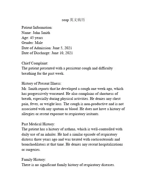
soap英文病历Patient Information:Name: John SmithAge: 45 yearsGender: MaleDate of Admission: June 5, 2021Date of Discharge: June 10, 2021Chief Complaint:The patient presented with a persistent cough and difficulty breathing for the past week.History of Present Illness:Mr. Smith reports that he developed a cough one week ago, which has progressively worsened. He also complains of shortness of breath, especially during physical activities. He denies any chest pain, fever, or weight loss. The cough is non-productive and is not associated with any sputum or blood. He does not have a history of allergies or recent exposure to respiratory irritants.Past Medical History:The patient has a history of asthma, which is well-controlled with daily use of an inhaler. He had a similar episode of respiratory distress three years ago and was treated with corticosteroids and bronchodilators at that time. He denies any recent hospitalizations or surgeries.Family History:There is no significant family history of respiratory diseases.Social History:Mr. Smith is a non-smoker and does not consume alcohol regularly. He works as an office manager and is not exposed to any occupational hazards. He lives with his wife and two teenage children. He denies any recent travel or contact with sick individuals.Physical Examination:Upon examination, the patient appears in no acute distress. Vital signs are stable with a temperature of 98.6°F (37°C), blood pressure of 120/80 mmHg, heart rate of 80 beats per minute, and respiratory rate of 16 breaths per minute. Auscultation of the lungs reveals bilateral wheezing and decreased breath sounds in the lower lung fields. There is no evidence of cyanosis or clubbing. The cardiovascular and abdominal examinations are within normal limits.Diagnostic Tests:A chest X-ray was ordered to evaluate the patient's respiratory symptoms. The X-ray showed bilateral diffuse patchy infiltrates, consistent with bronchial asthma. Pulmonary function tests were performed, revealing a decreased forced expiratory volume in one second (FEV1) and forced vital capacity (FVC), indicating obstructive lung disease.Assessment and Plan:The patient's symptoms, physical examination findings, and diagnostic test results are consistent with a diagnosis of exacerbation of bronchial asthma. The patient was started on a short course of oral corticosteroids, a short-acting bronchodilator,and an inhaled corticosteroid. He was also provided education regarding trigger avoidance and proper inhaler technique. Close follow-up was scheduled to monitor his response to treatment and adjust the management plan if necessary.Follow-Up:The patient will be seen for a follow-up visit in two weeks to evaluate his response to treatment and adjust his medication regimen if needed. He was instructed to monitor his lung function at home using a peak flow meter and seek medical attention if there is a significant decrease in his peak flow readings or if his symptoms worsen. The importance of regular follow-up visits and adherence to the prescribed medication regimen was emphasized. Summary:Mr. Smith, a 45-year-old male with a history of asthma, presented with a persistent cough and difficulty breathing. A diagnosis of exacerbation of bronchial asthma was made based on his symptoms, examination findings, and diagnostic tests. The patient was started on appropriate treatment and provided with education regarding trigger avoidance and inhaler technique. Close follow-up was arranged to monitor his response to treatment and ensure optimal management.。
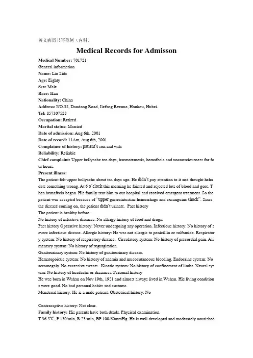
英文病历书写范例(内科)Medical Records for AdmissonMedical Number: 701721General informationName: Liu SideAge: EightySex: MaleRace: HanNationality: ChinaAddress: NO.35, Dandong Road, Jiefang Rvenue, Hankou, Hubei.Tel: 857307523Occupation: RetiredMarital status: MarriedDate of admission: Aug 6th, 2001Date of record: 11Am, Aug 6th, 2001Complainer of history:patient’s son and wifeReliability: ReliableChief complaint: Upper bellyache ten days, haematemesis, hemafecia and unconsciousness for fo ur hours.Present illness:The patient felt upper bellyache about ten days ago. He didn’t pay attention to it and thought heha date something wrong. At 6 o’cloc k this morning he fainted and rejected lots of blood and gore. T hen hemafecia began. His family sent him to our hospital and received emergent treatment. So the patient was accepted because of “upper gastrointestine hemorrhage and exsanguine shock”. Since the disease coming on, the patient didn’t urinate. Past historyThe patient is healthy before.No history of infective diseases. No allergy history of food and drugs.Past history Operative history: Never undergoing any operation. Infectious history: No history of s evere infectious disease. Allergic history: He was not allergic to penicillin or sulfamide. Respirator y system: No history of respiratory disease. Circulatory system: No history of precordial pain. Ali mentary system: No history of regurgitation.Genitourinary system: No history of genitourinary disease.Hematopoietic system: No history of anemia and mucocutaneous bleeding. Endocrine system: No acromegaly. No excessive sweats. Kinetic system: No history of confinement of limbs. Neural sys tem: No history of headache or dizziness. Personal historyHe was born in Wuhan on Nov 19th, 1921 and almost always lived in Wuhan. His living condition s were good. No bad personal habits and customs.Menstrual history: He is a male patient. Obstetrical history: NoContraceptive history: Not clear.Family history: His parents have both deads. Physical examinationT 36.5℃, P 130/min, R 23/min, BP 100/60mmHg. He is well developed and moderately nourished.Active position. His consciousness was not clear. His face was cadaverous and the skin was not sta ined yellow. No cyanosis. No pigmentation. No skin eruption. Spider angioma was not seen. No pi tting edema. Superficial lymph nodes were not found enlarged. HeadCranium: Hair was black and white, well distributed. No deformities. No scars. No masses. No ten derness.Ear: Bilateral auricles were symmetric and of no masses. No discharges were found in external au ditory canals. No tenderness in mastoid area. Auditory acuity was normal.Nose: No abnormal discharges were found in vetibulum nasi. Septum nasi was in midline. No nare s flaring. No tenderness in nasal sinuses. Eye: Bilateral eyelids were not swelling. No ptosis. No e ntropion. Conjunctiva was not congestive. Sclera was anicteric. Eyeballs were not projected or dep ressed. Movement was normal. Bilateral pupils were round and equal in size. Direct and indirect p upillary reactions to light were existent.Mouth: Oral mucous membrane was not smooth, and there were ulcer can be seen. Tongue was in midline. Pharynx was congestive. Tonsils were not enlarged.Neck: Symmetric and of no deformities. No masses. Thyroid was not enlarged. Trachea was in mi dline. ChestChestwall: Veins could not be seen easily. No subcutaneous emphysema. Intercostal space was nei ther narrowed nor widened. No tenderness.Thorax: Symmetric bilaterally. No deformities. Breast: Symmetric bilaterally.Lungs: Respiratory movement was bilaterally symmetric with the frequency of 23/min. thoracic e xpansion and tactile fremitus were symmetric bilaterally. No pleural friction fremitus. Resonance was heard during percussion. No abnormal breath sound was heard. No wheezes. No rales. Heart: No bulge and no abnormal impulse or thrills in precordial area. The point of maximum imp ulse was in 5th left intercostal space inside of the mid clavicular line and not diffuse. No pericardi al friction sound. Border of the heart was normal. Heart sounds were strong and no splitting. Rate 150/min. Cardiac rhythm was not regular. No pathological murmurs.Abdomen: Flat and soft. No bulge or depression. No abdominal wall varicosis. Gastralintestinal ty pe or peristalses were not seen. Tenderness was obvious around the navel and in upper abdoman. T here was not rebound tenderness on abdomen or renal region. Liver and spleen was untouched. No masses. Fluidthrill negative. Shifting dullness negative. Borhorygmus not heard. No vascular mur murs. Extremities: No articular swelling. Free movements of all limbs.Neural system: Physiological reflexes were existent without any pathological ones. Genitourinary system: Not examed. Rectum: not exanedInvestigationBlood-Rt: Hb 69g/L RBC 2.70T/L WBC 1. 1G/L PLT 120G/L History summary1. Patient was male, 80 years old2. Upper bellyache ten days, haematemesis, hemafecia and unconsciousness for four hours.3. No special past history.4. Physical examination: T 37.5℃, P 130/min, R 23/min, BP 100/60mmHg Superficial lymph node s were not found enlarged. No abdominal wall varicosis. Gastralintestinal type or peristalses were not seen. Tenderness was obvious around the navel and in upper abdoman. There was not rebound tenderness on abdomen or renal region. Liver and spleen was untouched. No masses. Fluidthrill ne gative. Shifting dullness negative. Borhorygmus not heard. No vascular murmurs. No other positive signs. 5. investigation information:Blood-Rt: Hb 69g/L RBC 2.80T/L WBC 1.1G/L PLT 120G/LImpression: upper gastrointestine hemorrhage Exsanguine shock出院小结(DISCHARGE SUMMARY), ===============Department of GastroenterologyChanghai Hospital,No.174 Changhai Road Shanghai, China Phone: 86-21-25074725-803 DISCHARGE SUMMARYDA TE OF ADMISSION: October 7th, 2005 DA TE OF DISCHARGE: October 12th, 2005 ATTE NDING PHYSICIAN: Yu Bai, MD PA TIENT AGE: 18ADMITTING DIAGNOSIS:V omiting for unknown reason: acute gastroenteritis?BRIEF HISTORYA 18-year-old female with a complaint of nausea and vomiting for nearly one month who was see n at Department of Gastroenterology in Changhai Hospital, found to have acute gastroenteritis and non-atrophic gastritis. The patient was subsequently recovered and discharged soon after medicati on.REVIEW OF SYSTEMShe has had no headache, fever, chills, diarrhea, chest pain, palpitations, dyspnea, cough, hemopty sis, dysuria, hematuria or ankle edema.PAST MEDICAL HISTORYShe has had no previous surgery, accidents or childhood illness.SOCIAL HISTORY: She has no history of excessive alcohol or tobacco use.FAMIL Y HISTORYShe has no family history of cardiovascular, respiratary and gastrointestinal diseases. PHYSICAL EXAMINA TIONTemperature is 37, pulse 80, respirations 16, blood pressure 112/70. General: Plump girl in no app arent distress. HEENT: She has no scalp lesions. Her pupils are equally round and reactive to light and accommodation. Extraocular movements are intact. Sclerae are anicteric. Oropharynx is clear. There is no thyromegaly. There is no cervical or supraclvicular lymphadenopathy. Cardiovascular: Regular rate andrhythm, normal S1, S2. Chest: Clear to auscultation bilateral. Abdomen: Bowel sounds present, no hepatosplenomagaly. Extremities: There is no cyanosis, clubbing or edema. Neurologic: Cranial n erves II-XII are intact. Motor examination is 5/5 in the bilateral upper and lower extremities. Sens ory, cerebellar and gait are normal.LABORATORY DATAWhite blood cells count 5.9, hemoglobin 111g/L, hematocrit 35.4. Sodium 142, potassium 4.3, chl oride 106, CO2 25, BUN 2.6mmol/L, creatinine 57μmol/L, glucose 4.1mmol/L, Albumin 36g/L. Endoscopic ExamChronic non-atrophic gastritisHOSPITAL COURSEThe patient was admitted and placed on fluid rehydration and mineral supplement. The patient im proved, showing gradual resolution of nausea and vomiting. The patient was discharged in stable c ondition.DISCHARGE DIAGNOSIS Acute gastroenteritisChronic non-atrophic gastritisPROGNOSISGood. No medications needed after discharge. But if this patient can not get used to Chinese food, she had better return to UK as soon as possible to prevent the relapse of acute gastroenteritis. The patient is to follow up with Dr. Bai in one week. ___________________________ Yu Bai, MD D: 12/10/2005。
英语病历模板范文Patient Identification:Date of Birth: [DOB]Sex: [Male/Female]Patient ID: [Unique Identifier]Chief Complaint:[Patient's primary concern or reason for the visit, e.g., "Severe headache for the past 3 days"]History of Present Illness:[Detailed account of the onset, duration, severity, and any associated symptoms of the current illness. Include any treatments already attempted.]Past Medical History:[List any previous medical conditions, surgeries, or hospitalizations.]Medications:[List all current medications, including dosages andfrequency.]Allergies:[Note any known allergies to medications, foods, or environmental factors.]Family Medical History:[Provide information on any significant medicalconditions in the patient's family.]Social History:[Include relevant lifestyle factors such as smoking status, alcohol consumption, exercise habits, and occupation.]Review of Systems:[Briefly summarize the patient's current state inrelation to various body systems, e.g., "No chest pain, no shortness of breath."]Physical Examination:[Record findings from the physical examination, including vital signs, general appearance, and specific observations related to the chief complaint.]Assessment:[Summarize the likely diagnosis or condition based on the information gathered.]Plan:[Outline the proposed treatment plan, including medications, referrals, follow-up appointments, and any necessary tests or procedures.]。
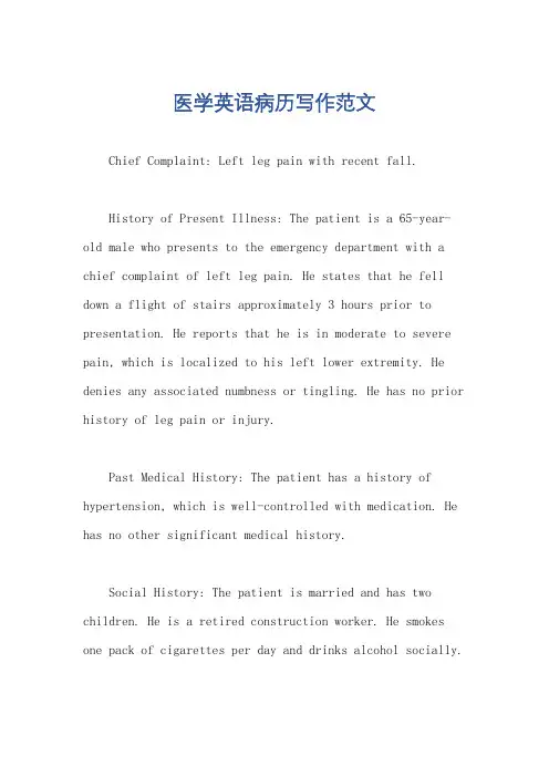
医学英语病历写作范文Chief Complaint: Left leg pain with recent fall.History of Present Illness: The patient is a 65-year-old male who presents to the emergency department with a chief complaint of left leg pain. He states that he fell down a flight of stairs approximately 3 hours prior to presentation. He reports that he is in moderate to severe pain, which is localized to his left lower extremity. He denies any associated numbness or tingling. He has no prior history of leg pain or injury.Past Medical History: The patient has a history of hypertension, which is well-controlled with medication. He has no other significant medical history.Social History: The patient is married and has two children. He is a retired construction worker. He smokes one pack of cigarettes per day and drinks alcohol socially.Family History: The patient's father has a history of coronary artery disease. His mother has a history of Alzheimer's disease.Physical Examination:Vital signs: Blood pressure 140/80 mmHg, heart rate 80 bpm, respiratory rate 18 breaths per minute, temperature 98.6°F (37°C).General: The patient is in moderate distress due to pain. He is alert and oriented to person, place, and time.HEENT: Normocephalic and atraumatic. Pupils are equal and reactive to light. Extraocular movements are intact. No conjunctival injection or discharge. Tympanic membranes are intact and mobile.Neck: Supple with full range of motion. No masses or tenderness.Chest: Auscultation reveals clear breath soundsbilaterally. No wheezes, rales, or rhonchi.Cardiovascular: Regular rate and rhythm. No murmurs, rubs, or gallops.Abdomen: Soft and non-tender. No masses or organomegaly.Extremities: Left lower extremity: Examination reveals swelling and tenderness of the left knee. There is a palpable step-off deformity of the lateral aspect of theleft knee. Active and passive range of motion is limiteddue to pain. Distal pulses are palpable and capillaryrefill is brisk. Sensation is intact. Right lower extremity: Examination reveals no abnormalities.Neurological Examination:Mental status: Alert and oriented to person, place,and time. No deficits in attention, memory, or language.Cranial nerves: No deficits.Motor: Strength is 5/5 in both upper and lower extremities. No atrophy or fasciculations.Sensory: Sensation is intact to light touch, pinprick, and temperature in all four extremities.Diagnostic Studies:X-ray of the left knee: The X-ray shows a displaced lateral tibial plateau fracture.Assessment:Left knee pain.Displaced lateral tibial plateau fracture.Plan:The patient will be admitted to the hospital for further evaluation and treatment.He will be placed in a knee immobilizer and will be started on pain medication.Orthopedic surgery will be consulted for further management.。
Union Hospital affiliated to Huazhong University of Science and TechnologyAdmission Record 0000337023Department: Respiratory Medicine Area: J17 Respiratory Medicine Bed No. 109031 Case No. 1565825Name: Hou Deguang Gender: Male Date of Birth:15/9/ 1936 Age:78 Nationality: ChinaID No. 42021 Ethnicity: Han Occupation: other Marital status: MarriedAddress: Nanchong,Sichuan Tel No.Source of History: Patient herself Reliability: ReliableAdmission Date & Time: 4/11/2021 14:36Chief Complaint: Found pleural effusion for about 2 months.Present Illness: The patient received the chest CT scan in the Wuhan Traditional Medicine Hospital two months ago and found right-side pleural effusion, right-sidepulmonary atelectasis. After that, he was hospitalized in the EndocrinologyDept of our hospital for poor management of blood glucose level. On thisadmission, He received the thoracocentesis, and the laboratory examinationresults indicated the large possibility of tuberculous pleural effusion. Nospecial treatment was given at that time. The patient was aware of a sense ofpolypnea after long walk, without cough, expectoration, night sweats, chestdistress, thoracalgia, wheeze, dyspnea and can lie down to sleep at night. Thereturn-visit in the clinic at October 13th showed that there were a few pleuraleffusion on the right side and is hard to be localized. Now the patient came toour hospital for further treatment and was admitted as “Pleural effusionorigin unknown〞.Since the onset of the disease, the patient’s sp irit, appetite and sleep arenormal. Nocturia for 1 time per night. Stool are as usual. No obvious weightand physical strength change.Past History: General Health Status: Relatively bad; Respiratory Syste m: Chronic bronchitis for about 10 years; Circulatory System: Hypertension for about 20years, highest reached 180/95mmHg, took Amlodipine orally 5mg qd, BP managementis good. Diagnosed of coronary heart disease in 2007, underwentintracoronary stent implantation in 2021, 3 stents were implanted; DigestiveSystems: None; Urinary System: Benign prostatic hyperplasia for about 5 years,Diabetic nephropathy for 3 years; Hematologic System: Thrombocytopenia for 2years; Endocrine System: None. Nervous System: Lacunar infarction in 2021;Motor System: None; Infection History: No infection of hepatitis and TB. Others:None special; Preventive Inoculation: In accordance with the stateplan;Operation History:underwent intracoronary stent implantation in 2021, 3stents was implantated; Blood Transfusion History:None; Traumatic History:None; Allergic History: None;Personal History: Habitual Residence: Hubei; Residential Environment: No exposure history to toxic substances and infected water; Travelling History: None; Smoking History:Smoking for about 40 years, 3 cigarettes per day. Quit smoking in 2021;Drinking History: Drinking for 40 years, 150g-350g per day, Quit drinking in2021;Marital History: Married,Menstrual History: MaleFamily History:Father is deceased, mother is deceased. No other infective and hereditary diseases.Physical ExaminationVital Signs: T:℃. P:86 bpm, regular. R: 20min, regular. BP: 132/74 mmHg. Height: 164cm.Weight: 64kg. Expression: Normal. Development: Well. Nutritional status: Fairly.Consciousness: Conscious. Spirit: Well. Gait: Normal. Position: Active.Coordination with Examination: Cooperative.Skin and Lymph Nodes:No jaundice. Some scattered scratch in hands and abdomen, No subcutaneous bleeding, edema, nodules or unusual pigmentation. Liverpalm(-). Spider angioma(-). No swelling of general superficial lymphnodes.HEENT(Head, Eye, Ear, Nose, Throat): Normal skull. No baldness, no scars. Eyes: No ptosis.Conjuctiva normal. The pupils are round, symmetric and responsive to lightand accommodation is normal. Ears: Externally normal. Canals clear. Drumsnormal. Noses: No abnormalities noted. Month and Throat: lips red, tongue red,no swelling of tonsils.Neck: Motion free. Thyroid is not enlarged. No abnormal pulsations. Trachea in middle. Carotid: Pulse is normal. Hepatojugular reflux sign(-). Vascular bruit: None.Chest and Lung:Normal contour. Breast normal. Inspection: respiratory movement symmetric and regular. Palpation: Normal and symmetric. No pleural friction fremitus. Percussion: both sides resonance. Auscultation: right-side breath sounds weaken, left-side is normal. No moist or dry rales. No pleural friction rubs.Heart:No protrusion of precordium. Normal apical impulse. No thrill. No enlarged cardiac dullness border. Heart rate: 88bpm, rhythm normal. No abnormal and extra cardiac sounds or cardiac murmurs. No peripheral vascular signs.Abdomen:Flat abdomen. No gastric or intestinal pattern. No visible peristalsis. Normal bowel sound. No rigidity. No mass palpable. No tenderness and rebound tenderness. Liver and spleen are not palpable. Kidneys are not palpable. No percussion tenderness over kidney regions. No shifting dullness.Rectum: Normal anus and perineum.Genitourinary System: Normal.Neural System: Normal.Extremities: No joint disease. Muscle strength normal. Pathological reflex (-).Specialty Examination: Right-side breath sounds weaken, left side normal. No moist or dry rales, No swelling of general superficial lymph nodes. No edema inneither lower extremities.Accessory Examination:Discharge record of Endocrinology Dept. of our hospital at September 2021; Clinic examination at October 13th: a few pleural effusion on theright side and is hard to be localized.History summary: 1. Hou Deguang, male, 78 yr.2. Admitted for 〞Found plaural effusion for about 2 months〞.3. T:℃. P:86 bpm, regular. R: 20min, regular. BP: 132/74 mmHg.Expression: Normal. Spirit clear. Cardiac sounds normal, HR: 72 bpm, rhythmnormal, No abnormal and extra cardiac sounds or cardiac murmurs. Right-side breathsounds weaken, left side normal. No moist or dry rales, no pleural friction rubs.Flat abdomen. No rigidity.4. Special examination:Trachea in middle. Contour symmetric.Respiratory movement regular. Right-side breath sounds weaken, left side normal. Nomoist or dry rales, no pleural friction rubs.5. Accessory Examination: Discharge record of Endocrinology Dept of ourhospital at September 2021; Clinic examination at October 13th: a few pleuraleffusion on the right side and is hard to be localized.6. Past history: Respiratory Syste m: Chronic bronchitis for about 10years; Circulatory System: Hypertension for about 20 years, highest reached180/95mmHg, took Amlodipine orally 5mg qd, BP management is good. Diagnosed ofcoronary heart disease in 2007, underwent intracoronary stent implantation in2021, 3 stents was implantated; Digestive Systems: None; Urinary System: Benignprostatic hyperplasia for about 5 years, Diabetic nephropathy for 3 years;Hematologic System: Thrombocytopenia for 2 years; Endocrine System: None.Nervous System: Lacunar infarction in 2021; Motor System: None;InfectionHistory: No infection of hepatitis and TB. Others: None special; PreventiveInoculation: In accordance with the stateplan; Operation History:underwentintracoronary stent implantation in 2021, 3 stents was implantated; BloodTransfusion History: None; Traumatic History: None; Allergic History: None; Impression: 1. Right-side pleural effusion origin unknown: TB? Tumor?2. II diabetes mellitus, Diabetic nephropathy3. Hypertension III, high risk4. Coronary heart disease, post-intracoronary stent implantation5. Lacunar infarction6. Thrombocytopenia7. Benign prostatic hyperplasiaRecorder: Cheng LongDate & Time: 4/11/2021 16:14Checker: Xu JuanjuanDate & Time: 5/11/2021 10:22。
英语作文病历模板英文回答:Medical History Template。
Patient Information。
Name:Date of Birth:Gender:Address:Phone Number:Email Address:Reason for Visit。
What brings you to the clinic today?Medical History。
Past Medical History。
Do you have any past medical conditions?Have you ever been hospitalized or had surgery?Do you currently take any medications?Do you have any allergies?Family Medical History。
Do any of your close family members have any medical conditions?Have any of your close family members passed away at a young age due to illness?Social History。
What is your occupation?Are you currently married or in a relationship? Do you have any children?Do you smoke, drink alcohol, or use drugs?Physical Examination。
General Appearance:Height:Weight:BMI:Vital Signs:Blood pressure: Pulse:Respiratory rate: Temperature:Cardiovascular:Heart rate:Heart sounds:Blood pressure: Respiratory:Respiratory rate: Lung sounds:Abdomen:Girth:Soft and non-tender: Liver span:Musculoskeletal:Range of motion:Strength:Reflexes:Skin:Color:Texture:Turgor:Assessment。
英文病历报告作文模板英文:Medical Record Report。
Name: John Smith。
Age: 35。
Gender: Male。
Date of Admission: 05/01/2021。
Date of Discharge: 05/07/2021。
Chief Complaint:The patient complained of a persistent cough and shortness of breath.History of Present Illness:The patient had a persistent cough and shortness of breath for two weeks. He tried to treat himself with over-the-counter medication but his symptoms did not improve. He decided to seek medical attention when his cough became more severe and he started to experience chest pain.Past Medical History:The patient has a history of asthma and seasonal allergies. He has been hospitalized in the past for asthma exacerbations.Physical Examination:On physical examination, the patient had wheezing and crackles in his lungs. His oxygen saturation was 92% on room air.Diagnostic Tests:A chest X-ray showed bilateral infiltrates consistent with pneumonia. A COVID-19 test was negative.Treatment:The patient was started on antibiotics for pneumonia and given nebulizer treatments for his asthma exacerbation. He was also given supplemental oxygen to maintain his oxygen saturation above 94%.Outcome:The patient's symptoms improved with treatment and he was discharged home after a week in the hospital.中文:病历报告。
住院病历英语作文{z}Title: Hospital Admission RecordHospital Admission RecordPatient Information:ame: [Patient"s Name]Age: [Patient"s Age]Gender: [Patient"s Gender]Date of Admission: [Date]Medical History:The patient, [Patient"s Name], a [Patient"s Age]-year-old [Patient"s Gender], was admitted to our hospital on [Date] due to [reason for admission].The patient has a history of [previous medical conditions, if any], and is currently taking [list of medications, if any].Physical Examination:Upon admission, the patient was found to have [physical examination findings, such as vital signs, general appearance, and specific organ system assessments].The patient complained of [list of symptoms, if any], and there were signs of [any observed abnormalities or symptoms].Diagnosis:Based on the patient"s medical history and physical examination, the following diagnosis was made: [list of diagnosed conditions or diseases].Treatment Plan:The patient was recommended to undergo [list of treatments, such as surgery, medication, or therapy] for the management of their condition.The treatment plan was discussed with the patient and/or their guardian, and informed consent was obtained.Progress Notes:[List of any significant changes in the patient"s condition, response to treatment, and any other relevant updates during the hospital stay].Discharge Summary:The patient, [Patient"s Name], was discharged from our hospital on [Date].At the time of discharge, the patient"s condition had improved significantly, and they was deemed stable for discharge.The patient was provided with discharge instructions, including any necessary medications, follow-up appointments, and lifestyle modifications.It is recommended that the patient continue to follow up with their primary care physician or designated healthcare provider for further monitoring and management of their condition.Any concerns or exacerbation of symptoms should prompt immediate medical attention.Please note that this document is a sample hospital admission record and should be customized according to the specific patient"s information and clinical details.。
自发性气胸住院病历模板范文英文回答:Spontaneous pneumothorax is a condition where air accumulates in the space between the lung and the chest wall, causing the lung to collapse. I experienced this condition recently and was hospitalized for treatment. Here is a template of my medical record during my hospitalization:Patient Name: [Your Name]Age: [Your Age]Gender: [Your Gender]Date of Admission: [Admission Date]Date of Discharge: [Discharge Date]Chief Complaint:I presented to the emergency department with sudden onset of sharp chest pain on the right side, which worsened with deep breaths. I also had difficulty breathing and felt lightheaded.History of Present Illness:I was at home when I suddenly felt a sharp pain in my right chest. The pain was so severe that it made itdifficult for me to take deep breaths. I also noticed that my breathing became more rapid and shallow. I felt lightheaded and had to sit down to catch my breath. The pain persisted for several hours, so I decided to go to the hospital.Past Medical History:I have never experienced any significant medical problems in the past. I do not have any history of lung diseases or previous episodes of pneumothorax.Physical Examination:Upon examination, I was found to have decreased breath sounds on the right side of my chest. My chest was slightly asymmetrical, with decreased movement on the right side. A chest X-ray confirmed the diagnosis of a spontaneous pneumothorax.Treatment:I was admitted to the hospital for further management.A chest tube was inserted into my right chest to remove the accumulated air and re-expand the lung. I was also given supplemental oxygen to help with my breathing. Pain medication was administered to alleviate my chest pain.Progress:Over the course of my hospitalization, my symptoms gradually improved. The chest tube was removed after a few days when the lung was fully re-expanded. I was able tobreathe comfortably and my chest pain resolved. I was discharged with instructions to follow up with my primary care physician for further evaluation and to discuss the possibility of preventive measures to reduce the risk of recurrence.中文回答:自发性气胸是一种空气在肺与胸壁之间积聚,导致肺部塌陷的状况。
英语写病历作文模板 Patient History Template。
英文回答:General Information。
Name:Age:Gender:Occupation:Address:Phone number:Emergency contact:Medical History。
Past medical history: List any previous illnesses, surgeries, hospitalizations, or accidents.Family medical history: Note any history of chronic diseases, such as heart disease, cancer, or diabetes, in the patient's family.Allergies: List any known allergies to medications, foods, or other substances.Medications: List all current medications, including prescription drugs, over-the-counter medications, and herbal supplements.Social history: Discuss the patient's lifestyle, including diet, exercise, smoking, alcohol use, and drug use.Present Illness。
Chief complaint: State the patient's primary reasonfor seeking medical attention.History of present illness: Describe the onset, duration, severity, and progression of the patient's symptoms.Physical Examination。
住院病历英语作文English:The hospitalization medical record serves as a crucial document in the continuum of patient care, encapsulating vital information about the patient's condition, treatment plan, and progress throughout their hospital stay. It typically includes demographic details, such as the patient's name, age, and contact information, alongside a comprehensive medical history detailing any pre-existing conditions, allergies, and medications. The presenting complaint and pertinent physical examination findings are documented, followed by diagnostic test results, including laboratory investigations, imaging studies, and other specialized tests. The treatment regimen, encompassing medications, procedures, surgeries, and therapeutic interventions, is meticulously recorded, along with any adverse reactions or complications encountered during the hospitalization. Moreover, the medical record serves as a communication tool among healthcare professionals, ensuring continuity of care and facilitating interdisciplinary collaboration. Ultimately, it plays a pivotal role in clinical decision-making, quality assessment, and medico-legal matters, serving as a repository of essential clinicalinformation essential for optimal patient management and healthcare delivery.Translated content:住院病历是患者医疗过程中不可或缺的文件,记录了患者在住院期间的病情、治疗方案和进展等重要信息。
Division: __________ Ward: __________ Bed: _________ Case No. ___________ Name: ______________ Sex: __________ Age: ___________ Nation: ___________ Birth Place: ________________________________ Marital Status:____________ Work-organization & Occupation: _______________________________________ Living Address & Tel: _________________________________________________ Date of admission: _______Date of history taken:_______ Informant:__________Chief Complaint: ___________________________________________________ History of Present Illness:___________________________________________________________ ___________________________________________________________ ___________________________________________________________ ___________________________________________________________ ___________________________________________________________ ___________________________________________________________ ___________________________________________________________ ___________________________________________________________ ___________________________________________________________ ___________________________________________________________ ___________________________________________________________ ___________________________________________________________ ___________________________________________________________ ___________________________________________________________Past History:General Health Status: 1.good 2.moderate 3.poorDisease history: (if any, please write down the date of onset, brief diagnosticand therapeutic course, and the results.)Respiratory system:1. None2.Repeated pharyngeal pain3.chronic cough4.expectoration:5. Hemoptysis6.asthma7.dyspnea8.chest pain_______________________________________________________________ Circulatory system:1.None2.Palpitation3.exertional dyspnea4..cyanosis5.hemoptysis6.Edema of lower extremities7.chest pain8.syncope9.hypertension _______________________________________________________________ Digestive system:1.None2.Anorexia3.dysphagia4.sour regurgitation5.eructation6.nausea7.Emesis8.melena9.abdominal pain 10.diarrhea11.hematemesis 12.Hematochezia 13.jaundice_______________________________________________________________ Urinary system:1.None2.Lumbar pain3.urinary frequency4.urinary urgency5.dysuria6.oliguria7.polyuria8.retention of urine9.incontinence of urine10.hematuria 11.Pyuria 12.nocturia 13.puffy face_______________________________________________________________ Hematopoietic system:1.None2.Fatigue3.dizziness4.gingival hemorrhage5.epistaxis6.subcutaneous hemorrhage_______________________________________________________________ Metabolic and endocrine system:1.None2.Bulimia3.anorexia4.hot intolerance5.cold intolerance6.hyperhidrosis7.Polydipsia8.amenorrhea9.tremor of hands 10.character change 11.Marked obesity12.marked emaciation 13.hirsutism 14.alopecia15.Hyperpigmentation 16.sexual function change_______________________________________________________________ Neurological system:1.None2.Dizziness3.headache4.paresthesia5.hypomnesis6. Visual disturbance7.Insomnia8.somnolence9.syncope 10.convulsion 11.Disturbance of consciousness12.paralysis 13. vertigo_______________________________________________________________ Reproductive system:1.None2.others_______________________________________________________________ Musculoskeletal system:1.None2.Migrating arthralgia3.arthralgia4.artrcocele5.arthremia6.Dysarthrosis7.myalgia8.muscular atrophy_______________________________________________________________ Infectious Disease:1.None2.Typhoid fever3.Dysentery4.Malaria 4.Schistosomiasis4.Leptospirosis 7.Tuberculosis 8.Epidemic hemorrhagic fever9.others_______________________________________________________________ Vaccine inoculation:1.None2.Yes3.Not clearVaccine detail __________________________________________ Trauma and/or operation history:Operations:1.None2.YesOperation details:_______________________________________ Traumas:1.None2.YesTrauma details:_________________________________________ Blood transfusion history:1.None2.Yes ( 1.Whole blood 2.Plasma3.Ingredient transfusion)Blood type:____________ Transfusion time:___________Transfusion reaction1.None2.YesClinic manifestation:_____________________________ Allergic history:1.None2.Yes3.Not clearallergen:________________________________________________clinical manifestation:_____________________________________Personal history:Custom living address:____________________________________________ Resident history in endemic disease area:_____________________________ Smoking: 1.No 2.YesAverage ___pieces per day; about___yearsGiving-up 1.No 2.Yes (Time:_______________________) Drinking: 1.No 2.YesAverage ___grams per day; about ___yearsGiving-up 1.No 2.Yes(Time:________________________) Drug abuse:1.No 2.YesDrug names:_______________________________________ _______________________________________________________________ Marital and obstetrical history:Married age: __________years old Pregnancy ___________timesLabor _______________times(1.Natural labor: _______times 2.Operative labor: ________times3.Natural abortion: ______times4.Artificial abortion: _______times5.Premature labor:__________times6.stillbirth__________times)Health status of the Mate:1.Well2.Not fineDetails: _______________________________________________ Menstrual history:Menarchal age: _______ Duration ______day Interval ____daysLast menstrual period: ____________ Menopausal age: ____years oldAmount of flow: 1.small 2. moderate 3. largeDysmenorrheal: 1. presence 2.absence Menstrual irregularity 1. No 2.Yes Family history: (especially pay attention to the infectious and hereditary diseaserelated to the present illness)Father: 1.healthy 2.ill:________ 3.deceased cause: ___________________ Mother:1.healthy 2.ill:________ 3.deceased cause: ___________________ Others: ________________________________________________________ The anterior statement was agreed by the informant.Signature of informant: Datetime:Physical ExaminationVital signs:Temperature:______0C Blood pressure:_______/_______mmHg Pulse: _____ bpm (1.regular 2.irregular_____________________________) Respiration: ___bpm (1.regular 2.irregular____________________________) General conditions:Development: 1.Normal 2.Hypoplasia 3.HyperplasiaNutrition: 1.good 2.moderate 3.poor 4.cachexiaFacial expression: 1.normal 2.acute 3.chronic other_____________________ Habitus: 1.asthenic type 2.sthenic type 3.ortho-thenic typePosition: 1.active 2.positive pulsive 4.other_______________________ Consciousness: 1.clear 2.somnolence 3.confusion 4.stupor 5.slight coma6.mediate coma7.deep coma8.deliriumCooperation: 1Yes 2.No Gait: 1.normal 2.abnormal______Skin and mucosa:Color: 1.normal 2.pale 3.redness 4.cyanosis 5.jaundice 6.pigmentationSkin eruption:1.No 2.Yes( type: __________distribution:__________________) Subcutaneous bleeding: 1.no 2.yes (type:_______distribution:______________) Edema:1. no 2.yes ( location and degree________________________________) Hair: 1.normal 2.abnormal(details_____________________________________) Temperature and moisture: normal cold warm dry moist dehydration Liver palmar : 1.no 2.yes Spider angioma (location:________________) Others: __________________________________________________________ Lymph nodes: enlargement of superficial lymph node:1.no2.yesDescription: ________________________________________________ Head:Skull size:1.normal 2.abnormal (description:____________________________) Skull shape:1.normal 2.abnormal(description:___________________________) Hair distribution :1.normal 2.abnormal(description:______________________) Others:___________________________________________________________ Eye: exophthalmos:___________eyelid:____________conjunctiva:__________ sclera:________________Cornea:_______________________Pupil: 1.equally round and in size 2.unequal (R______mm L_______mm)Pupil reflex: 1.normal 2.delayed (R___s L___s ) 3.absent (R___L___)others:______________________________________________________ Ear: Auricle 1.normal 2.desformation (description:_______________________) Discharge of external auditory canal:1.no 2.yes (1.left 2.right quality:_____)Mastoid tenderness 1.no 2.yes (1.left 2.right quality:__________________)Disturbance of auditory acuity:1.no 2.yes(1.left 2.right description:_______) Nose: Flaring of alae nasi :1.no 2.yes Stuffy discharge 1.no 2.yes(quality______) Tenderness over paranasal sinuses:1.no 2.yes (location:_______________) Mouth: Lip______________Mucosa_____________Tongue________________ Teeth:1.normal 2. Agomphiasis 3. Eurodontia 4.others:____________________Gum :1.normal 2.abnormal (Description____________________________)Tonsil:___________________________Pharynx:_____________________Sound: 1.normal 2.hoarseness 3.others:_____________________________ Neck:Neck rigidity 1.no 2.yes (______________transvers fingers)Carotid artery: 1.normal pulsation 2.increased pulsation 3.marked distention Trachea location: 1.middle 2.deviation (1.leftward_______2.rightward______) Hepatojugular vein reflux: 1. negative 2.positiveThyroid: 1.normal 2.enlarged _______ 3.bruit (1.no 2.yes ________________) Chest:Chest wall: 1.normal 2.barrel chest 3.prominence or retraction:( left________right_________Precordial prominence__________) Percussion pain over sternum 1.No 2.YesBreast: 1.Normal 2.abnormal _______________________________________ Lung:Inspection: respiratory movement 1.normal 2.abnormal_____________ Palpation: vocal tactile fremitus:1.normal 2.abnormal _______________ pleural rubbing sensation:1.no 2.yes______________________Subcutaneous crepitus sensation:1.no 2.yes________________ Percussion:1. resonance 2. Hyperresonance &location_____________3 Flatness&location_________________________________4. dullness & location:_______________________________5.tympany &location:_______________________________lower border of lung: (detailed percussion in respiratory disease) midclavicular line : R:_____intercostae L:_____intercostaemidaxillary line: R:______intercostae L:_____intercostaescapular line: R:______intercostae L:_____intercostaemovement of lower borders:R:_______cmL:__________cm Auscultation: Breathing sound : 1.normal 2.abnormal _______________Rales:1.no 2.yes__________________________________ Heart: Inspection:Apical pulsation: 1.normal 2.unseen 3.increase 4.diffuseSubxiphoid pulsation: 1.no 2.yesLocation of apex beat: 1.normal 2.shift (______ intercosta,distance away from left MCL______cm) Palpation:Apical pulsation:1. normal 2.lifting apex impulse 3.negative pulsationThrill:1.no 2.yes(location:___________ phase:_________________)Percussion: relative dullness border: 1.normal 2.abnormalAuscultation: Heart rate:___bpm Rhythm:1.regular 2.irregular_______ Heart sound: 1.normal 2.abnormal________________________Extra sound: 1.no 2.S3 3.S4 4. opening snapP2_________ A2_________Pericardial friction sound:1.no 2.yesMurmur: 1.no 2.yes (location____________phase_____________quality______intensity________ transmission___________effects of position_________________________________effects of respiration______________________________ Peripheral vascular signs:1.None2.paradoxical pulse3.pulsus alternans4. Water hammer pulse5.capillary pulsation6.pulse deficit7.Pistol shot sound8.Duroziez signAbdomen:Inspection: Shape: 1.normal 2.protuberance 3.scaphoid 4.frog-bellyGastric pattern 1.no 2.yes Intestinal pattern 1.no 2.yesAbdominal vein varicosis 1.no 2.yes(direction:______________ )Operation scar1.no 2.yes ________________________________ Palpation: 1.soft 2. tensive (location:____________________________)Tenderness: 1.no 2.yes(location:_______________________)Rebound tenderness:1.no 2.yes(location:________________)Fluctuation: 1.present 2.abscentSuccussion splash: 1.negative 2.positiveLiver:_______________________________________________Gallbladder: __________________Murphy sign:____________Spleen:______________________________________________Kidneys:____________________________________________Abdominal mass:______________________________________Others:______________________________________________ Percussion: Liver dullness border: 1.normal 2.decreased 3.absentUpper hepatic border:Right Midclavicular Line ________IntercostaShift dullness:1.negative 2.positive Ascites:_____________degreePain on percussion in costovertebral area: 1.negative 2.positve ____ Auscultation: Bowel sounds : 1.normal 2.hyperperistalsis 3.hypoperistalsis4.absence Gurgling sound:1.no 2.yesVascular bruit 1.no 2.yes (location_____________________) Genital organ: 1.unexamined 2.normal 3.abnormalAnus and rectum: 1.unexamined 2.normal 3.abnormalSpine and extremities:Spine: 1.normal 2.deformity (1.kyphosis 2.lordosis 3.scoliosis)3.Tenderness(location______________________________)Extremities:1.normal 2.arthremia & arthrocele (location_________________)3.Ankylosis (location__________)4.Aropachy: 1.no 2.yes5.Muscular atrophy (location_______________________) Neurological system:1.normal 2.abnormal_______________________________ _____________________________________________________________________ Important examination results before hospitalized___________________________________________________________ ___________________________________________________________ ___________________________________________________________ ___________________________________________________________ ___________________________________________________________ Summary of the history:______________________________________ ___________________________________________________________ ___________________________________________________________ ___________________________________________________________ ___________________________________________________________ ___________________________________________________________ ___________________________________________________________ ___________________________________________________________ ___________________________________________________________ Initial diagnosis:_____________________________________________ ___________________________________________________________ ___________________________________________________________ ___________________________________________________________ ___________________________________________________________Recorder:Corrector:。