Induction of cell cycle arrest and apoptosis in human nasopharyngeal carcinoma cells by ZD6474,an inhibitor of VEGFR tyrosine kinase with additional activity against EGFR tyrosine kinase
Xia Xiao1,Jiangxue Wu1*,Xiaofeng Zhu1,Peng Zhao1,Jinlin Zhou1,Quentin Qiang Liu1,Limin Zheng1,Musheng Zeng1, Ranyi Liu1and Wenlin Huang1,2,3*
1State Key Laboratory of Oncology in South China,Cancer Center,Sun Yat-Sen University,Guangzhou510060,
People’s Republic of China
2Institute of Microbiology,Chinese Academy of Science,Beijing100080,People’s Republic of China
3Guangzhou Doublle Bio-product Inc.,Guangzhou516003,People’s Republic of China
ZD6474is a vascular endothelial growth factor receptor(VEGFR) and epidermal growth factor receptor(EGFR)tyrosine kinase in-hibitor.The present study was undertaken to investigate the direct antiproliferative effect of ZD6474on human nasopharyngeal car-cinoma(NPC)in vitro and the antitumor activity on NPC xeno-grafts in vivo.Results indicated that ZD6474treatment inhibited EGFR phosphorylation and led to a dose-and time-dependent decrease in NPC cell(CNE-1,CNE-2and C666-1)proliferation. Further investigation demonstrated G0/G1cell cycle arrest in all 3cell lines,which was associated with an upregulation of p21and/ or p27,and downregulation of CDK4,CDK6and CDK2.ZD6474 treatment also induced apoptosis in CNE-1and CNE-2cells.The apoptosis mechanisms involved reduction of Bcl-2and/or Bcl-X L, induction of Bak and/or Bax,and activation of caspases-3,-9and/ or-8.The in vivo antitumor activity was evaluated in CNE-2and C666-1xenografted nude mice.Administration of ZD6474(25–100mg/kg/day,once-daily,p.o.)produced a dose-dependent inhi-bition of tumor growth and prolonged survival in both models. This study suggests that ZD6474exerts direct antiproliferative effects on NPC cell lines in vitro by inducing G0/G1arrest and ap-optosis,and potent antitumor effects on NPC xenografts in vivo.It indicates that ZD6474may offer a new and effective treatment for human NPC.
'2007Wiley-Liss,Inc.
Key words:vascular endothelial growth factor receptor;epidermal growth factor receptor;tyrosine kinase inhibitor;ZD6474;nasopharyn-geal carcinoma;cell cycle;apoptosis
Receptor tyrosine kinases(RTKs)are crucial elements in many cell regulatory processes and are altered or overexpressed in human carcinogenesis.A variety of therapeutic agents have been developed to inhibit the activities of RTKs.A recent highlight is the development of quinazoline-derived agents that are selective ATP competitors of RTKs.ZD6474is an orally bioavailable, small molecule,anilinoquinazoline derivative that is selective for VEGFR-2(KDR)and is currently in Phase III clinical develop-ment.ZD6474has an IC50of 0.04l M against the isolated KDR enzyme and blocks VEGF-induced signaling in endothelial cells.1 Consistent with inhibition of VEGF signaling and angiogenesis, once-daily oral dosing of ZD6474produced signi?cant broad-spectrum antitumor activity in a panel of histologically diverse human tumor xenografts in nude mice.2In addition to its antian-giogenic properties,ZD6474inhibits,to a less extent,the activity of epidermal growth factor receptor(EGFR)tyrosine kinase(IC50 50.5l M).3Overexpression of EGFR or its ligands has been reported in many human tumors.4The aberrant EGFR tyrosine ki-nase activity leads to increased tumor cell proliferation,survival, invasiveness and overexpression of proangiogenic factors such as VEGF.5–8Therefore,ZD6474has the potential to inhibit2pivotal pathways in tumor growth by targeting tumor growth indirectly, via inhibition of VEGF-dependent tumor angiogenesis and endo-thelial cell survival,and targeting tumor growth directly,via inhi-bition of EGFR-dependent tumor cell proliferation and survival.9 The ability of ZD6474to inhibit tumor cell proliferation was examined directly in several cancer cell lines(ovarian,colon, gastric and breast)in vitro with IC50values ranging from0.09to 17.7l M.10It seems that the direct antiproliferative effect of ZD6474is highly tissue-and cell-type speci?c.Thus,elucidation of the mechanisms underlying the direct antitumor effects of ZD6474on particular cancer cells may provide an opportunity to target speci?c patient subsets who may obtain further bene?t from this pharmacological activity.
Nasopharyngeal carcinoma(NPC)is a major malignant disease of the head/neck region that is endemic within the young popula-tion of South China and Southeast Asia.The current treatment reg-imens of ionizing radiation combined with cisplatin chemotherapy yields a5-year survival rate of 70%,11,12indicating that new therapies to combat this disease are urgently needed.
The purpose of this study is to examine the direct antiprolifera-tive effects of ZD6474,with its additional activity against EGFR tyrosine kinase,on NPC cell growth in vitro and the underlying molecular basis.In vivo,the antitumor effect of ZD6474on NPC cell xenograft models is also evaluated.
Material and methods
Reagents
ZD6474was synthesized according to a patent(number:wo 2,003,039,551)procedure.ZD6474:1H NMR(DMSO d6):8.64(s, 1H),8.07(s,1H),7.74(d,1H),7.53(m,2H),7.32(s,1H),4.09(d, 2H),3.99(s,3H),3.50(d,2H),3.02(t,2H),2.78(s,3H),2.16(m, 1H),2.05(d,2H),1.65(m,2H).ESI-MS m/z475(M11);Anal. Calcd for C22H24N4O2BrF0.5H2O1.8HCl:C,48.2;H,5.0;N, 10.1;Found C,47.8;H,5.1N10.0.
Cell lines
Three human NPC cell lines were studied:CNE-1(well-differ-entiated NPC cell line,EBV negative),CNE-2(poorly differenti-ated NPC cell line,EBV negative)and C666-1(undifferentiated NPC cell line,EBV positive).13–16C666-1was a kind gift from Dr.Saiwah Tsao(University of Hong Kong,Hong Kong,People’s Republic of China).Cells were cultured in RPMI1640supple-mented with10%fetal bovine serum(Gibco,Paisley,UK),100 units/ml penicillin and100l g/ml streptomycin at37°C in a5%
This article contains supplementary material available via the Internet at https://www.doczj.com/doc/0f146956.html,/jpages/0020-7136/suppmat.
Grant sponsor:National Basic Research Program of China(973Pro-gram);Grant number:2004CB518801;Grant sponsor:China Postdoctoral Science Foundation;Grant number:2004046156;Grant sponsor:Hi-tech Research and Development Program of China(863Program);Grant num-ber:2006AA02Z489;Grant sponsor:Guangzhou Science Foundation; Grant number:2004Z3-E4011.
The?rst two authors contributed equally to this work.
*Correspondence to:Cancer Center,Sun Yat-Sen University,Guang-zhou510060,PRC.Fax:186-20-8734-3146.
E-mail:wl_huang@https://www.doczj.com/doc/0f146956.html,
Received23January2007;Accepted after revision22May2007
DOI10.1002/ijc.22955
Published online13July2007in Wiley InterScience(www.interscience. https://www.doczj.com/doc/0f146956.html,).
Int.J.Cancer:121,2095–2104(2007)
'2007Wiley-Liss,Inc.
Publication of the International Union Against
Cancer
CO2humidi?ed atmosphere.Experiments were carried out in the log phase of growth.For experiments on synchronized cultures, cells were synchronized in G0/G1phase by culturing in RPMI 1640without serum for24hr.Cells were then released to reenter the cell cycle by the addition of10%fetal bovine serum,either in the presence or in the absence of ZD6474,and were harvested at the indicated time points.
MTT assay
Cells were seeded in96-well plate at a density of23103–23 104cells/well,allowed to recover for16–24hr and then treated using complete media with or without ZD6474at different con-centrations(0.1–25.6l M).After the indicated incubation time, cell viability was measured by MTT assay.Brie?y,20l l of MTT stock solution(5mg/ml in PBS)was added into each well and incubated for4hr in an incubator(37°C,5%CO2).The plate was centrifuged at1,800rpm for5min at4°C and the medium was removed.One hundred microliter of DMSO per well was added to dissolve the formazan crystal,and the absorbance was recorded on a microplate reader at of570/630nm wave length.
Cell cycle analysis
Cell cycle analysis was performed as described before.17 Brie?y,both?oating and adherent cells were harvested after ZD6474treatment for the indicated time courses,washed with ice-cold PBS and?xed overnight with70%ethanol at4°C,fol-lowed by resuspension in500l l of PBS.After addition of10l l RNase(10mg/ml),cells were left for30min at37°C and stained with10l l propidium iodide(1mg/ml).Then,cells were analyzed by a Coulter Epics Elite?ow cytometer(Beckman-Coulter, Miami,FL).
Annexin-V/7-AAD binding assay
For Annexin-V/7-AAD binding assay,both?oating and adher-ent cells were collected after ZD6474treatment for48hr and washed with ice-cold PBS.Cells were resuspended in100l l of binding buffer,and stained with10l l of FITC-Annexin V and 20l l of7-AAD.After incubation on ice in the dark for15min, every sample was diluted with385l l13binding buffer and im-mediately analyzed by a Coulter Epics Altra?ow cytometer (Beckman-Coulter).
Caspase activity assay
Cells were plated in175-cm2?asks.After incubation with ZD6474for the desired time,?oating and adherent cells were har-vested and combined.Preparation of cell lysates and measurement of caspase-8,caspase-9and caspase-3activity were performed with Caspase-8Assay Kit,Caspase-9Assay Kit and Caspase-3 Cellular Activity Assay Kit(Calbiochem,La Jolla,CA)according to the manufacturer’s instruction.
Antibodies
All of the antibodies,excluding EGFR(Cell Signaling Technol-ogy,Beverly,MA),Bcl-X L and p-BAD(Ser136)(SAT,CA)and GAPDH(Kangcheng,Shanghai,China)were obtained from Santa Cruz(Berkeley,CA).
Western blotting analysis and immunoprecipitation
Cell lysates were prepared by extracting proteins with RIPA lysis buffer containing Protease Inhibitor Cocktail II(state,Lake Placid,NY).Protein estimation was performed using Bradford method.Twenty to40l g total cell protein extracts were resolved by electrophoresis using NuPAGE14–12%Bis-Tris Gel(Invi-trogen,Carlsbad,CA)and transferred to PVDF membrane. SeeBlue1prestained standard(Invitrogen)was used to determine product size.The membrane was blocked with either5%BSA or 5%nonfat milk in Tris-buffered saline and then incubated with primary antibodies for4hr at room temperature followed by washing3times for5min each with TBST.Blots were incubated with horseradish peroxidase-conjugated secondary antibody for1 hr at room temperature.Bands were detected using enhanced chemiluminescence system(Cell Signaling,Danvers,MA)and quanti?ed by densitometry using Bio-rad Quantity One software (Bio-rad,Hercules,CA).For immunoprecipitations,500l g of total protein from cell lysates was incubated with1l g primary antibody for2hr at4°C.Twenty microliter of appropriate protein G Plus-Agarose suspension(Santa Cruz)was added and then incu-bated at4°C on a rocker platform overnight.The precipitates were washed4times with ice-cold PBS buffer,resuspended in43LDS sample buffer(Invitrogen)and resolved using NuPAGE14–12% Bis-Tris Gel followed by immunoblot analysis.
Tumor xenograft models
Female BALB/c nude mice(6-to8-week-old,weighing>18g) were obtained from Shanghai Slike experimental animals Co. (Shanghai,China;animal experimental license No.SCXKhu2003-0008).Mice were housed in air-?ltered laminar?ow cabinets with 12-hr light/dark cycles,provided with sterilized food and water ad libitum.After1-week adaptation,mice were injected s.c.in the scapular region with23106CNE-2cells or13107C666-1cells in100l l of serum-free media.When tumors reached a volume of 30–50mm3,mice were randomly allocated into4groups(n56) and received(p.o.)either ZD6474(25–100mg/kg/day)or vehicle (sterilized water),administered once daily at0.1ml/10g body weight.Tumor volume was assessed twice weekly by calipers measurement of tumor diameters and calculated according to the formula:V5L3W2/2(L,length;W,width).After3.5weeks treatment,mice were killed and tumors were resected and weighed.For survival studies(n510),mice were dosed(p.o., once-daily)with either ZD6474(25–100mg/kg/day)or vehicle (sterilized water)for25days,and then the tumors were allowed to grow.Mice either were found dead or were killed when tumors were estimated by palpation to reach10%body weight or individ-ual mice appeared stressed by weight loss,ruf?ed fur and/or leth-argy.All the animal experiments were conducted in accordance with‘‘Guidelines for the Welfare of Animals in Experimental Neoplasia’’.
Statistical analysis
Results were evaluated using2-tailed Student’s t test with SPSS 13.0software(SSPS,Chicago,IL),unless otherwise speci?ed. Results of survival were evaluated using Kaplan Meier survival analysis.A p value<0.05was considered statistically signi?cant.
Results
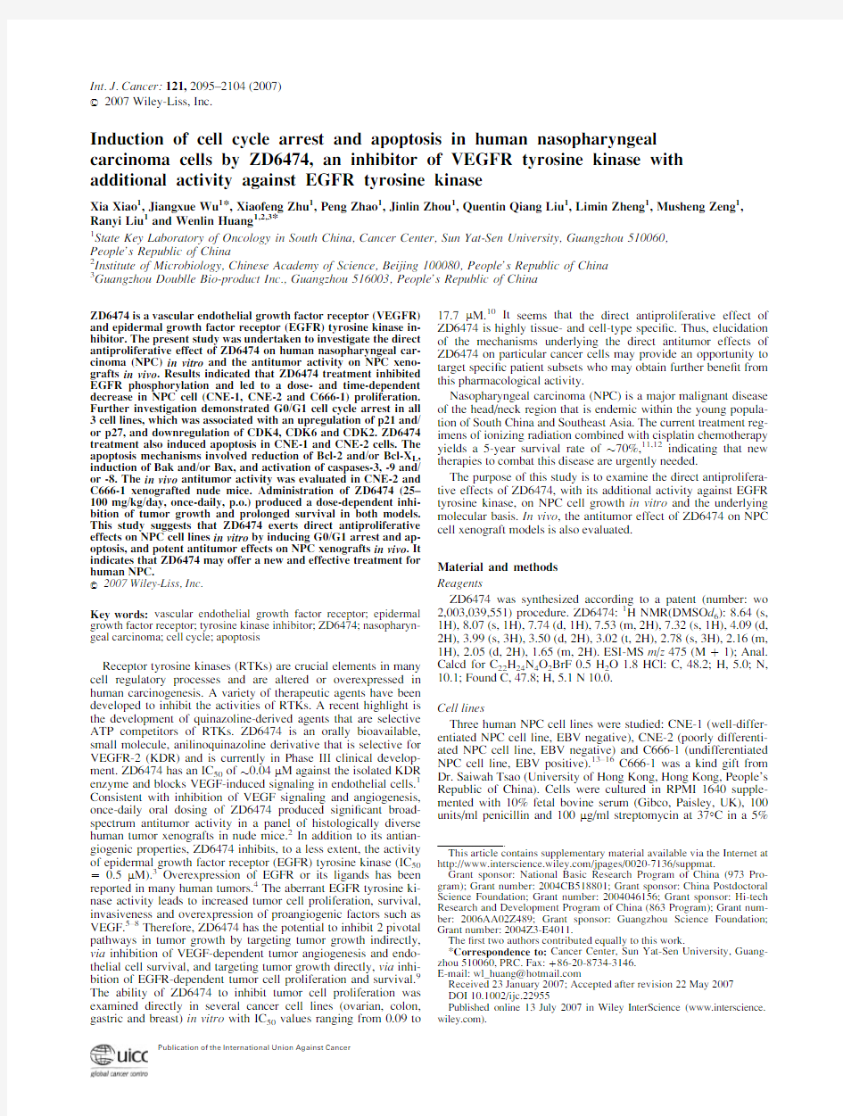
ZD6474treatment inhibits the tyrosine phosphorylation of EGFR VEGFR1,VEGFR2and EGFR expression was evaluated in3 NPC cell lines at protein levels.Results showed that both VEGFR1and VEGFR2proteins were undetectable in all the NPC cell lines(data not shown).The relative expression of EGFR in all 3cell lines was as shown in Figure1a.Densitometric analysis showed that the relative abundance of EGFR was1.1,1.23and 0.66in CNE-1,CNE-2and C666-1,respectively,after normaliz-ing with GAPDH.Figure1b showed that ZD6474treatment inhib-ited the tyrosine phosphorylation of EGFR within15min in CNE-1cells.These results were obtained after immunoprecipitation with an anti-EGFR antibody and subsequent Western blotting with an anti-phosphotyrosine antibody of the cell extracts treated with 3l M ZD6474.Similar results were also observed in CNE-2and C666-1(data not shown).
Effect of ZD6474on EGFR downstream signaling pathways
To further identify the effect of ZD6474on EGFR downstream signaling that might contribute to the observed direct growth inhi-bition,we examined the phosphorylation of several key regulators involved.Figure1c exhibited that the phosphorylation of Akt was signi?cantly inhibited by ZD6474treatment in all3cell lines.
2096XIAO ET AL.
Consistent with the inhibition of Akt activity,phosphorylation of GSK-3b ,a target of the Akt kinase,was reduced (Fig.1c ).Figure 1d showed that the phosphorylation of ERK1/2was signi?cantly inhibited by ZD6474treatment in CNE-2and C666-1cells.Simi-lar result was also observed in CNE-1(data not shown).
Antiproliferative effects of ZD6474on human NPC cell lines
We evaluated the effect of ZD6474on the growth of human NPC cell lines,CNE-1,CNE-2and C666-1using MTT assay.ZD6474treatment caused a signi?cant growth inhibition in NPC cell lines in a dose-and time-dependent manner.After 24hr of incubation,ZD6474had minor effects on cell growth (data not shown).After 48hr of incubation,the IC 50values of CNE-1,CNE-2and C666-1were 3.6,6.2and 23.4l M,respectively (data not shown).Moreover,the corresponding IC 50values,examined after 72hr of incubation,were 2.3,3.6and 4.86l M,respectively (Fig.2).In addition,the result indicated that among the 3NPC cell lines tested,CNE-1is the most sensitive cell line to the antipro-liferative effect of ZD6474.For example,after exposure to 3.2l M ZD6474for 48hr,the proliferation of CNE-1,CNE-2and C666-1decreased to 55.3,69.4and 77.5%,respectively,compared with
untreated cells (data not shown).The corresponding relative growth rates were further reduced to 43.3,52.4and 68.2%after the same concentration of ZD6474treatment for 72hr (Fig.2).
ZD6474induces cell cycle arrest in NPC cell lines
Based on the results of MTT assay,ZD6474concentrations at 1.5–6l M for CNE-1and CNE-2and at 3–9l M for C666-1,which inhibited the growth of corresponding cell lines by 25–75%over a time period of 72hr,were adopted for further studies.To examine whether the antiproliferative effect of ZD6474on NPC cell lines was partly mediated via speci?c cell cycle arrest,we investigated the cell cycle phase distribution of ZD6474-treated cells by ?ow cytometric analysis.The results revealed that ZD6474treatment caused an accumulation of cells in the G0/G1fraction in all the 3cell lines,and signi?cant alterations were observed by 24hr of treatment (Fig.3a ).For example,after expo-sure to ZD6474(6l M in CNE-1and CNE-2;9l M in C666-1)for 24hr,the percentage of G0/G1phase cells increased from (52.366)%to (79.466.9)%,(45.2610.2)%to (59.967.6)%and (54.961.3)%to (58.761.5)%in CNE-1,CNE-2and C666-1cells (Fig.3a ,upper panel;p <0.05),respectively,with a
proportional
F IGURE 1–Effect of ZD6474on EGFR signaling pathways.(a )Western blotting analysis of EGFR expression in CNE-1,CNE-2and C666-1.The GAPDH serves as a loading control.(b )ZD6474inhib-its the phosphorylation of EGFR in CNE-1cells.(c )ZD6474inhibits the phosphorylation of Akt and its downstream signaling in all 3cell lines.(d )ZD6474inhibits the phosphorylation of ERK1/2in CNE-2and C666-1.Cells were treated with ZD6474for the indi-cated time periods.Following har-vesting,cells were lysed and proc-essed for immunoprecipitation or Western blotting analysis using antibodies directed against EGFR,p-Try-20,p-Akt1/2/3,p-GSK3b ,p-Bad (ser136),ERK1/2and p-ERK1/2as described in Materials and Methods.
2097
ANTIPROLIFERATIVE AND ANTITUMORAL EFFECTS OF ZD6474
reduction in the S and G2-M phase fractions.Moreover,the G0/G1cell cycle arrest induced by ZD6474exhibited in a dose-de-pendent manner in CNE-1(Fig.3a ,lower panel;p <0.05),CNE-2and C666-1(data not shown).
To further dissect the G0/G1cell cycle arrest of ZD6474,C666-1cells were synchronized in G0/G1phase by serum starvation.Then cells reentered into cell cycle by addition of serum,and the cell cycle progression was monitored in the presence or absence of ZD6474.The results showed that untreated cells rapidly exited G0/G1phase and proceeded through S and G2-M phase (Fig.3b ,top).ZD6474-treated cells,however,progressed more slowly (Fig.3b ,bottom).At 16hr,70.7%of the ZD6474-treated cells remained in G0/G1fraction compared with 60.3%of the untreated cells.By 24hr,in contrast to only 53.5%of the untreated cells in G0/G1phase,69%of the ZD6474-treated cells were still delayed in G0/G1phase.Similar results were obtained in CNE-1and CNE-2cells (data not shown).
Effect of ZD6474on proteins regulating the G1to S transition in the cell cycle
To investigate the molecular mechanisms involved in the G0/G1cell cycle arrest caused by ZD6474treatment,a number of key molecules regulating cells from the G1to the S phase of the cell cycle were examined.These include cyclins and its catalytic part-ners,the CDKs and inhibitors of CDKs.The results demonstrated that,in CNE-1cells,ZD6474treatment resulted in a signi?cant reduction in CDK4,CDK6and CDK2levels after 24–72hr of incubation (Fig.3c ,upper panel).In CNE-2cells,the same treat-ment resulted in a moderate decrease in CDK6and CDK2levels,with little or no effect on CDK4expression (Fig.3c ,upper panel).In C666-1cells,a detectable decrease in CDK4and CDK6expres-sion was observed (Fig.3c ,upper panel).In addition to the altera-tions in CDKs expression,the inhibitors of CDKs,including the Cip/Kip and INK4family of proteins,were also regulated by ZD6474treatment.As shown in Figure 3c lower panel,treatment with ZD6474resulted in a marked increase in p21and p27levels in CNE-1and CNE-2cells,and these results could be detected as early as 6hr and became apparent after 16hr.However,in C666-1cells,a signi?cant decrease in p21levels and a slight decrease in p27levels were observed (Fig.3c ,lower panel).No effect on p57was detected in all 3cell lines.Furthermore,the results also revealed that,in the 3NPC cell lines we tested,the protein of p16was undetectable and the other INK4family members,p15,p18,p19,were unaltered (data not shown).ZD6474-treated NPC cell lines did not show any detectable changes in the levels of cyclin D1and cyclin E (data not shown).
Determination of ZD6474-induced apoptosis in human NPC cell lines
To investigate the fate of ZD6474-treated NPC cell lines,an Annexin-V/7-AAD binding assay was performed (Fig.4a ).The results exhibited an increased percentage of early apoptotic cells (Annexin-V-positive)after ZD6474treatment for 48hr in CNE-2cells (p <0.05).In the same way,ZD6474treatment of CNE-2cells also induced a signi?cant increase in the fraction of late apo-ptotic/necrosis cells (Annexin-V/7-AAD double-positive,p <0.05).For example,after treatment with 6l M ZD6474for 48hr,the percentage of early apoptotic CNE-2cells was (12.261.1)%when compared with (2.561.2)%of the control group (Fig.4a ;p <0.05),and the percentage of late apoptotic/necrosis CNE-2cells was (12.761.8)%in contrast to (0.560.2)%of the control group (Fig.4a ;p <0.05).Similar result was obtained in CNE-1cells (data not shown).No data indicative of apoptosis induced by ZD6474in C666-1cells were obtained.
Furthermore,we evaluated the involvement of various caspases including caspase 3(the executioner caspase),caspase 8(the ini-tiator of the extrinsic pathway)and caspase 9(the initiator of the intrinsic pathway)during ZD6474induced apoptosis in NPC cells.As shown in Figure 4b ,ZD6474treatment signi?cantly increased caspase-3and -9activities in CNE-1and CNE-2cells (p <0.05).Additionally,ZD6474treatment also led to caspase-8activation in CNE-1(Fig.4b ;p <0.05).
Effect of ZD6474on proteins regulating apoptosis
To elucidate the molecular basis responsible for ZD6474-induced apoptosis,cells were treated with ZD6474from 24to 72hr,and then the expression of Bcl-2family proteins were exam-ined by Western blotting analysis.As shown in Figure 4c ,in CNE-1cells,a marked decrease in Bcl-2and Bcl-X l levels and a moderate increase in the expression of Bak and Bax which are known to heterodimer with Bcl-2or Bcl-X l to favor apoptosis were detected.In CNE-2cells,ZD6474treatment resulted in a moderate increase in Bak and Bax levels,with no effect on Bcl-2and Bcl-X l expressions (Fig.4c ).In addition,Bid,the BH3domain-only protein,which can be cleaved by caspase-8and also translocate to mitochondria to induce cytochrome c release and mitochondrial damage,was found reduced in full length in
CNE-1
F IGURE 2–Effect of ZD6474on the proliferation of CNE-1,CNE-2and C666-1cells.Cells were treated with ZD6474at doses ranging from 0.1to 25.6l M for 72hr and the cell viability was measured by MTT assay.Data are given as relative growth rates compared with untreated control group.Each point represents mean of 3independent experiments conducted in triplicate;bars,SE.*p <0.05,compared with cells treated with medium alone.
2098
XIAO ET AL.
cells and unaltered in CNE-2cells (Fig.4c ).Furthermore as a result of blocking,the phosphorylation of Akt by ZD6474treat-ment,the levels of phospho-Bad (ser 136)decreased in all 3cell lines (Fig.1c ).
ZD6474inhibits the growth of human NPC xenografts
Since the majority of NPC biopsies belong to undifferentiated cell type,CNE-2and C666-1were focused on for in vivo antitu-mor studies.The experiments were performed twice,and the results of representative experiments were presented.As shown in Figure 5a ,once-daily oral administration of ZD6474produced a signi?cant dose-dependent inhibition of tumor growth in both models,compared with the control (p <0.05).The inhibition rates of ZD6474-treated groups were assessed by tumor weight (Fig.5b ;p <0.05).The growth of tumors after ZD6474treatment was signi?cantly slower than that of the control groups (p <0.05).In the CNE-2-exnograft model,the inhibition rates of 100,50and 25mg/kg/day group were 86.4,47.3and 33.5%,respectively,and in the C666-1-exnograft model,the corresponding inhibition
rates
F IGURE 3–ZD6474delays G0/G1cell cycle progression in NPC cell lines.(a )FACS analysis of the cell cycle distribution of asynchronized NPC cell lines.Upper panel,NPC cells were treated with the indicated concen-trations of ZD6474for 24hr.Lower panel,CNE-1cells were treated with increasing concentra-tions of ZD6474(1.5–6l M)for 24hr.The total population of cells in each phase of the cell cycle is shown.Column,mean of 3inde-pendent experiments;Bars,SD.*p <0.05,the proportion of cells in G0/G1phase after ZD6474treat-ment versus that after vehicle treat-ment.(b )representative FACS analysis of the cell cycle distribu-tion of synchronized C666-1cells,which were released to reenter the cell cycle either in the absence (top)or in the presence (bottom)of 9l M ZD6474.The experiments were repeated 3times.(c )Western blotting analysis of proteins involved in G0/G1cell cycle arrest.Synchronized NPC cells were treated with ZD6474(4.5l M in CNE-1and CNE-2,and 6l M in C666-1)for the indicated time periods.Following harvesting,cells were lysed and processed for Western blotting analysis using antibodies directed against CDK2,CDK4,CDK6,p21,p27and p57as described in Materials and Methods.[Color ?gure can be viewed in the online issue,which is available at https://www.doczj.com/doc/0f146956.html,.]
2099
ANTIPROLIFERATIVE AND ANTITUMORAL EFFECTS OF ZD6474
were 91.5,57.1and 43.6%,respectively.The in vivo antitumor studies also showed that ZD6474treatment was well tolerated with only slight effects on body weight (particularly at dose of <50mg/kg/day)and no adverse effects on clinical condition (even at 100mg/kg/day for 25days)(data not shown).
ZD6474treatment prolongs the survival of mice bearing human NPC xenografts
The long-term outcome of ZD6474treatment in human NPC-xenografted mice was determined by survival rates.The experi-ment was conducted twice,and the results of representative experiment were shown (Fig.5c ).Our data suggested that,in each exnograft model,survival durations signi?cantly prolonged after ZD6474treatment when compared with that of control group (p <0.05,Kaplan-Meier).For CNE-2cell-xenografted mice,the me-dian survival of 100,50,25mg/kg/day and vehicle-treated groups were 6063.2,4662.4,3661.6and 3261.9days,respec-tively.For C666-1cell-xenografted mice,the corresponding median survival was 6865.5,5564.7,4362.4and 3861.6days,respectively (Fig.5c ).Discussion
Tumor cell proliferation and angiogenesis are 2crucial pro-cesses for solid tumor growth.18Activation of VEGFR-2is neces-sary and suf?cient to VEGF-induced pathological angiogenesis and vascular permeability.Abnormal EGF/EGFR signaling path-way is closely related with tumor cell proliferation and survival.ZD6474is a small molecule tyrosine kinase inhibitor with activity
against both VEGFR-2and EGFR.1,3By inhibition of EGFR tyro-sine kinase activity,ZD6474could impart a direct inhibitory effect on tumor cell growth and survival.However,this direct in-hibitory action of ZD6474may be highly cell-type speci?c,and need further investigation to elucidate which subset of tumors may be sensitive to this agent and the involved mechanisms.
In this study,we report for the ?rst time that ZD6474exerts direct antiproliferative effect on NPC cells via induction of G0/G1phase arrest and apoptosis in vitro ,and potent antineoplastic effects on CNE-2and C666-1xenograft models in vivo .More importantly,the molecular mechanisms controlling the direct anti-proliferative effect are well de?ned.
There are some papers that have reported the sensitivities of several cancer cell lines to the direct antiproliferative activity of ZD6474in vitro .However,in different papers,different methods were used to measure the sensitivity.To date,a human lung tumor cell line,PC-9,was con?rmed to be hypersensitive to the direct antiproliferative action of ZD6474(IC 5050.09l M)because of harboring a 15-bp deletion in the gene encoding the EGFR.Although compared with PC-9,NPC cell lines did not appear to be hypersensitive to the direct antiproliferative effect of ZD6474,we have showed that the proliferation of the 3NPC cell lines with overexpression of EGFR were directly and signi?cantly inhibited in response to low doses of ZD6474.19,20
Furthermore,we have found that G0/G1cell cycle arrest con-tributed to the antiproliferative effect of ZD6474.Investigation of the cki-cyclin-cdk machinery exhibited that ZD6474treatment led to a marked increase in both p21and p27expression in CNE-1and CNE-2cells (Fig.3c ,lower panel).Conversely,a
moderate
F IGURE 3–(C ONTINUED )
2100
XIAO ET AL.
decrease in p21protein level was observed in C666-1(Fig.3c ,lower panel).p21occupies a central position in the G1phase cell cycle progression by acting as a general cyclin-dependent kinase inhibitor.21–25p21can exert a negative effect on G1progression by inhibiting the activity of cyclin E/CDK2complexes,but can also facilitate G1phase progression as an assembly factor for cyclin D/CDK4complexes.26Extending the original function of p21as a cyclin-dependent kinase inhibitor,ample evidence sup-port that p21is a central player at the interface of signaling com-plexes involved in cell cycle control,tumor suppression,cellular stress,and apoptosis.27Detjen et al .27reported that downregula-tion of p21in hepatocellular carcinoma cells functions as a novel and critical determinant of an alternative,IFN-g -responsive growth inhibitory pathway.Additionally,recent research has shown that p21can positively or negatively regulate gene expres-sion involved in growth arrest.24,25In the current study,the precise role of p21in ZD6474-induced growth arrest in C666-1cells remains unclear but warrants further investigation.Apart from the G0/G1phase arrest,CNE-1and CNE-2cells also underwent apoptosis in response to ZD6474treatment,as con-?rmed by Annexin-V/7-AAD binding assay.Furthermore,we pro-vide evidence that ZD6474treatment increased the activity of cas-pase-9and caspase-3in CNE-1and CNE-2cells (Fig.4b ).In addi-tion,the activity of caspase-8increased in CNE-1cells (Fig.4b ).This is an important observation because caspases are central regu-lators of apoptosis.Caspase-8and caspase-9represent 2distinct apoptotic pathways;one is the death receptor-mediated,and the other is mitochondria-mediated pathway.28–31The former pathway is triggered by ligation of death receptor like Fas and TNFR.In contrast,the mitochondrial pathway responds to anticancer agent and environmental stresses.32Thus,the data from the present study suggest that ZD6474-induced apoptotic pathway is variable depending on cell types.In CNE-2,it is mediated via mitochon-drial pathway.Whereas,in CNE-1,the mitochondria-controlled and the caspase-8-dependent pathway are both involved,with a reduction in the full length of Bid indicating a link between
them.
F IGURE 4–ZD6474treatment induced apoptosis in CNE-1and CNE-2cells.(a )Annexin-V/7-AAD binding assay analysis of apoptosis in ZD6474-treated CNE-2cells.The graphs are representative of two duplicate experiments,and the percentage of Annexin-V-positive (lower-right quadrant)and Annexin-V/7-AAD double-positive (upper-right quadrant)from these experiments are given.(b )Effect of ZD6474treatment on the activities of caspase-3,-9and -8.Cells were treated with 6l M ZD6474for 48hr.Caspases assay was performed as described in Materials and Methods.Columns,mean of 3independent experiments;Bars,SE.*p <0.05versus vehicle-treated cells.(c )Western blotting analysis of the expression of Bcl-2family members in response to ZD6474.Cells were treated with 6l M ZD6474for the indicated time.Following harvest-ing,cells were lysed and processed for Western blotting analysis using antibodies directed against Bcl-2,Bcl-X L ,Bak,Bax and Bid as described in Materials and Methods.[Color ?gure can be viewed in the online issue,which is available at https://www.doczj.com/doc/0f146956.html,.]
2101
ANTIPROLIFERATIVE AND ANTITUMORAL EFFECTS OF ZD6474
F IGURE 5–Antitumor effect of ZD6474on BALB/c nude mice bearing human NPC xenografts.Xenografts were established s.c.in athymic mice and allowed to reach a volume of 0.3–0.5cm 3before treatment.Once-daily oral administration of ZD6474or vehicle was then started and continued for the duration of the experiment.(a )Effect of ZD6474on the growth of tumor xenografts.ZD6474produced a dose-dependent inhi-bition of tumor xenograft growth in each of the models examined.Data points represent a mean from 6mice;Bars,SD.Signi?cant dose-depend-ent reduction of tumor volume was observed.*p <0.05versus control.(b )Antitumor activity of ZD6474on CNE-2and C666-1xenografts assessed by tumor weight.Mice were killed after 3.5weeks of treatment and tumors were resected and weighted.Columns,average weight of tu-mor from 6mice;Bars,SD.*,p <0.05versus control.(c )Survival of BALB/c nude mice bearing CNE-2or C666-1xenografts after administra-tion of ZD6474.Signi?cant survival prolongation was observed in ZD6474-treated mice compared with the control group (control versus ZD6474group;p <0.05,Kaplan-Meier).[Color ?gure can be viewed in the online issue,which is available at https://www.doczj.com/doc/0f146956.html,.]
2102
XIAO ET AL.
Akt is an important effector of EGFR signaling.In ZD6474-treated NPC cells,Akt phosphorylation was inhibited within15 min(Fig.1c).Many studies have demonstrated that Akt is involved in cell cycle regulation by preventing GSK3b-mediated phosphorylation and degradation of cyclin D1,33–35and by nega-tively regulating the cyclin-dependent kinase inhibitor p21and p27.36–38Additionally,Akt has been shown to promote cell sur-vival and suppress apoptosis via its ability to phosphorylate Bad (at Ser136)and subsequently liberate the Bcl-2family.39Our results suggest that a similar mechanism may be involved in NPC cells,and that inhibition of Akt phosphorylation by ZD6474may affect both cell cycle progression and apoptosis.
In vivo study,ZD6474exhibits potent antitumor activity in CNE-2and C666-1xenografts models.Immunohistochemical analysis of CNE-2tumor xenograft showed that mice treated with 25–100mg/kg/day ZD6474had marked decreases in tumor micro-vessel count and tumor cell proliferation(supplementary data,Ta-ble I;Fig.1;p<0.05).The potential mechanisms may be explained by an effective double-target blockage.In addition to the primary direct antiangiogenic effect by blockage of VEGFR-2 signaling in endothelial cells,interfering with EGFR signaling may also contribute to the observed tumor growth inhibition.As we demonstrate in the present study,ZD6474imparts direct anti-proliferative effect on NPC cell lines by blockage of EGFR activa-tion.Moreover,several studies have shown that blockage of EGFR activation,by either antibodies or tyrosine kinase inhibitors;dose-dependently inhibited the production of angiogenic factors such as VEGF in tumor cells.6,40–44Similarly,in the present study, immunohistochemical evaluation of the production of VEGF which was performed on CNE-2tumors revealed a signi?cant and dose-dependent reduction in the percentage of VEGF-staining pos-itive CNE-2cells(supplementary data,Table I;Fig.1;p<0.05). So we may conclude that ZD6474could also exert additional indi-rect antiangiogenic effects on human NPC xenograft tumors by inactivation of EGFR activity.In addition,EGFR activation is of-ten linked with tumor invasion and metastasis;therefore,these processes might be affected by EGFR inhibition with ZD6474.
In summary,the current study is the?rst systematic effort to assess the direct inhibitory effects of ZD6474,a p.o.bioavailable VEGFR-2tyrosine kinase inhibitor with additional activity against EGFR tyrosine kinase,on human NPC,and to de?ne the underly-ing mechanisms.Results shed signi?cant light on the molecular basis of cell cycle arrest and apoptosis induction by ZD6474,and may provide a speci?c platform for future therapy of human NPC.
Acknowledgements
We thank Miss Jingjing Wu and Miss Hongyun Jia(Sun Yat-Sen University,Guangzhou,People’s Republic of China)for help-ful discussion.We also thank Miss Han Liu and Miss Jiemin Chen (Sun Yat-Sen University,Guangzhou,People’s Republic of China)for their excellent technical assistance.
References
1.Hennequin LF,Stokes ES,Thomas AP,Johnstone C,Ple PA,Ogilvie
DJ,Dukes M,Wedge SR,Kendrew J,Curwen JO.Novel4-anilino-quinazolines with C-7basic side chains:design and structure activity relationship of a series of potent,orally active.VEGF receptor tyro-sine kinase inhibitors.J Med Chem2002;45:1300–12.
2.Tuccillo C,Romano M,Troiani T,Martinelli E,Morgillo F,De Vita
F,Bianco R,Fontanini G,Bianco RA,Tortora G,Ciardiello F.Antitu-mor activity of ZD6474,a vascular endothelial growth factor-2and epidermal growth factor receptor small molecule tyrosine kinase in-hibitor,in combination with SC-236,a cyclooxygenase-2inhibitor.
Clin Cancer Res2005;11:1268–76.
3.Ciardiello F,Caputo R,Damiano V,Caputo R,Troiani T,Vitagliano
D,Carlomagno F,Veneziani BM,Fontanini G,Bianco AR,Tortora
G.Antitumor effects of ZD6474,a small molecule vascular endothe-
lial growth factor receptor tyrosine kinase inhibitor,with additional activity against epidermal growth factor receptor tyrosine kinase.Clin Cancer Res2003;9:1546–56.
4.Ciardiello F,Tortora G.A novel approach in the treatment of cancer:target-
ing the epidermal growth factor receptor.Clin Cancer Res2001;729:58–70.
5.Salomon DS,Brandt R,Ciardiello F,Normanno N.Epidermal growth
factor-related peptides and their receptors in human malignancies.
Crit Rev Oncol Hematol1995;19:183–232.
6.Perrotte P,Matsumoto T,Inoue K,Kuniyasu H,Eve BY,Hicklin DJ,
Radinsky R,Dinney CP.Anti-epidermal growth factor receptor anti-body C225inhibits angiogenesis in human transitional cell carcinoma growing orthotopically in nude mice.Clin Cancer Res1999;5:257–65.
7.Goldman CK,Kim J,Wong WL,King V,Brock T,Gillespie GY.
Epidermal growth factor stimulates vascular endothelial growth factor production by human malignant glioma cells:a model of glioblastoma multiforme pathophysiology.Mol Biol Cell1993;4:121–33.
8.Gille J,Swerlick RA,Caughman SW.Transforming growth factor-a-
induced transcriptional activation of the vascular permeability factor (VPF/VEGF)gene requires AP-2-dependent DNA binding and trans-activation.EMBO J1997;16:750–9.
9.Ryan AJ,Wedge SR.ZD6474—a novel inhibitor of VEGFR and
EGFR tyrosine kinase activity.Br J Cancer2005;92(Suppl1):S6–S13.
10.Arao T,Fukumoto H,Takeda M,Tamura T,Saijo N,Nishio K.Small
in-frame deletion in the epidermal growth factor receptor as a target forZD6474.Cancer Res2004;64:9101–4.
11.Li JH,Li P,Klamut H,Liu FF.Cytotoxic effects of Ad5CMV-p53
expression in two human nasopharyngeal carcinoma cell lines.Clin Cancer Res1997;3:507–14.
12.Altun M,Fandi A,Dupuis O,Cvitkovic E,Krajina Z,Eschwege F.Un-
differentiated nasopharyngeal cancer(UCNT):current diagnostic and therapeutic aspects.Int J Radiat Oncol Biol Phys1995;32:859–77. 13.Weinrib L,Li JH,Donovan J,Huang D,Liu FF.Cisplatin chemother-
apy plus adenoviral p53gene therapy in EBV-positive and-negative nasopharyngeal carcinoma.Cancer Gene Ther2001;8:352–60.14.Li JH,Lax SA,Kim J,Klamut H,Liu FF.The effects of combining
ionizing radiation and adenoviral p53therapy in nasopharyngeal car-cinoma.Int J Radiat Oncol Biol Phys1999;43:607–16.
15.Li JH,Shi W,Chia M.Ef?cacy of targeted FasL in nasopharyngeal
carcinoma.Mol Ther2003;8:964–73.
16.Cheung ST,Huang DP,Hui AB.Nasopharyngeal carcinoma cell line
(C666-1)consistently harbouring Epstein-Barr virus.Int J Cancer 1999;83:121–6.
17.Wu J,Xiao X,Zhao P,Xue G,Zhu Y,Zhu X,Zheng L,Zeng Y,
Huang W.Minicircle-IFN g induces antiproliferative and antitumoral effects in human nasopharyngeal carcinoma.Clin Cancer Res 2006;12:4702–13.
18.McCarty MF,Wey J,Stoeltzing O,Liu W,Fan F,Bucana C,Mans-
?eld PF,Ryan AJ,Ellis LM.ZD6474,a vascular endothelial growth factor receptor tyrosine kinase inhibitor with additional activity against epidermal growth factor receptor tyrosine kinase,inhibits orthotopic growth and angiogenesis of gastric cancer.Mol Cancer Ther2004;3:1041–8.
19.Zhu XF,Liu ZC,Xie BF,Li ZM,Feng GK,Yang D,Zeng YX.
EGFR tyrosine kinase inhibitor AG1478inhibits cell proliferation and arrests cell cycle in nasopharyngeal carcinoma cells.Cancer Lett 2001;169:27–32.
20.Sun Y,Fry DW,Vincent P,Nelson JM,Elliott W,Leopold WR.
Growth inhibition of nasopharyngeal carcinoma cells by EGF receptor tyrosine kinase inhibitors.Anticancer Res1999;19:919–24.
21.Prueitt RL,Boersma BJ,Howe TM,Goodman JE,Thomas DD,Ying
L,P?ester CM,Yfantis HG,Cottrell JR,Lee DH,Remaley AT,Hof-seth LJ,et al.In?ammation and IGF-I activate the Akt pathway in breast cancer.Int J Cancer2007;120:796–805.
22.Das A,Banik NL,Ray SK.Mechanism of apoptosis with the involve-
ment of calpain and caspase cascades in human malignant neuroblas-toma SH-SY5Y cells exposed to?avonoids.Int J Cancer2006;119: 2575–85.
23.Desbois-Mouthon C,Cacheux W,Blivet-Van Eggelpoel MJ,Barbu
V,Fartoux L,Poupon R,Housset C,Rosmorduc O.Impact of IGF-1R/EGFR cross-talks on hepatoma cell sensitivity to ge?tinib.Int J Cancer2006;119:2557–66.
24.Coqueret O.New roles for p21and p27cell-cycle inhibitors:a func-
tion for each cell compartment?Trends Cell Biol2003;13:65–70. 25.Cheng M,Olivier P,Diehl JA,Fero M,Roussel MF,Roberts JM,
Sherr CJ.The p21(Cip1)and p27(Kip1)CDK‘inhibitors?are essential activators of cyclin D-dependent kinases in murine?broblasts.
EMBO J1999;18:1571–83.
26.Sherr CJ,Roberts JM.CDK inhibitors:positive and negative regula-
tors of G1-phase progression.Genes Dev1999;13:1501–12.
27.Detjen KM,Murphy D,Welzel M,Farwig K,Wiedenmann B,Rose-
wicz S.Downregulation of p21(waf/cip-1)mediates apoptosis of human hepatocellular carcinoma cells in response to interferon-gamma.Exp Cell Res2003;282:78–89.
2103
ANTIPROLIFERATIVE AND ANTITUMORAL EFFECTS OF ZD6474
28.Deveraux QL,Roy N,Stennicke HR,Van Arsdale T,Zhou Q,Srini-
vasula SM,Alnemri ES,Salvesen GS,Reed JC.IAPs block apoptotic events induced by caspase-8and cytochrome c by direct inhibition of distinct caspases.EMBO J1998;17:2215–23.
29.Kluck RM,Bossy-Wetzel E,Green DR,Newmeyer DD.The release
of cytochrome c from mitochondria:a primary site for Bcl-2regula-tion of apoptosis.Science1997;275:1132–6.
30.Green DR.Apoptosis.Death deceiver.Nature1998;396:629–30.
31.Park MT,Choi JA,Kim MJ,Um HD,Bae S,Kang CM,Cho CK,
Kang S,Chung HY,Lee YS,Lee SJ.Suppression of extracellular sig-nal-related kinase and activation of p38MAPK are two critical events leading to caspase-8-and mitochondria-mediated cell death in phyto-sphingosine-treated human cancer cells.J Biol Chem2003;278: 50624–34.
32.Gross A,Jockel J,Wei MC,Korsmeyer SJ.Enforced dimerization of
BAX results in its translocation,mitochondrial dysfunction and apo-ptosis.EMBO J1998;17:3878–85.
33.Rossig L,Badorff C,Holzmann Y,Zeiher AM,Dimmeler S.Glyco-
gen synthase kinase-3couples AKT-dependent signaling to the regu-lation of p21Cip1degradation.J Biol Chem2002;277:9684–9.
34.Cross DA,Alessi DR,Cohen P,Andjelkovich M,Hemmings BA.In-
hibition of glycogen synthase kinase-3by insulin mediated by protein kinase B.Nature1995;378:785–9.
35.Diehl JA,Cheng M,Roussel MF,Sherr CJ.Glycogen synthase ki-
nase-3b regulates cyclin D1proteolysis and subcellular localization.
Genes Dev1998;12:3499–511.
36.Harper JW,Adami GR,Wei N,Keyomarsi K,Elledge SJ.The p21
Cdk-interacting protein Cip1is a potent inhibitor of G1cyclin-de-pendent kinases.Cell1993;75:805–16.
37.Vivanco I,Sawyers CL.The phosphatidylinositol3-Kinase AKT
pathway in human cancer.Nat Rev Cancer2002;2:489–501.38.Blain SW,Massague J.Breast cancer banishes p27from nucleus.Nat
Med2002;8:1076–8.
39.Kumar R,Hung MC.Signaling intricacies take center stage in cancer
cells.Cancer Res2005;65:2511–15.
40.Bruns CJ,Solorzano CC,Harbison MT,Ozawa S,Tsan R,Fan D,
Abbruzzese J,Traxler P,Buchdunger E,Radinsky R,Fidler IJ.Block-ade of the epidermal growth factor receptor signaling by a novel tyro-sine kinase inhibitor leads to apoptosis of endothelial cells and ther-apy of human pancreatic carcinoma.Cancer Res2000;60:2926–35. 41.Bruns CJ,Harbison MT,Davis DW,Portera CA,Tsan R,McConkey
DJ,Evans DB,Abbruzzese JL,Hicklin DJ,Radinsky R.Epidermal growth factor receptor blockade with C225plus gemcitabine results in regression of human pancreatic carcinoma growing orthotopically in nude mice by antiangiogenic mechanisms.Clin Cancer Res2000;6: 1936–48.
42.Ciardiello F,Damiano V,Bianco R,Bianco C,Fontanini G,De Lau-
rentiis M,De Placido S,Mendelsohn J,Bianco AR,Tortora G.Antitu-mor activity of combined blockade of epidermal growth factor recep-tor and protein kinase A.J Natl Cancer Inst1996;88:1770–6.
43.Ciardiello F,Bianco R,Damiano V,Fontanini G,Caputo R,Pomatico
G,De Placido S,Bianco AR,Mendelsohn J,Tortora G.Antiangio-genic and antitumor activity of anti-epidermal growth factor receptor C225monoclonal antibody in combination with vascular endothelial growth factor antisense oligonucleotide in human GEO colon cancer cells.Clin Cancer Res2000;6:3739–47.
44.Ciardiello F,Caputo R,Bianco R,Damiano V,Fontanini G,Cuccato
S,De Placido S,Bianco AR,Tortora G.Inhibition of growth factor production and angiogenesis in human cancer cells by ZD1839 (Iressa),a selective epidermal growth factor receptor tyrosine kinase inhibitor.Clin Cancer Res2001;7:1459–65.
2104XIAO ET AL.
肿瘤综述 摘要肿瘤(Tumor) 是机体在各种致癌因素作用下,局部组织的某一个细胞在基因水平上失去对其生长的正常调控,导致其克隆性异常增生而形成的新生物。学界一般将肿瘤分为良性和恶性两大类。 关键词肿瘤良性与恶性治疗预后 一、肿瘤的概念 肿瘤(tumor, neoplam )是一种基因病,但并非是遗传的。它是指细胞在致癌因素作用下,基因发生了改变,失去对其生长的正常调控,导致单克隆性异常增生而形成的新生物。根据肿瘤的生物学特性及其对机体的危害性的不同,肿瘤可分为良性肿瘤和恶性肿瘤两大类。前者生长缓慢,与周围组织界限清楚,不发生转移,对人体健康危害不大。后者生长迅速,可转移到身体其他部位,还会产生有害物质,破坏正常器官结构,使机体功能失调,威胁生命。 二、肿瘤的组织形态学 (一)、肿瘤的一般形态 (1)肿瘤的肉眼观形态 肉眼观肿瘤的形态多种多样,并可在一定程度上反映肿瘤的良恶性。 1)肿瘤的数目和大小:肿瘤的数目、大小不一。多为一个,有时也可为多个。肿瘤的大小与肿瘤的性质(良性、恶性)、生长时间和发生部位有一定关系。生长于体表或较大体腔内的肿瘤有时可生长得很大,而生长于密闭的狭小腔道内的肿瘤一般较小。肿瘤极大者,通常生长缓慢,多为良性;恶性肿瘤生长迅速,短期内即可带来不良后果,因此常长不大。
2)肿瘤的形状:肿瘤的形状多种多样,有息肉状(外生性生长)、乳头状(外生性生长)、结节状(膨胀性生长)、分叶状(膨胀性生长)、囊状(膨胀性生长)、浸润性包块状(浸润性生长)、弥漫性肥厚状(外生伴浸润性生长)、溃疡状伴浸润性生长。形状上的差异与其发生部位、组织来源、生长方式和肿瘤的良恶性密切相关。 3)肿瘤的颜色:一般肿瘤的切面呈灰白或灰红色,视其含血量的多寡、有无出血、变性、坏死等而定。有些肿瘤会因其含有色素而呈现不同的颜色。因此可以根据肿瘤的颜色推断为何种肿瘤。如脂肪瘤呈黄色,恶性黑色素瘤呈黑色,血管瘤呈红色或暗红色 4)肿瘤的硬度:与肿瘤的种类、肿瘤的实质与间质的比例及有无变性、坏死有关。实质多于间质的肿瘤一般较软;相反,间质多于实质的肿瘤一般较硬。瘤组织发生坏死时较软,发生钙化或骨化时则较硬。脂肪瘤很软,骨瘤很硬。 (2)肿瘤的组织结构 肿瘤的组织结构多种多样,但所有的肿瘤的组织成分都可分为实质和间质两部分。 1)肿瘤的实质:肿瘤实质是肿瘤细胞的总称,是肿瘤的主要成分。它决定肿瘤的生物学特点以及每种肿瘤的特殊性。通常根据肿瘤的实质形态来识别各种肿瘤的组织来源,进行肿瘤的分类、命名、和组织学诊断,并根据其分化成熟程度和异型性大小来确定肿瘤的良恶性和肿瘤的恶性程度。
内皮抑素 内皮抑素(endostatin) 是目前作用最强、实验效果最好的肿瘤血管生 成抑制剂,近年来倍受关注,目前在美国已进行Ⅰ期和Ⅱ期临床试验,并有 可能成为新一代抗肿瘤药物。 1 内皮抑素的结构特点 内皮抑素是1997 年O’Reilly 等[1]从培养的小鼠内皮细胞瘤( EOMA) 上清中分离纯化的一种内源性血管生成抑制剂,为20 kd 分子量蛋白质。氨基酸序列分析显示:内皮抑素为胶原18 分子C 末端部分,共184 个氨基酸。在体液(血清、尿液) 中也可分离出天然内皮抑素分子。内皮抑素是胶原18 的降解产物,降解过程至少包括两步酶解,参与酶解的可能有弹性蛋白酶、 组织蛋白酶L 和基质金属蛋白酶[2] 。进一步晶体结构分析发现:内皮抑素结构表面有一由11 个精氨酸残基组成的碱性区域,为肝素结合位点,这解 释了内皮抑素对 肝素的高亲和力特性,也可能是通过该区域与血管生成因子竞争结合 肝素,起到抑制血管生成作用。但也有研究表明内皮抑素与血管壁的结合不 依赖于肝素结合位点,且与FGF - 2 无竞争性抑制作用[3] 。此外,在内皮 抑素序列中发现由其N 端第1 ,3 ,11 位3 个组氨酸及第76 位的天冬氨 酸残基组成的锌离子结合位点,锌与内皮抑素的N 端环绕形成一个二聚体 结构。最初认为内皮抑素与锌离子结合对其抗血管生成活性很重要,但后来通过基因修饰方法去除锌离子结合位点的研究表明,内皮抑素抑制内皮细 胞的迁移及肿瘤的生长并不依赖锌离子结合位点[4] 。 2 内皮抑素的生物学功能 2. 1 内皮抑素对血管内皮细胞的抑制作用内皮抑素能特异性抑制血管内皮细胞在bFGF 诱导下的增殖,抑制内皮细胞的迁移,诱导内皮细胞凋亡,但对非内皮细胞,如平滑肌细胞、3T3 成纤维细胞、Lewis 肺癌细胞等 均无抑制作用[5] 。Kim 等[6] 研究也证明,内皮抑素能抑制人脐静脉内皮细胞穿透人工基底膜的能力,且与抑制效果呈剂量依赖关系。 2. 2 内皮抑素对血管生成的抑制作用目前,多种实验都能证实内皮 抑素对生长的血管产生抑制作用,而对静止的血管组织不起作用。 O’Reilly 等[1] 通过鸡胚绒毛尿囊膜(CAM) 实验,用大肠杆菌或杆状病 毒表达的内皮抑素均显示出对鸡胚血管生成有明显抑制作用,且未见毒性 反应。Bloch 等[7]研究证明内皮抑素并不影响小鼠伤口愈合、伤口收缩、伤口感染及伤口上皮再生,但能减少肉芽组织的形成。Yin 等[8]将携带内 皮抑素基因的重组慢病毒注入由TNF 诱导的小鼠初期类风湿性关节炎的关 节内,结果显示内皮抑素可抑制关
基因表达谱测序 背景介绍 基因表达谱分析利用HiSeq 2000高通量测序平台对mRNA进行测序,获得10M读长为49nt的原始reads,每一个reads可以对应到相应的转录本,从而研究基因的表达差异情况。与转录组测序相比,基因表达谱分析要求的读长更短,测序通量更小,仅可用于基因表达差异的研究。该方法具有定量准、可重复性高、检测阈值宽、成本低等特点,能很好的替代以往的数字化表达谱分析。 技术路线
生物信息学分析 送样要求 样品要求 1. 所需Total RNA 的量均不少于 20μg/文库,Total RNA 可以保存在DEPC 处理过的水中、75%的乙醇、异丙醇中,具体以什么方式保存请注明。 2. 如提供实验材料为动物组织材料,样品质量需大于2g ; 3. 如提供实验材料为植物样品,样品质量需大于4g ; 4. 如提供实验材料为培养细胞,请提供1×10⁷培养好的细胞; 5. 如提供实验材料为血液样品,请提供≥2ml 的样品。 我们强烈建议在送样的同时客户做好备份,以备后续实验之用。 样品纯度要求 1. OD 260/OD 280在1.8- 2.0之间,RNA 无降解、28S 和18S 核糖体RNA 条带非常亮且清晰(其
大小决定于用于抽提RNA的物种类型),28S的密度大约是18S的2倍;Agilent 2100检测仪分析RNA完整性数据RIN≥8。 2. 无蛋白质、基因组DNA污染,如有污染请去蛋白并进行DNase I处理。 请提供至少一种样品的凝胶电泳或者Agilent 2100检测仪检测图片,并注明其浓度、体积、OD260/OD280、溶剂名称、制备时间、物种来源以及特别备注。最终以我方定量、质检为准。 样品采集 为了保证提取RNA的完整性,确保后续实验的顺利进行,请务必确保样品的新鲜,对于如何确保样品的新鲜针对不同的样品获取材料的方法如下: 1. 动物组织:从活体上迅速的取下组织(切成黄豆粒大小的块状),每切成一个黄豆粒大小的块状立即放入液氮中,重复上述操作,直至足够提取总RNA的量;准备一个50ml的离心管,做相应的标记(样品名称、编号、客户姓名、时间),最好既在管盖上做好标记,也在管壁上做好相应的标记,先放入液氮中预冷2-3min,拿出离心管(离心管的下部分还是保持在液氮中),打开离心管的盖子,将液氮中黄豆粒大小的块状收集进离心管中。 2. 植物组织: (1)如所采集的是果实、麦穗等体积偏大的样品,收集样品请参照1.动物组织取样方法;(2)如采集的是叶片等体积偏小的样品,请尽量采集嫩叶、幼芽等,每采集一片叶片立即放入液氮中,直至足够提取总RNA的量,后续操作请参照动物组织的采集。 (3)如是植物的花,在采集花骨朵的时候请尽量不要采集到花萼、叶片等,每采集一个花骨朵请立即放入液氮中,直至足够提取总RNA的量;后续操作请参照动物组织的采集。3. 如提供实验材料为菌丝体,请取500μl的菌液于1.5ml离心管中,离心去上清,剩余菌丝体放入液氮或干冰中,请提供不少于5管的菌丝体。 样品运输 从液氮中取出准备好的样品,请立即放入干冰中,并用干冰掩埋好样品。请填写完整订单,放入自封袋中与样品一起邮寄。为防止RNA的降解,请确保干冰的量足够运送到目的地。我们强烈建议在寄送RNA样品时将RNA保存在75%的乙醇或异丙醇中。 如是特殊样品,关于送样量和保存问题请与我们联系沟通,以便双方共同协商解决。 提供结果 根据客户需求,提供不同深度的信息分析结果。
全基因组表达谱分析方法(DGE)----基于新一代测序技术的 技术路线 该方法首先从每个mRNA的3’端酶切得到一段21bp的TAG片段(特异性标记该基因);然后通过高通量测序,得到大量的TAG序列,不同的TAG序列的数量就代表了相应基因的表达量;通过生物信息学分析得到TAG代表的基因、基因表达水平、以及样品间基因表达差异等信息。技术路线如下: 1、样品准备: a) 提供浓度≥300ng/ul、总量≥6ug、OD260/280为1.8~2.2的总RNA样品; 2、样品制备(见图1-1): a) 类似SAGE技术,通过特异性酶切的方法从每个mRNA的3’末端得到一段21bp 的特异性片段,用来标记该基因,称为TAG; b) 在TAG片段两端连接上用于测序的接头引物; 3、上机测序: a) 通过高通量测序每个样品可以得到至少250万条TAG序列; 4、基本信息分析: a) 对原始数据进行基本处理,得到高质量的TAG序列; b) 通过统计每个TAG序列的数量,得到该TAG标记的基因的表达量; c) 对TAG进行注释,建立TAG和基因的对应关系; d) 基因在正义链和反义链上表达量间的关系; e) 其它统计分析; 5、高级信息分析: a) 基因在样品间差异表达分析; b) 库容量饱和度分析;
c) 其它分析; 测序优势 利用高通量测序进行表达谱研究的优势很明显,具体如下: 1.数字化信号:直接测定每个基因的特异性表达标签序列,通过计数表达标签序列的数目来确定该基因的表达量,大大提高了定量分析的准确度。整体表达差异分布符合正态分布,不会因为不同批次实验引起不必要的误差。 2.可重复性高:不同批次的表达谱度量准确,能够更准确的进行表达差异分析。 3.高灵敏度:对于表达差异不大的基因能够灵敏的检测其表达差异;能够检测出低丰度的表达基因。 4.全基因组分析,高性价比:由于该技术不用事先设计探针,而是直接测序的方式,因此无需了解物种基因信息,可以直接对任何物种进行包括未知基因在内的全基因组表达谱分析,因此性价比很高。 5.高通量测序:已有数据表明,当测序通量达到200万个表达标签时,即可得到样本中接近全部表达基因的表达量数据,而目前每个样本分析可以得到300 万~600万个表达标签。
外科分子生物学 Molecular Biology of Surgery 外科学发展历史,强调分子生物学对外科学发展的重要性以引起学习兴趣 History of Surgery 解剖外科学 Anatomy Surgery 病理外科学 Pathological Surgery 病理生理外科学 Patho-physiological Surg 细胞分子生物学外科学 Cell Molecular Biology of Surgery (CMBS) 外科细胞分子学定义和研究范围 Cell Molecular Surgery 从现代细胞分子生物学角度,以细胞分子生物学技术为手段研究外科疾病的病因、发病机制、诊断、治疗和预防 The science study on the etiology, mechanisms, diagnosis , treatment and prevention of the diseases using cell biology and molecular biology 问题 ? 肿瘤的发病机理是什么? ? 什么是基因重组药物? ? 什么是基因治疗? ? 同样是胃癌病人,为何化疗敏感性差异很大? 第一节 基因的结构与功能 Structure & Function of Gene 概念 基因:编码一条多肽链或一个RNA分子所必需的全部DNA序列。就是一个转录单位. Gene:DNA Fragment for coding a peptide molecule ( a trancript unit )
外显子 exon 内含子 intron 增强子 enhancer 启动子 promoter、沉寂子(silencer) 前导序列 5’ untranslated regions 5’UTR 终止序列 3’ untranslated regions 3’UTR 基因按其功能可分为: 结构基因:可被转录成mRNA,进而翻译成多肽链,构成各种结构蛋白质、酶和激素等。 调控基因:指某些可调节控制结构基因表达的基因 Classification of genes as its function: Structural gene Regular gene 基因组(genome):细胞内所有的基因总和 结构基因(structural gene):与成熟mRNA的5’和3’对应的基因区域基因表达gene expression:基因产生功能分子的过程 transcript into RNA and/or translate into protein 转录Transcription :以DNA为模板合成mRNA的过程 翻译Translation:以mRNA为模板合成多肽链的过程 DNA复制Replication Gene Expression Transcription: RNA synthesis using a DNA-dependent RNA polymerase Translation: RNA directs Polypeptide synthesis 基因表达调控Regulation of Gene Expression 基因组水平:基因组结构改变 Genome: Genomic DNA structural change 转录水平:转录激活、转录延长
小鼠表达谱芯片及服务 热点推荐 芯片名称:Agilent SurePrint G3 Mouse Gene Expression 8x60K HOT! 芯片介绍:安捷伦基于G3平台最新设计的小鼠表达谱芯片。涵盖39,430 条Entrez Gene RNAs 外,及16,251条lincRNA。除了检测蛋白编码RNA表达量变化,还能检测非编码lincRNA 的表达量变化。探针设计参照的数据库为:RefSeq Build 37;Ensembl Release 55;Unigene Build 176;GenBank (April 2009);RIKEN 3。lincRNA探针是Agilent和John Rinn 实验室(麻省理工学院-哈佛大学Broad研究所)共同设计的。 Agilent 小鼠表达谱芯片服务 芯片名称:Agilent SurePrint G3 Mouse Gene Expression 8x60K NEW! 芯片介绍:安捷伦基于G3平台最新设计的小鼠表达谱芯片。涵盖39,430 条Entrez Gene RNAs 外,及16,251条lincRNA。除了检测蛋白编码RNA表达量变化,还能检测非编码lincRNA 的表达量变化。探针设计参照的数据库为:RefSeq Build 37;Ensembl Release 55;Unigene Build 176;GenBank (April 2009);RIKEN 3。lincRNA探针是Agilent和John Rinn 实验室(麻省理工学院-哈佛大学Broad研究所)共同设计的。 芯片推荐:Agilent Whole Mouse Genome Oligo Microarray(4×44K) 芯片介绍:Agilent小鼠全基因组表达谱芯片,真正代表小鼠基因组中所有已知基因及其产生的转录本,代表了超过41,174 个小鼠基因和转录本。设计该产品所用的序列信息源于UCSC、NIA、RefSeq、Ensembl、Unigene和RIKEN等数据库,而且绝大多数探针经过Agilent专利的实验验证程序的检验和优化。 Affymetrix 小鼠表达谱芯片服务 芯片名称:GeneChip Mouse Genome 430 2.0 Array 详细介绍:涵盖了39,000个转录本,代表34,000个的小鼠基因。序列信息基于GeneBank、dbEST、RefSeq,The sequence clusters 在UniGene database (Build 107, June 2002)创建,并通过了Whitehead Institute for Genome Research (MGSC, April 2002)小鼠基因组进行了分析比较。 芯片推荐:Affymetrix GeneChip HT MG-430 PM Array Plate 芯片介绍:该款芯片信息与Affymetrix 小鼠基因组430 2.0芯片相同。涵盖了39,000个转录本,代表34,000个的小鼠基因。序列信息基于GeneBank、dbEST、RefSeq,The sequence clusters 在UniGene database (Build 107, June 2002)创建,并通过了Whitehead Institute for Genome Research (MGSC, April 2002)小鼠基因组进行了分析比较。 Phalanx小鼠表达谱芯片及服务 芯片名称:Phalanx MOA V5 Mouse OneArray? 芯片介绍:源自台湾工业研究院专利生产技术,依据美国食品药品管理局(FDA)制定的生物芯片质量评估标准MAQC计划规范,总探针数27,294个,基因探针数26,423个,参考数据库:RefSeq release 42;Ensemble release 59。 Illumina小鼠表达谱芯片服务 芯片推荐:Illumina Mouse WG-6 expression beadchips
基因表达谱芯片的数据分析(2012-03-13 15:25:58)转载▼ 标签:杂谈分类:生物信息 摘要 基因芯片数据分析的目的就是从看似杂乱无序的数据中找出它固有的规律, 本文根据数据分析的目的, 从差异基因表达分析、聚类分析、判别分析以及其它分析等角度对芯片数据分析进行综述, 并对每一种方法的优缺点进行评述, 为正确选用基因芯片数据分析方法提供参考. 关键词: 基因芯片; 数据分析; 差异基因表达; 聚类分析; 判别分析 吴斌, 沈自尹. 基因表达谱芯片的数据分析. 世界华人消化杂志2006;14(1):68-74 https://www.doczj.com/doc/0f146956.html,/1009-3079/14/68.asp 0 引言 基因芯片数据分析就是对从基因芯片高密度杂交点阵图中提取的杂交点荧光强度信号进行的定量分析, 通过有效数据的筛选和相关基因表达谱的聚类, 最终整合杂交点的生物学信息, 发现基因的表达谱与功能可能存在的联系. 然而每次实验都产生海量数据, 如何解读芯片上成千上万个基因点的杂交信息, 将无机的信息数据与有机的生命活动联系起来, 阐释生命特征和规律以及基因的功能, 是生物信息学研究的重要课题[1]. 基因芯片的数据分析方法从机器学习的角度可分为监督分析和非监督分析, 假如分类还没有形成, 非监督分析和聚类方法是恰当的分析方法; 假如分类已经存在, 则监督分析和判别方法就比非监督分析和聚类方法更有效率。根据研究目的的不同[2,3], 我们对基因芯片数据分析方法分类如下: (1)差异基因表达分析: 基因芯片可用于监测基因在不同组织样品中的表达差异, 例如在正常细胞和肿瘤细胞中; (2)聚类分析: 分析基因或样本之间的相互关系, 使用的统计方法主要是聚类分析; (3)判别分析: 以某些在不同样品中表达差异显著的基因作为模版, 通过判别分析就可建立有效的疾病诊断方法. 1 差异基因表达分析(difference expression, DE) 对于使用参照实验设计进行的重复实验, 可以对2样本的基因表达数据进行差异基因表达分
恩度(重组人血管内皮抑制素注射液) 【药品名称】 商品名称:恩度 通用名称:重组人血管内皮抑制素注射液 英文名称:Recombinant Human Endostatin Injection 【成份】 主要成分:重组人血管内皮抑制素,来源:大肠杆菌工程菌发酵产品,辅料:醋酸钠,冰醋酸,甘露醇。 【适应症】 本品联合NP化疗方案用于治疗初治或复治的Ⅲ/Ⅳ期非小细胞肺癌患者。 【用法用量】 本品为静脉给药,临用时将本品加入250~500 毫升生理盐水中,匀速静脉点滴,滴注时间3~4小时。与NP化疗方案联合给药时,本品在治疗周期的第1~14日,每天给药一次,每次7.5 毫克/m2(1.2x105U/m2),连续给药14天,休息一周,再继续下一周期治疗。通常可进行2~4个周期的治疗。临床推荐医师在患者能耐受的情况下可适当延长本品的使用时间。 【不良反应】 在Ⅰ~Ⅲ期临床研究中,共有470例晚期非小细胞肺癌(NSCLC)患者使用了本品,常见的药物不良反应(>1/100,<1/10)主要有心脏不良反应,少见的药物不良反应(>1/1000,<1/100)主要有消化系统反应、皮肤及附件的过敏反应。1.心脏反应:用药初期少数患者可出现轻度疲乏、胸闷、心慌,绝大多数不良反应经对症处理后可以好转,不影响继续用药,极个别病例因上述症状持续存在而停止用药。发生心脏不良反应的患者共有30例(6.38%),
主要表现为用药后第2~7天内发生心肌缺血,心脏不良反应均为Ⅰ、Ⅱ度或轻 【禁忌】 心、肾功能不全者慎用。 【注意事项】 1.过敏体质或对蛋白类生物制品有过敏史者慎用; 2.有严重心脏病或病史者,包括:有记录的充血性心力衰竭病史、高危性不能控制的心率失常、需药物治疗的心绞痛、临床明确诊断心瓣膜疾病、心电图严重心肌梗塞病史以及顽固性高血压者慎用。本品临床使用过程中应定期进行心电检测,出现心脏不良反应者应进行心电监护; 3.本品为无色澄明液体,如遇有浑浊、沉淀等异常现象,则不得使用。包装瓶有损坏、过期失效不能使用。【孕妇及哺乳期妇女用药】本品尚未在孕妇及哺乳期妇女中使用,也未进行动物生殖毒性研究,需要时应在医师严密观察下使用。【儿童用药】本品尚无儿童患者用药研究资料,确实需要用药时,应在医生指导下使用。【老年用药】对有严重心脏病史的老年肿瘤患者,应在医师严密观察下应用。 【特殊人群用药】 儿童注意事项: 本品尚无儿童患者用药研究资料,确实需要用药时,应在医生指导下使用。 妊娠与哺乳期注意事项: 本品尚未在孕妇及哺乳期妇女中使用,也未进行动物生殖毒性研究,需要时应在医师严密观察下应用。 老人注意事项: 对有严重心脏病史的老年肿瘤患者,应在医师严密观察下应用。 【药物相互作用】
. 精品 重组人血管内皮抑制素的注意事项 来源: 百济药房药讯 作者:百济动态 浏览:99 发布时间:2012-9-1 7:18:00 药品图片 药品名称 及通用名 生产厂家及规格 价格及优惠 恩度 重组人血管内皮抑制素注射液 山东先声麦得津生物制药有限公司 规格:15mg/3ml/支(2.4×105 U/支)/支 百济会员价:950元 导读:重组人血管内皮抑制素在2004年8月,III 期临床研究获得满意的试验结果,历时9年、耗资1亿的国家一类新药重组人血管内皮抑制素(ENDOSTAR)终于研发成功,标志着我国在血管抑制剂类抗肿瘤药物研发领域已经走在世界前列。 重组人血管内皮抑制素是麦得津生物工程股份有限公司和清华大学联合研制成功的新药,公司科研主力由留学美国的博士团队组成。通过创造性地对Endostatin 进行改构,成功攻克了Endostatin 蛋白复性这一世界性技术难题,不仅稳定性提高,半衰期延长。2004年8月,III 期临床研究获得满意的试验结果,历时9年、耗资1亿的国家一类新药重组人血管内皮抑制素(ENDOSTAR )终于研发成功,标志着我国在血管抑制剂类抗肿瘤药物研发领域已经走在世界前列。 重组人血管内皮抑制素的注意事项: 1.过敏体质或对蛋白类生物制品有过敏史者慎用; 2.有严重心脏病或病史者,包括:有记录的充血性心力衰竭病史、高危性不能控制的心率失常、需药物治疗的心绞痛、临床明确诊断心瓣膜疾病、心电图严重心肌梗塞病史以及顽固性高血压者慎用。重组人血管内皮抑制素临床使用过程中应定期进行心电检测,出现心脏不良反应者应进行心电监护; 3. 重组人血管内皮抑制素为无色澄明液体,如遇有浑浊、沉淀等异常现象,则不得使用。包装瓶有损坏、过期失效不能使用。 百济药师温馨提醒:重组人血管内皮抑制素用法用量: 重组人血管内皮抑制素为静脉给药,临用时将重组人血管内皮抑制素加入250~500 ml 生理盐水中,匀速静脉点滴,滴注时间3~4小时。与NP 化疗方案联合给药时,重组人血管内皮抑制素在治疗周期的第1~14日,每天给药一次,每次7.5 mg /m2(1.2×105U/m2),连续给药14天,休息一周,再继续下一周期治疗。通常可进行2~4个周期的治疗。医师在患者能耐受的情况下可适当延长重组人血管内皮抑制素的使用时间。
基因表达谱数据 基因表达谱可以用一个矩阵来表示,每一行代表一个基因,每一列代表一个样本(如图1)。所有基因的表达谱数据在“gene_exp.txt ”文件中存储,第一列为基因的entrez geneid ,第2~61列是疾病样本的表达,第62~76列是正常样本的表达。 图1 基因表达谱的矩阵表示 寻找差异表达的基因: 原理介绍: 差异表达分析是目前比较常用的识别疾病相关miRNA 以及基因的方法,目前也有很多差异表达分析的方法,但比较简单也比较常用的是Fold change 方法。它的优点是计算简单直观,缺点是没有考虑到差异表达的统计显著性;通常以2倍差异为阈值,判断基因是否差异表达。Fold change 的计算公式如下: normal Disease x x c Fold = _ 即用疾病样本的表达均值除以正常样本的表达均值。 差异表达分析的目的:识别两个条件下表达差异显著的基因,即一个基因在两个条件中的表达水平,在排除各种偏差后,其差异具有统计学意义。我们利用一种比较常见的T 检验(T-test )方法来寻找差异表达的miRNA 。T 检验的主要原理为:对每一个miRNA 计算一个T 统计量来衡量疾病与正常情况下miRNA 表达的差异,然后根据t 分布计算显著性p 值来衡量这种差异的显著性,T 统计量计算公式如下: n s n s x x t normal Disease normal Disease miRNA //22+-= 对于得到的显著性p 值,我们需要进行多重检验校正(FDR ),比较常用的是BH 方法(Benjamini and Hochberg, 1995)。
RNA-Seq名词解释 1.index 测序的标签,用于测定混合样本,通过每个样本添加的不同标签进行数据区分,鉴别测序样品。 2.碱基质量值 (Quality Score或Q-score)是碱基识别(Base Calling)出错的概率的整数映射。碱基质量值越高 表明碱基识别越可靠,碱基测错的可能性越小。 3.Q30 碱基质量值为Q30代表碱基的精确度在99.9%。 4.FPKM(Fragments Per Kilobase of transcript per Million fragments mapped) 每1百万个map上的reads中map到外显子的每1K个碱基上的fragment个数。计算公式为 公式中,cDNA Fragments 表示比对到某一转录本上的片段数目,即双端Reads数目;Mapped Reads(Millions)表示Mapped Reads总数, 以10为单位;Transcript Length(kb):转录本长度,以kb个碱基为单位。 5.FC(Fold Change) 即差异表达倍数。 6.FDR(False Discovery Rate) 即错误发现率,定义为在多重假设检验过程中,错误拒绝(拒绝真的原(零)假设)的个数占所有被拒绝 的原假设个数的比例的期望值。通过控制FDR来决定P值的阈值。 7.P值(P-value) 即概率,反映某一事件发生的可能性大小。统计学根据显著性检验方法所得到的P 值,一般以P<0.05 为显著,P<0.01为非常显著,其含义是样本间的差异由抽样误差所致的概率小于0.05或0.01。 8.可变剪接(Alternative splicing)
康成生物全基因组表达谱芯片技术服务 康成生物为您提供全基因组表达谱芯片技术服务,您只需要提供保存完好的组织或细胞标本,康成的芯片技术服务人员就可为您完成全部实验操作, 并提供完整的实验报告。根据您的需要您可选择不同厂家提供的全基因组表达谱芯片,包括Phalanx , Agilent和NimbleGen。 Phala nx 全基因组表达谱芯片 华联生物科技开发的标准规格的高密度基因组芯片(Phalanx Whole Genome Microarray)在开发过程中透过台湾工业技术研究院与英国 Sanger Institute等国外权威研究机构合作,从设计到生产再到实验的各个步骤中均执行严格标准,采用创新技术,广泛吸收现有芯片的优点,使得其生产的高密度基因组芯片获得了优异的国际品质。康成生物为您提供华联生物高密度基因组芯片及全程技术服务。 Phalanx Slide TM专利片基处理技术 华联生物的高密度基因组芯片,探针设计采用台湾工业技术研究院特有探针设计软件平台( Integrated Massive Probes Optimal Recognition Tool ,IMPORT )。在芯片的制作过程中,华联生物应用表面化学专利技术( PhalanxSlide TM Technology )对片基表面进行处理,使得片基与寡核苷酸探针的亲和活力更高,背景噪音更低,点阵的均一性更强。 高速的PhalanxArray探针布放技术 华联生物在点样过程中,采用非接触式基因探针布放技术,并以方阵基因探针高速布放技术(PhalanxArray Technology)之优势,大量生产。PhalanxArray 同时使用196个排列整齐的PhalanxJets,在一张芯片上布放39,200个均一的探针。PhalanxArray能够布放多达1,000,000张高 密度芯片,布放效率和产量是目前市场上一般芯片布放系统的100倍。 先进的PhalanxJet TM专利点样技术 华联生物开发出独特的PhalanxJet TM系统,结合其先进的非接触式基因探针布放技术和专利的片基处理技术,保证了探针布放的高重复性。尤其重要 的是,PhalanxJet TM系统可以最大限度的避免探针布放中可能的探针交叉污染。每个单独的PhalanxJet TM包含200个独立的点样针,分别对应不同 的探针,在布放时彼此独立,不会相互干扰。 严谨的检测探针和控制探针设计 华联生物的的高密度基因组芯片,寡核苷酸探针均经过严格筛选,能特异性检测数据库中的基因,灵敏度高,特异性强。人类基因组表达谱芯片,探针 信息主要基于数据库UniGene V.175版,同时整合了各大重要数据库信息。小鼠基因组表达谱芯片,探针信息基于数据库MEEBO (Mouse Exonic Evidence Based Oligonucleotide) 。 华联生物的高密度基因组芯片,实验控制探针设计严谨,包括GAM,OGAM,CGAMs,IHCs,ITQC,ETQC等等,并且还采用了多家公司已经设计好的芯片检测探针,如SpotReport Oligo Array 验证系统,Stratagene 的Alien Oligo Array 验证系统,以及Ambion 公司的ArrayControl Sense Oligo Spots系统等等,从而全面检测样品质量,杂交反应效果,标记反应效果等。使得芯片质量与实验效果得到双重保障。 生物芯片质量评估标准MAQC规范 依据美国食品药物管理局(FDA)与国际上主要生物芯片企业协商制定的生物芯片质量评估标准MAQC计划规范,华联全基因组表达谱芯片各项指标,
真核mRNA测序是基于HiSeq平台,对真核生物特定组织或细胞在某个时期转录出来的所有mRNA进行测序,既可研究已知基因,亦能发掘新基因,全 面快速地获得mRNA序列和丰度信息。真核mRNA测序方法可以分为:有参考转录组、无参考转录组以及数字基因表达谱(DGE)三大类。 技术参数 案例解析 [案例一] mRNA和small RNA转录组揭示新合成异源六倍体小麦杂种 优势的动态部分同源调控 诺禾致源携手中国农业科学院作物科学研究所,利用转录组测序技术,对杂交亲本、新合成异源六倍体小麦的幼苗、穗和种子进行了mRNA和smallRNA测序及信息分析,发现新合成异源六倍体小麦绝大部分基因表现为12类基因表达模式,包括加性表达,少部分的基因表现为非加性,基因的非加性表现出非常强的发育时期特异性,与生长势密切相关;miRNA的丰度随着倍性的增加逐渐下降,新合成异源六倍体小麦中非加性表达的 miRNA也同样表现出亲本显性表 达,miRNA的表达敏感性与生长势和适应性密切相关。该研究揭示了不同倍性 非对等杂种优势的分子基础。 [案例二] 磷酸三(2,3-二氯丙基)酯(TDCPP)对四膜虫生长繁殖的 抑制作用与核糖体相关 诺禾携手华中农业大学,利用转录组测序和信息分析技术,研究了TDCPP处理组和对照组差异基因表达,并对差异表达基因进行KEGG通路分析,发现核糖体基因通路显著富集, 同时伴随胞浆和粗面内质网上核糖体数量减少体积增大。这些探索表明四膜虫可以作为TDCPP反应的生物指标,为后续研究TDCPP作用其他生物的毒理机制提供了新视角。 [案例三] 转录组揭示寄主植物与宿主之间进行RNA交换的机制 参考文献 菟丝子被称作勒死草,会用被称作吸根的专用器官穿透宿主组织与其建立联系,可以吸取宿主的水份与营养物质,也能吸取RNA(mRNA)分子。本研究分别选取菟丝子和拟南芥及番茄的共生体茎上的三段组织进行转录组学的研究,发现寄生植物与寄主之间mRNA的转移量很大且是一种双向转移的模式;两种宿主相比,更多的拟南芥RNA被转移到菟丝子植物之中,而且菟丝子与拟南芥之间较自由的交换,可表明调节菟丝子吸根选择性的机制可能是宿主特异性的,从而揭示了寄主与宿主之间进行RNA转移的遗传机制。 [1] Li A, Liu D, Wu J, et al . mRNA and small RNA transcriptomes reveal insights into dynamic homoeolog regulation of allopolyploid heterosis in nascent hexaploid wheat [J]. The Plant Cell, 2014: tpc. 114.124388.[2] Jing Li, John P , Giesy, Liqin Yu, et al . Effects of Tris (1,3-dichloro-2-propyl) Phosphate (TDCPP) in Tetrahymena Thermophila: Targeting the Ribosome. Scientific Reports. 2015, 5:10562. [3] Kim G, LeBlanc M L, et al . Genomic-scale exchange of mRNA between a parasitic plant and its hosts [J]. Science, 2014, 345(6198): 808-811. 图1 非加性表达miRNA与亲本显性表达miRNA的 等级聚类分析和两者的关联 图2 显著富集的KEGG通路 图3 菟丝子与拟南芥、番茄转移RNA和非转移RNA的表达和富集分析 样品要求文库类型测序策略数据量类型 分析内容 项目周期 真核有参转录组测序 真核无参转录组测序 6 Gb、8 Gb、10 Gb、12 Gb clean data 6 M clean reads 3 Gb clean data 项目数据至少12 Gb clean data 数字基因表达谱(DGE) HiSeq PE150 HiSeq PE150 HiSeq SE50HiSeq PE125普通转录组文库; 链特异性转录组文库 40天50天30天 35天(有参)45天(无参) RNA样品总量≥1.5 μg; RNA样品浓度≥50 ng/μL 参考基因组比对 新转录本预测可变剪切分析SNP/InDel分析 基因表达水平分析RNA-seq整体质量评估 转录因子注释GO/KEGG富集分析蛋白互作网络分析基因共表达网络构建可视化结果展示 参考转录组拼接 转录本/Unigene长度统计 基因功能注释NR,NT,Swiss Prot GO,KEGG,KOG Protein Family CDS预测分析SNP/SSR分析
2006年许昌市优质课教案 【目的要求】 1、掌握肿瘤、转移的概念 2、掌握肿瘤的生长方式和转移途径 3、理解肿瘤血道转移的途径 4、熟悉肿瘤的一般形态 【授课内容及时间分配】 1、复习发热的相关内容(4分钟) 2、由肿瘤病例导入并提出问题(5分钟) 3、肿瘤的概念及一般形态(10分钟) 4、肿瘤的生长与扩散(18分钟) 5、小结(5分钟) 6、作业(3分钟) 【讲授重点】 1、肿瘤的概念 2、肿瘤的生长方式与转移途径 【讲授难点】 1、肿瘤的转移 【教学方法】 充分利用多媒体并采用PBL教学模式,从病例着手提出问题,紧密结合临床,运用启发式教学法。 【教具】 媒体教学课件、激光教鞭等。 【教材及参考书】 1.许俊业杜华贞主编《病理学基础》第一版,郑州:河南科学技术出版社,2002 2.张敏吉主编《病理学》第三版北京:人民卫生出版社. 1996 3.李玉林主编《病理学》第六版北京:人民卫生出版社. 2003 【教学步骤】 一、复习发热相关内容 以提问的方式进行复习: 发热的时相分为体温上升期、高温持续期和体温下降期。
王某,男性,25岁,发热3天。到一小诊所就诊,医生测得其体温37.8℃,经简单检查未发现其它明显体征,于是医生为患者进行解热降温处理,请问该医生处理的得当吗?为什么? 答:医生处理的不当。因为对原因不明的发热病人,如果体温不是太高,不应随便解热,以免延误诊断与治疗。 二、肿瘤病例及提出问题 病史摘要:某女,38岁,上腹部隐痛2年余;近半年来腹痛加剧,经常呕吐,食欲极差;近半个月来出现低热而收入住院。 体检:消瘦,面色苍白,体温37.8℃,脉搏80次/min,血压100/80mmHg,两侧颈部、左锁骨上及腋窝淋巴结肿大,两肺可闻及湿啰音。肝大,脐下两指。 胸透:双侧肺叶见大量直径1~3cm大的致密阴影,边界清楚。 B超:肝组织上有数个直径2cm左右的结节,边界清楚。 入院后经抗感染、抗结核治疗均不见好转。半小时前排黑色大便,呕吐大量鲜血,昏迷,抢救无效,死亡。 尸检摘要:双侧肺叶表面见数个直径2cm大小灰白色肿块,质硬,边界清楚。胃贲门处有一4x4x5cm肿块,沿胃壁浸润生长,灰白质硬,表面有溃疡,出血。胃周围淋巴结、颈部及腋下淋巴结肿大,质硬,切面灰白色。肝大,表面可见数个lxlx2cm的灰白色肿块,质硬,与周围组织界限清楚。腹膜表面较粗糙,见数个直径0.5-lcm的结节,灰白色。 诊断:贲门部溃疡型胃癌 提出的问题: 1.胃肿块的形态怎样?(肿瘤的大体形态) 2.浸润生长是怎么回事?(肿瘤的生长方式) 3.腹膜为何出现结节?(种植性转移) 4.肺和肝为什么会出现阴影和结节?(转移) 三、肿瘤概念及一般形态 概述: 肿瘤是常见病、多发病。可分为良性和恶性两大种类,恶性肿瘤俗称癌症。 全世界每年约有700万人死于恶性肿瘤。我国城市居民死因第一位,农村居民死因第三位。 肺癌、鼻咽癌、胃癌、食管癌、大肠癌、肝癌、乳腺癌、宫颈癌、白血病和淋巴瘤等是我国危害严重的恶性肿瘤,是防治的重点。 概念:
重组人血管内皮抑素联合NP方案治疗晚期非小细胞肺癌 王 蕾1,黄 兴2,刘晓洪1,王越华1 C li nical observati on of rh-endostati n(Endostar,YH-16)co m bi ned w ith NP regi m en in treat m ent of advanced non-sm all cell lung cancer patients WANG Lei1,HUANG X i n g2,LI U X i a o-hong1,WANG Yue-hua1 1D epart m ent of O ncology,2D e p ar t m ent of U ro logy,X iangfan CentralH osp it al,X iangfan441021,China. Ab stract O b jective:T o investi gate t he c li n ica l effects and tox ic it y o f rh-endosta ti n(Endostar,YH-16)co m b i ned w ith N P reg i m en in trea t m ent o f advanced non-s m a ll ce ll l ung cancer(NSCLC)pa ti ents.M ethods:F ift y si x h isto l og icall y o r cy t o log i ca lly confir m ed stage IIIB and I V N SCLC pa ti ents were rando m l y div i ded into control g roup (n=29):NP(v i norelbi ne25mg/m2on day1and day5,c isplati n30m g/m2on day2t o4),and tr i a l group(n= 27):Endostar pl us N P(v inore l b i ne25mg/m2on day1and day5,c i sp lati n30mg/m2on day2to4,Endo sta r 7.5m g/m2on day1t o14)ev ery3w eeks.A fter2-4cycles,t he cli nica l effec ts and tox i c ity w ere ev al ua ted.Re su lts:T he response rate w as20.69%i n contro l g roup and37.04%i n trial g roup(P<0.05);c li n ica l benefit rate w as62.07%in contro l g roup and74.07%i n tr i a l group(P<0.05).There w ere no si gnificant diff e rences i n the i nci dence o f neutropenia,thrombocy topenia,nausea,vo m iti ng,ph l eb itis,hypohepa tia and rena l dysfuncti on.Con clusi on:T he comb i nati on of Endostar w ith N P reg i m en is safe and can i m prove the e fficacy rate o f advanced N SCLC patients. K ey w ords recomb i nant hu m an endo sta ti n;NP reg i m en;non-s m all cell lung cancer M odern O nco logy2010,18(08):1531-1532 摘要 目的:比较重组人血管内皮抑素(Endo star,YH-16,恩度)联合NP方案和单纯化疗治疗晚期非小细 胞肺癌(NSCLC)的临床疗效及不良反应。方法:56例经细胞学或病理学确诊的晚期N SCLC患者,随机分为 两组:单纯N P方案组(对照组)29例,长春瑞宾(NVB)25m g/m2,d 1、5,顺铂(DD P)30m g/m2,d 2-4 ,21d为一个 周期;恩度+NP方案组(试验组)27例,化疗方案同对照组,恩度在治疗周期的第1-14日,每天给药一次,每次7.5mg/m2。2-4周期治疗结束后对近期临床疗效、不良反应进行评价。结果:对照组有效率为20.69%,临床受益率62.07%;试验组有效率为37.04%,临床受益率74.07%,两组相比,差异有统计学意义(P< 0.05)。试验组白细胞减少、胃肠道反应、肝功能损伤等不良反应发生率与对照组无明显差异。结论:恩度+ N P方案能明显提高晚期N S CLC的有效率,且安全性较好。 关键词 重组人血管内皮抑素;NP方案;非小细胞肺癌 中图分类号 R734.2 文献标识码 A DO I:10.3969/.j i ssn.1672-4992.2010.08.23 文章编号 1672-4992-(2010)08-1531-02 肺癌是严重威胁人类健康与生命的常见恶性肿瘤之一,发病率逐年上升。目前,全世界每年有超过120万的肺癌新发病例,其中75%-80%为非小细胞肺癌(N SCLC)[1]。以铂类为基础的化疗已成为治疗晚期N SCLC的主要方法,但其疗效尚不令人满意。重组人血管内皮抑素(Endostar,YH -16,恩度)为内源性抗血管生成因子,具有抗血管生成及肿瘤生长作用[2]。我们采用恩度联合N P方案治疗晚期N SCLC27例,临床效果满意,报告如下。 收稿日期 2009-11-25 修回日期 2009-12-31 作者单位 襄樊市中心医院1肿瘤科,2泌尿外科,湖北 襄樊 441021 作者简介 王蕾(1979-),女,湖北襄樊人,主治医师,硕士在读,主要从事肿瘤内科工作。E-m ai:l youyou z h i w ei@ 126.co m 1 资料和方法 1.1 一般资料 2007年2月至2008年10月我科收治晚期N SCLC患者56例,均经细胞学或病理学证实为III b -IV期的初治或复治患者,治疗前血常规、肝肾功能及心电图均正常,预计生存期超过3个月,均有可评价疗效的肿瘤观察指标。56例患者随 机分为两组:单纯NP方案组(对照组)29例,恩度+NP方案组(试验组)27例。两组在性别、年龄、ECOG评分、病理类型、临床分期等方面差异无显著性意义(P>0.05)(见表1)。 1.2 治疗方法 对照组:长春瑞宾(NV B)25mg/m2,d 1、5 ,顺铂(DD P) 30mg/m2,d 2-4 ,21d为一个周期;试验组:化疗方案同对照组,恩度在治疗周期的第1-14日,每天给药一次,每次7.5m g/m2。2-4周期治疗结束后对其近期临床疗效、不良反应进行评价。 1531 现代肿瘤医学 2010年08月 第18卷第08期