Heat-shock cognate 70 is required for the activation of heat-shock factor 1
- 格式:pdf
- 大小:320.18 KB
- 文档页数:8
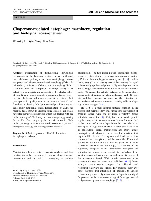
REVIEWChaperone-mediated autophagy:machinery,regulation and biological consequencesWenming Li •Qian Yang •Zixu MaoReceived:12July 2010/Revised:7October 2010/Accepted:8October 2010/Published online:26October 2010ÓSpringer Basel AG 2010Abstract Degradation of dysfunctional intracellular components in the lysosome system can occur through three different pathways,i.e.,macroautophagy,micro-autophagy and chaperone-mediated autophagy (CMA).In this review,we focus on CMA,a type of autophagy distinct from the other two autophagic pathways owing to its selectivity,saturability and competitivity by which a subset of long-lived cytosolic soluble proteins are directly deliv-ered into the lysosomal lumen via specific receptors.CMA participates in quality control to maintain normal cell functions by clearing ‘‘old’’proteins and provides energy to cells under nutritional stress.Deregulation of CMA has recently been shown to underlie some diseases,especially neurodegenerative disorders for which the decline with age in the activity of CMA may become a major aggravating factor.Therefore,targeting aberrant alteration in CMA under pathological conditions could serve as a potential therapeutic strategy for treating related diseases.Keywords CMA ÁLysosome ÁHsc70ÁLamp2a ÁAutophagy ÁUnibiquitinIntroductionMaintaining a balance between protein synthesis and deg-radation is absolutely essential for proper cellular function,homeostasis and survival in a changing extracellularenvironment.The two major protein degradation mecha-nisms in eukaryotes are the ubiquitin-proteasome system (UPS)and the autophagy-lysosome system [1,2].Collec-tively,they (1)exert quality control by clearing damaged or incorrectly synthesized proteins,(2)recycle proteins that are no longer needed into constitutive amino acid compo-nents,(3)mount the cellular defense by breaking down components of various invading pathogens,and (4)regu-late cellular response to stress or the alteration in extracellular micro-environments,assisting cells in adapt-ing to new changes [3,4].The UPS is a multi-subunit protease complex in the cytosol that permits entry and subsequent degradation of proteins tagged with one or more covalently bound ubiquitin molecules [5].Ubiquitin is a small protein highly conserved from yeast to man.It was first described in the context of protein degradation,but later shown to participate in regulation of other cellular processes,such as endocytosis,signal transduction and DNA repair.Conjugation of ubiquitin is a complex reaction that requires E1,E2and E3enzymes,and leads to the for-mation of an isopeptide bond between the C-terminal glycine of ubiquitin and the 3-amino group of a lysine residue of the substrate protein [6,7].Subunits of the regulatory complex of the proteasome recognize the ubiquitin tag,remove it and mediate the unfolding of the substrates required to gain access to the catalytic region of the proteasome barrel.With certain exceptions,most proteasome substrates have short half-lives [6,8].Inter-estingly,recent studies suggest that ubiquitin and lysosomal pathways are linked.For example,new evi-dence suggests that attachment of ubiquitin to various cellular cargos not only constitutes a degradation signal for proteasome,but also serves to signal for cargo removal by lysosomal system via autophagy [2,9].W.Li ÁQ.Yang ÁZ.Mao (&)Departments of Pharmacology and Neurology,Emory University School of Medicine,Atlanta,GA 30322,USAe-mail:zmao@Cell.Mol.Life Sci.(2011)68:749–763DOI 10.1007/s00018-010-0565-6Cellular and Molecular Life SciencesAutophagy is a conserved cellular ‘‘self-eating’’process that involves sequestration and delivery of cytosolic components to the lysosome for degradation and recycling [10].This evolutionarily conserved process can be cate-gorized into three classes depending on their respective sequestration and delivery mechanism (Fig.1)[11].In macroautophagy,a double-membrane vesicle termed the autophagosome is formed to engulf long-lived proteins and organelles.The autophagosome is subsequently fused with a lysosome,releasing its cargo for degradation by lyso-somal hydrolases.The resultant nucleotides,amino acids and fatty acids are eventually recycled back into the cytosol for reuse.In microautophagy,the sequestration of cytosolic content is facilitated by direct invagination or exvagination of lysosomal membrane,and subsequent budding of the invaginated vesicles into the lysosomal lumen releases the sequestered cytosolic material.In con-trast to the vesicle-mediated substrate delivery of macro and microautophagy,chaperone-mediated autophagy (CMA)targets and delivers substrate proteins directly across the lysosomal membrane via the specific receptor [9,12].Only proteins containing a consensus peptide sequence are recognized by a chaperone complex.This CMA sub-strate-chaperone complex locates to the lysosome through interaction with the lysosome receptor and translocates the substrate across the lysosomal membrane with the assis-tance of lysosomal chaperones on the lumenal side [12,13].In this review,we first describe the machinery of CMA,summarize the regulation mechanisms involved in activation of CMA,and finally discuss the biological consequences of CMA under various physiological and pathological conditions,and relevant therapeutic strategies via regulating CMA.History of CMAIn the early 1980s,Professor Dice’s group first reported that radiolabeled RNase A introduced into the cytoplasm of human fibroblasts by using erythrocyte-mediated microin-jection or osmotic lysis of pinosomes was degraded with a half-life of approximately 90h in the presence of serum,whereas in response to serum deprivation its rate of deg-radation was enhanced 1.6-fold [14].This enhanced breakdown following serum withdrawal was highly selec-tive and based on a feature present within the N-terminal 20amino acids of RNase A [15].During subsequent years,the essential motifs related to KFERQ were identified in proteins serving as substrates of this selective degradation pathway,which is referred to as the selective pathway for degradation of cytosolic proteins by lysosomes [16,17].In 1989,a 73-kDa heat shock cognate protein (Hsc73,now commonly referred toas Hsc70)was found to bind to KFERQ-like regions in intracellular proteins that are tar-geted for lysosomal degradation in response to serum deprivation [18].In 1994,isolated rat liver lysosomes were used to probe the selective binding and uptake of RNase A and glyceraldehyde-3-phosphate dehydrogenase.Their uptake and degradation by lysosomes were progressively activated in rat liver by starvation [19].In 1996,Lamp2a,also named Lgp96(lysosomal glycoprotein of 96kDa),was identified as a receptor for the selective import and degradation of proteins within lysosomes [20].In 1997,an intralysosomal Hsc73was determined to be required for the selective pathway of lysosome-mediated protein deg-radation [21].Since 2000,this pathway has been formally named chaperone-mediated autophagy (CMA)[22,23].Throughout the subsequent decade,new substrates,moreFig.1Autophagy refers to the conserved degradation of intracellular components by lysosomes.In mammals,three types of autophagy have been described:macroautophagy,microautophagy and CMA.Adapted from [11]with permission750W.Li et al.detailed machinery,new components,physiological roles and associated diseases for CMA have been extensively investigated and elucidated [3].Machinery of CMACMA uses the unique and distinctive machinery from the other two autophagy pathways to carry out this process [3,24].The basic machinery consists of at least three types of proteins,including (1)chaperone proteins,which are responsible for recognizing substrates based on their spe-cific motifs and delivering them to lysosomes,(2)receptor proteins,which bind and transport/pull substrates into lysosome lumens,and (3)substrate proteins,which are a subset of soluble cytosolic proteins containing specific motifs related to KFERQ.Following activation of CMA,these three subsets of proteins collaborate and complete the process (Fig.2)[3,25].Workshop—lysosomeLysosomes are the primary catabolic compartment of eukaryotic cells.The name lysosome derives from the Greek words lysis,which means dissolution or destruction,and soma,which means body [24].Lysosomes were dis-covered by the Belgian cytologist Christian de Duve in 1949[26].They are created by the addition of hydrolyticenzymes to early endosomes from the Golgi apparatus [24].The membrane around a lysosome allows the digestive enzymes to work at pH 5.1–5.5,which is optimal to these acidic hydrolases.Lysosomes fuse with vacuoles and dis-pense their enzymes within digesting the contents [27].A healthy cell is dependent on the proper targeting of newly synthesized lysosomal proteins.Two classes of proteins are essential for the function of lysosomes:soluble lysosomal hydrolases (i.e.,acid hydrolases)and integral lysosomal membrane proteins (LMPs)[24].Lysosomal acidic pH and the large variety of hydrolases present in the lysosomal lumen (including proteases,lipases,glycosi-dases and nucleases)confer upon this organelle its high capacity of degradation and mediate complete breakdown of all types of molecules.In addition to bulk degradation,lysosomal hydrolases are involved in antigen processing,degradation of the extracellular matrix and initiation of apoptosis [24,28].LMPs reside mainly in the lysosomal limiting membrane and have diverse functions,including acidification of the lysosomal lumen,protein import from the cytosol,membrane fusion and transport of degradation products to the cytoplasm [29].The main LMPs are lyso-some-associated membrane protein 1and 2(Lamp1and 2),lysosomal integral membrane protein 2and tetraspanin CD63[24].Substrates can reach lysosomes via heteroph-agy,in which the cargo to be degraded originates at the plasma membrane or extracellularly,or via autophagy,for cargo located in the cytosol [3,24,30].Fig.2The diagram ofproposed mechanisms of CMA:a Hsc70with co-chaperones recognizes a KFERQ-related peptide in cytosolic substrate proteins;b the complex binds to the Lamp2a receptor on the lysosomal membrane;c the substrate protein is unfolded before traversing the lysosomal membrane;d lys-Hsc70pulls the substrate into the lysosome matrix;e the substrate protein is degraded by lysosomal proteases;f the Hsc70-cochaperone complex is released from the lysosomal membrane;g Hsc70is available to bind to another CMA substrate.Adapted from [25]with permissionBasics of chaperone-mediated autophagy 751Chaperones—Hsc70and partnersHsc70,a73-kDa protein,is the constitutive member of the heat shock protein70family of chaperone[19,25].Hsc70s have been found to be involved in many cellular processes including dissociation of clathrin and assembly proteins [25,31].Binding of Hsc70to substrate proteins is regu-lated by ATP binding and hydrolysis,and the ADP-bound form of Hsc70has the highest affinity for protein substrates for CMA[19,32].Hsc70is located in the cytosol or in the lumen of lysosomes.Cytosolic Hsc70(cyt-Hsc70)can recognize a peptide sequence including the KFERQ motif in CMA substrate proteins and aid in their transport to the lysosomal receptor[3,19].Cyt-Hsc70docked to the lysosomal membrane helps to unfold substrate proteins,a necessary step for their entry into lysosomes[33].Other co-chaperones interact with Hsc70and regulate its activities.Hsp40may activate the ATPase activity of Hsc70to facilitate substrate binding;and the Hsp70-interacting protein(Hip)stimulates the assembly of Hsc70 with Hsp40and the protein substrate[25].Cell division cycle48(Cdc48)can also enhance the activity of Hsc70-Hsp40complexes[32,34].Hsc70-Hsp90organizing pro-tein acts as an adapter between Hsc70and Hsp90,which recognize unfolded regions within proteins and prevent substrate protein aggregation[25,32].There are co-chap-erones of Hsp90that may be in the molecular chaperone complex.Activator of Hsp90ATPase is a family of heat shock proteins that activates the ATPase activity of Hsp90 and thereby stimulates both protein binding and release [32,35].The Bcl2-associated athanogene1protein(Bag-1) was initially described as a co-chaperone of Hsc70that uncouples the ATPase cycle from substrate binding[36]. But several subsequent studies showed that it functions as a nucleotide exchange factor that stimulates substrate release [37].The carboxyl terminus of Hsc70-interacting protein (Chip),which can regulate protein refolding[38],acts as a chaperone-associated ubiquitin ligase to stim-ulate the degradation of Hsc70client proteins[39].The chaperone complex present on the cytosolic face of lyso-somal membranes is linked to Lamp2a by the substrate protein via a site that is different from sites of interaction for the molecular chaperone complex.Each of the com-ponents in the molecular chaperone complex is required for transport of substrates into the lysosome lumen[25,32].Both Hsc70and Hsp90have been found to be also present in the lysosomal lumen.Lysosomal-Hsc70(lys-Hsc70)is required for the CMA pathway[21].The more active population of lysosomes contains abundant lys-Hsc70,whereas the less active ones contain little Hsc70. The latter group of lysosomes can be made more active for CMA if they are allowed to take up Hsc70.A role for lys-Hsc70in the uptake of substrate proteins has been demonstrated in cultured confluent humanfibroblasts.It has been speculated that lys-Hsc70may be required to pull proteins into the lysosomal lumen because of analogous roles of Hsp70s in the translocation of proteins into the endoplasmic reticulum,mitochondria and chloroplasts [21,25].Nearly half of the lysosomal Hsp90s associate with the lumenal side of the lysosomal membrane where this chaperone may contribute to the stabilization of essential components of the translocation complex when it is organized into a multimeric structure[3,40].Receptor—Lamp2aIn1996,a lysosomal membrane glycoprotein Lgp96 (called Lamp2a in human)was identified as a receptor for binding and uptake of lysosome substrates[20].Lamp2a is one of the three splice variants of the Lamp2gene that gives rise to three single-span membrane proteins, Lamp2a,b and c.These variants all have a common highly N-glycosylated lumenal region,but possess different transmembrane and C-terminal cytosolic tail regions. Lamp2a has a short cytosolic tail(GLKRHHTGYEQF)to which CMA substrate proteins bind[25].The positively charged residues in the Lamp2a cytosolic tail are important for binding of substrate proteins.However,the specific amino acids on the CMA substrate proteins required for receptor binding remain elusive[41].Binding of substrate proteins to Lamp2a is a rate-limiting step for the CMA process,and overexpression of Lamp2a in Chinese hamster ovary cells can increase CMA mp2a is not limited to acting as a classic receptor.Increasing evidence shows that Lamp2a is involved in many other aspects of the CMA process such as substrate translocation[3,25,40]. Lamp2deficiency in mice causes extensive accumulation of autophagic vacuoles in many tissues[42].Contrary to the proposed receptor function of Lamp2a in CMA,con-fluent mouse embryonicfibroblasts deficient in lamp2 appear to have normal levels of CMA-associated lysosomal proteolysis after prolonged serum withdrawal,suggesting that Lamp2a may not be the only receptor for CMA [29,43].Substrates—KFERQ motifSubstrates for CMA are defined by an amino acid sequence motif related to KFERQ[3,16].This pentapeptide target-ing motif wasfirst identified in microinjected RNase A[44].It consists of a glutamine(Q)preceded or followed bya combination of four amino acids that are basic(R,K), acidic(D,E),or bulky and hydrophobic(F,I,L,V)resi-dues.In some cases,Q may be substituted by the related N [45].Antibodies raised against KFERQ can immunopre-cipitate30%of cytosolic proteins in mammalian cells[46].752W.Li et al.Without denaturation,more than80%of cytosolic pro-teins that contain KFERQ motifs can be recognized by the antibody to this motif,indicating that most KFERQ motifs in these proteins are exposed.However,certain proteins such as aldolase B have hidden KFERQ motifs due to multimeric formation.The may dissociate the aldolase tetramer and result in the exposure of KFERQ motif[25,47].In addition,certain monomers may have hidden KFERQ sequences that may be exposed following partial unfolding.Therefore,the presence of a KFERQ sequence alone in the primary structure of a protein is not sufficient for determining them as substrates of CMA[48]. Such identification requires rigorous experimental proof. Recently,we have identified MEF2D,a protein known to promote neuronal survival as a CMA substrate.Interest-ingly,MEF2D uses a set of over-lapping imperfect KFERQ motifs to mediate its interaction with Hsc70[49]. Characteristics of CMACompared to macro-and microautophagy,which occurs in a wide range of eukaryotes including mammals,plants and fungi,CMA has only been described in mammals.This process has some distinctive features.SelectivityLysosome-mediated degradation had traditionally been perceived as a process performed in bulk with poor selectivity.The initial idea of selective autophagy origi-nated from the observation that starvation in animals or serum removal in cultured cells accelerates the degradation of particular cytosolic proteins in lysosomes but not others[50].Selectivity thus became one of the hallmarks of CMA[51].CMA mediates selective targeting of non-essential proteins for degradation to obtain the amino acids required for the synthesis of essential proteins.The intrinsic selec-tivity of CMA is also well suited for the removal of specific proteins damaged during stress without interfering with nearby normally functioning forms of the same protein[51, 52].This selectivity is achieved by making the KFERQ motifs in the altered protein accessible to the chaperone but inaccessible when it is properly folded or pare to CMA,macroautophagy has traditionally been consid-ered as a nonselective degradation process.However, recent evidence suggests that this view needs modification. New experimental data demonstrate that some macro-autophagic processes,termed chaperone-assisted selective autophagy,are assisted by chaperone proteins and can be selective in targeting protein complexes,organelles and microbes[2].In this process,macroautophagy has been proposed to be initiated by a selective ubiquitylation of cellular targets and followed by recognition via autophagic ubiquitin adaptors such as p62,NBR1and HDAC6.These molecules can mediate docking of ubiquitinated proteins or damaged organelles to autophagosomes and lysosomes, thereby ensuring their selective degradation[2,53,54].SaturabilityThe unusual characteristics of CMA are not limited to its selectivity.As the mechanism for cargo delivery was revealed,it became evident that,in contrast to the other forms of autophagy,vesicle formation was not required in CMA.Instead,the substrate proteins were translocated across the lysosomal membrane[55].The receptor Lamp2a is mainly responsible for this translocation.Because of the requirement for receptor binding before translocation can occur,this delivery process becomes saturable.In contrast to Hsc70,which is often in excess in the cytosol,levels of Lamp2a are limiting for CMA and hence subjected to tight regulation[23].CompetitivityThe selectivity and saturability of CMA directly lead to the competitive binding of CMA substrates.During the process of CMA,different substrates compete for binding Hsc70 and limited pool of Lamp2a[23,56].Several studies have shown that degradation of some substrates is slowed down by overexpression of other substrates.This has been proposed to underlie the pathogenesis of some diseases [49,57].For example,both the wild-type a-synuclein and neuronal transcription factor MEF2D are substrates of CMA.Elevated levels of wild-type a-synuclein may reduce the degradation of MEF2D via CMA[49].Regulation of CMAThe signal transduction pathways involved in the regula-tion of CMA from cell membranes to cytosolic chaperone proteins and lysosomes remain largely elusive[3].The p38 MAPK inhibitor can partly prevent the activation of CMA, implicating this pathway in CMA[45].On the other hand, the extensively investigated local regulation of CMA activity in the lysosomes has precise,fine-tuned mecha-nisms[3,25].Regulation of CMA via Lamp2aThe level of Lamp2a at the lysosomal membrane is pro-portional to the activity of CMA.Therefore,changes in the Lamp2a level at the lysosomal membrane can quickly regulate the activity of CMA.The level of Lamp2a at theBasics of chaperone-mediated autophagy753lysosomal membrane may be changed by synthesis,deg-radation and redistribution[41,58].De novo synthesis of Lamp2a or its by protein synthesis inhibitors can directly change the level of Lamp2a in cells,but the physiological or pathological relevance remains unclear [3,59].The distribution of Lamp2a at the lysosomal membrane is regulated by its dynamic association with discrete membrane lipid microdomains[60].Under basal conditions,sequestration of Lamp2a in cholesterol-enri-ched regions favors its cleavage by two proteases, including an unidentified metalloprotease at the membrane and a serine protease—cathepsin A,which associates dynamically with the lumenal side of the lysosomal mem-brane[60,61].Exclusion of Lamp2a from these regions allows its multimerization(see below),a step required for the uptake of CMA substrate by lysosomes.Interestingly, the degradation of Lamp2a can be reduced by the presence of substrates or stimuli that induce the activation of CMA. This blockage in degradation,rather than the increase of synthesis,to increase Lamp2a at the lysosomal membrane is particularly advantageous to cells with limited access to amino acids such as during nutrient deficit[3,62].The levels of Lamp2a can be further increased at the lysosomal membrane through the mobilization of the pool normally resident in the lysosomal lumen.The exact nature of this luminal pool of Lamp2a remains unclear,but intact molecules of this protein exist inside lysosomes,and a gradual decrease in the percentage of Lamp2in this com-partment occurs as activation of CMA persists beyond1 day[3,60].Membrane chaperones and an intact membrane potential are needed for the mobilization of Lamp2a from the lumen to membrane[3].Fractionation studies have shown that luminal Lamp2a associates with lipid,indicat-ing the possible existence of luminal Lamp2a-containing micelles.It is thought that these micelles fuse or integrate into the lysosomal membrane under specific stress,result-ing in the incorporation of Lamp2a in the membrane and exposure of its C terminus to the cytosol[60,63].Lamp2a can undergo cycles of rapid assembly into a 700-kDa protein complex at the lysosomal membrane. Monomers of Lamp2a at the lysosomal membrane can accept substrate proteins,and this interaction drives the organization of Lamp2a into multimeric complexes needed for substrate translocation into lysosomes.Once the sub-strate protein reaches the lysosomal lumen,Lamp2a will disassemble from the multimeric complex to enable sub-sequent rounds of substrate binding[3,40].This continuous assembly and disassembly of Lamp2a from the multimeric translocation complex highlights the impor-tance of the lateral mobility of this protein in the lysosomal membrane[40].Chaperone proteins located at both sides of the lysosomal membrane may regulate the lateral mobility of Lamp2a.Lys-Hsc70induces disassembly of Lamp2a from the700-kDa complex once the substrate has crossed the membrane.A lysosome-associated form of the glial fibrillary acidic protein(GFAP),a component of the intermediatefilament network,associates to Lamp2a once it is organized into multimers and contributes to stabilizing the CMA translocation complex against the disassembling activity of Hsc70,whereas GTP-mediated release of elongation factor-1a from the lysosomal membrane pro-motes self-association of GFAP,disassembly of the CMA translocation complex and the consequent decrease in CMA[12,64].In addition,Hsp90at the lumenal side is also required to preserve the stability of Lamp2a during the transition[40].Regulation of CMA via Hsc70The other limiting lysosomal component is lys-Hsc70.As indicated in the previous section,the presence of this chaperone at the luminal side of the membrane is necessary for substrate translocation[18].Levels of lys-Hsc70 increase gradually with increasing CMA activity,although the mechanisms modulating this increase are still poorly understood.Although Hsc70contains two KFERQ sequences and is a putative substrate of CMA[21,25],it appears that neither CMA nor macroautophagy is involved in delivering lys-Hsc70to lysosomes[3,65].It is possible that this chaperone reaches lysosomes through maturation of late endosomes,a compartment in which high levels of luminal Hsc70have been detected,thus highlighting a possible relationship between CMA and endocytosis [3,65,66].Regulation of CMA via macroautophagyThe activity of CMA is also directly modulated by changes in other autophagic and proteolytic systems inside the cell. Cells in culture respond to CMA blockage by upregulating macroautophagy.Similarly,blockage of macroautophagy results in constitutive activation of CMA[65].These pathways are clearly not redundant,as CMA is,for example,unable to degrade organelles normally turned over by macroautophagy,whereas macroautophagy lacks the selectivity of CMA in the degradation of individual soluble cytosolic proteins[67].Nevertheless,the compen-satory activation of one form of autophagy when the other is compromised allows cells to preserve homeostasis,at least under basal conditions.Additionally,blockage of either form of autophagy also has a direct impact on pro-teasomal activity[3].The molecular mechanisms that regulate crosstalk between these two different pathways are currently under investigation.In the case of the interrela-tionship between macroautophagy and CMA,continuous fusion of autophagosomes to lysosomes when754W.Li et al.macroautophagy is upregulated results in transient dissi-pation of the lumenal lysosomal pH,which negatively affects lys-Hsc70stability.In fact,although lys-Hsc70is normally stable at pH ranges of5.2–5.4,changes in pH values above5.6in the lysosomal lumen result in its rapid degradation in this compartment.The reduced levels of lys-Hsc70in those lysosomes decrease their capability to perform CMA[68].Regulation of CMA via UPSUPS and autophagy were long viewed as independent, parallel degradation systems with no point of intersection. Increasing evidence shows that the UPS and autophagy are functionally interrelated catabolic processes.Specifi-cally,these degradation systems share certain substrates and regulatory molecules,and show coordinated and,in some contexts,compensatory function.For example,the neuronal protein a-synuclein can be degraded by the UPS,macroautophagy and CMA.Under conditions in which the UPS is compromised,enhanced degradation by CMA and macroautophagy may become critical to maintaining pools of amino acids for protein synthesis and may protect against the accumulation of a toxic species[1,3].On the other hand,during the acute stages of CMA blockage,there is an accumulation of poly-ubiquitinated proteins,often in the form of protein aggregates,attributable to the observed reduction in their removal through the proteasome system,and the under-lying mechanisms remain to be clarified[69]. Interestingly,recent evidence suggests that ubiquitin may play a key role in the crosstalk between proteasome-mediated degradation and selective autophagy[2,70]. Furthermore,ubiquilin,a ubiquitin-like protein,functions to regulate macroautophagy by facilitating maturation of LC3protein,and is also a substrate of CMA,indicating that ubiquilin may also be at a crossroad between protein degradation pathways[71,72].However,detailed mechanisms are under investigation.Physiological relevanceSelective degradation of cytosolic proteins via CMA con-tributes to both quality control(housekeeping)and response to stress[2,3].CMA was initially identified as an inducible pathway in response to stress.However, increasing evidence shows that there is a certain level of CMA activity under basal conditions.Most cell types analyzed to date display some level of continuous CMA activity detectable in the absence of typical CMA-inducing conditions.Basal CMA requires participation of the same effectors at the lysosomal membrane—the membrane chaperones and the protein translocation complex.It has been speculated that there is a difference between basal and inducible CMA.Whether the regulation of basal and inducible CMA occurs through different signaling mecha-nisms is under investigation[65].However,both basal and inducible CMA may have physiological relevance.Recycling and quality controlThe delivery of intracellular substrates such as misfolded proteins and damaged organelles from the cytosol to the lysosomes for degradation is crucial for cell survival. Under physiological conditions,renewal of cytosolic pro-teins is needed to maintain their normal function through recycling their old versions[4].Besides bulk autophagy (microautophagy and macroautophagy),CMA efficiently facilitates the transit of specific proteins from the cyto-plasm to lysosomes and is responsible for their recycling.If this process is inhibited,excessive old and dysfunctional proteins will accumulate in the cytosol and disturb the physiological functions of cells[3,49].Immune responseDurable adaptive immunity is dependent on CD4?T cell recognition of the major histocompatibility complex (MHC)class II molecules that display peptides from exogenous and endogenous antigens.Specialized antigen-presenting cells use the endosomal/lysosomal systems to internalize exogenous antigens,which can then be pre-sented on MHC to CD4?T cells.Beside the proteasome and macroautophagy in processing and MHC loading with endogenous and exogenous antigens,CMA has also been recently investigated in antigen processing/presentation. Cells with reduced levels of Lamp2a or Hsc70exhibit decreased presentation of cytoplasmic epitopes on class II molecules[73].Conversely,an increase in cytoplasmic autoantigen presentation is observed upon overexpression of either Lamp2a or Hsc70.Furthermore,there is cross-talk between autophagy pathways in the expression of MHC class II molecules.Macroautophagy can deliver antigens into autophagosomes for processing by acidic proteases before MHC class II presentation.However,other endog-enous antigens are processed by cytoplasmic proteases, yielding fragments that translocate via CMA into the endosomal network to intersect MHC class II.This cross-talk,particularly in response to stress,appears to balance the relative efficiency of each pathway,limits redundancy and gives MHC class II broader access to antigens within different intracellular compartments.Whether alteration in CMA activity such as its reduction with age could con-tribute to the altered immune response requires further investigation[74].Basics of chaperone-mediated autophagy755。

84猪业科学 SWINE INDUSTRY SCIENCE 2013年 第12期猪轮状病毒属于呼肠孤病毒科轮状病毒属,是引起多种新生动物和幼龄动物腹泻的重要肠道病原之一,也是感染1~5日龄仔猪或断奶仔猪的常见病原。
以厌食、呕吐、腹泻和脱水、体重减轻为特征,引起仔猪的死亡,成年猪掉膘,饲料报酬降低,增加人工费和药费的开支等。
若与传染性胃肠炎和大肠杆菌混和感染则病情加重,死亡率增高。
1 猪轮状病毒形态成熟完整轮状病毒粒子略呈圆形,没有囊膜,为65~75 nm 的二十面体对称。
电镜观察,病毒的中央为核酸构成的芯髓,呈致密的六角形棱心,直径为37~40 nm。
轮状病毒粒子由3层衣壳组成:内衣壳(VP2)、中间层衣壳(VP6)和外层衣壳(VP4和VP7)。
中间层衣壳(VP6)有32个呈放射状排列的圆柱形壳粒组成,壳粒由此向外呈辐射状排列。
外衣壳厚约20 nm,为连接于壳粒末端的光滑薄膜状结构,使该病毒形成车轮状外观,轮状病毒为此而得名(见图1、图2)。
在感染的粪样和细胞培养物中存在2种形式的病毒粒子,只有3层衣壳的光滑型(S 型)粒子,具有感染性[1],直径为75 nm。
外层衣壳被除去,就会导致感染性丧失,表明钙离子通过使外层衣壳VP7稳定,对轮状病毒粒子的稳定性起着关键作用[2]。
不同类型的病毒,稳定外层衣壳所用钙的浓度不一样[3]。
没有外层衣壳的双层粒子(R 型)是很小的,边缘粗糙,直径为65 nm。
病毒核酸基因由11 个不连续的 猪轮状病毒病原学研究进展王振玲1,李显和2(1.北京农业职业学院,北京 房山 102442;2.北京市大兴区魏善庄镇动物防疫畜牧水产技术推广站,北京 大兴 102611)摘 要:轮状病毒不仅感染猪、牛、羊、禽等动物,同时还感染人。
轮状病毒的持续存在,不仅严重威胁养殖业的发展,同时威胁到肉食品和人类的安全,成为世界科学家们研究的焦点。
对轮状病毒形态、分类、理化性质和生物学特性、致病机理等病原学等内容进行了综述。

热休克蛋白70与心肌保护研究现状(作者:___________单位: ___________邮编: ___________)【关键词】热休克蛋白质70 心肌缺血心肌再灌注损伤/治疗热休克蛋白(heat shock protein, HSP)是指细胞在应激原特别是环境高温诱导下所产生的,由热休克基因所编码合成的,序列高度保守的一组蛋白质。
近年来研究发现,以热、缺血缺氧为应激原或通过基因调控的方法诱导心肌细胞产生热休克蛋白,可使心肌具有抗热、抗缺血缺氧的保护作用。
热休克蛋白按其分子量不同分为多个家族,其中,热休克蛋白70(HSP70)备受重视,已成为新近较重视的内源性心肌保护途径之一,在对抗心肌缺血再灌注损伤中发挥着极为重要的作用。
综合近几年的研究热点,现就其特性及其在心肌保护方面的研究现状进行综述。
1 HSP的一般概况1962年Ritossa[1]首次从果蝇在热应激环境中诱导合成而命名,随后的研究发现,除高温外,缺血、低氧、重金属盐及大多数病理状态下都能诱导细胞产生热休克蛋白,因此又称应激蛋白,但习惯上仍沿用热休克蛋白这一名称。
根据分子量大小和同源程度可分为HSP110,HSP90,HSP70,HSP60及小分子量HSP等几个家族,每个家族又有多个成员。
HSP70是HSP家族中最保守、最主要、含量最丰富,也是近年来研究备受关注的一类。
HSP70家族(分子量:72~80)成员最多,共有21种蛋白质,是一组进化上高度保守的应激蛋白。
按表达情况可分为结构型HSP70和诱导型HSP70,生理情况下,正常细胞可表达结构型HSP70,应激情况下略有增加。
结构型HSP70又可分为两种,即葡萄糖调节蛋白78(glucose-regulated protein, GRP78),这种蛋白存在于内质网腔内;另一种是GRP75,主要存在于线粒体内。
诱导型HSP70即HSP72,仅出现于应激细胞,通常在正常细胞中并不表达或表达量甚少,但是在热应激或其它应激原的作用下则表达迅速增加[2]。
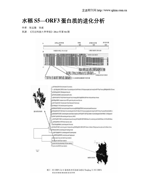
水稻S5—ORF3蛋白质的进化分析作者:张达瀚张屹来源:《河北科技大学学报》2014年第04期摘要:生殖隔离在形成新物种和物种同一性的保持中有重要作用,因而在水稻优化育种中有重要的意义。
S5位点是一个已克隆的控制水稻生殖隔离的基因,它产生的S5-ORF3,ORF4和ORF5蛋白质共同调控着indica-japonica杂交后代的育性,特别是S5-ORF3蛋白质在打破水稻亚种indica-japonica之间的生殖隔离与促进物种间的基因交流中发挥重要作用。
针对S5-ORF3的进化分析对于研究它的功能和起源很重要,但目前尚无报道。
本文基于序列和进化分析,提出S5-ORF3为一种定位在内质网中的新的HSP70家族蛋白,但缺乏HSP70的C端的多肽结合结构域,推测S5-ORF3可能通过影响alpha-淀粉酶的合成来作用于内质网压力;找到了ORF3的19条同源蛋白,并用极大似然算法构建了可靠的进化树,发现水稻的S5-ORF3与节节麦的luminal-binding蛋白进化关系最为密切;作了置信度100%的S5-ORF3蛋白质的三维结构预测,预测了居中的配体绑定位点;发现了水稻的S5-ORF3与其他HSP70蛋白相比独特的基序,为进一步研究S5-ORF3的作用机理和演化历史提供了线索和数据支持。
关键词:水稻;S5-ORF3 蛋白;进化树;正向选择;配体结合中图分类号:Q816 文献标志码:A生殖隔离限制了物种间的基因交流,一般来说,水稻的2个亚种indica和 japonica间产生的后代是不育的[1-2]。
研究发现,在水稻基因组的S5位点有一个与生殖隔离相关的“破坏与保护蛋白系统”,该系统由3个紧密连锁的基因ORF3,ORF4,ORF5组成,这三者共同调控着indica-japonica杂交后代的育性,ORF4+与ORF5+导致了内质网的压力,这种压力导致杂交水稻雌配子败育,而ORF3+作为保护者又会阻止内质网压力的产生,从而抑制雌配子败育并打破生殖隔离,使indica和japonica之间的基因可以交流[3]。

收稿日期:1999-10-20;修订日期:2000-12-30基金项目: 国家自然科学研究基金资助(39770824)作者简介:范云霞(1964-),女(汉族),河南平顶山市,中国协和医科大学生物化学硕士研究生审校者:中国协和医科大学肿瘤研究所 黄常志026 热休克蛋白与免疫应答范云霞(中国协和医科大学肿瘤研究所,北京 100021))摘要:近年研究发现,热休克蛋白参与免疫应答过程,在抗原受体成熟、抗原加工、呈递等多方面起作用,并且这种作用具有潜在的临床应用前景,本文综述这一领域的研究现状。
关键词:热休克蛋白;免疫应答;抗原受体;抗原加工呈递文章编号:1001-103X(2001)02-0062-03 中图分类-号:R392 11 文献标识码:A 热休克蛋白(Heat Shock Protein,HSP)是一组具有重要生理功能,高度保守的蛋白质分子家族。
生理、病理及环境因素等都可诱导热休克蛋白产生,故又称为应激蛋白(Stress Protein)。
根据分子量大小和同源程度可分为HSP110、HSP90、HSP70、HSP60、小分子HSP 及泛素等几个家族。
热休克蛋白的生物学功能广泛,不仅表现在应激条件下维持细胞必需的蛋白质空间构象,保护细胞生命活动,以确保细胞生存,而且在蛋白质折叠、跨膜运输、转位、细胞骨架及核骨架稳定等基本功能方面发挥重要作用,调节这些蛋白的活性和功能。
而自身并不参与大分子蛋白组成,故称为 分子伴侣 。
本文主要综述HSP70、HSP90、泛素等在抗原受体装配、抗原加工与呈递等方面的作用。
1 HSP70、HSP90、泛素组成和结构HSP70家族成员广泛存在细胞的各个亚细胞结构、胞浆及胞核中,如:组成性表达HSC70(Heat Shock Cognate Protein,HSC70),热诱导表达HSP70,定位于内体和质膜PBP72/74(Peptide Binding Protein,PBP),线粒体基质中GRP75(Glucose Regulated Protein,GRP),内质网腔丰富存在BiP(Immunoglobulin hea vy chain binding Protein,BiP)。
HSP70的研究进展及其在生物医学中的应用作者:刘嘉敏赵元莙来源:《教育教学论坛》2017年第50期摘要:HSP70是热休克蛋白家族的重要组成成员,均属机体内的一类应激蛋白,具有保护细胞和生命体的重要作用。
本文从HSP70家族各成员的研究概况、生物学功能及其在生物医学方面的应用予以简要综述,以期为中小学的素质教育提供生物医学方面的普及知识。
关键词:热休克蛋白;分类;功能;生物医学;应用中图分类号:G642.0 文献标志码:A 文章编号:1674-9324(2017)50-0061-02热休克蛋白(HSPs)是指在机体遇高温刺激及其他一些环境压力,如营养缺乏、氧压、重金属、强紫外线照射及细菌等生物污染有害刺激后,诱导机体所产生的高度保守的、可帮助细胞耐受这些环境压力并对机体起着重要防御作用的一组蛋白质。
依据分子量大小,热休克蛋白可分为HSP100、HSP90、HSP70、HSP60、HSP40、小HSP家族和泛素等家族。
目前,热休克蛋白70(HSP70)是热休克家族(HSPs)中研究最多的应激蛋白,该类蛋白在原核生物和真核生物中广泛存在。
一、HSP70的分类及功能在生物细胞中,HSP70含量丰富且分布广泛,它们不仅分布于细胞质中,有些还存在于细胞器中。
HSP70 家族主要包括四类蛋白质:(1)诱导型 HSP70(也称HSP72),存在于细胞核中,在正常细胞内表达量较少,细胞发生应激后,表达量迅速增加,与ATP具有高亲和力。
(2)结构型HSC70(Heat shock cognate 70,也称HSP 73),存在于细胞浆中,在所有的细胞内组成性表达,是哺乳动物细胞内的结构蛋白,外界应激原刺激后只有少量增加,与ATP 也具有高亲和力。
(3)葡萄糖调节蛋白78(GRP78)(也称HSPA5)位于内质网上,应激条件下稍有表达,参与多种细胞生命过程。
(4)葡萄糖调节蛋白75(HSP75)(也称HSPA9),主要位于线粒体内,其基因能够在多种组织中表达,且表达量和细胞自身的能量代谢有关。
收稿日期:2013-11-05基金项目:国家自然科学基金项目(81041088) *通信作者:E-mail: hchen@ 细胞内吞的研究进展范真真,陈虹*,黄秉仁(中国医学科学院&北京协和医学院基础医学研究所医学分子生物学国家重点实验室,北京 100005)摘要:细胞外基质的各种分子经细胞膜进入真核细胞是一个复杂的过程。
细胞内吞是通过细胞质膜的变形运动将细胞外物质转运入细胞内的过程。
不同的细胞内吞途径需要不同的蛋白质分子参与,引起不同的信号转导通路。
目前认为细胞内吞和膜转运是细胞对其信号转导过程的一种精密的组织安排,细胞内吞在细胞信号转导,维持机体动态平衡方面起着重要作用。
细胞内吞途径通常可以分为网格蛋白依赖的内吞和非网格蛋白依赖的内吞,其中后者包括陷窝蛋白依赖和非陷窝蛋白依赖的内吞,以及巨胞饮介导的内吞。
本文将就这几种主要细胞内吞途径及与细胞信号转导通路关系的研究进展予以介绍。
关键词:细胞内吞;网格蛋白;陷窝蛋白;发动蛋白;巨胞饮Research progress of cell endocytosisFAN Zhenzhen, CHEN Hong*, HUANG Bingren(The National laboratory of Medical Molecular Biology, Department of Biochemistry & Molecular Biology,Institute of Basic Medical Sciences, Chinese Academy of Medical Sciences & School of Basic Medicine,Peking Union Medical College, Beijing 100005, China)Abstract: It is a complex process for the movement of multiple extracellular matrix molecules crossing the eukaryotic cell membrane. Endocytosis is such a process, through which extracellular materials can be transported across the membrane. Different endocytosis pathways require the participation of diverse protein factors, which can activate different signaling pathways. Currently, endocytosis and membrane trafficking are considered to be precise and highly organized in cellular signaling transduction system. In addition, endocytosis plays key roles in cellular signaling and homeostasis. In general, endocytosis can be divided into two types: the clathrin-dependent and clathrin-independent endocytosis pathways. The clathrin-independent endocytosis pathways could be subdivided into three categories: caveolin-dependent endocytosis, caveolin-independent endocytosis and macropinocytosis. In this review, we will outline the research advance in the relationship be-tween the main endocytosis pathways and cell signaling transduction.Key Words: cell endocytosis; clathrin; caveolin; dynamin; macropinocytosis细胞内吞作用(endocytosis)长期以来一直被认为是细胞将营养物质和与膜相关分子内置化的一个简单过程。
生物技术进展2019年㊀第9卷㊀第4期㊀332~340CurrentBiotechnology㊀ISSN2095 ̄2341进展评述Reviews㊀收稿日期:2019 ̄03 ̄18ꎻ接受日期:2019 ̄04 ̄24㊀作者简介:闫东科ꎬ助教ꎬ研究方向为生物化学与分子生物学ꎮE ̄mail:983069525@qq.comꎮ∗通信作者:闫东科与吕平为共同通信作者ꎮ吕平ꎬ教授ꎬ研究方向为生物技术ꎮE ̄mail:Bestman_0429@163.com低氧诱导因子及其抑制剂研究进展闫东科∗ꎬ㊀吕㊀平∗天津职业大学生物与环境工程学院食品生物技术系ꎬ天津300350摘㊀要:低氧(hypoxia)是生物体内常见的生理和病理现象ꎬ也是常见的实体肿瘤微环境ꎮ人和其他哺乳动物体内存在一系列应对缺氧胁迫的调节机制ꎬ而低氧诱导因子(hypoxia ̄induciblefactorsꎬHIFs)是这些调节机制中的关键调控因子ꎬ是一类参与细胞缺氧应答反应的转录因子家族ꎮ大量研究表明ꎬHIFs与生物体的生长㊁发育㊁血管生成㊁红细胞生成㊁细胞代谢和自噬以及肿瘤的发生㊁发展㊁转移侵袭㊁放化疗抗性和癌症患者的不良预后等密切相关ꎮ因此ꎬ进一步加强HIFs及其抑制剂的相关研究对于肿瘤的认识与治疗具有重要意义ꎮ从HIFs的蛋白质结构㊁稳定性调控㊁转录激活调控及其靶向抑制剂的研究进展等方面进行综述ꎬ以期为靶向HIFs的肿瘤治疗提供新线索ꎬ为新型HIFs抑制剂的研究与开发提供参考ꎮ关键词:低氧诱导因子ꎻ稳定性调控ꎻ转录活性ꎻ靶向抑制剂ꎻ肿瘤治疗DOI:10.19586/j.2095 ̄2341.2019.0027ProgressonHypoxia ̄inducibleFactoranditsInhibitorsYANDongke∗ꎬLYUPing∗DepartmentofFoodBiotechnologyꎬSchoolofBiologicalandEnvironmentalEngineeringꎬTianjinVocationalInstituteꎬTianjin300350ꎬChinaAbstract:Hypoxiaisacommonphysiologicalandpathologicalphenomenoninvivoandacommonmicroenvironmentofsolidtumors.Thereareaseriesofregulationmechanismsinhumansandothermammalstodealwithhypoxiastress.Andhypoxia ̄induciblefactors(HIFs)arethekeyregulatorsoftheseregulationmechanismsꎬandtheyareafamilyoftranscriptionfactorsinvolvedincellularhypoxiaresponse.ExtensivestudieshaveshownthatHIFsarecloselyrelatedtogrowthanddevelopmentoforganismsꎬangiogenesisꎬerythropoiesisꎬcellmetabolismandautophagyꎬandoccurrenceꎬdevelopmentꎬmetastasisandinvasionꎬradiochemotherapyresistanceoftumoraswellaspoorprognosisofcancerpatients.ThereforeꎬitisofgreatsignificancetofurtherstrengthentheresearchonHIFsandtheirinhibitorsfortheunderstandingandtreatmentoftumors.TheprogressofproteinstructureꎬstabilityregulationꎬtranscriptionalactivationregulationandtargetedinhibitorsofHIFswasreviewedꎬinordertoprovidenewinsightsinHIFstargetedtumortherapyandofferreferencesfortheresearchanddevelopmentofnewHIFsinhibitors.Keywords:HIFsꎻstabilityregulationꎻtranscriptionalactivityꎻtargetedinhibitorꎻtumortherapy㊀㊀低氧是生物体内常见的生理和病理现象ꎬ常发生于早期胚胎发育㊁肿瘤形成㊁急慢性血管疾病和慢性阻塞性肺病等ꎮ为了应对低氧胁迫ꎬ生物体内存在一系列调节机制ꎬ而在这些调节途径中ꎬHIFs是最主要的一类介导低氧适应性应答反应的转录因子家族ꎮHIFs调控的靶基因产物涉及红细胞生成㊁血管生成以及细胞的增殖㊁代谢和凋亡等多个方面ꎬ因而在人和哺乳动物的低氧适应生理性应答反应中发挥着重要作用ꎻ而HIFs在机体的低氧适应病理性应答反应中的作用更值得关注ꎬ如HIFs可调控肿瘤细胞的代谢重编程㊁抑制肿瘤细胞凋亡㊁诱导自噬促进肿瘤细胞存活ꎬ并与新生血管生成㊁上皮间质转化(epithelial ̄mesenchymaltransitionꎬEMT)㊁转移侵袭㊁放化疗抗性㊁pH稳态㊁自分泌㊁肿瘤干细胞(tumorstemcellꎬCSC)维持和肿瘤预后等密切相关ꎬ从而起到促进肿瘤发生发展的作用[1~4]ꎮ因此ꎬ加强HIFs生物学功能与特性及其. All Rights Reserved.作用调控机制的研究ꎬ是开发新型抗癌药物的关键ꎮ本文将从HIFs的蛋白质结构㊁稳定性调控㊁转录激活调控及其靶向抑制剂的研究进展等方面进行综述ꎬ以期为靶向HIFs的肿瘤治疗提供新线索ꎬ为新型HIFs抑制剂的研究与开发提供参考ꎮ1㊀HIFs蛋白质结构人和其他哺乳动物体内共存在3种HIFs家族成员ꎬ即HIF ̄1㊁HIF ̄2和HIF ̄3ꎬ三者均为由受氧气浓度调控的HIF ̄α亚基(HIF ̄1α㊁HIF ̄2α和HIF ̄3α)和组成型表达的HIF ̄1β亚基(又称芳香烃受体核转运蛋白ꎬarylhydrocarbonreceptornucleartranslocatorꎬARNT)组成的异源二聚体ꎮ3种HIF ̄α亚基(HIF ̄αs)和HIF ̄1β亚基的N端均含有碱性螺旋-环-螺旋(basichelix ̄loop ̄helixꎬbHLH)结构域㊁PAS(Per/ARNT/Sim) ̄A和PAS ̄B结构域(图1)ꎬ其中ꎬbHLH ̄PAS结构域介导HIFs的异源二聚化以及HIFs与靶基因增强子或启动子上的低氧应答元件(hypoxiaresponseele ̄mentꎬHREꎬ5ᶄ ̄A/GCGTG ̄3ᶄ)结合[5]ꎮHIF ̄1α和HIF ̄2α亚基的C端含有氧依赖的降解结构域(oxygen ̄dependentdegradationdomainꎬODD)和2个转录激活结构域N ̄TAD和C ̄TAD(N/C ̄terminalactivationdomain)ꎮHIF ̄3α亚基的C端含有ODD结构域和N ̄TAD结构域ꎬ但缺少C ̄TAD结构域(图1)ꎮODD结构域中的LAPYIXMD基序在HIF ̄αs与肿瘤抑制蛋白VHL(vonHippel ̄LindautumorsuppressorproteinꎬpVHL)的结合中起关键作用[6]ꎻN ̄TAD结构域在HIF ̄αs的靶基因调控特异性中起主导作用ꎻC ̄TAD结构域具有富集辅助因子p300/CBP形成转录激活复合物的作用[7]ꎮ此外ꎬHIF ̄1α和HIF ̄2α亚基的C端存在双向核定位信号(nuclearlocalizationsignalꎬNLS)ꎬ以确保二者定位于细胞核[8]ꎻHIF ̄3α亚基的C端存在2段功能上冗余的NLSꎬ且第一段NLS的核定位功能强于第二段[9]ꎮHIF ̄3α亚基由于选择性剪接㊁转录启动子不同和翻译起始密码子位点不同等原因ꎬ存在多种剪接变异体[10]ꎮ如人源HIF ̄3α(hHIF ̄3α)有8种剪接变异体[11]ꎬ其中全长的hHIF ̄3α1含有亮氨酸拉链(leucinezipperꎬLZIP)基序和存在LA ̄PYIXMD基序的ODD结构域(图1)ꎻ而hHIF ̄3α4剪接体仅含有bHLH ̄PAS结构域(图1)ꎬ且对HIF ̄1α和HIF ̄2α具有负调控作用[12]ꎮHIFs除了通过与靶基因上的HRE直接结合激活转录ꎬ还可通过干扰其他转录因子(如Myc㊁p53和Notch等)的活性间接影响基因表达[13]ꎮ如HIF ̄1和HIF ̄2竞争性地与Notch受体的胞内结构域(NotchintracellulardomainꎬNICD)相互作用ꎬ当HIF ̄2α与NICD相互作用时ꎬ将抑制Notch信号通路的活性ꎬ而低氧对胶质瘤干细胞(gliomastemcellsꎬGSC)的增殖和自我更新等的促进作用是通过激活Notch信号通路实现的ꎻ与此相反ꎬ当HIF ̄1α与NICD相互作用时ꎬ将活化Notch信号通路ꎬ增加低氧时Notch靶基因的表达ꎬ促进GSC的增殖[14]ꎮ2㊀HIFs的稳定性调控2.1㊀氧依赖稳定性调控机制HIFs作为细胞内氧动态平衡的感受器ꎬHIF ̄αs亚基的稳定性受到氧浓度的严格监控ꎬ氧浓度图1㊀HIF ̄αs㊁HIF ̄1β㊁hHIF ̄3α1和hHIF ̄3α4的结构Fig.1㊀StructuresofHIF ̄αsꎬHIF ̄1βꎬhHIF ̄3α1andhHIF ̄3α4.注:ID:抑制结构域(inhibitotydomain)ꎮ333闫东科ꎬ等:低氧诱导因子及其抑制剂研究进展. All Rights Reserved.的变化可引起HIF ̄αs亚基蛋白质水平的改变ꎬ这种氧依赖的稳定性调控机制以O2/PHDs/pVHL降解途径较为经典ꎮ常氧时ꎬ由pVHL㊁延伸蛋白ElonginB和ElonginC等组成的E3泛素连接酶复合物催化HIF ̄1/2α亚基中的Lys残基发生多聚泛素化修饰ꎬ而后由26S蛋白酶体降解ꎬ其中pVHL直接与HIF ̄1/2α亚基结合ꎮ在多聚泛素化修饰发生前ꎬHIF ̄1/2α亚基ODD结构域中特异的Pro残基[15] (如HIF ̄1α中的P402和P564残基ꎬHIF ̄2α中的P405和P531残基)被以O2㊁α ̄酮戊二酸为底物ꎬ以Fe2+㊁抗坏血酸盐为辅酶的脯氨酸羟化酶(prolylhydroxylasedomainproteinsꎬPHDs)羟基化ꎮ羟基化修饰作用促进HIF ̄1/2α亚基与pVHL的结合ꎮ该降解途径称为O2/PHDs/pVHL途径(图2)ꎮPHD是该途径的关键限速酶ꎬ在哺乳动物体内存在4种PHDꎬ即PHD1~4ꎬ其中PHD2主要负责调节HIF ̄1α的降解ꎬPHD1和PHD3主要负责调节HIF ̄2α的降解[16]ꎮDuan[6]在对脊椎动物中HIF ̄αs亚基的氨基酸序列同源性进行分析时发现ꎬhHIF ̄3α1中的P406/P492/L502残基和斑马鱼HIF ̄3α1中的P393/P493/L503残基均对应于hHIF ̄1α亚基中被PHD羟基化的氨基酸位点和与pVHL结合的氨基酸位点ꎮ上述氨基酸残基突变后可提高相应HIF ̄3α1蛋白的稳定性ꎮ推测全长HIF ̄3α亚基及包含有ODD结构域的HIF ̄3α剪接体也可经O2/PHDs/pVHL途径降解ꎮ图2㊀HIF ̄α亚基的O2/PHDs/pVHL降解途径Fig.2㊀DegradationpathwayofHIF ̄αbyO2/PHDs/pVHL.㊀㊀低氧时ꎬ细胞内琥珀酸㊁延胡索酸㊁活性氧(reactiveoxygenspeciesꎬROS)等的积累以及CoCl2㊁二甲基乙二酰基甘氨酸(dimethyloxalylg ̄lycineꎬDMOG)㊁铁离子螯合剂等化学物质均可抑制PHD的羟基化活性ꎬ从而阻断O2/PHDs/pVHL降解途径ꎬ使HIFs得以稳定ꎮ其中ꎬDMOG为α ̄酮戊二酸的结构类似物ꎬ是PHD的竞争性抑制剂[17]ꎻ而PHD的催化中心含有Fe2+ꎬ所以铁离子螯合剂也可抑制其活性ꎮ2.2㊀非氧依赖稳定性调控机制近年来的研究表明ꎬHIF ̄α亚基的稳定性除受上述经典氧依赖降解途径的调控外ꎬ还受到非氧依赖机制的调控ꎮHIF ̄αs亚基的非氧依赖稳定性调控机制以分子伴侣介导的自噬(chaperone ̄mediatedautoph ̄agyꎬCMA)使HIF ̄1α亚基在溶酶体中被降解为代表ꎮCMA是一种选择性自噬ꎬ负责降解胞质中近30%的因氧化损伤的可溶性蛋白质ꎬ这些蛋白质均含有KFERQ样五肽基序[18]ꎮ在CMA介导的溶酶体降解HIF ̄1α亚基的途径中ꎬ分子伴侣HSPA8/HSC70通过识别HIF ̄1α中的KFERQ样基序与其结合ꎬ在使HIF ̄1α亚基肽链伸展后将其转运至CMA受体-溶酶体相关膜蛋白2A(lysoso ̄mal ̄associatedmembraneprotein2AꎬLAMP2A)处ꎬ433生物技术进展CurrentBiotechnology. All Rights Reserved.LAMP2A介导HIF ̄1α亚基转位进入溶酶体腔ꎬ最终被溶酶体中的酸性蛋白酶系降解[19](图3)ꎮHIF ̄1α亚基的这种稳定性调控机制是非氧依赖的ꎬ其中组成型热休克蛋白70(heatshockcognate70ꎬHSC70)和LAMP2A是该机制中的核心组分ꎮ当利用质子泵抑制剂巴伐洛霉素抑制溶酶体膜上的质子泵V ̄ATPase或利用弱碱化合物氯喹中和溶酶体中的酸性环境时ꎬ均可升高HIF ̄1α亚基的活性和蛋白质水平ꎮ而当过表达促进溶酶体发生的转录因子TFEB或利用强心苷类化合物地高辛激活CMA以及血清饥饿或葡萄糖饥饿时ꎬ均导致HIF ̄1α亚基的活性和蛋白质水平降低[19] (图3)ꎮ图3㊀由CMA介导的溶酶体途径降解HIF ̄1αFig.3㊀DegradationofHIF ̄1αthroughlysosomalpathwaymediatedbyCMA.CHIP/STUB1(carboxyterminusofHsp70in ̄teractingprotein)是HIF ̄1α亚基与HSC70㊁LAMP2A结合所必需的ꎮSTUB1既具有分子伴侣结合活性又具有E3泛素连接酶活性ꎬ其N端含有3个三十四肽重复(tetratricopeptiderepeatꎬTPR)结构域ꎬC端含有U ̄box结构域ꎬTPR结构域通过与胞质中的分子伴侣(HSPA8和HSPA4)相互作用使STUB1起到辅助分子伴侣活性ꎬU ̄box结构域起到的是E3泛素连接酶活性ꎬ这2种活性均为HIF ̄1α和LAMP2A结合所必需的[20]ꎮ另外ꎬ在长期低氧情况下ꎬ热休克蛋白70(heatshockprotein70ꎬHSP70)也可通过招募STUB1选择性促进HIF ̄1α亚基被多聚泛素化修饰后由蛋白酶体降解ꎬ这说明STUB1在HIF ̄1α亚基的降解机制选择(CMA介导的溶酶体降解ꎬ多聚泛素化修饰后蛋白酶体降解)中起到关键作用ꎮ值得注意的是ꎬ虽然HSP70和HSC70在氨基酸序列上具有86%的相似性[21]ꎬ但二者与HIF ̄1α相互作用时的分子机制不同ꎬ事实上ꎬHIF ̄1α的N端与HSC70的C端结合ꎬ而HIF ̄1α的C端与HSP70的N端结合[19]ꎮHIF ̄1α亚基还受到其他非氧依赖机制的调控ꎮ如Adam等[22]在乳腺癌细胞系MCF ̄7中发现一种E3泛素连接酶SIAH1/2(seven ̄in ̄absentiahomolog1/2)以不受O2水平影响的方式通过降低自身底物PHD3的稳定性ꎬ维持HIF ̄1α亚基的水平ꎬ促进乳腺癌细胞的转移和侵袭ꎮ研究人员在人脑胶质瘤细胞系U251中也观察到ꎬ低氧时SI ̄AH1使PHD3稳定性下降ꎬHIF ̄1α不被降解ꎬ从而促进胶质瘤细胞的转移㊁侵袭[23]ꎮ此外ꎬ活化的蛋白激酶C1受体RACK1㊁精脒/精胺N1 ̄乙酰转移酶SSAT1㊁钙调磷酸酶(calcineurin)㊁低氧相关因子(hypoxia ̄associatedfactorꎬHAF)㊁分化型胚胎软骨发育基因SHARP1和HSP70/CHIP(carboxyterminusofHsp70interactingprotein)也以非氧依赖的方式调节HIF ̄1α亚基的蛋白酶体降解[19]ꎮ3㊀HIFs的转录激活调控HIFs作为转录因子调控人和哺乳动物体内数百种靶基因的表达ꎬ涉及红细胞生成㊁血管生成以及细胞的代谢㊁凋亡㊁迁移和自噬等众多生理过程ꎬ而这些生理过程与肿瘤的发生发展联系紧密ꎮ如自噬在肿瘤发展中具有维持癌变的作用ꎬ而HIF ̄1α可通过诱导BNIP3表达介导肿瘤细胞的自噬过程[7]ꎻHIF ̄1α还能激活多药耐药基因MDR1(multidrugresistancegene1)的表达ꎬMDR1编码一种跨膜P ̄糖蛋白(P ̄glycoproteinꎬPgp)ꎬ从而将化疗药物泵出肿瘤细胞ꎬ使肿瘤细胞具有化疗抗性ꎻHIF ̄2α可通过减少放疗产生的ROS促进肿瘤细胞存活ꎮHIF ̄αs亚基转录活性的调控常通过羟基化㊁磷酸化㊁去乙酰化修饰等来实现ꎬ这些修饰作用通过533闫东科ꎬ等:低氧诱导因子及其抑制剂研究进展. All Rights Reserved.影响HIF ̄αs亚基对p300/CBP的亲和力㊁与HIF ̄1β亚基的二聚化㊁与pVHL的相互作用ꎬ对HIFs的转录激活功能起到正向或负向的调控作用ꎮ3.1㊀羟基化HIF ̄1α和HIF ̄2α亚基的转录活性受到一种天冬氨酸羟化酶(又称HIF ̄1α抑制因子ꎬfactor ̄inhibitingHIF ̄1αꎬFIH ̄1)的调控ꎬFIH ̄1的催化功能与PHDs类似ꎬ是O2㊁α ̄酮戊二酸和Fe2+依赖的ꎮ常氧时ꎬFIH ̄1可分别羟基化hHIF ̄1α亚基C ̄TAD结构域中的Asn803残基和hHIF ̄2α亚基C ̄TAD结构域中的Asn851残基[7](图4A)ꎬ阻断HIF ̄1/2α与p300/CBP的结合ꎬ从而抑制HIF ̄1和HIF ̄2的转录激活功能ꎻ而HIF ̄3α亚基由于缺少C ̄TAD结构域ꎬFIH ̄1无法通过羟基化修饰调节其转录活性ꎮ缺氧或有CoCl2㊁DMOG㊁铁离子螯合剂等存在时ꎬFIH ̄1活性被抑制ꎬ未发生羟基化修饰的HIF ̄1/2α和HIF ̄1β亚基成功富集p300/CBPꎬ从而激活靶基因转录(图4B)ꎮ图4㊀HIF ̄1/2的转录激活调控Fig.4㊀RegulationoftranscriptionalactivationofHIF ̄1/2.3.2㊀磷酸化促分裂原活化蛋白激酶(mitogen ̄activatedproteinkinaseꎬMAPK)途径中的有丝分裂原蛋白激酶p42/p44对HIF ̄1α亚基Thr796残基和HIF ̄2α亚基Thr844残基的磷酸化可增强C ̄TAD结构域与CBP/p300的相互作用ꎬ使HIF ̄1和HIF ̄2的转录活性明显上调ꎮp42/p44还可通过磷酸化HIF ̄1α亚基S641和S643残基抑制HIF ̄1α与出核蛋白CRM1的相互作用ꎬ促进HIF ̄1α在细胞核中的积累[24]ꎬ从而提高HIF ̄1的蛋白质水平ꎮ而酪蛋白激酶1(caseinkinase1ꎬCK1)对HIF ̄1α亚基PAS ̄B结构域中的Ser247残基的磷酸化作用可抑制HIF ̄1α与HIF ̄1β亚基的结合ꎬ减少HIF ̄1α靶基因的表达[7]ꎮ3.3㊀去乙酰化目前ꎬNAD+依赖的组蛋白去乙酰化酶Sirtuins1(Sirt1)对HIF ̄1α亚基活性的调控尚无定论[25]ꎮ起初ꎬ研究发现Sirt1特异性对HIF ̄2α亚基起到去乙酰化作用ꎬ从而增强其转录活性ꎬ而对HIF ̄1α亚基没有作用ꎮ后来ꎬ有研究表明ꎬ常氧时ꎬSirt1通过去除HIF ̄1α亚基Lys674残基上的乙酰基阻碍其对p300的富集ꎬ从而抑制HIF ̄1α亚基的转录活性[26]ꎻ低氧时ꎬ细胞内氧化还原反应产生的NAD+减少ꎬSirt1去乙酰化酶活性降低ꎬ解除了对HIF ̄1α的抑制作用ꎬ而HIF ̄1α亚基Lys674残基上的乙酰化修饰由p300/CBP相关因子(p300/CBP ̄associatedfactorꎬPCAF)负责催化ꎬPCAF具有拮抗Sirt1去乙酰化酶活性的能力[27]ꎮ再后来研究发现ꎬ肝癌细胞系(hepatocellularcar ̄cinomaꎬHCC)中的Sirt1可促进HIF ̄1α亚基的积累并对其转录活性起到正向调控作用[28]ꎮ此外ꎬ低氧环境对Sirt1活性的影响也不明确ꎮ一种机制认为ꎬ低氧时ꎬ细胞内的NAD+减少ꎬ抑制了Sirt1的活性ꎻ另一种机制认为ꎬ低氧使Sirt1的表达以一种低氧诱导因子依赖性的方式上升ꎮ3.4㊀其他调控机制除上述3种修饰作用可调控HIF ̄αs亚基转录活性外ꎬ还有其他机制也可调控HIFs的转录活633生物技术进展CurrentBiotechnology. All Rights Reserved.性ꎮ如哺乳动物Sirt家族(Sirt1~7)中的另一成员Sirt7通过分别与HIF ̄1α和HIF ̄2α亚基间的相互作用(这种相互作用被认为是直接的物理作用)ꎬ可在蛋白质水平上对二者起到非氧依赖的负调控作用[29]ꎮSirt7起到的这种使HIF ̄1α和HIF ̄2α降解的调控作用ꎬ具体机制尚不清楚ꎬ但这一过程不需要Sirt7发挥去乙酰化酶活性ꎬ也不需要O2/PHDs/pVHL降解途径中的Pro残基被羟基化修饰ꎬ同时也没有蛋白酶体和溶酶体的参与ꎮ抑癌基因p53对HIF ̄1α亚基活性也具有调控作用ꎬ主要取决于缺氧的程度和持续时间[30]ꎮ常氧时ꎬp53和HIF ̄1α的蛋白质水平较低ꎻ轻度缺氧时ꎬp53处于失活状态ꎬ而HIF ̄1α开始积累并保持在一定水平ꎬ同时激活抑癌基因p21的表达ꎬ从而引发短暂的细胞周期停滞和细胞低氧适应ꎻ中度缺氧时ꎬHIF ̄1α亚基的表达水平进一步升高ꎬ使p21持续表达引发细胞衰老ꎻ严重缺氧时ꎬp53开始逐渐积累并和HIF ̄1α竞争性地与p300结合ꎬ从而减弱HIF ̄1的转录活性ꎬ同时p53减轻抗凋亡基因(如miR ̄17 ̄92)对促凋亡基因(如BIM)的抑制作用ꎬ引发细胞凋亡ꎻ极端缺氧时ꎬp53导致HIF ̄1α经O2/PHDs/pVHL途径降解ꎬ同时促进细胞凋亡ꎮ此外ꎬCSC中的HIF ̄1α和HIF ̄2α亚基活性还受到磷脂酰肌醇3 ̄激酶信号途径(PI3K ̄AKT途径)的调节ꎮ该途径通过对HIF ̄1α亚基的正向调控激活CSC存活相关基因(如糖酵解酶类基因)ꎬ同时抑制抑癌基因p53的活性ꎬ从而促进CSC的存活ꎻ该途径还可通过激活HIF ̄2α亚基提高其下游CSC干性相关基因Oct ̄4㊁Sox ̄2等的表达水平ꎬ促进CSC的干性维持[31]ꎮ4㊀HIFs抑制剂目前ꎬ已将HIFs抑制剂作为靶向抗癌药物的重要研究内容ꎬ大多数HIFs抑制剂的靶向目标为HIF ̄1α和HIF ̄2αꎬ尚未开发出针对HIF ̄3α的特异性抑制剂[6]ꎮ4.1㊀影响HIF ̄αmRNA或HIF ̄α蛋白合成的抑制剂人工合成的反义寡脱氧核苷酸EZN ̄2968含有16个与hHIF ̄1αmRNA100%互补的核苷酸残基ꎬ其可以剂量依赖性方式下调hHIF ̄1α亚基的表达ꎬ且在浓度为5nmol/L时具有完全抑制活性ꎮEZN ̄2968与HIF ̄2αmRNA具有3个碱基对错配ꎬ故对HIF ̄2α亚基的抑制作用较弱ꎮ针对EZN ̄2968的Ⅰ期临床试验肿瘤活检结果表明ꎬEZN ̄2968降低了HIF ̄1α亚基和靶基因的mRNA水平[32]ꎮmicroRNAs(miRNAs)可通过与HIF ̄αmRNA的相互作用来调控HIF ̄α的合成ꎮ如miR ̄145[33]和miR ̄558[34]通过碱基互补配对原则分别与HIF ̄2αmRNA的3ᶄ端非编码区和5ᶄ端非编码区结合来抑制其转录翻译过程ꎮHutt等[35]在HCC细胞中发现组蛋白去乙酰化酶抑制剂(histonedeacetylasesinhibitorsꎬHDACis)伏立诺他能通过抑制HDAC9以一种eIF3G(真核翻译起始因子ꎬeukaryotictranslationinitiationfactor)依赖的翻译机制降低HIF ̄1α亚基的蛋白质水平ꎮ伏立诺他和另一种HDACis罗米地辛已被美国FDA批准用于治疗皮肤T细胞淋巴癌ꎮ喜树碱的半合成类似物拓扑替康(topotecanꎬTPT)是拓扑异构酶Ⅰ(topoisomeraseIꎬTopI)的抑制剂ꎬ可在翻译水平上抑制HIF ̄1α亚基的产生ꎬ喜树碱类药物可用于小细胞肺癌和卵巢癌等的临床治疗[36]ꎮ雌激素代谢物2 ̄甲氧基雌二醇(2 ̄Methoxyestradiolꎬ2ME2)可通过抑制HIF ̄1α和HIF ̄2α亚基的翻译合成过程以及二者的核易位过程来阻碍肿瘤生长和血管生成[37]ꎮ此外ꎬShukla等[38]发现HIF ̄1α通过上调胞苷三磷酸合成(cytidinetriphosphatesynthaseꎬCTPS1)和转酮醇酶(transketolaseꎬTKT)的表达介导胰腺癌细胞对吉西他滨的耐药性ꎬ而当利用地高辛抑制HIF ̄1α亚基的翻译过程后ꎬ胰腺癌细胞对吉西他滨的敏感性增强ꎮ4.2㊀影响HIF ̄α亚基稳定性或二聚化的抑制剂格尔德霉素(geldanamycinꎬGDM)及其合成衍生物17 ̄烯丙基氨基格尔德霉素(17 ̄allylamino ̄17 ̄demethoxygeldanamycinꎬ17 ̄AAG)可通过抑制热休克蛋白90(heat ̄shockprotein90ꎬHSP90)的活性使HIF ̄α亚基因无法正确折叠和定位ꎬ从而以不依赖pVHL的方式降解ꎮ另一种小分子HSP90抑制剂EC154相较于17 ̄AAG具有更强的733闫东科ꎬ等:低氧诱导因子及其抑制剂研究进展. All Rights Reserved.抑制HSP90活性的能力[39]ꎮHIF ̄αs和HIF ̄1β亚基中的PAS结构域参与了HIFs异源二聚体的组装ꎮ因此ꎬ靶向PAS结构域的小分子可影响HIF ̄αs与HIF ̄1β的二聚化ꎮ消毒灭菌剂吖啶黄素(acriflavine)通过结合至HIF ̄αs亚基PAS ̄B结构域和HIF ̄1β亚基PAS ̄A结构域相结合的界面处ꎬ破坏HIFs异源二聚体的稳定性[40]ꎮ环肽抑制剂(环 ̄CLLFVY)选择性作用于HIF ̄1α亚基PAS ̄B结构域ꎬ从而破坏HIF ̄1的二聚化过程ꎬ而对HIF ̄2的二聚化过程没有影响[41]ꎻ化合物PT2385选择性作用于HIF ̄2α亚基PAS ̄B结构域ꎬ而对HIF ̄1没有影响[42]ꎻ双环化合物OX3可结合至HIF ̄2α亚基PAS ̄B结构域的疏水口袋内ꎬ影响HIF ̄2的构象稳定和HRE序列结合活性ꎬ但对HIF ̄1几乎没有影响[5]ꎮ4.3㊀影响HIFs与DNA结合的抑制剂HIFs主要是通过与靶基因中的HRE序列结合发挥转录激活作用ꎮ利用染色质免疫共沉淀技术(ChIPassay)对人脑胶质瘤细胞系U251进行体外研究证实ꎬ棘霉素(echinomycin)可特异性抑制HIF ̄1与VEGF启动子区的HRE序列(5ᶄ ̄TACGTG ̄3ᶄ)结合ꎬ从而抑制缺氧诱导的VEGF的表达[43]ꎬ但棘霉素的临床试验效果欠佳ꎮ此外ꎬ靶向HRE序列的HIF ̄1抑制剂还有聚酰胺类化合物㊁多柔比星和柔红霉素[44]ꎮ4.4㊀影响HIFs转录复合物形成的抑制剂来自真菌黑毛菌属毛壳菌的毛壳菌素(chet ̄omin)可通过作用于p300CH1结构域中的锌结合位点使Zn2+外排ꎬ改变CH1结构域的构象ꎬ从而破坏p300与HIF ̄1α的相互作用[45]ꎮReece等[46]证实毛壳菌素以剂量依赖性方式使分泌型VEGF㊁乳酸脱氢酶A(lactatedehydrogenaseAꎬLDHA)和烯醇酶1(enolase1ꎬENO1)的表达下降ꎬ最终导致鼠前列腺癌异种移植细胞的生长受到明显抑制ꎮ硼替佐米的抗肿瘤活性是通过增强天冬氨酸羟化酶FIH与HIF ̄1α的结合ꎬ破坏HIF ̄1α对p300的富集作用[47]ꎮ此外ꎬ抗血小板凝集剂YC ̄1和噻唑烷酮化合物也都是通过破坏HIF ̄αs与p300间的相互作用ꎬ来抑制HIFs对靶基因的转录激活活性ꎬ而根据YC ̄1的结构设计合成出的化合物CJ ̄3k也可高效抑制HIF ̄1α的活性[48]ꎮ5㊀展望目前ꎬ大多数靶向特异性生长因子及其受体或通路的药物在应用于癌症治疗的临床试验后ꎬ最终会使癌症产生相应抗性ꎬ而实体肿瘤内部的异质性和克隆进化也可能使肿瘤获得相应抗性ꎮ相比较而言ꎬ因为HIFs参与了肿瘤细胞分化㊁肿瘤细胞代谢重编程㊁肿瘤血管生成并促进了肿瘤转移和治疗抗性ꎬ所以靶向肿瘤组织的缺氧表型被认为是治疗癌症的更为有效的方法ꎮ如前所述ꎬHIF ̄1α㊁HIF ̄2α和HIF ̄3α在蛋白质结构㊁稳定性调控和转录激活调控上均有相似之处ꎬ但三者在不同类型肿瘤的发生㊁发展中表现出功能关系上的复杂性ꎮ如Jiang等[49]发现HIF ̄1α和HIF ̄2α在宫颈癌细胞系CaSki的存活㊁凋亡和细胞周期中具有类似的作用ꎬ在单一抑制HIF ̄1α或HIF ̄2α的表达时ꎬ均可将CaSki的细胞周期阻断在G1期ꎮ再如ꎬ对膀胱癌T24细胞系的体外研究表明ꎬ长时间低氧情况下ꎬHAF表达水平升高并起到E3泛素连接酶的作用ꎬ同时激活NF ̄κB途径ꎬ使HIF ̄1α以非氧依赖的方式经多聚泛素化蛋白酶体途径降解ꎻ而此时HIF ̄2α的表达补偿性增加ꎬ从而使T24细胞加速恶化并有利于T24干细胞标志物的维持[50]ꎮ这表明HIF ̄1α和HIF ̄2α之间存在代偿性机制ꎮ另外ꎬ过表达HIF ̄1α亚基可减慢pVHL缺陷型肾细胞癌(renalcellcarcinomaꎬRCC)异种移植细胞的生长ꎬ而过表达HIF ̄2α亚基可促进RCC移植细胞的生长ꎮ这表明在一些肿瘤中ꎬHIF ̄1α和HIF ̄2α起相反作用[51]ꎮ因此ꎬ根据不同肿瘤研发出针对不同HIFs家族成员的特异性靶向抑制剂药物应是今后的一个主攻方向ꎮ同时ꎬ为了提高临床治疗效果ꎬHIFs抑制剂应与放疗㊁化疗和免疫治疗等联合应用于临床试验ꎮ参㊀考㊀文㊀献[1]㊀PinheiroCꎬMiranda ̄GoncalvesVꎬLongatto ̄FilhoAꎬetal..Themetabolicmicroenvironmentofmelanomas:PrognosticvalueofMCT1andMCT4[J].CellCycleꎬ2016ꎬ15(11):1462-1470.[2]㊀SerockiMꎬBartoszewskaSꎬJanaszak ̄JasieckaAꎬetal..miR ̄833生物技术进展CurrentBiotechnology. All Rights Reserved.NAsregulatetheHIFswitchduringhypoxia:Anoveltherapeutictarget[J].Angiogenesisꎬ2018ꎬ21(2):183-202. [3]㊀KlausAꎬFathiOꎬTatjanaTWꎬetal..Expressionofhypoxia ̄associatedproteinHIF ̄1αinfollicularthyroidcancerisassoci ̄atedwithdistantmetastasis[J].Pathol.Oncol.Res.ꎬ2018ꎬ24(2):289-296.[4]㊀ZhuGHꎬHuangCꎬFengZZꎬetal..Hypoxia ̄inducedsnailexpressionthroughtranscriptionalregulationbyHIF ̄1alphainpancreaticcancercells[J].Digest.Dis.Sci.ꎬ2013ꎬ58(12):3503-3515.[5]㊀WilkinsSEꎬAbboudMIꎬHancockRLꎬetal..Targetingprotein ̄proteininteractionsintheHIFsystem[J].ChemMed ̄Chemꎬ2016ꎬ11(8):773-786.[6]㊀DuanC.Hypoxia ̄induciblefactor3biology:Complexitiesandemergingthemes[J].Am.J.Physiol.CellPh.ꎬ2016ꎬ310(4):C260-C269.[7]㊀Saint ̄MartinAꎬCastañeda ̄PatlánMꎬRobles ̄FloresM.Theroleofhypoxia ̄induciblefactorsincancerresistance[J].J.CellSignal.ꎬ2017ꎬ2:154.[8]㊀KallioPJ.Signaltransductioninhypoxiccells:Induciblenu ̄cleartranslocationandrecruitmentoftheCBP/p300coactivatorbythehypoxia ̄induciblefactor ̄1alpha[J].EMBOJ.ꎬ2014ꎬ17(22):6573-6586.[9]㊀YaoQꎬZhangPꎬLuLꎬetal..NuclearlocalizationofHif ̄3alpharequirestworedundantNLSmotifsinitsuniqueC ̄ter ̄minalregion[J].FEBSLett.ꎬ2018ꎬ592(16):2769-2775. [10]㊀XiangXꎬKylieJꎬGunseliOꎬetal..HIF ̄3α1promotescolor ̄ectaltumorcellgrowthbyactivationofJAK ̄STAT3signaling[J].Oncotargetꎬ2016ꎬ7(10):11567-11579.[11]㊀PengZꎬYanBꎬLingLꎬetal..Anoxygen ̄insensitiveHif ̄3αisoforminhibitsWntsignalingbydestabilizingthenuclearb ̄catenincomplex[J].eLifeꎬ2016ꎬ14(5):e08996. [12]㊀YangSLꎬWuCꎬXiongZFꎬetal..Progressonhypoxia ̄in ̄duciblefactor ̄3:Itsstructureꎬgeneregulationandbiologicalfunction[J].Mol.Med.Rep.ꎬ2015ꎬ12(2):2411-2416. [13]㊀WigerupCꎬPåhlmanSꎬBexellD.Therapeutictargetingofhy ̄poxiaandhypoxia ̄induciblefactorsincancer[J].Pharmacol.Therapeut.ꎬ2016ꎬ164(1):152-169.[14]㊀HuYYꎬFuLAꎬLiSZꎬetal..Hif ̄1αandHif ̄2αdifferenti ̄allyregulateNotchsignalingthroughcompetitiveinteractionwiththeintracellulardomainofNotchreceptorsingliomastemcells[J].CancerLett.ꎬ2014ꎬ349(1):67-76.[15]㊀ZhaoJꎬDuFꎬShenGꎬetal..Theroleofhypoxia ̄induciblefactor ̄2indigestivesystemcancers[J].CellDeathDis.ꎬ2015ꎬ6:e1600.[16]㊀WielockxBꎬGrinenkoTꎬMirtschinkPꎬetal..Hypoxiapathwayproteinsinnormalandmalignanthematopoiesis[J].Cellsꎬ2019ꎬ8(2):155-165.[17]㊀SemenzaGL.Targetinghypoxia ̄induciblefactor1tostimulatetissuevascularization[J].J.Invest.Med.ꎬ2016ꎬ64(2):361-363.[18]㊀XilouriMꎬStefanisL.Chaperonemediatedautophagyinaging:Starvetoprosper[J].AgeingRes.Rev.ꎬ2016ꎬ32:13-21. [19]㊀HubbiMEꎬHuHꎬKshitizꎬetal..Chaperone ̄mediatedauto ̄phagytargetshypoxia ̄induciblefactor ̄1alpha(HIF ̄1alpha)forlysosomaldegradation[J].J.Biol.Chem.ꎬ2013ꎬ288(15):10703-10714.[20]㊀FerreiraJVꎬFofoHꎬBejaranoEꎬetal..STUB1/CHIPisre ̄quiredforHIF1Adegradationbychaperone ̄mediatedautophagy[J].Autophagyꎬ2013ꎬ9(9):1349-1366.[21]㊀SossSEꎬRoseKLꎬHillSꎬetal..BiochemicalandproteomicanalysisofubiquitinationofHsc70andHsp70bytheE3ligaseCHIP[J].PLoSONEꎬ2015ꎬ10(5):e0128240.[22]㊀AdamMGꎬMattSꎬChristianSꎬetal..SIAHubiquitinligasesregulatebreastcancercellmigrationandinvasionindependentoftheoxygenstatus[J].CellCycleꎬ2015ꎬ14(23):3734-3747.[23]㊀ShiHꎬZhengBꎬWuYꎬetal..UbiquitinligaseSiah1promotesthemigrationandinvasionofhumangliomacellsbyregulatingHIF ̄1αsignalingunderhypoxia[J].Oncol.Rep.ꎬ2015ꎬ33(3):1185-1190.[24]㊀KietzmannTꎬMennerichDꎬDimovaEY.Hypoxia ̄induciblefactors(HIFs)andphosphorylation:Impactonstabilityꎬlocal ̄izationꎬandtransactivity[J].Front.CellDev.Biol.ꎬ2016ꎬ4:11.[25]㊀JooHYꎬYunMꎬJeongJꎬetal..SIRT1deacetylatesandsta ̄bilizeshypoxia ̄induciblefactor ̄1alpha(HIF ̄1alpha)viadirectinteractionsduringhypoxia[J].Biochem.Bioph.Res.Co.ꎬ2015ꎬ462(4):294-300.[26]㊀LeeSDꎬKimWꎬJeongJWꎬetal..AK ̄1ꎬaSIRT2inhibitorꎬdestabilizesHIF ̄1alphaanddiminishesitstranscrip ̄tionalactivityduringhypoxia[J].CancerLett.ꎬ2016ꎬ373(1):138-145.[27]㊀SubbaramaiahKꎬIyengarNMꎬMorrowMꎬetal..ProstaglandinE2down ̄regulatessirtuin1(SIRT1)ꎬleadingtoelevatedlevelsofaromataseꎬprovidinginsightsintotheobesity ̄breastcancerconnection[J].J.Biol.Chem.ꎬ2019ꎬ294(1):361-371.[28]㊀CalganiAꎬDelleMonacheSꎬCesarePꎬetal..Leptincontrib ̄utestolong ̄termstabilizationofHIF ̄1alphaincancercellssubjectedtooxygenlimitingconditions[J].CancerLett.ꎬ2016ꎬ376(1):1-9.[29]㊀HubbiMEꎬHuHꎬKshitizꎬetal..Sirtuin ̄7inhibitstheactiv ̄ityofhypoxia ̄induciblefactors[J].J.Biol.Chem.ꎬ2013ꎬ288(29):20768-20775.[30]㊀ZhouCHꎬZhangXPꎬLiuFꎬetal..ModelingtheinterplaybetweentheHIF ̄1andp53pathwaysinhypoxia[J].Sci.Rep.ꎬ2015ꎬ5(1):13834-13843.[31]㊀SchoningJPꎬMonteiroMꎬGuW.Drugresistanceandcancerstemcells:Thesharedbutdistinctrolesofhypoxia ̄induciblefactorsHIF1alphaandHIF2alpha[J].Clin.Exp.Pharmacol.Physiol.ꎬ2017ꎬ44(2):153-161.[32]㊀JeongWꎬRapisardaAꎬParkSRꎬetal..PilottrialofEZN ̄2968ꎬanantisenseoligonucleotideinhibitorofhypoxia ̄induciblefactor ̄1alpha(HIF ̄1alpha)ꎬinpatientswithrefrac ̄torysolidtumors[J].CancerChemoth.Pharm.ꎬ2014ꎬ73(2):343-348.[33]㊀ZhangHꎬPuJꎬQiTꎬetal..MicroRNA ̄145inhibitsthegrowthꎬinvasionꎬmetastasisandangiogenesisofneuroblastomacellsthroughtargetinghypoxia ̄induciblefactor2alpha[J].933闫东科ꎬ等:低氧诱导因子及其抑制剂研究进展. All Rights Reserved.Oncogeneꎬ2014ꎬ33(3):387-397.[34]㊀QuHꎬZhengLꎬSongHꎬetal..microRNA ̄558facilitatestheexpressionofhypoxia ̄induciblefactor2alphathroughbindingto5ᶄ ̄untranslatedregioninneuroblastoma[J].Oncotargetꎬ2016ꎬ7(26):40657-40673.[35]㊀HuttDMꎬRothDMꎬVignaudHꎬetal..Thehistonedeacety ̄laseinhibitorꎬvorinostatꎬrepresseshypoxiainduciblefactor1alphaexpressionthroughtranslationalinhibition[J].PLoSONEꎬ2014ꎬ9(8):e106224.[36]㊀ColtellaNꎬValsecchiRꎬPonenteMꎬetal..Synergisticleuke ̄miaeradicationbycombinedtreatmentwithretinoicacidandHIFinhibitionbyEZN ̄2208(PEG ̄SN38)inpreclinicalmodelsofPML ̄RARαandPLZF ̄RARα ̄drivenleukemia[J].Clin.CancerRes.ꎬ2016ꎬ21(16):3685-3694.[37]㊀MaLꎬLiGꎬZhuHꎬetal..2 ̄Methoxyestradiolsynergizeswithsorafenibtosuppresshepatocellularcarcinomabysimultaneouslydysregulatinghypoxia ̄induciblefactor ̄1and ̄2[J].CancerLett.ꎬ2014ꎬ355(1):96-105.[38]㊀ShuklaSKꎬPurohitVꎬMehlaKꎬetal..MUC1andHIF ̄1alphasignalingcrosstalkinducesanabolicglucosemetabolismtoimpartgemcitabineresistancetopancreaticcancer[J].CancerCellꎬ2017ꎬ32(1):71-87.[39]㊀YuTꎬTangBꎬSunX.Developmentofinhibitorstargetinghy ̄poxia ̄induciblefactor1and2forcancertherapy[J].YonseiMed.J.ꎬ2017ꎬ58(3):489-496.[40]㊀WuDꎬPotluriNꎬLuJꎬetal..Structuralintegrationinhypoxia ̄induciblefactors[J].Natureꎬ2015ꎬ524(7565):303-308.[41]㊀MirandaEꎬNordgrenIKꎬMaleALꎬetal..AcyclicpeptideinhibitorofHIF ̄1heterodimerizationthatinhibitshypoxiasig ̄nalingincancercells[J].J.Am.Chem.Soc.ꎬ2013ꎬ135(28):10418-10425.[42]㊀ScheuermannTHꎬLiQꎬMaHWꎬetal..Allostericinhibitionofhypoxiainduciblefactor ̄2withsmallmolecules[J].Nat.Chem.Biol.ꎬ2013ꎬ9(4):271-276.[43]㊀SriLMꎬSudhanaSM.MoleculardockingbasedscreeningofasimulatedHIF ̄1proteinmodelforpotentialinhibitors[J].Bioinformationꎬ2017ꎬ13(11):388-393.[44]㊀PortugalJ.ChallengingtranscriptionbyDNA ̄bindingantitumordrugs[J].Biochem.Pharmacol.ꎬ2018ꎬ155:336-45. [45]㊀JayatungaMKꎬThompsonSꎬMckeeTCꎬetal..InhibitionoftheHIF1alpha ̄p300interactionbyquinone ̄andindandioned ̄mediatedejectionofstructuralZn(II)[J].Eur.J.Med.Chem.ꎬ2015ꎬ94:509-516.[46]㊀ReeceKMꎬRichardsonEDꎬCookKMꎬetal..Epidithio ̄diketopiperazines(ETPs)exhibitinvitroantiangiogenicandinvivoantitumoractivitybydisruptingtheHIF ̄1α/p300complexinapreclinicalmodelofprostatecancer[J].Mol.Cancerꎬ2014ꎬ13(1):91-103.[47]㊀ZimnaAꎬKurpiszM.Hypoxia ̄induciblefactor ̄1inphysiologicalandpathophysiologicalangiogenesis:Applicationsandtherapies[J].Biomed.Res.Int.ꎬ2015:549412. [48]㊀MasoudGNꎬWangJꎬChenJꎬetal..DesignꎬsynthesisandbiologicalevaluationofnovelHIF1αinhibitors[J].AnticancerRes.ꎬ2015ꎬ35(7):3849-3859.[49]㊀JiangLꎬShiSꎬShiQꎬetal..SimilarityinthefunctionsofHIF ̄1alphaandHIF ̄2alphaproteinsincervicalcancercells[J].Oncol.Lett.ꎬ2017ꎬ14(5):5643-5651.[50]㊀GuanZꎬDingCꎬDuYꎬetal..HAFdrivestheswitchofHIF ̄1αtoHIF ̄2αbyactivatingtheNF ̄κBpathwayꎬleadingtoma ̄lignantbehaviorofT24bladdercancercells[J].Int.J.Oncol.ꎬ2014ꎬ44(2):393-402.[51]㊀Martínez ̄SáezOꎬBorauPGꎬAlonso ̄GordoaTꎬetal..Targe ̄tingHIF ̄2αinclearcellrenalcellcarcinoma:Apromisingtherapeuticstrategy[J].Crit.Rev.Oncol.Hemat.ꎬ2017ꎬ111:117-123.043生物技术进展CurrentBiotechnology. All Rights Reserved.。
Biochem.J.(2005)392,145–152(Printed in Great Britain)doi:10.1042/BJ20050412145 Heat-shock cognate70is required for the activation of heat-shock factor1 in mammalian cellsSang-Gun AHN*1,Soo-A KIM*,Jung-Hoon YOON*and Panayiotis VACRATSIS†1*Department of Pathology,Chosun University College of Dentistry,Gwangju501-759,South Korea,and†Department of Chemistry/Biochemistry,University of Windsor, Windsor,Ontario,Canada N9B3P4HSF1(heat-shock factor1)plays an essential role in mediating the appropriate cellular response to diverse forms of physiological stresses.However,it is not clear how HSF1is regulated by interacting proteins under normal and stressful conditions.In the present study,Hsc70(heat-shock cognate70)was identified as a HSF1-interacting protein using the TAP(tandem affinity puri-fication)system and MS.HSF1can interact with Hsc70in vivo and directly in vitro.Interestingly,Hsc70is required for the regulation of HSF1during heat stress and subsequent target gene expression in mammalian cells.Moreover,cells transfected with siRNAs (small interfering RNAs)targeted to Hsc70showed greatly de-creased HSF1activation with expression of HSF1target genes being dramatically reduced.Finally,loss of Hsc70expression in cells resulted in an increase in stress-induced apoptosis.These results indicate that Hsc70is a necessary and critical regulator of HSF1activities.Key words:chaperone,heat-shock cognate70(Hsc70),heat-shock factor1(HSF1),heat-shock protein70(Hsp70),stress response.INTRODUCTIONVarious physiological and cellular stresses can disrupt essential cellular signalling pathways and inhibit protein synthesis leading to dramatic increases in the levels of unfolded proteins,disruption of the cytoskeleton and loss of mitochondrial function[1,2].The cellular stress response induces the expression of highly conserved Hsps(heat-shock proteins)that serve as molecular chaperones to accelerate refolding of damaged proteins and protect native proteins from unfolding under stressful conditions[3–5]. Stress-induced expression of Hsp genes is carried out by the actions of the HSFs(heat-shock factors),a family of transcription factors remarkably conserved from yeast to human.In baker’s yeast Saccharomyces cerevisiae,a single HSF gene is essential for cell viability under all conditions and is required for both basal and stress induced transcription of Hsp genes[6,7].Mammals have multiple distinct HSF genes,encoding isoforms denoted HSF1, HSF2and HSF4.HSF1is the predominant HSF isoform that responds to thermal and oxidative stress to activate the expression of Hsp genes[7,8].In unstressed cells,HSF1largely localizes in the cytoplasm as an inactive monomer.In response to cellular stress,HSF1undergoes the transition from a monomer to a homo-trimer form and localizes to the nucleus,where it acquires DNA-binding and transactivation activity[8,9].Interacting proteins play a crucial role in the HSF1regulatory pathway.Under normal physiological conditions,HSF1activation is repressed by multichaperone complexes consisting of Hsps [10].Recent reports demonstrated that HSF1is negatively regu-lated by Hsp90or Hsp90multiprotein complexes[11–14].In response to cellular stress,denatured proteins accumulate and compete with HSF1for binding to the Hsp90complex.In this situation,the Hsp90–HSF1complex is interrupted,and free HSF1 is activated[11–14].Hsp70has also emerged as a key factor for HSF1regulation.Several studies showed that Hsp70interacts directly with HSF1and attenuates heat-shock response[15–18]. Also,HSBP1(heat-shock-factor-binding protein1)has been reported to interact with the HSF1trimer together with Hsp70 in order to suppress HSF1transcriptional activity[19].Clearly, there are a number of distinct steps in the HSF1activation pathway that involve many molecular participants.Although several HSF1 regulatory proteins have been identified,the molecular mech-anism of HSF1activation remains to be elucidated.In the present paper,we identified Hsc70(heat-shock cognate 70)as an HSF1-interacting protein by the TAP(tandem affinity purification)system followed by MS.Although it has been re-ported that HSF1and Hsc70associate in high-molecular-mass complexes in the cytoplasm of unstressed cells,the functional significance of this interaction has not been characterized[20].We demonstrate that the C-terminal region of HSF1interacts directly with the substrate-binding domain of Hsc70.Interestingly,the complex of HSF1and Hsc70is co-localized into the nucleus after heat-shock treatment.We also show that Hsc70is required for the trimerization of HSF1and HSF1-mediated gene expression in response to heat shock.Collectively,the present study demon-strates a previously unreported functional role of Hsc70for HSF1 activation.MATERIALS AND METHODSCell culture,transfection and heat-shock treatmentHEK-293(human embryonic kidney)and mEF(mouse embryo-nicfibroblast)cells were cultured at37◦C in a humidified atmosphere with5%CO2.DMEM(Dulbecco’s modified Eagle’s medium)supplemented with10%(v/v)foetal bovine serum was used as a growth medium for all cell lines.mEF cells derived from congenic WT(wild-type)mice and hsf1−/−mice were a gift from Dr Ivor Benjamin(Southwestern Medical Center,UniversityAbbreviations used:EGS,ethylene glycol bis(succinimidyl succinate);EMSA,electrophoretic mobility-shift assay;GST,glutathione S-transferase;HEK, human embryonic kidney;Hsc70,heat-shock cognate70;HSE,heat-shock element;HSF,heat-shock factor;HSF1(FL),full-length HSF1;Hsp,heat-shock protein;MALDI,matrix-assisted laser-desorption ionization;mEF,mouse embryonicfibroblast;PSD,post-source decay;RNAi,RNA interference;siRNA, small interfering RNA;TAP,tandem affinity purification;TEV,tobacco etch virus;TOF,time offlight;WT,wild type.1Correspondence can be addressed to either of these authors(email ahnsg@chosun.ac.kr or vacratsi@uwindsor.ca).c 2005Biochemical Society146S.-G.Ahn and othersof Texas,Dallas,TX,U.S.A.).Cells were transfected using the FuGENE6reagent(Roche Molecular Biochemicals)according to the manufacturer’s instructions.For the heat-shock treatment, culture plates were wrapped with Parafilm TM and immersed in the water bath for the times and temperatures specified in the Figure legends.PlasmidsVectors for the expression of bacterial recombinant GST(gluta-thione S-transferase)-tagged full-length HSF1[HSF1(FL)]and C-terminal deleted HSF1[HSF1-(1–290)]fusion proteins have been described in[21].The open reading frame of Hsc70(HSPA8, 1941nucleotides)corresponding to GenBank®accession number BC016179was amplified by PCR using human skeletal muscle cDNA as a template(Clontech).A vector for the expression of bacterial recombinant His6-tagged Hsc70was created using the pET21a vector(Stratagene).The pET-Hsc70vector was created by inserting a DNA fragment containing the Hsc70open reading frame without a stop codon into the5 BamHI and3 XhoI sites of pET21a in-frame with the His6tag.Mammalian expression vectors for C-terminally TAP-tagged HSF1(FL)and C-terminal-deleted HSF1(amino acids1–290)proteins were created by inserting cDNA fragments into the5 KpnI and3 NotI sites of pCDNA3.1-TAP.Vectors for the expression of C-terminally TAP-tagged full-length Hsc70and C-terminal-deleted Hsc70proteins (amino acids1–386and1–510)in mammalian cells were created by inserting cDNA fragments encoding each of these proteins into the5 KpnI and3 NotI sites of pCDNA3.1-TAP.Mammalian expression vector for N-terminally FLAG-tagged Hsc70protein was created by inserting the cDNA fragment containing the complete open reading frame into the5 KpnI and3 XbaI sites of pCDNA3.1-NF.TAP purificationHEK-293cells were transfected with plasmids that expressed C-terminally TAP-tagged HSF1(FL)or HSF1-(1–290)respectively. HSF1(FL)–TAP and HSF1-(1–290)–TAP fusion proteins were purified according to the TAP-tagged protein purification pro-cedure[22,23],with some modifications.Briefly,36h after trans-fection,cells were washed twice with cold PBS and lysed by soni-cation.Cell lysates were cleared by centrifugation at10000g for 10min.The supernatants were allowed to bind IgG–Sepharose for2h at4◦C using a Bio-Rad disposable column.The IgG–Sepharose column was washed with TNP buffer(10mM Tris/ HCl,pH8.0,150mM NaCl and0.1%Nonidet P40)lacking protease inhibitors.TEV(tobacco etch virus)cleavage was per-formed using rTEV protease(Amersham Biosciences)accord-ing to the manufacturer’s instruction.TEV-cleaved proteins were transferred to the column containing calmodulin beads (Amersham Biosciences)and incubated for2h at4◦C.After the beads were washed with calmodulin-binding buffer,the bound proteins were boiled in SDS/PAGE sample buffer.MSThe gel-separated proteins of interest were excised and in-gel digested with trypsin as described in[24].In-gel digestion was also performed on gel pieces that were excised from the similar mobility region from the untransfected control.The pool of tryptic peptides from the samples was analysed by MALDI(matrix-assisted laser-desorption ionization)–TOF(time-of-flight)MS in linear positive mode to generate a peptide mass map using α-cyano-4-hydroxycinnaminic acid(saturated solution in50%acetonitrile with0.1%trifluoroacetic acid)as the UV-absorbing matrix.MALDI–TOF MS was performed on a V oyager-DE Pro time-of-flight instrument(Applied Biosystems),equipped with a nitrogen laser operating at337nm.All mass spectra were externally calibrated with bradykinin and insulin and internally calibrated with trypsin autolysis peaks.The peptide mass values were used to search a non-redundant database(NCBInr)using the software tool MS-Digest.Selected peptides were subjected to PSD (post-source decay)analysis to obtain partial peptide sequencing (MS/MS)information.MALDI–PSD analysis was performed as described in[24].ImmunoblottingHEK-293cells were transfected with either pCDNA3.1-HSF1-TAP or pCDNA3.1-FLAG-Hsc70mammalian expression vector. At48h after transfection,whole cell lysates were prepared as described previously[25].Proteins were resolved by SDS/PAGE (12%gels)and immunoblotted using the following antibodies: anti-Hsp70(Santa Cruz Biotechnology),anti-actin(C-11,Santa Cruz Biotechnology),anti-Hsc70(sc-24,Santa Cruz Biotechno-logy),anti-Hsp27(SPA-815,StressGen),and anti-HSF1(a gift from Dr Carl Wu,National Institutes of Health,Bethesda,MD, U.S.A.).In vitro HSF1cross-linking analysisFor cross-linking experiments,whole-cell extracts were prepared by thawing frozen cell pellets in HEG buffer[20mM Hepes, pH7.9,0.5mM EDTA,10%(v/v)glycerol containing0.42M NaCl,1.5mM MgCl2and protease inhibitor cocktail(Roche Molecular Biochemicals)].Cells were dispersed by repeated pipetting,and were incubated on ice for15min.Cell extracts were cleared by centrifugation at12000g for15min at4◦C,and supernatants(30µg of protein)were used for HSF1trimerization studies.EGS[ethylene glycol bis(succinimidyl succinate)]cross-linking was carried out as described previously[26].EGS was added to thefinal concentrations specified in Figure legends and incubated at room temperature(25◦C)for30min.After quenching the cross-linking reactions with excess1M Tris/HCl, pH7.5,samples were resolved by SDS/PAGE(6%gels)and analysed by immunoblotting using anti-HSF1antibody.EMSA(electrophoretic mobility-shift assay)Nuclear extracts were prepared from heat-shock-treated HEK-293 cells as described previously[25].Extract(5µg)was incubated with32P-labelled HSE(heat-shock element)consensus sequence oligonucleotide for15min at room temperature in binding buffer[20mM Hepes,pH7.6,5mM EDTA,1mM dithiothreitol, 150mM KCl,50mM(NH4)2SO4and1%(v/v)Tween-20]. Following native5%PAGE,HSF1–HSE DNA complexes were visualized by autoradiography.siRNA(small interfering RNA)experimentsiRNA was carried out as described previously[27,28].A19-nucleotide double-stranded siRNA was generated against Hsc70 using oligonucleotide(5 -CAGCACGGAAAAGTCGAGA-3 ) and inserted into the5 XhoI and3 XbaI sites of the pSuppressor-Neo vector(Imgenex).HEK-293cells were transfected with RNAi(RNA interference)expression vector for Hsc70(pSuppres-sorNeo-Hsc70)using FuGENE6reagent according to the manufacturer’s instructions.Cells were harvested48h afterc 2005Biochemical SocietyHsc70is involved in HSF1activation147Figure 1Identification of HSF1-interacting proteins(A )HEK-293cells were transfected with either pCDNA3.1-TAP (C)or pCDNA3.1-HSF1-TAP (HSF1)expression vector.At 48h after transfection,TAP-tagged HSF1and its associated proteins were purified using the TAP system.Co-purified proteins were separated by SDS/PAGE,and the gel was stained with Coomassie Blue.Two bands excised for MALDI–TOF MS analysis are marked with an asterisk (*).Molecular-mass sizes are indicated in kDa.(B )MALDI–TOF MS of the tryptic digests of Hsc70.Shown in bold are peptide masses unique to p71(upper band in A )that were sequenced by MALDI–PSD (labelled MS/MS).(C )HEK-293cells were transfected with TAP-tagged HSF1-(1–290),TAP-tagged HSF1(FL)expression vectors or empty vector control (C).At 48h after transfection,cell extracts were subjected to immunoprecipitation using the TAP system,and endogenous Hsc70was detected by Western blot (WB)analysis using anti-Hsc70antibodies.(D )Purified GST-tagged bacterial recombinant HSF1-(1–290)or HSF1(FL)(1µg)were incubated with equimolar His 6-tagged bacterial recombinant Hsc70.HSF1was pulled-down using glutathione–agarose beads.Co-precipitated proteins were resolved by SDS/PAGE and detected by Western blot (WB)analysis using anti-His antibodies.C,empty vector control.(E )HEK-293cells were transfected with TAP-tagged HSF1(FL)expression vector.At 48h after transfection,cells were untreated (−)or heat-shocked at 42◦C (+)for 1h.Cell extracts were subjected to immunoprecipitation using the TAP system,and endogenous Hsc70was detected by Western blot analysis using anti-Hsc70antibodies.transfection.Cell lysates were separated by SDS/PAGE (10%gels)and analysed by immunoblotting as described above.Apoptosis measurementApoptotic cells were assayed using annexin V–FLUOS and propidium iodide (Roche Molecular Biochemicals)and visualized using a Nikon Eclipse E800automated fluorescent microscope equipped with a digital camera or analysed by flow cytometry.Apoptosis was quantified using a cell death detection ELISA kit (Roche Molecular Biochemicals).Relative apoptosis correlates with absorption at 405nm with a reference wavelength of 490nm according to the manufacturer’s instructions.RESULTSIdentification of HSF1-interacting proteinsTo investigate the regulatory mechanism of HSF1,we searched for potential proteins that interact with HSF1using the TAP system.HEK-293cells were transfected with C-terminally TAP-tagged HSF1mammalian expression vector (pCDNA3.1-HSF1-TAP),and proteins associated with HSF1were purified by the two-step affinity purification method as described in the Materials and methods section.The proteins present in the eluted fraction were concentrated and resolved on an SDS/12%polyacrylamide gel and visualized by Coomassie Blue staining.Interestingly,two pro-tein bands of approx.70kDa showed a strong interaction with HSF1(Figure 1A).To identify these two proteins,MALDI–TOF MS was performed as described in the Materials and methods section.The corresponding mobility region of the vector control lane was also used as a negative control.Peptide values unique to the sample were used to search the NCBInr database.Also,MALDI–PSD was employed to obtain partial sequence information on prominent peptides that were detected in the MALDI–TOF spectrum.The peptide mass fingerprint data,along with the MALDI–PSD data,identified Hsc70(Figure 1A,upper band)and Hsp70(Figure 1A,lower band)as the proteins inter-acting specifically with HSF1.Because of the high sequence similarity between Hsc70and Hsp70,the MALDI–PSD MS/MSc 2005Biochemical Society148S.-G.Ahn and othersdata was essential to obtain unique sequence information for unambiguous identification(Figure1B).Since the Hsp70interaction with HSF1has been well char-acterized,we focused on the interaction between HSF1and Hsc70.To confirm the interaction between HSF1and Hsc70, HEK-293cells were transfected with an expression vector for TAP-tagged HSF1(pCDNA3.1-HSF1-TAP).Western blot ana-lysis using antibodies specific for Hsc70confirmed the inter-action between HSF1and endogenous Hsc70(Figure1C,right-hand panel).Interestingly,HSF1-(1–290),encompassing the N-terminal DNA-binding domain and coiled-coil motif,did not interact with Hsc70.These results suggest that the interaction between HSF1and Hsc70is mediated by the C-terminal region of HSF1which includes its transactivation domain.To investigate whether the interaction between HSF1and Hsc70 is direct,we examined the binding in vitro using GST pull-down assays.GST-tagged HSF1(FL)and HSF1-(1–290)were expressed in bacteria and purified.Bacterial recombinant His6-tagged Hsc70was also purified.As shown in Figure1(D),Hsc70 specifically interacts with HSF1(FL).However,HSF1-(1–290) did not show any interaction with Hsc70,confirming that the C-terminal domain of HSF1is required for binding with Hsc70. We next examined whether the interaction between HSF1 and Hsc70is maintained during heat stress.HEK-293cells were transfected with the TAP-tagged HSF1expression vector (pCDNA3.1-HSF1-TAP).At48h after transfection,cells were heat shocked for1h at42◦C.Western blot analysis using anti-Hsc70antibody showed that the interaction between HSF1 and Hsc70was not affected by heat stress(Figure1E).Hsc70interacts with HSF1through its C-terminal regionHsc70consists of three domains(Figure2A):an N-terminal ATPase domain(amino acids1–383),a proximal substrate-binding domain(amino acids384–612),containing a coiled-coil domain,and a domain of unknown function(amino acids613–646),which includes the binding site for the cofactor Hsp40.To identify the region of Hsc70that is necessary for the interaction with HSF1,we constructed several TAP-tagged Hsc70deletion mutants and examined their ability to interact with endo-genous HSF1in HEK-293cells.As shown in Figure2B(upper panel),HSF1interacts with full-length Hsc70,but not with Hsc70 (amino acids1–386)(which contains the ATPase domain only) and Hsc70(amino acids1–510)(which contains the ATPase domain and substrate-binding domain minus the coiled-coil do-main).These results suggest that the C-terminal domain of the Hsc70is necessary for its interaction with HSF1.Hsc70induces HSF1trimerization and DNA-binding activity in vivo Under non-stressful conditions,HSF1is found predominantly as a cytoplasmic monomer that lacks specific DNA-binding activity. When cells are stressed,HSF1homotrimerizes,acquires DNA-binding activity,translocates from the cytoplasm to the nucleus, is hyperphosphorylated and becomes transcriptionally competent. Thefinding that HSF1interacts with Hsc70raises the possibility that Hsc70may regulate the activation of HSF1.To examine whether the Hsc70can modulate the HSF1 trimerization,cross-linking experiments were performed using whole-cell extracts prepared from HEK-293cells that expressed FLAG-tagged Hsc70.As shown in Figure3(A),heat shock induces HSF1trimerization in control cells.Interestingly,over-expression of Hsc70increased HSF1trimerization further under the heat shock treatment(Figure3A).Furthermore,while HSF1 exists as a monomer in non-heat-shocked cells,HSF1trimer-ization can be moderately induced in the absence of heatshock Figure2Hsc70interacts with HSF1through its C-terminal region(A)Schematic diagram of Hsc70domain organization.Hsc70is composed of an N-terminal ATPase domain,a central substrate-binding domain and a C-terminal domain of unknown function.The substrate-binding domain contains a coiled-coil domain that is likely to facilitate protein–protein interactions.Domain boundaries were obtained from the SMART and COIL programs.(B)HEK-293cells were transfected with the indicated TAP-tagged expression vectors or empty vector control(C).At48h after transfection,TAP-tagged Hsc70was purified,and co-precipitated endogenous HSF1was analysed by Western blot analysis using anti-HSF1 antibodies(upper panel).The expression level of proteins in the transfected cells was monitored by Western blot analysis using anti-Hsc70antibodies(lower panel).Molecular-mass sizes are given in kDa.when Hsc70was overexpressed(Figure3A).We next examined the potential role of Hsc70in stress-inducible HSF1DNA-binding activity.HEK-293cells were transfected with FLAG-tagged Hsc70expression vector(pCDNA3.1-FLAG-Hsc70),and the DNA-binding activity of HSF1was examined by EMSA. As shown in Figure3(B),overexpression of Hsc70results in a significant induction of HSF1DNA-binding activity in the ab-sence of heat shock.Collectively,these results demonstrate that the Hsc70–HSF1interaction may positively regulate HSF1 activation.Hsc70is localized to the nucleus upon heat shockThe distribution of endogenous Hsc70was examined using WT and hsf1−/−mEF cells.To assess the distribution of Hsc70, cellular lysates were fractionated,and Western blot analysis was performed before and after exposure to heat shock.Under the non-stress conditions,both HSF1and Hsc70were predominantly localized in the cytoplasm of WT mEF cells(Figure4A).Upon heat shock,endogenous HSF1and Hsc70redistributed to the nucleus(Figure4A).Immunoblotting using the same extracts assured that the c-fos localized in the nucleus,whereas actin remained largely in the cytoplasm under both stressed and non-stressed conditions(Figure4A).In contrast,Hsc70remained largely in the cytoplasm upon heat shock in hsf1−/−mEF cells, suggesting that Hsc70co-localizes with HSF1to the nucleus under heat-stressed conditions(Figure4B).We examined further the subcellular distribution of HSF1and Hsc70usingfluorescence microscopy.hsf1−/−mEF cells were co-transfected with pEGFP-HSF1and pCDNA3.1-FLAG-Hsc70mammalian expressionc 2005Biochemical SocietyHsc70is involved in HSF1activation149Figure3Hsc70affects HSF1-trimerization and DNA-binding activitiesHEK-293cells were transfected with either pCDNA3.1(C)or pCDNA3.1-FLAG-Hsc70mammalian expression vector.At48h after transfection,cells were untreated(−)or heat shocked at42◦C(+) for1h.(A)Whole-cell extracts(20µg)were subjected to in vitro cross-linking experiment with2mM EGS.HSF1was detected by Western blot analysis using anti-HSF1antibodies.The positions of HSF1monomers,dimers,trimers and putative hexamers(6×)are shown on the right.(B)Nuclear extracts were prepared,and HSF1DNA-binding activity was measured by EMSA using32P-labelled HSF1DNA-binding fragments known as HSEs.The position of the HSF1–HSE complex and the free HSE DNA fragments are shown on the right.C,empty vectorcontrol.Figure4HSF1and Hsc70are co-localized in the nucleus after heat shock (A)WT and(B)hsf1−/−mEF cells were heat-shocked at42◦C for1h.Cytosolic(C)and nuclear(N)fractions were prepared and resolved by SDS/PAGE.HSF1,Hsc70,actin and c-fos were detected by Western blot analysis.(C)hsf1−/−mEF cells were co-transfected with pEGFP-HSF1and pCDNA3.1-FLAG-Hsc70mammalian expression vectors.At48h after transfection,cells were untreated(Control)or heat-shocked at42◦C for1h.Cells werefixed and immunostained using anti-FLAG antibodies.Nuclei were stained with DAPI(4,6-diamidino-2-phenylindole).vectors.At48h after transfection,cells were heat shocked for 1h at42◦C,and indirect immunofluorescence analysis was performed.As shown in Figure4(C),HSF1and Hsc70were found predominately in the cytoplasm under non-stressed conditions and translocated to the nucleus upon heat stress.Hsc70regulates heat-induced HSF1activationThe role of Hsc70in the activation of HSF1in vivo was evaluated using specific siRNAs to knockdown the expression of Hsc70.Transfection of the RNAi expression vector for Hsc70(pSuppressorNeo-Hsc70)led to a marked inhibition of endogenous Hsc70expression in HEK-293cells(Figure5A).As shown in Figure5(B),transfected cells with Hsc70siRNA vector did not significantly prevent HSF1–HSE complex formation under non-stressed conditions.In fact,a slight increase in HSF1DNA binding was observed,suggesting a possible compensatory path-way is activated in response to knocking down Hsc70expression. However,under heat-shock conditions,knocking down Hsc70 expression strongly prevented HSF1DNA-binding activity,sug-gesting that Hsc70is required for HSF1activation in response to heat shock.Previous reports have clearly demonstrated the important role of mammalian HSF1in stress-induced Hsp gene expression.To examine the role of Hsc70on HSF1target gene expression,HEK-293cells were transfected with Hsc70siRNA vector or an empty siRNA vector.At48h after transfection,cells were heat shocked for1h and recovered for the indicated time periods to activ-ate the expression of Hsp70and Hsp27.As shown in Figure5(C), the typical expression of Hsp70and Hsp27was induced upon heat-shock treatment in control cells.Interestingly,under non-stressed conditions,Hsc70knockdown cells expressed a low level of Hsp70compared with WT cells,again demonstrating a possible compensatory response to the loss of Hsc70expression.However, cells transfected with Hsc70siRNA vector showed a severe reduction of target gene expression upon heat stress(Figure5C), clearly indicating Hsc70involvement in the transcriptional activation function of HSF1during cellular insults.c 2005Biochemical Society150S.-G.Ahn andothersFigure 5Depletion of endogenous Hsc70by RNAi inhibits heat-stress-mediated HSF1activationHEK-293cells were transiently transfected with a pSuppressorNeo (C)or pSuppressorNeo-Hsc70RNAi expression vector.(A )At 48h after transfection,total cell extracts were prepared,and Hsc70and actin protein levels were detected by Western blot analysis.(B )At 48h after transfection,cells were untreated (−)or heat-shocked at 42◦C (+)for 1h.Nuclear extracts were prepared,and HSF1DNA-binding activity was measured by EMSA using 32P-labelled HSF1DNA-binding fragments known as HSEs.The position of the HSF1–HSE complex and the free HSE DNA fragments are shown on the right.(C )At 48h after transfection,cells were untreated (−)or heat-shocked at 42◦C (+)for 1h,followed by recovery at 37◦C for 24or 48h.Total cell extracts were prepared,and Hsp70,Hsp27,HSF1and actin protein levels were detected by Western blot analysis.We next examined whether knocking down Hsc70protein ex-pression affected stress-induced apoptosis using cell death detec-tion ELISA analysis.Control cells allowed recovering for 24or 48h after heat-shock treatment showed a low level of cell death (Figure 6A).However,Hsc70-siRNA-vector-transfected cells showed an increased susceptibility to programmed cell death by heat stress (Figure 6A).Annexin V–FLUOS analysis also re-vealed an elevated number of apoptotic cells in Hsc70-siRNA-vector-transfected cells compared with empty-vector-transfected control cells following heat stress (Figure 6B).DISCUSSIONMany biochemical and genetic studies have demonstrated that the mammalian HSF1,and the corresponding Drosophila HSF respond to stress to activate target gene transcription [7,8,26].The activation of mammalian HSF1is a multi-step process that involves the conversion of a cytoplasmically localized monomer,which binds to HSEs with low affinity,into a homotrimer,fol-lowed by nuclear translocation,high-affinity binding to HSEs and target gene activation [9].Consistent with the transient nature of Hsp target gene activation in the stress response,trimerized HSF1is ultimately converted back into the cytosolic monomer [11,12].In the present study,we characterized a novel mechanism of HSF1activation.Hsc70was identified as a strong HSF1-interacting protein using a strenuous TAP system followed by MS.We showed that Hsc70is required for HSF1to become activated and target expression of appropriate genes during heat stress.Hsc70is best known as a molecular chaperone because of its major role in protein folding [1,3,4,29].Two members of theHsp70family,Hsc70and Hsp70,have a high degree of sequencehomology.Several groups have recently reported that the stress-inducible Hsp70binds with the HSF1transactivation domain as a part of a down-regulation of the heat-shock response [15–18].Since HSF1induces Hsp70gene expression in response to heat stress,it has been suggested that Hsp70may function in a negative-feedback mechanism to return HSF1to its inactive monomeric state [18].Although it has been reported that HSF1and Hsc70associate in a high-molecular-mass complex in the cytoplasm of unstressed cells [20],the role of Hsc70in HSF1regulation is unknown.Although both Hsc70and Hsp70interact tightly with HSF1,the function of their respective interactions with HSF1is probably different [15,20].First,Hsc70and Hsp70have different expression patterns;for example,Hsp70expression is induced by stress,whereas Hsc70is constitutively expressed in cells.Secondly,although both Hsc70and Hsp70have been shown to interact directly with lipid bilayers and promote membrane protein folding and polypeptide translocation,Hsc70has a more dramatic effect than Hsp70[30].Also,Hsc70and Hsp70differently acquire immunogenic peptides during oxidative stress.Hsp70associates with peptides more quantitatively than Hsc70under oxidative conditions [30].Moreover,the secondary structure of Hsp70is more dramatically changed than Hsc70under oxidative conditions [31].Although the overall structures of these two proteins are very similar,they have considerable differences in their C-terminal domains.This domain may therefore mediate substrate specificity and specific biological functions [32–34].We observed that HSF1interacts directly with Hsc70under both unstressed and heat-stressed conditions.Heat stress did not affect the level of HSF1–Hsc70interaction (Figure 1E).Why the overexpression of Hsc70did not fully activate HSF1in unstressed cells (Figure 3)is likely to be due to the involvement of additional regulatory associating proteins.Hsp90has emerged as a keyc 2005Biochemical Society。