徕卡DVM视频显微镜介绍及应用
- 格式:pdf
- 大小:12.40 MB
- 文档页数:11
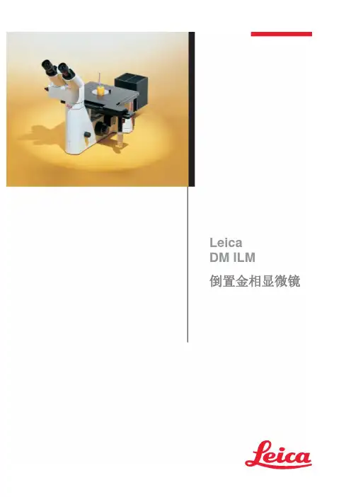
LeicaDM ILM倒置金相显微镜Leica DM ILM适用于来料、生产控制检查,检查样品制备过程和金相教学。
为您提供最优质的光学器件和最高效的显微镜系统是我们的责任。
您需要一台能快速准确显示结果的材料显微镜系统。
徕卡显微镜系统旨在减少您观察的时间,降低您的操作难度,同时提供最优化的结果。
- 徕卡品牌意味着高标准的光学性能。
HC (和谐系统) 光学系统的开发将光学显微镜标准Array提到了一个新的高度。
宽广的物镜选择范围保证了材料显微镜惊人的成像质量。
高性价比的HI Plan EPI 新物镜系列将明亮高反差与一流分辨率和优化的坚固的,耐腐蚀铸铝机身,漆面清洁,表面光滑。
显微镜的T型机身提供高稳定性,即使在高放大倍率下放置较重的样品,也能保证整体的稳健。
充足手部动作的空间是您操作灵活。
6 V 35 W照明此外,可另配外置的12V100W卤素灯反射光照明器或透射光。
根据要求,两个灯也可以同时安装开启,为明场和荧光工作。
新的灯光系统新的入射光照明系统插拔式3位反射棱镜组,很容易交换入射光及反射光照明。
滤光片组两个固定在显微镜内32mm直径的过滤器,以及一个可选的50厘米直径的中间过滤器优化观察图像。
3层机械移动载物台载物台适合不同形状和大小的样本。
几乎可以接受所有的样本量,并允许非破坏性的大型部件显微镜检查。
247X230mm 大载物台可以放置宽、高以及重的样品,内有150X150mm 的矩形台板插入可以完全移除。
小样本放在有80mm ,40mm ,30mm 和20mm 的内孔空白宽度。
同时有旋钮转动样本。
高承载能力 - 宽调节范围双层载物台支持高达样品负有重8Kg 。
XY 方向的调节范围在60 X40mm ,可迅速扫描试样。
矩形台板有4个垂直调整位,防止因制样不平对聚焦产生影响。
4新的照明系统可接纳不同类型的光源(灯泡灯丝或电弧放电),确保最佳的光强度和均匀性光通量。
外置和内置都按照克勒照明原理。
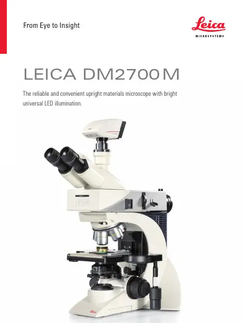
From Eye to InsightLeica DM2700 MThe reliable and convenient upright materials microscope with bright universal LED illumination.3 LEIca DM2700 M – ThE rELIabLE, convEnIEnT uprIghT MaTErIaLs MIcroscopESimple and Reliable:The Leica DM2700 MSee materials in the best lightExamine materials in the best light. The universal white light (4500 K) LED illumination in combination with renowned high quality Leica optics provide the ideal inspection tool for all of your quality assessments. The Leica DM2700 M demonstrates how simple and reliable microscopy can be, while at the same time, helps to improve your workflow so you can concentrate on the task at hand.›Brilliance›Reliability›Flexibility›Easy Documentation4LeD illumination, Daylight at a Touch The universal microscope illumination for a wide range of industrial applicationsA powerful light source for Brightfield, Darkfield, Differential Contrast, and Polarized Images on the right: Universal LED illumination.Light applications:› white light, constant color temperature› true color imaging› entirely adjustment-free› and lasts 20 yearsEnter the future of microscopy.5LEIca DM2700 M – ThE rELIabLE, convEnIEnT uprIghT MaTErIaLs MIcroscopE image Brilliance You can count OnTop of the line opticsA microscope is as good as its optics. This statement is still true for in today's digital Image on the right: n PLAn EPI series of objectives (plan achromat)world. The brand Leica Microsystems has always represented the highest standards in optical performance. Leica objectives are innovative, cost-effective, and always pro-vide high-contrast, pin sharp images as seen through the eyepieces and captured with digital cameras. They combine brilliance and sharp contrast with high resolution and optimized image fields. Whatever your work demands Leica Microsystems offers anaffordable solution.6Reliability You can count OnWe don’t make a science out of microscope operationUncomplicated AnD easy to understand functionality is built into the design of theLeica DM2700 M. Concentrate on your work, not on microscope adjustments.Ferrit C60, etched, undereutectoid, ferrit-perlite grain structure. LED brightfield, 500xBEnEFITSThe combination of Leica's color codedobjectives and the aperture diaphragmresults in the Color Coded DiaphragmAssistant (CCDA). With it, the basicsetting of resolution, contrast, and depth of field is simple and straightforward.The built-in focus stop, the heightadjustable focus knobs, as well as the three-gear focus mechanism for coarse, fine, and ultra-fine focusing make the Leica DM2700 M convenient and reliable for daily use. It can dramatically speed up your work processes while minimizingoperating errors. Using a manual microscope has never been easier.Accept no compromises when it comes to operation, performance, and features. The Leica DM2700 M is sturdy, durable, and ergonomically designed for ease ofuse and user comfort.7LEIca DM2700 M– ThE rELIabLE, convEnIEnT uprIghT MaTErIaLs MIcroscopE Flexibility Means Saving MoneyVersatility for all samplesThe Leica DM2700 M is a flexible upright microscope system for Brightfield (BF),Darkfield (DF), DIfferential Interference Contrast (DIC), Qualitative Polarization (PoL), and Fluorescence (FLUo) applications. In addition to all incident light applications, theLeica DM2700 M can also be equipped with transmitted light.Image on the right: Leica DM2700 M with Ergo Tube, upright/non-reversed image, 100%-0%, 50%-50%, 0%-100% beamsplitter.oBJECTIVE TURRETS There are three objective turrets tochoose from: The BF/DF M32 nosepieceholds up to five objectives, while the (BF/FLUo) can accommodate six or seven objectives.FLExIBLE In EVERY APPLICATIon Specimens with a size of up to 100 × 100 mm – such as foils, wafers or PCBs – and a thickness of up to 80 mm, such asmachined components, can be examined using the comprehensive stage program.KEEP TRACK oF YoUR SAMPLESThe Macro objective enables you to see almost 40 mm of the sample at a glance. The ideal addition for fast orientation and overview documentation.8Documentation SimplifiedDocumenting, saving, and retrieving imagesLeica digital cameras, optimized for reflected light applications, in combinationwith Leica Application Suite (LAS) image acquisition and archiving software, ensureconvenient and efficient documentation of your results.Upper image: Coated and annealed brass sample in LED brightfield-oblique contrast, 500x,Lower image: Plastic composite material in LED brightfield-oblique contrast, 200x. 3D-like image due to oblique illumination.QUICK AnD PRECISE AnALYSIS oF MATERIALS DATALeica Steel Expert, Phase & grain Expert, and Cleanliness Expert are dedicatedsoftware packages that providehigh-quality solutions, particularly in environments that require high samplethroughput and automated operation. With a modular structure, the functional-ity ranges from simple, interactive to automated photogrammetry; for example, characterizing metal surfaces or particle analysis.9 Chapter – SubChapter10(Dimensions in mm)Dimensions Leica DM2700 M331.4410505.5302.8365.8Undereutectoid, ferrit-perlite grain structure LED brightfield-oblique contrast, n Plan 100x Undereutectoid, ferrit-perlite grain structure LED brightfield contrast, n Plan 100xLEIca DM2700 M – ThE rELIabLE, convEnIEnT uprIghT MaTErIaLs MIcroscopE11 SpecificationsLeica DM2700 Morder no.: English 11914788 ∙ Copyright © by Leica Microsystems CMS gmbh,Wetzlar, germany, 2016. Subject to modifications. LEICA and the Leica Logo are registered trademarks of Leica Microsystems IR gmbh.Leica Microsystems – an international company with a strong network of worldwide customer services:Leica Microsystems operates globally in three divisions, where we rank with the market leaders.LIFE SCIEnCE DIVISIonThe Leica Microsystems Life Science Division supports the imaging needs of the scientific community with advanced innovation and technical expertise for the visualization, measurement, and analysis of microstructures. our strong focus on understanding scientific applications puts Leica Microsystems’ customers at the leading edge of science.InDUSTRY DIVISIonThe Leica Microsystems Industry Division’s focus is to supportc ustomers’ pursuit of the highest quality end result. Leica Microsystems provide the best and most innovative imaging systems to see, measure, and analyze the microstructures in routine and research industrial applications, materials science, quality control, forensic science inves-tigation, and educational applications.MEDICAL DIVISIonThe Leica Microsystems Medical Division’s focus is to partner with and support surgeons and their care of patients with the highest-quality, most innovative surgical microscope technology today and into the future.The statement by Ernst Leitz in 1907, “With the User, For the User,” describes the fruitful collaboration with end users and driving force of innovation at Leica Microsystems. We have developed five brand values to live up to this tradition: Pioneering, high-end Quality, Team Spirit, Dedication to Science, and Continuous Improvement. For us, living up to these values means: Living up to Life .active worldwide Tel. Fax australia ∙ north ryde+61 2 8870 3500 2 9878 1055austria ∙ vienna +43 1 486 80 50 0 1 486 80 50 30belgium ∙ Diegem +32 2 790 98 50 2 790 98 68canada ∙ concord/ontario +1 800 248 0123 847 405 0164Denmark ∙ ballerup +45 4454 0101 4454 0111France ∙ nanterre cedex+33 811 000 664 1 56 05 23 23germany ∙ Wetzlar +49 64 41 29 40 00 64 41 29 41 55Italy ∙ Milan +39 02 574 861 02 574 03392Japan ∙ Tokyo +81 3 5421 2800 3 5421 2896Korea ∙ seoul+82 2 514 65 43 2 514 65 48netherlands ∙ rijswijk+31 70 4132 100 70 4132 109people’s rep. of china ∙ hong Kong +852 2564 6699 2564 4163∙ shanghai +86 21 6387 6606 21 6387 6698portugal ∙ Lisbon +351 21 388 9112 21 385 4668singapore +65 6779 7823 6773 0628spain ∙ barcelona +34 93 494 95 30 93 494 95 32sweden ∙ Kista+46 8 625 45 45 8 625 45 10switzerland ∙ heerbrugg +41 71 726 34 34 71 726 34 44united Kingdom ∙ Milton Keynes +44 800 298 2344 1908 246312usa ∙ buffalo grove/lllinois+1800 248 0123847 405 0164。
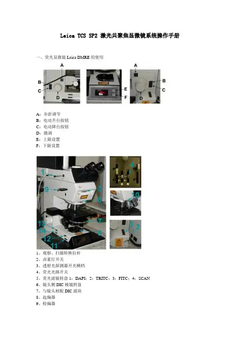
Leica TCS SP2 激光共聚焦显微镜系统操作手册一、荧光显微镜Leica DMRE的使用A:步距调节B:电动升台按钮C:电动降台按钮D:微调E:上限设置F:下限设置1、观察、扫描转换拉杆2、卤素灯开关3、透射光探测器开光横档4、荧光光路开关5、荧光滤镜转盘1:DAPI;2:TRITC;3:FITC;4:SCAN6、镜头侧DIC棱镜转盘7、与镜头相配DIC滤块8、起偏器9、检偏器10、镜头侧DIC棱镜微调旋钮11、光强调节纽12、减光滤光片13、孔径光阑14、视场光阑选择合适的镜头Leica TCS SP2 镜头配置镜头类型使用介质放大倍率/数值孔径编号HC PL APO CS DRY 10X /0.4 506511HC PL APO CS DRY 20X/ 0.7 506513HC PL APO CS DRY 40 X 0.85/ CORR 506140HC PL APO Ibd.BC OIL 63X /1.4 506192Leica TCS SP2 AOBS 镜头配置镜头类型使用介质放大倍率/数值孔径编号HC PL FLUOTAR DRY 10X/0.3 506505HC PL FLUOTAR DRY 20X/0.5 506503HCX PL APO OIL 40X/1.25-0.75 506176HC PL APO Ibd.BC OIL 63X/1.4 506192 HCX PL APO CS OIL 100X/1.40-0.70 506038 WATER HCX APO L U-V-I WA TER 63X/0.9 506148荧光观察荧光光路开关至“O”位,转轮至“1.0×”位,转换拉杆完全推进,荧光滤块换到相应号位(1:DAPI;2:TRITC;3:FITC;4:SCAN)图像输出扫描荧光光路开关至“I”位,转轮至“UV”位,转换拉杆完全拉出,荧光滤块换到4:SCAN)荧光观察(以油镜观察为例)1、样品正面朝上正确放在显微镜样品台上,点上镜油。
Leica DM 显微镜操作指南主机右下角电源,左下角调亮度,最下面光源部分可调视场光阑大小,聚光镜上面有孔径光阑一般调到最上面-适合高倍镜最大光亮度主机功能注解明场:通常观察配置使用,配合不同光源上的滤色片,达到良好观察效果因为卤素灯为钨丝发光(色温偏黄红),灯丝容易成像在中央-需要在低倍镜下调节光源照明位置使之充满整个观察市场,高倍镜下视场小就无需调节日光型滤色片-提高色温,补偿黄色就变成日光型,为人眼习惯色温绿色滤色片-背景绿色灰度滤色片-降低70%的光强,不改变色温暗场:提高分辨率,暗场片使光散射入玻片,使原先明亮部分变暗,增强反衬效果,需要有暗场片及聚光镜相差:观察活细胞及无染色的特殊微生物片-通过在聚光镜上的相差环和对应的相差物镜上的镜头的相差环,产生反差效果达到分辨透明物质(折射率与介质不同以分开)的观察效果荧光:通过汞灯电源上的汞灯(水银通过超高压激发发出弧灯)通过各种不同的滤块A滤块11513824-紫外滤色块-激发波长为BP 340-380nm-发射波长为400nm (看到为紫色)(倒置镜中可用A滤块替代明场装盘通道)I3滤块11513828-蓝色滤色块-激发波长为BP 450-490nm-发射波长为510nm (看到为绿色)N2.1滤块11513832-绿色滤色块-激发波长为BP 515-560nm-发射波长为580nm(看到为红色)汞灯灯箱中安装汞灯要注意调节汞灯的照明角度,使之均匀汞灯电源上有汞灯计时器,每次关闭时待汞灯冷却时方能重新打开(30分钟以上)否则容易烧坏汞灯,汞灯寿命一般200小时-500小时不等,超过时间汞灯并未烧坏,只是汞灯效能降低,需要专业工程师协助更换汞灯卤素灯-长效能的使用时间一般在2000小时左右,100块钱左右1个,如果长期在高能耗(年度最高情况观察)可能损耗较快显微镜的使用方法和注意事项1.取镜与放置:取镜时,用右手握住镜臂,左手托住镜座,使镜体保持直立,以防滤镜和目镜滑脱甩出。
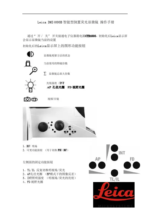
Leica DMI4000B智能型倒置荧光显微镜操作手册通过“开/关”开关接通电子仪器箱电源CTR4000,初始化后Leica显示屏会显示显微镜当前的设置初始化后的Leica显示屏上的图形功能按钮显微镜观察方法的状态当前使用的物镜倍数∑显微镜总放大倍数光线强度/ INTAP 孔径光圈FD视野光圈视频/目镜1、BF/ 明场2、可变功能按钮(用于切换PH \ BF)左侧面的固定功能按钮1、TL/IL 反复切换明视场/荧光2、AP孔径光圈(BF模式下的图像反差)3、INT照明强度(明视场/荧光的亮度)4、FD视野光圈右侧面的固定功能按钮1、开启荧光虑块盒-——(切勿按下以免造成意外)2、FLUO 切换至荧光观察方法3、FLUO>PH 切换至实时荧光与相差二、荧光观察开启通过“开/关”开关接通EL6000荧光光源、开启后指示灯呈黄绿色显微镜荧光自动控制按钮1 荧光虑块A 颜色表达蓝2 荧光虑块I3 颜色表达绿3 荧光虑块N21 颜色表达红4、SHUTTER 荧光挡板(阻挡或释放荧光)其它固定功能按键为空一、操作步骤1、安放标本微微抬起显微镜镜臂,使标本有足够的空间进入载物台,防止撞坏聚光镜,然后将标本放在载物台上,固定样品,转动载物台X、Y控制器的旋钮,使要观察的标本对准通光孔中央,整个操作过程必需轻起轻放,以免对显微镜造成损坏2、调焦调焦时,不应在高倍镜下直接调焦,先旋转粗调焦旋钮慢慢降低镜筒,并从侧面仔细观察,直到看清标本物像时停止,再用细调焦旋钮回调清晰,从低倍镜转至高倍时,只需略微调动细调焦旋钮,即可使物像清晰3、照明强度的调节:通过显微镜左侧INT固定按钮调节,使光线亮度适中4、荧光观察时,开启荧光光源,根据选择合适虑块进行观察(建议荧光照明强度为100%)5、拍照功能切换左上端的拉杆导入计算机,进入软件相关操作6、观察完毕后,移去样品关闭图像软件后再关闭显微镜电源箱的开关按钮,并将照明强度调至最弱,以防止下次开机时瞬间过强电流烧坏光源灯,清理台面,盖上防尘罩二、注意事项1、显微镜不论在使用或者存放时应在稳固的平台上,同时应避免灰尘、潮湿、过冷、过热及含有酸碱性的蒸汽2、所有镜头表面必须保持清洁,落在镜头表面的灰尘,可用吸耳球吹去,也可用软毛刷轻轻的掸去掉。
徕卡显微镜的操作规程徕卡显微镜操作规程徕卡显微镜(Leica microscope)是一种用于观察微小物体结构和细胞的仪器。
具备高分辨率、高对比度和高清晰度的特点,所以在医学、生物学、材料科学等领域得到广泛应用。
正确的操作徕卡显微镜对于获得准确的观察结果非常重要。
以下是使用徕卡显微镜的基本操作规程:1. 准备阶段:a. 确保工作台面干净整洁。
b. 检查显微镜是否正常工作:确认光源、电源是否连接正常,各部分是否可靠、灵活。
2. 标本处理:a. 准备好待观察的标本。
b. 标本表面应干净,无灰尘或杂质。
c. 根据需要做好标本的染色、切片或加热等处理。
3. 调整目镜:a. 将目镜放置在显微镜上,并调节至合适位置。
b. 调节目镜对焦,使得目镜视野内的标记线条清晰可见。
4. 放置标本:a. 将标本放置在显微镜载物台上。
b. 使用载物台移动手柄,调整标本位置至合适观察范围。
5. 聚焦标本:a. 将低倍物镜放置于物镜转盘上,并通过转盘选择低倍放大倍数。
b. 使用聚焦手轮,调节缓慢转动物镜直到标本处于清晰对焦状态。
6. 切换放大倍数:a. 借助转盘,选择高倍物镜,再次通过聚焦手轮将标本调整到清晰对焦状态。
7. 调整照明:a. 使用光源调节开关,调整适当的照明亮度,并确保光线均匀。
b. 当使用偏光显微镜时,可通过偏光滤光片调节和改善观察效果。
8. 观察标本:a. 使用眼睛或通过连接数字摄像头的计算机观察标本。
b. 使用显微镜台上的移动手柄,控制标本的滑动和旋转。
9. 调整视野和对比度:a. 使用光孔调节光源入射角度,改变光线方向,调整视野亮度和对比度。
b. 使用聚焦手轮继续调整标本对焦,确保图像清晰。
10. 清洁和保养:a. 使用尘埃清除器或吹气球轻轻吹除标本表面的灰尘。
b. 使用专用擦拭布或纯棉棉签擦拭显微镜镜片,注意不要使用化学溶剂。
以上是使用徕卡显微镜的基本操作规程。
使用前请务必熟悉操作手册,并按照规程正确操作,以保证观察结果的准确性和显微镜的正常使用寿命。
报告打印机
显微镜照明调节
显微镜主体
计算机
显微镜模式设置开关
图1
图2
图3
图4图5图6图7
手动变
换倍率
图8
(3)点击“焊接”分析软件专用模块(图8),进入拍照测量界面;点击“新建项目”(图9),设置测量数据存储;点击“报告模板”(图10),选择对应产品的报告输出模板。
水平放置测量样件,调节高度和照明亮度(图11),点击“抓拍”,结合辅助线与测量工具测量
要测的要素。
完成所有测量后,点击“生成报告”(图12);在表格报告中填写产品信息(图1图10
图9 图11
图12
图13
图14
2)、调节高度,使预览达到最清晰状态,点击抓拍。
3)、依次在图像(图15)选取均匀分布的
&R0=(Ra-Ra0)/Ra0×100%; Ra=读数平均值;
图15
4)、检测结果输出记录:完成检查后,将数据结果记录在“表1中,附在本规程后,存档备份。
徕卡徕卡测量显微镜安全操作及保养规程1. 引言徕卡徕卡测量显微镜是一种常用的实验室设备,广泛应用于生物学、医学、物理学等领域。
为了确保设备的正常运行和延长使用寿命,正确的操作和适当的保养非常重要。
本文档将介绍徕卡徕卡测量显微镜的安全操作规程以及保养要点。
2. 安全操作规程2.1 准备工作在使用徕卡徕卡测量显微镜之前,需要进行以下准备工作: - 保证工作区域安全整洁,避免杂物堆放。
- 确保设备稳固放置在平坦的台面上。
- 检查电源线是否连接稳固,插头是否完好。
- 检查显微镜光源是否正常工作。
2.2 操作步骤正确的操作徕卡徕卡测量显微镜可以保证观察到清晰的图像,并确保个人安全。
以下是操作步骤的简要说明: 1. 打开显微镜电源并调整亮度。
2. 调节目镜和物镜,使得目标物体清晰可见。
3. 使用焦距调节按钮调整焦距,以获得更清晰的图像。
避免过度调节导致破坏物镜。
4. 使用准确的测量标尺对目标物体进行测量。
5. 使用标本盖片保护目标物体,避免触碰镜头或者目标物体。
6. 使用移动平台进行样本移动,避免使用手指直接触碰样本或镜头。
7. 使用合适的孔径和倍率进行观察,避免使用过大倍率导致图像模糊或畸变。
2.3 注意事项在操作徕卡徕卡测量显微镜之前,需要注意以下事项: 1. 镜头和样本可能会产生热量,避免触碰以防烫伤。
2. 在操作过程中,尽量避免震动和冲击,以免影响图像质量。
3. 注意保持适当的姿势,避免长时间弯腰或者歪曲身体造成不适。
4. 在调节焦距时,轻轻旋转,避免过大力度的调整。
3. 保养规程适当的保养可以延长徕卡徕卡测量显微镜的使用寿命,并保证其正常工作。
以下是一些保养要点: 1. 定期清洁镜头和样本盖片,使用干净而柔软的棉纱或者镜头纸轻轻擦拭。
2. 避免使用有机溶剂或强酸碱溶液清洁镜头,以免损坏镀膜和镜片。
3. 存放在干燥、通风的环境中,避免受潮或过热。
4. 定期检查电源线和插头的连接情况,并保持清洁干燥。
Leica DM ILInverted Microscope –for all Routine and Laboratory Applications2Leica Design by Ernest Igl /Christophe ApothélozLeica DM IL – compact inverted microscope for laboratory routine The new inverted Leica microscope blends ergonomy, a compactdesign and effective contrasting methods into a system for virtuallyunlimited life science applications.The integration of Leica HCS* optics extends the range of objectivesfor inverted microscopy.For the first time, high-quality relief contrast can be producedwithout special objectives with our new Integrated ModulationContrast (IMC) technique.Optimised phase contrast and brilliant incident light fluorescencemake the Leica DM IL the number one choice in contrastingmicroscopes.3With its unbeatable modularity, ergonomy and free view of thespecimen, teamed with newly developed and optimised contrastingtechniques, the Leica DM IL offers you a top level introduction toinverted microscopy.The adaption of the Leica DM IL to infinity optics allows theintegration of Leica HCS* components for superb image resolution,brilliant contrast and precise colour rendering. The DM IL is theinverted equivalent of our successful upright DM microscopes ofthe L and R class.The Leica DMIL is a microscope for all applications in microbiologyor the cell culture laboratory. A universal inverted microscope forroutine use: stable and space-saving, flexible and upgradablewith optics from Leica’s research microscopes.* Harmonic Component System Microinjection of oocytes in mouseRNA microinjection of frog oocytes(Xenopus)4The stand The stand of the Leica DM IL microscope is stable, aluminium-cast and excellently designed. There are two versions for biological applications:The DM IL for brightfield, phase and modulation contrast and the DM IL with additional incident light fluorescence unit.Appreciated by users for many years, the stable T-shaped micro-scope base offers plenty of valuable space round the microscope and ensures comfortable, fatigue-free microscopy. The micro-scope’s footprint is optimised to provide the necessary room for experiments, and all controls are ergonomically located. Many different components can be adapted to suit individual require-ments. The high stability, low centre of gravity and four vibration damping feet eliminate vibrations even in extreme conditions. The excellent stability of the DM IL also makes it the ideal solution for photography with long exposure times. In addition, finite element calculations and thorough practical testing in a wide variety of applications guarantee focusing which is not only ultra precise but also stable over long periods of time.The Leica DMIL contrasting microscopeThe System Discussion unitCardiac muscle cells5Due to its modularity, the Leica DM IL is particularly suitable forliving cell microscopy.Its modern, practical design, the integration of state-of-the-art,top quality optics and the excellent standard of the adaptedcontrasting techniques prove useful in research tasks as well asroutine applications.You will be convinced by the DM IL’s first-class technology andmany innovative and practical ideas.We at Leica believe microscopy should be a pleasurable experienceand designed the DM IL to be associated with enjoyment at thelaboratory workplace.Trinocular tube with DC 100Ergonomic phototubeTrinocular tube with MPS DM IL with illumination column the other way round6Nosepiece focusingSamples are focused with the quadruple objective nosepiece.The reliability, stability and precision of the focusing is not influenced by the microscope stage and the samples on it or by accessory components such as the object guide and manipulators.Illumination system The compact brightfield illumination unit is attached to a column and can be comfortably adjusted. Setting the correct height for the condenser used or the specimen on the stage is facilitated by markings along the column. The pre-centred, extremely powerful 6V 35W halogen lamp provides optimal illumination even of critical specimens. The transmitted light illumination concept is rounded off by the integration of the contrast slide (for phase and modulation contrast), the module for light filters with 32 mm diameter and the aperture diaphragm. The microscope stage with illumination arm can be turned round by 180°and is freely accessable for sample positioning from three sides.Light filters The filter module on the illumination column accommodates 32 mm diameter filters in a spoon-shaped holder. We are constantly adding to our wide range of filters, enabling you to selectively optimise the illumination for observation and image documentation.Built-in 6V 35W power supply The 6V 35W power supply, which provides the full lamp power including on/off switch, status indicator and brightness adjustment,is fully integrated in the microscope stand. Apart from the ergonomic advantages, this saves space on the microscope desk and allows the microscope to be easily picked up as a unit and moved elsewhere.The Technology7CondensersThe Leica DM IL offers you a choice of two condensers.The S 90/0.23 condenser with a free working distance of 90 mm and a numerical aperture of 0.23 is designed for brightfield and phase contrast and is particularly suitable for specimens in bulky laboratory vessels.The S 55/0.35 condenser with a free working distance of 55 mm and a numerical aperture of 0.35 for bulky containers is designed for brightfield, phase and integrated modulation contrast (IMC)and is particularly suitable for higher magnifications or thick specimens.Without a condenser, the maximum free working distance is 200 mm.Stage and accessoriesThe DM IL offers a wide variety of stages with a whole array of accessories and different inserts for your specimen vessels:The standard stage is a fixed stage plate of 252 x 212 mm. The stage can be widened by 70 mm on both sides by adapter plates. The interchangeable stage inserts (20-50 mm) allow smaller petri dishes to be used as well without losing the focus when the objective nosepiece is rotated.Object guides can be attached to both the left and right of the stage and have a minimum adjustment range of 83 x 127 mm. The control of the coaxial drive is in an ergonomically low position so that you can rest your hands on the desk while scanning specimens.The object guides accommodate special and multi-purpose frames for all types of culture vessels.A heating stage up to max. 45°C, a 3-plate mechanical stage and scanning stages round off the range of stages for the DM IL.Micromanipulation ...... and scanning stage8DM LB tubeEyepieces and objectivesThe optics are the heart of every microscope and decisive for the quality of information. We have set new standards here by intro-ducing our HC optics.The Leica DM IL is designed for brightfield, phase contrast, Inte-grated Modulation Contrast (IMC) and incident light fluorescence.All infinity-corrected high performance objectives in the Leica range with 25 mm screw thread are compatible with the DM IL microscope.Even earlier-type Leica objectives can be adapted for use on the DM IL. We offer a wide range of special objectives for inverted microscopy applications with long free working distances (L objectives) and/or with correction mounts (Corr objectives) to compensate for different vessel thicknesses. The latest Leica optics brochure features our whole range of objectives.Depending on the tube configuration, there is a wide choice of eyepieces with magnifications 10x, 12.5x, 15x, 16x or 25x, suitable for fields of view up to 20 mm. Besides special high-point eyepieces for eyeglass wearers, we also supply eyepieces with adjustable eyelens (M eyepieces), into which different types of graticule can be inserted.The DM IL range also comprises many different observation and photo tubes. The tubes are interchangeable and can be individually rotated by 360°in the tube mount and then fixed in position. All tubes are fitted with an infinity tube lens 1x. The following tubes are used on the DM IL:•Binocular tube ILB, with 45°viewing angle, for eyepieces with 23.2 mm outer diameter •Trinocular (photo)tube ILT, with 45°viewing angle, for eyepieces with 23.2 mm outer diameter, with vertical photo/TV exit with switchable light path for either 100% visual or 100% photo/TV.The position of the photo/TV exit 88 mm to the side of the tube has the special advantage that it allows an unobstructed view of the specimen.Other tubes from the Leica DM L range can be used via an IL/L adapter:•Binocular tube HC LB 45°viewing angle •Binocular tube HC LVB 0-35°ergotube •Trinocular (photo)tube HC L1T 45°viewing angle •Trinocular (photo)tube HC L3T 45°viewing angle •Trinocular (photo)tube HC LV1T 0-35°ergotubeThe Optics351+364 mn UVAr 488mn 568mn 647mn Ar/Kr 4905205575766486703504004505005506006507009Developments in diagnostics and med./biol. research (e.g. fluores-cence applications) and the increasing use of video technology and electronic image processing have to be paralleled by intelligent technical adaptations of the microscope system.The new HCS optics concept introduced with the Leica DM R microscope meets this requirement. It is the result of an integral system consideration, harnessing all technological potential.The abbreviation HCS stands for Harmonic Component System.Its special features are:•well-balanced optical and mechanical fitting dimensions•harmonious balance of all optical system components, i.e. the parameters contributing to the microscope’s performance (objectives, tube lenses, tubes, eyepieces, TV cameras/adapters,etc.) have been harmonised throughout the entire optic system.This has created scope for even greater optical opportunities.The HCS system is the answer to your application requirements not only today, but in future, too.Rat testicles, IMC10The Leica DM IL is the microscope for all requirements in the cell culture lab. A universal inverted microscope for routine application: stable and space-saving, flexible and upgradable with optics from Leica research microscopes.BrightfieldThe whole range of objectives from 4x-100x magnification can be used for brightfield applications. Samples in almost any kind of vessel can be examined with or without a condenser, while a 6V 35W halogen lamp ensures optimal illumination.Phase contrastIn vivo/ in vitro microscopy specimens are mostly living cultures or microorganisms and are examined under sterile conditions. Contrast of the transparent tissue can only be enhanced by optical methods.Phase contrast is a useful technique for high-contrast imaging of unstained specimens. The phase contrast technique used by Leica on the DM IL has been optimised for inverted microscopy applications and produces equally excellent contrast in watery solutions and of dry preparations in petri dishes.Spinal cord, catContrastingLymphocyte toxicity testdouble staining, strongly positiveLymphocyte toxicity testdouble staining, weakly positive11Human brain Femtotip microinjection needle (Photo: Eppendorf)FibroblastsIMC (Integrated Modulation Contrast)The innovative technique of Integrated Modulation Contrast (IMC)now introduced by Leica in the DM IL is based on Hoffman’sprinciple and produces this contrast without the need for specialobjectives–ordinary brightfield or phase contrast objectives canbe used. The Leica IMC provides a high-contrast, 3D image oftransparent objects similar to that of interference contrast. Plasticculture vessels do not impair the quality of the image as thetechnique is polarisation-neutral.The diaphragm slide on the side of the illumination and theswitchable modulator in the intermediate image of the pupilproduce the type of contrast named after Hoffman without modi-fying the objectives. High contrast, high resolution, a halo-freerelief image of either stained or unstained specimens make Leica’sIMC a new standard in the class of inverted routine microscopy.FluorescenceThe fluorescence model of the DM IL reflects the growing signifi-cance of fluorescence for in vivo/in vitro microscopy. The maincomponents of this configuration are an incident light axis integrat-ed in the microscope stand, incorporating a fluorescence slide forthree filter cubes. A wide range of light sources with multi-lens,chromatically corrected collectors brighten up even the weakestfluorescence. The fluorescence filter cubes comprise an optimallymatched combination of excitation, reflection, band-pass andbarrier filters. We are constantly updating our range of filtercubes to keep pace with the latest challenges in biology andmedicine.Transmitted light techniques can be used simultaneously or inalternation in order to clearly allocate fluorescent and non-fluorescent structures.The Leica DM IL offers you a powerful system for immunology,cytopathology, virology – in fact wherever fluorescence techniquesare used in combination with inverted microscopy.Leica Microsystems – the brand for outstanding products Microscopes Compound Stereo Surgical Laser Scanning Photomicrography Video Microscopy Measuring Microscopes Advanced Systems Image Analysis Spectral Photometry Automated Inspection Stations Measurement Systems Electron Beam Lithography Laboratory equipment Tissue Processors Embedding Systems Routine- & Immunostaining Coverslippers Refractometers Microtomes Sliding, Rotary & Disc Cryostats Ultramicrotomes EM Sample Preparation Leica Microsystems’ Mission is to be the world’s first-choice provider of innovative solutions to our customers’ needs for vision, measurement, lithography and analysis of microstructures.Leica, the leading brand for microscopes and scientific instruments, has developed from five brand names, all with a long tradition: Wild, Leitz, Reichert, Jung and Cambridge Instruments. Leica symbolizes not only tradition, but also innovation.Leica Microsystems – an international company with a strong network of customer servicesAustralia:North Ryde/NSW Tel. +61 2 9879 9700Fax +61 2 9817 8358Austria:Vienna Tel. +43 1 495 44 160Fax +43 1 495 44 1630Canada:Willowdale/Ontario Tel. +1 416 497 2860Fax +1 416 497 8516Denmark:Herlev Tel. +45 4454 0101Fax +45 4454 0111Finland:Espoo Tel. +358 9 6153 555Fax +358 9 5022 398France:Rueil-Malmaison Tel. +33 1 473 285 85Fax +33 1 473 285 86CedexGermany:Bensheim Tel. +49 6251 136 0Fax +49 6251 136 155Italy:Milan Tel. +39 0257 486.1Fax +39 0257 40 3273Japan:Tokyo Tel. +81 3 5435 9600Fax +81 3 5435 9618Korea:Seoul Tel. +82 2 514 65 43Fax +82 2 514 65 48Netherlands:Rijswijk Tel. +31 70 4132 100Fax +31 70 4132 109Portugal:Lisbon Tel. +351 1 388 9112Fax +351 1 385 4668Republic of China:Hong Kong Tel. +852 2564 6699Fax +852 2503 4826Singapore:Tel. +65 779 7823Fax +65 773 0628Spain:Barcelona Tel. +34 93 494 95 30Fax +34 93 494 95 32Sweden:Sollentuna Tel. +46 8 625 45 45Fax +46 8 625 45 10Switzerland:Glattbrugg Tel. +41 1 809 34 34Fax +41 1 809 34 44United Kingdom:Milton Keynes Tel. +44 1908 246 246Fax +44 1908 609 992USA:Deerfield/Illinois Tel. +1 847 405 0123Fax +1 847 405 0030and representatives of Leicain more than 100 countries.Leica Microsystems Wetzlar GmbH Ernst-Leitz-Straße D-35578 Wetzlar (Germany)Tel. +49 (0)6441-290Fax +49 (0)6441-292599 ©L e i c a M i c r o s y s t e m s W e t z l a r G m b H •E r n s t -L e i t z -S t r a ße •35578 W e t z l a r •T e l. (06441) 29-0 •F a x (06441) 29-2599 G e d r u c k t a u f c h l o r f r e i g e b l e i c h t e m P a p i e r .B e s t e l l -N u m m e r n d e r A u s g a b e n i n : D e u t s c h 913 761•E n g l i s c h 913 762 •F r a n z ös i s c h 913 763 •I t a l i e n i s c h 913 764 •S a c h -N r . 501-068 G e d r u c k t i n D e u t s c h l a n d X I /98/A X /B .H .。
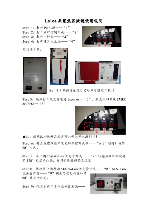
Leica共聚焦显微镜使用说明Step 1: 打开PC电源——“1”Step 2: 打开汞灯控制开关——“2”Step 3: 打开中控盒——“3”Step 4: 打开计算机主机——“4”,启动计算机。
注:计算机操作系统启动后方可继续开机!!Step 5: 顺序打开激光器电源Scanner—“5”,激光冷却系统LASER Ar/ArKr—“6”★注:须确认冷风开启后方可打开激光电源!!!!!Step 6: 将上图虚线框中激光功率控制旋钮——“6.5”顺时针旋转60°左右。
Step 7: 将上图所示488 nm激光管开关——“7”钥匙沿顺时针旋转约120°至显示灯亮,再将钥匙旋回竖直位置Step 8: 依次将上图所示543/594 nm激光管开关——“8”和633 nm 激光管开关——“9”钥匙沿顺时针旋转约90°至显示灯亮。
Step 9:依次打开半导体激光器电源——“10”和405 nm激光管开关——“11”,(“11”的开启方式同“8”、“9”。
)Leica共聚焦显微镜关机顺序及注意事项Step 1: 关闭软件→拷贝Data→关闭计算机Step 2: 依照开机顺序倒序关闭各个开关,由”11”→”7”, ”5”→”1”。
★注:(1)冷风——”6”不能立即关闭!!须在关闭所有硬件设备30min 后方可关闭,以使激光管完全冷却,延长使用寿命。
(2)PC电源——“1”须在电脑确定关闭后方可关闭!!数据采集Step 1: 实验前扫描样本片观察激光通路是否正常工作。
Step 2: 上样,须轻起轻落科勒照明系统,选择合适倍数物镜,通过FLUO键选择相应荧光通道,调节载物台和焦距,找到合适视野。
Step 3: 点击由Visual→Scan,点击选择扫描模式,如xy, xyz, xyt, xyzt, xyλ etc..点击选择扫描格式(一般为默认值512*512)。
Step 4: 点击打开Beam path setting窗口,对光路和检测器进行设置。
M220莱卡显微镜操作指南一、准备工作1.将显微镜放置在平稳的工作台上,确保显微镜不会晃动或倾斜。
2.连接电源线,确保电源稳定并接地。
3.打开显微镜电源开关,待显微镜系统启动完毕。
二、调节光源1.打开光源开关,调节亮度控制旋钮,确保光源亮度适中。
2.使用透射光源时,打开透射光开关,待光源预热后再进行观察。
三、调节物镜1.使用低倍物镜(如4X或10X)进行初步调焦。
2.用手轻轻转动物镜转盘,选择高倍物镜(如40X或100X)。
3.使用粗调焦旋钮,将物镜快速向下旋转,直到样本大致清晰可见。
4.使用细调焦旋钮,慢慢向上转动,逐渐调焦,直到样本完全清晰可见。
四、调节样本台1.使用样本台高度调节旋钮,调整样本与物镜的最佳焦距。
2.使用样本台平移旋钮,调整样本位置,使其位于视野中心。
五、调节眼镜1.调节眼镜间距,使两个眼睛看到的图像重合。
2.使用眼镜调焦旋钮,调节眼镜仪表内部物镜与眼睛的最佳距离,确保清晰度。
六、观察样本1.使用目镜观察器,选择合适的目镜放大倍数。
2.使用样本台旋转旋钮,观察样本不同方位的图像。
3.使用显微镜平移旋钮,移动物镜位置,观察不同位置的样本区域。
七、调节对比度和饱和度1.使用对比度调节旋钮,调整样本中不同部分的对比度,使其更加清晰。
2.使用饱和度调节旋钮,调整图像的颜色饱和度和亮度。
八、关闭显微镜1.关闭光源开关,使光源熄灭。
2.关闭透射光开关,如有使用透射光源。
3.关闭显微镜电源开关。
4.拔掉电源线。
注意事项:1.在操作显微镜前,应确保手部干净,以免污染样本或任何光学部件。
2.在操作过程中,应避免撞击或强烈震动,以免损坏显微镜。
3.使用高倍物镜时,需谨慎,以防止物镜与样本接触,损坏样本。
4.使用时应根据具体要求选择合适的光源和物镜。
5.在放置显微镜时,应选择平稳的工作台,并确保显微镜稳定。
6.在清洁显微镜镜片时,应使用适当的清洁剂和软布,避免划伤镜片。
以上是M220莱卡显微镜的操作指南,通过正确操作和维护显微镜,可以保证其正常使用和延长使用寿命。
Leica DM4B荧光显微镜操作规程一.操作步骤1.打开电源,连接电脑(DM4B确认USB白色数据线连接好电脑背后口)2.放好样本,找到合适视野,从低倍到高倍观察(物镜转盘),调好焦距。
3.液晶屏说明-每次机器启动都会保留最上次操作设置4.STATUS为目前机器状态--TL表示目前是透射光状态5.AP-孔径光栏,视场光栏,光强显示在状态中,通过机身左侧键盘控制6.BF-明场,正常染色的样品,用的比较多7.PH-相差观察方法-适应于透明非染色薄样品,有些样慢反射会反射亮光8.FL-荧光-呈现荧光的效果,适用细胞特异性染色效果/机身左侧下部TL/IL点击切换明场和荧光切换,或直接点击软件切换二.拍照摄取步骤1.确认数码CCD和电脑连接线是否连接;(注意显微镜镜先开机再开软件)2.明场状态下观察样品请调节合适的照明【充足的合理曝光可以调节显微镜照明或曝光时间。
可用目镜盖盖住目镜以避免日光灯影响-有一横线】3.把三目镜筒如果有拉杆(需要把拉杆拉到拍照位置,一般往外拉)4.确认选择软件中摄取界面的MIC显微镜控制面板部分5.白平衡设置选择自动白平衡;一般自动曝光(如果反应慢是样品照明不足,可增加照明-切换到手动,超过0.5秒是相机需时间照明)6.饱和度一般150,伽马值一般玻片0.6(明场状态),荧光或其他状态可以适当拉高去除背景噪音(每次使用完需要回复正常值,避免其它人留意)7.调整完毕,选择采集图像三.图像处理步骤可以后期进行图像各种处理,如曝光,剪切,注释等等四.图像分析步骤图像分析,添加标尺(图像浏览边上第1个图标点开),注意要合并原图添加注释等等保养与维护:D需要经常通电使用,注意湿度,否则需放到干燥器里封闭保存2.除湿机如有条件尽量长期开放,每天清空水箱3.防尘,每段工作期之后,应用软质防尘罩罩住,以避免显微镜和附属设备染灰尘。
灰尘和松放的污粒,可用软毛刷或不掉毛心棉布除掉。
顽固性的污物可用一块于净的棉布上普通水溶液、汽油或酒精擦掉。
全新智能型数字式显微镜Leica DM4000 M材料科学简捷的显微镜操作完美的微观世界影像让全新的徕卡DM数字式显微镜帮助您实现神圣的科学探索!便捷的显微观察,完美的科学探索外观:新技术新设计最吸引您的首先是徕卡智能型数字显微镜的全新的设计:稳重及曲线流畅的轮廓。
图像清晰,逼真一旦您使用了任何一款Leica全新数字显微镜,一定会对其爱不释手。
在同类显微镜中,Leica的新型数字显微镜能够提供最好的图像清晰度,景深和对比度。
操作简单,准确徕卡DM数字显微镜系列为用户提供了很多前所未有的解决方案。
其中最突出的一点就是帮助用户简化显微镜的操作,使显微镜的使用简单、准确、得心应手。
智能化的设计帮助您以简单的操作完成复杂的调节程序,实现多种多样的研究及诊断。
人体工程学,使显微镜操作更加轻松、舒适。
在我们的全新显微镜中,人机工程学被广泛应用。
您可以真真切切地感受它。
通过与德国研究所(Fraunhofer Institute)**的紧密合作,新型显微镜的设计达到了最先进的人机工程学的要求,使显微镜操作更加舒适、轻松。
智能型设计全自动光栏调节全新数字式显微镜可以自动识别观察技术和观察物镜,并将光栏调节在最佳状态。
无论透射光还是反射光路,光栏的调节都是自动的。
全自动光强度调节照明光的强度可以自动调节,这意味着在您转换不同放大倍数的物镜时,标本图像的亮度会保持不变,而且还不会产生炫光。
同时,如果您所得观察对照明光有特殊的要求,也可以根据情况调整光的强度。
智能系统将随时记忆您的调整状态。
全新:恒温色温控制(CCIC)对于您的视觉而言太暗,而对于您的相机而言又太亮,这时以往显微镜照明的问题。
不同的灯泡电压,带来不同的色温,使照相颜色失真。
Leica数字式显微镜的新型光路设计能够在光强的变化下自动保持恒定色温。
您不再需要使用中性密度滤片和日光滤片用以校正光强和色温。
高自动化的新型聚光镜我们的全自动聚光器可以满足最苛刻的要求。
徕卡工业显微镜安全操作及保养规程徕卡工业显微镜是一种高端的显微镜设备,广泛应用于科学研究和工业生产领域。
为了确保使用安全和设备长期使用寿命,我们需要按照以下操作和保养规程进行操作和维护。
安全操作规程1. 熟悉设备使用说明书在操作设备之前,首先应当认真阅读设备的使用说明书,了解设备结构、性能、操作规程以及安全事项。
如发现不理解的或不熟悉的内容,应当向技术人员咨询。
2. 检查设备完好性在每次操作设备之前,应当仔细检查设备的完好性,如镜头、支架、灯泡等是否有松动或损坏现象。
如发现问题,应当及时修理或更换设备。
3. 接通电源前的操作在接通电源前,应当确保所有电路已经正确接通,并检查所有的开关是否处于合适的位置,尤其是光源和电源是否开启。
开机后应当关注设备是否正常运转和有无异常声音,及时排查问题。
4. 戴好个人防护装备操作设备时应当戴好个人防护装备,如手套、护目镜、口罩等,以确保个人安全。
5. 操作设备前的准备工作在操作设备之前,应当准备好所需的样品以及辅助设备,以便顺利完成工作。
同时,在设备调节过程中应当轻拍设备以防止设备晃动。
6. 正确操作操作设备时应当按照使用说明书的要求进行,如施加适当的力度、调节镜头和照明等,根据需要调整成像效果。
7. 关机操作规范使用完毕之后,应当先将电源关闭,然后才能拆卸样品或放置相关工具,保证设备的完好性,并放置在干燥、凉爽的地方。
8. 特殊情况处理在操作设备时,如出现特殊情况,如设备故障等,应当立即停止操作,并进行相应的维修工作。
保养规程1. 设备日常清洁设备在使用过程中会产生灰尘,对设备的清洁工作十分重要。
在每次使用完毕后,应当用温水轻拭设备表面,并用干净的软布擦拭干净,以防止灰尘和污垢对设备的影响。
2. 镜头保养镜头是设备的核心部分,对于镜头应当特别注意保养。
使用过程中要避免碰撞和刮擦,并避免使用损坏镜头的低质量试剂。
如发现镜头有污垢,应当使用干净的棉花蘸取酒精擦拭,在清理过程中应当轻柔操作。
有了DVM6,无需苦苦寻觅,结果近在眼前,任何人都能成为显微镜应用专家。
徕卡第六代三维视频显微镜(DVM 6)是徕卡自主研发,颠覆传统的革命性产品。
数字技术已经在很多方面为我们的工作和日常生活带来了革命性变化,而这样的创新也将永无止境地进行下去。
工业质量控制领域对宏观成像、显微成像和图像处理均有着最严格的要求,创新的数字技术将使其尤为受益。
徕卡显微系统有限公司的数字显微镜在机动性和速度两方面开创了新维度。
在诸多应用场合下,它能为传统方式提供理想的质量控制辅助。
数字显微镜生成的图像将直接显示在高分辨率显示器上,因此您无需透过目镜观察。
变倍光学元件采用流线型设计,即使是极难触及的表面也能有效深入,相对于费时费力的传统显微镜检验技术,它甚至还能对大型静态部件进行无损检验。
Leica DVM6数字显微镜不仅具备出众的光学品质,还在量化分析方面表现卓著,无论是二维分析还是高级三维表面测量。
设备一体化设计,功能高度集成,外观给您简洁大方,接触使用给您不负众望!
无论您从事的是质量保证、失效分析、研发还是取证的工作,Leica DVM6视频显微镜都提供能快速、可靠和易用的解决方案。
光学组件-PLANAPO物镜
超高分辨率成像-1000万物理像素摄像头徕卡的数字图像和光学图像一样出色。
徕卡是光学行
业的先锋和精密显微成像的世界领先企业。
公司的历
史及那股提供最高细节度和清晰度样品图像的激情可
追溯到160多年前。
徕卡工程师不但消除了光学像差,
还不断争取获得更的高分辨率-DVM6 PLANAPO光学
镜头。
PLANAPO光学装置的优点:最大的光学校正能
力;至边界区非常详细的图像细节;在变焦范围内无
彩色边纹。
与利用插值和耗时像素移动的视频显微镜不同,DVM6的核心包含一个本地1000万像素摄像系统。
每秒37帧以上的快速实时图像显示可以让您的手和肉眼协调,以确保操作的舒适。
将摄像机与变焦模块相结合,则可提供完善的污染防护。
聚集驱动粗调/精调超高分辨率显示系统连接:
开关/电源/USB3.0/手动/
脚踏开关/聚光灯照明
16:1变倍比
真正无极连续变倍自动景深合成:EDOF
16:1变焦范围:极端放大倍数使观察分析更多样,只需
一个旋转动作,您便可以看到16倍的放大比例,最高放
大倍数达到2350倍。
秉承徕卡高端体式显微镜无极连续变倍能力,无论您调节红色变焦环到什么位置,DVM6都能精准对应当前倍率,并显示在屏幕上方。
无需繁琐的上下限设置,无需人为判断起始位置终止位
置,只需选择一键EDOF,即可在视野内完成高低景深
合成。
当您希望在高倍率下获得更大视野,只需一键数秒后视野扩展即可完成,并且徕卡DVM6视野扩展功能不仅满足二维三维视野扩展,而且视野扩展区域不受像素限制,最大
可延伸到XY扫描区域。
无限制视野扩展功能
您可以轻松地单手倾斜观察,便可只将注意力用在监视器上进行样品研究。
在默认情况下,倾斜轴与焦点对准,因此在- 60°至+ 60°的任何角度,您可以看到样品永远在焦点以内。
旋转载物台从全新的视角观察样品,有助于您找到要找的细节信息。
智能多角度观察系统可在屏幕上方显示实时的放大倍率、支架的倾斜角度、载物台的旋转角度,并且保存图片后,这些参数全部保留到图片文件夹,方便日后追溯。
当您支架倾斜过大,预警传感器会发出警示音,提醒您防止镜头碰触到载物台导致损坏。
测量软件丰富多样,可满足您日常检验的所有需求。
并且磁力功能,可以减少鼠标在屏幕上人为选取测量点的偏位,他可以自动检测目标位置的边缘,自动的更正测量点。
这样有效的降低了人为误差,从而保证测量尺寸的准确和可靠。
实时精准测量
实时精准测量
系统检测各种焦平面内的一系列图
像,并从图像堆栈自动计算全聚焦
图像,其中在清晰聚焦内显示所有
元素。
这种全聚焦图像包含每个像
素的高度信息。
因此,还可以将其
视为三维模型,以分析表面结构和
进行测量。
三维测量
您的照明选择决定了您看到的内容。
您可以根据样品、应用和任务选择不
同的集成LED照明选项。
在粗糙表面
全部或部分使用环形光,或者为扁平
反射样品选择同轴照明。
您还可以将
照明模式结合起来,以揭示您以前从
未见过的细节。
观察扁平反射样品
时,用于明/暗控制的四分之一波片
控制。
减小对比度,以强调轻微不平
整性,例如划痕。
徕卡DVM6视频显微镜应用图集汽车、金属行业
电子、半导体行业
非金属化工行业
制药、食品行业
高校科研行业
3D分析
文物,古玩行业
紫砂壶印章瓷器消光前瓷器消光后
玻璃纤维建材砂砾特种塑料
火器枪弹
工具痕迹
印章土壤
化纤,建材行业
动物,植物行业
公安,刑侦行业
DMS1000/300视频显微镜
一款伟大的视频显微镜,2年全球销量破2000台> 8:1 编码变倍– 可分档变倍
> 可变孔径TM技术
> 远心镜头– 可选
> 放大倍数从 6x 到 300x (22”Monitor)
> 5百万像素摄像头 (DMS1000)/3百万像素摄像头(DMS300)> 高清活图1980x1080px (@30fps)
> 图形定制叠加
> 独立模式或电脑联机模式
DCM8白光共焦干涉显微镜
共聚焦行业技术翘楚,Z轴分辨率行业最高0.1nm > 3合一的双核技术设备
> BF/DF (明场/暗场)光学显微镜
> Confocal (共焦)显微镜
> Interferometry (干涉)显微镜
> 超大Z轴精度:0.1nm-40mm
> 完美的彩色和3D图像合成技术
> 快速采图:3-10秒
> 无需耗材维护费用:智能双LED同光路照明11。