IPS Cells Are Man-Made Cancer cells
- 格式:pdf
- 大小:130.14 KB
- 文档页数:3
日本发现癌干细胞克星癌干细胞癌症干细胞(Cancer Stem Cell,CSC),又称癌干细胞、肿瘤干细胞,是指具有干细胞(Stem Cell)性质的癌细胞,也就是具有“自我复制”(Self-Renewal)以及“具有多细胞分化”(Differentiation)等能力。
通常这类的细胞被认为有形成肿瘤,发展成癌症的潜力,特别是随着癌症转移出去后,产生新型癌症的来源。
癌干细胞存在于癌细胞中,是癌症复发和转移的“元凶”。
由于存在数量极少,因此很难被发现,抗癌药物也很难对其产生效果。
最新新闻大麻药用价值被承认:杀死癌细胞提高放疗效果2015-04-13 19:59外媒称,日本三重大学研究生院医学系的科研小组发现了一种可以附着在癌干细胞上的可视化物质。
此项成果已在英国某科学杂志的电子版上刊登。
据《日本经济新闻》4月11日报道,由于这种物质具有发光特性,它附着在癌干细胞上之后,癌干细胞集中的区域就变得可见。
此外,这种可视化物质还有抑制癌干细胞增殖甚至导致其死亡的作用,可望成为有效治疗癌症的新方法。
...详情中文名癌干细胞外文名Cancer Stem Cell,CSC别称肿瘤干细胞细胞特质细胞被认为有形成肿瘤的潜力目录1重要特征▪自我更新▪高致瘤性▪分化潜能▪耐药性2治疗方法3研究进展1重要特征自我更新白血病中的癌干细胞白血病中的癌干细胞自我更新(self-renewal)是指一个细胞分裂为两个细胞,但其中一个子代细胞仍然保持与亲代细胞完全相同的未分化状态;而另一个子代细胞则定向(commit)分化,这种分裂称为不对称分裂(dissymmetricdivision)。
癌干细胞15crmo钢管与成体干细胞类似,也具有自我更新的特性。
认识正常干细胞自我更新的调节机制是理解肿瘤细胞增殖机制的基础,因为癌通常被认为是自我更新失控所致的疾病。
肿瘤干细胞自我更新的特性是造成肿瘤复发、转移及预后不良的主要原因。
正常干细胞通过不对称分裂在实现自我更新的同时,也通过分化、细胞周期的有序进行,为机体在生命过程中维持恒态(homeostasis)起了不可替代的作用。

ipscell名词解释
干细胞(ipscell)是一种具有潜在多能性的细胞,它们具有自我更新的能力,并且可以分化成各种类型的细胞。
这些细胞可以来源于胚胎(胚胎干细胞)或成体组织(成体干细胞)。
其中,诱导多能性干细胞(iPSC)是一种特殊类型的干细胞,它们是从成体细胞中重新编程而来的多能性细胞,可以分化成各种类型的细胞,如神经细胞、心脏细胞等。
iPSC 的发现为医学研究和治疗提供了新的可能性,因为它们可以为疾病建模、药物筛选和组织再生提供新的途径。
这些细胞对于研究和治疗许多疾病,包括癌症、心脏病和神经退行性疾病等,具有潜在的重要意义。


iPS细胞是什么?
iPS 是指体细胞经过导入特定的重编程因子而诱导为多能干细胞(induced pluripotent stem cells, iPS cells)。
2006年,日本科学家Yamanaka 及其研究小组首次选用逆转录病毒作为载体,向鼠成纤维细胞中导入了与多功能性有关的基因(Oct4、Sox2、c-Myc 和Klf4),之后选择多能性标志物Fbx15 筛选转染后的细胞,最终获得了具有与胚胎干细胞(ES)某些特性的多功能干细胞,并命名为诱导多能干细胞(iPS 细胞),此细胞在形态、基因和蛋白表达、细胞倍增能力、类胚体和畸形瘤生成能力、分化能力等都与胚胎干细胞极为相似。
次年,该研究小组又成功将人体细胞重编程为iPS 细胞,自此,在全球范围内拉开了 iPS 细胞研究的序幕。
与 ES 细胞相比,iPS 细胞的产生解决了几个重大问题。
首先,伦理学专家认为干细胞的产生不应该破坏任何一个胚胎,而iPS 细胞绕过了这个伦理学问题;其次,iPS 细胞可以由患者个体的成体细胞重编程得来,为个体化的疾病细胞模型提供了有力的基础;最后,由iPS 细胞诱导分化而来的成体细胞,可以应用于患者疾病的临床治疗,为再生医学提供一个崭新的思路。
诺奖发明iPS细胞对医疗技术的发展和影响干细胞技术在生命医学的发展中是衡量其发展水平的重要条件,从有关干细胞技术领域的专利拥有数量分析来看,美国拥有绝对优势,中国与日本相差较小,但我国干细胞研究及其应用市场,在世界干细胞领域内仍是重要力量。
iPS 细胞诱导多能干细胞(induced pluripotent stem cells),简称为iPS 细胞。
iPS细胞是通过对成熟体细胞“重新编程”培育出的干细胞,拥有与胚胎干细胞相似的分化潜力,能够分化成多种组织和器官。
但与胚胎干细胞相比,iPS细胞的获得方法相对简单,并且避开了一些伦理问题,因此有巨大的医疗价值。
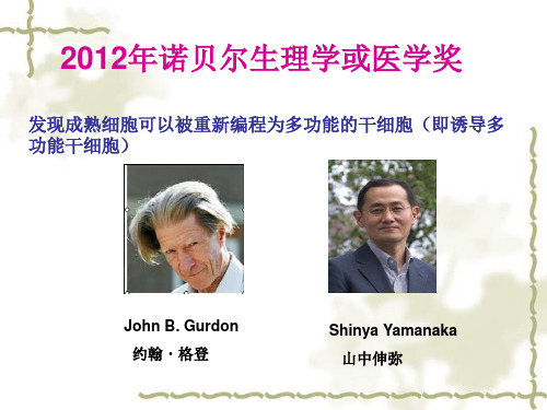
2006年日本京都大学Shinya Yamanaka在世界著名学术杂志《细胞》上率先报道了诱导多能干细胞的研究。
2012年10月8日,John B. Gurdon 与 Shinya Yamanaka 因此获得诺贝尔生理学和医学奖。
诱导多能干细胞iPS 细胞对医疗的发展和影响1. iPS 细胞对医疗的发展随着iPS 细胞功能的发现与实现,iPS 细胞对医疗的发展及影响也越深远。
从对医疗的发展来看,iPS 细胞的出现为解决众多医疗难题提供了优化方案及可能性,同时能够为疾病建模、药物筛选、再生医学等方面的研究奠定基础。
iPS 细胞对多种疾病治疗具有促进作用。
在治疗帕金森病中,间充质干细胞细胞移植治疗能够取得进步,而基于iPS 细胞诱导的间充质干细胞的增殖速度及免疫能力更具优势,对治疗帕金森病起到促进作用。
在 A 型血友病的治疗研究中,学者发现 iPS 细胞技术的基因修复方法的可行性,并且将此种方法成功应用在A 型血友病动物模型的治疗上,为 A 型血友病提供了潜在的治疗方案。
在治疗肿瘤的研究中,我国科学家突破了 T 细胞研究技术难题,通过iPS 细胞产生能够杀死肿瘤细胞的T 淋巴细胞。
此外,通过iPS 细胞在癌症治疗研究、心肌梗死、黄斑变性、血液系统疾病、红斑狼疮、缺氧缺血性脑病、1 型糖尿病等方面都存在未来应用医疗方面的可能性。
1.RTRadiotherapy,Radiation Therapy放疗,放射治疗放射治疗是利用放射线治疗肿瘤的一种方法,是当今治疗肿瘤的三大手段之一。
据统计,大约有60~70%恶性肿瘤患者需要接受放射治疗。
有些恶性肿瘤通过放疗可以得到根治,并可能获得同类同期肿瘤的手术治疗的疗效,且可保存所在的器官及其功能。
2.IMRTIntensity Modulated Radiation Therapy调强放射治疗调强放射治疗与以往放射治疗技术不同,它通过调节各个方向照射野的野内射线的强度产生非均匀照射野,达到肿瘤的高剂量三维适形分布和危及器官的低剂量分布,从而提高肿瘤的照射剂量,尽可能地减少危及器官和正常组织的受量,最终提高肿瘤局部的控制率,改善肿瘤患者的生存质量。
3.MLCMultiLeaf Collimator多叶准直器,多叶光栅MLC最初设计主要是用于替代射野挡铅,后来发展成了IMRT的基础,控制叶片运动可实现静态MLC和动态MLC调强。
4.QA & QCQuality Assurance & Quality Control质量保证和质量控制放射治疗的QA是指经过周密计划而采取的一系列必要的措施,保证放射治疗的整个服务过程中的各个环节按国际标准准确安全的执行。
这个简单的定义意味着质量保证有两个重要内容:质量评定,即按一定标准度量和评价整个治疗过程中的服务质量和治疗效果;质量控制,即采取必要的措施保证QA的执行,并不断修改服务过程中的某些环节,达到新的QA级水平。
5.AAPMAmerican Association of Physicists in Medicine美国医学物理学家协会6.SADSource to Axis Distance源轴距放射源到机架旋转或机器等中心的距离。
SSDSource to Surface Distance源皮距放射源到模体表面照射野中心的距离。
3DCRT、X刀、IMRT等技术都采用SAD技术,国内常规放疗正在普及SAD等中心照射技术,希望大家能尽早放弃SSD技术,只在某些特定情况下采用SSD技术。
STEM CELLS:人类干细胞可用于制造结肠癌疫苗
佚名
【期刊名称】《临床合理用药杂志》
【年(卷),期】2009(2)21
【总页数】1页(P32-32)
【关键词】人类干细胞;癌疫苗;结肠;制造;科学家
【正文语种】中文
【中图分类】R-052;R730.51
【相关文献】
1.Cell Stem Cell:支持干细胞与癌症间联系的新证据 [J], 生物谷
2.周琪在« Cell Stem Cell»发表单倍体干细胞研究新成果 [J],
3.山东大学 Cell stem cell 干细胞研究新成果 [J],
4.WJSC 6^(th) Anniversary Special Issues(2):Mesenchymal stem cells Brain mesenchymal stem cells:The other stem cells of the brain? [J], Florence Appaix;Marie-France Nissou;Boudewijn van der Sanden;Matthieu Dreyfus;Fran ois Berger;Jean-Paul Issartel;Didier Wion
5.Methods to produce induced pluripotent stem cell-derived mesenchymal stem cells: Mesenchymal stem cells from induced pluripotent stem cells [J], Victoria Dupuis;Elisa Oltra
因版权原因,仅展示原文概要,查看原文内容请购买。
Logical Biology 8 (1):16-18, 2008OPEN LETTER© Truthfinding Cyberpress IPS CELLS AND CANCERSIPS Cells Are Man-Made Cancer CellsShi V. LiuEagle Institute of Molecular MedicineApex, NC 27502, USACorresponding with SVL@(Received 2008-01-21; accepted 2008-02-13; published 2008-02-13*)HIGHLIGHTY amanaka’s iPS cells are expected to provide a great hope for therapeutic cloning. However, by definition and nature, these iPS cells are just some man-made cancer cells.ABSTRACTRecent high-profile publications have regarded iPS(induced pluripotent stem)cells as potential therapeutic stem cells for regenerative medicine. However, based on the fact that the inducing/reprogramming factors contained in iPS cells are known oncogenes and their expression have been associated with various cancers, I will announce iPS cells as man-made cancer cells. These safety-problematic cells should not be put into any therapeutic use.KEY WORDSY amanaka, iPS cells, Stem cell, Cancer, Reprogramming, Criticism, Negligence, Editorial misconductIn 2006 Takahashi and Y amanaka announced that, with just four factors (Oct3/4, Sox2, c-Myc and Klf4), they can produced pluripotent stem cells from mouse embryonic and adult fibroblast cultures (Takahashi and Y amanaka, 2006). Since then, these so-called “induced pluripotent stem (iPS) cells” have been reported in high-profile in various top journals despite some strong criticisms (Liu, 2008). Experts in the stem research field have heralded the creation of iPS cells as a milestone for therapeutic cloning (Zaehres and Scholer, 2007), even though the real safe therapeutic value of these magic cells have not yet proven at all.The therapeutic value of the first-generation iPS cells was actually seriously questioned due to the presence of Myc, a well known oncogene (Battey et al., 1983; Dominguez-Sola et al., 2007). Thus, efforts have been made to eliminate Myc, the unexpected ingredient in the stem cell cocktail (Knoepfler, 2008). It turned out, the so-called one of the “essential” four components of the original iPS cell cocktail is not essential at all because iPS cells were still “generated” without using Myc (Nakagawa et al., 2008; Y u et al., 2007).The elimination of Myc has been regarded as a major achievement in creating the second-generation iPS cells that are described as “safer” than the first-generation iPS cells (Pera and Hasegawa, 2008). However, are these Myc-free iPS cells really safe?Here I wish to raise a caution that all other three inducing/reprogramming factors still remained in the Takahashi/Yamanaka cocktail for second-generation iPS cells are all oncogenes by definition and their over-expression has all been associated with this or that kind of cancer.For examples, it has been shown that Oct3/4 gene is expressed in adult human breast stem cells (Trosko, 2006) and bladder cancer (Atlasi et al., 2007). Interestingly, some abnormal effects with the ectopic expression of Oct-4 was already known to a leading iPS research group (Hochedlinger 2005).Sox2 has been shown to be expressed in human gastric carcinoma (Li et al., 2004), stomach adenocarcinomas (Tsukamoto et al., 2005), pancreatic carcinoma (Sanada et al., 2006b), vater adenocarcinoma (Sanada et al., 2006a), malignant glioma (Schmitz et al., 2007), breast cancer (Rodriguez-Pinilla et al., 2007) and brain tumors (Phi et al., 2008).KLF4, .once believed to be a tumor-suppressor, is now also shown as an oncogene (Rowland and Peeper, 2006). KLF4 is often over-expressed in squamous cell carcinoma (Foster et al., 2005; Foster et al., 1999). Up to 70% of primary human breast cancers show over-expression of KLF4 (Foster et al., 2000) and nuclear localization of KLF4 has been found as a predictor of poor outcome of breast cancer (Pandya et al., 2004).In a different four-factor cocktail for “inducing” pluripotent stem cells, Nanog and Lin28 were used in addition to Oct4 and Sox2 (Y u et al., 2007). However, forced expression of Nanog in hematopoietic stem cells has been shown to result in a T-cell disorder (Tanaka et al., 2007).So far the known iPS cell cocktail component that has not been found in an association with cancer is Lin28. However, this component is shown as not absolutely required for “inducing” iPS cells (Y u et al., 2007).Thus, all essential iPS cell-inducing/reprogramming factors are oncogenes and their over-expression has been linked with cancers. When iPS cells are full of such oncogenes and the expression of these genes is already at elevated level in the iPS cells, how can we say these iPS cells are safe for therapeutic use?Come on! Let us face the reality and say iPS cells are man-made cancer cells.ReferencesAtlasi, Y., Mowla, S. J., Ziaee, S. A., and Bahrami, A. R. (2007). OCT-4, an embryonic stem cell marker, is highly expressed in bladder cancer. Int J Cancer 120, 1598-1602.Battey, J., Moulding, C., Taub, R., Murphy, W., Stewart, T., Potter, H., Lenoir, G., and Leder, P. (1983). The human c-myc oncogene: structural consequences of translocation into the IgH locus in Burkitt lymphoma. Cell 34, 779-787.Dominguez-Sola, D., Ying, C. Y., Grandori, C., Ruggiero, L., Chen, B., Li, M., Galloway, D. A., Gu, W., Gautier, J., and Dalla-Favera, R. (2007). Non-transcriptional control of DNA replication by c-Myc. Nature 448, 445-451.Foster, K. W., Frost, A. R., McKie-Bell, P., Lin, C. Y., Engler, J. A., Grizzle, W. E., and Ruppert, J. M. (2000). Increase of GKLF messenger RNA and protein expression during progression of breast cancer. Cancer Res 60, 6488-6495.Foster, K. W., Liu, Z., Nail, C. D., Li, X., Fitzgerald, T. J., Bailey, S. K., Frost, A. R., Louro, I. D., Townes, T. M., Paterson, A. J., et al. (2005). Induction of KLF4 in basal keratinocytes blocks the proliferation-differentiation switch and initiates squamous epithelial dysplasia. Oncogene 24, 1491-1500.Foster, K. W., Ren, S., Louro, I. D., Lobo-Ruppert, S. M., McKie-Bell, P., Grizzle, W., Hayes, M. R., Broker, T. R., Chow, L. T., and Ruppert, J. M. (1999). Oncogene expression cloning by retroviral transduction of adenovirus E1A-immortalized rat kidney RK3E cells: transformation of a host with epithelial features by c-MYC and the zinc finger protein GKLF. Cell Growth Differ 10, 423-434. Knoepfler, P. S. (2008). Why Myc? An unexpected ingredient in the stem cell cocktail. Cell Stem Cell 2, 18-21.Li, X. L., Eishi, Y., Bai, Y. Q., Sakai, H., Akiyama, Y., Tani, M., Takizawa, T., Koike, M., and Y uasa, Y. (2004). Expression of the SRY-related HMG box protein SOX2 in human gastric carcinoma. Int J Oncol 24, 257-263.Liu, S. V. (2008). The rapid publication of research on iPS cells and the strong suppression of criticism on publications of iPS cells. Top W atch 3, 1-4.Nakagawa, M., Y oyanagi, M., Tanabe, K., Takahashi, K., Ichisaka, T., Aoi, T., Okita, K., Mochiduki, Y., Takizawa, N., and Y amanaka,S.(2008).Generation of induced pluripotent stem cells without Myc from mouse and human fibroblasts.Nature Biotechnol 26, 101-106.Pandya, A. Y., Talley, L. I., Frost, A. R., Fitzgerald, T. J., Trivedi, V., Chakravarthy, M., Chhieng, D. C., Grizzle, W. E., Engler, J.A., Krontiras, H., et al. (2004). Nuclear localization of KLF4 is associated with an aggressive phenotype in early-stage breast cancer. Clin Cancer Res 10, 2709-2719.Pera, M. F., and Hasegawa, K. (2008). Simpler and safer cell reprogramming. Nat Biotechnol 26, 59-60.Phi, J. H., Park, S. H., Kim, S. K., Paek, S. H., Kim, J. H., Lee, Y. J., Cho, B. K., Park, C. K., Lee, D. H., and W ang, K. C. (2008). Sox2 Expression in Brain Tumors: A Reflection of the Neuroglial Differentiation Pathway. Am J Surg Pathol 32, 103-112. Rodriguez-Pinilla,S.M.,Sarrio,D.,Moreno-Bueno,G.,Rodriguez-Gil,Y.,Martinez,M.A.,Hernandez,L., Hardisson,D., Reis-Filho, J. S., and Palacios, J. (2007). Sox2: a possible driver of the basal-like phenotype in sporadic breast cancer. Mod Pathol 20, 474-481.Rowland, B. D., and Peeper, D. S. (2006). KLF4, p21 and context-dependent opposing forces in cancer. Nat Rev Cancer 6, 11-23. Sanada, Y., Y oshida, K., Konishi, K., Oeda, M., Ohara, M., and Tsutani, Y. (2006a). Expression of gastric mucin MUC5AC and gastric transcription factor SOX2 in ampulla of vater adenocarcinoma: comparison between expression patterns and histologic subtypes. Oncol Rep 15, 1157-1161.Sanada, Y., Y oshida, K., Ohara, M., Oeda, M., Konishi, K., and Tsutani, Y. (2006b). Histopathologic evaluation of stepwise progression of pancreatic carcinoma with immunohistochemical analysis of gastric epithelial transcription factor SOX2: comparison of expression patterns between invasive components and cancerous or nonneoplastic intraductal components. Pancreas 32, 164-170. Schmitz, M., Temme, A., Senner, V., Ebner, R., Schwind, S., Stevanovic, S., W ehner, R., Schackert, G., Schackert, H. K., Fussel, M., et al. (2007). Identification of SOX2 as a novel glioma-associated antigen and potential target for T cell-based immunotherapy. Br J Cancer 96, 1293-1301.Takahashi, K., and Y amanaka, S. (2006). Induction of pluripotent stem cells from mouse embryonic and adult fibroblast cultures by defined factors. Cell 126, 663-676.Tanaka, Y., Era, T., Nishikawa, S., and Kawamata, S. (2007). Forced expression of Nanog in hematopoietic stem cells results in a gammadeltaT-cell disorder. Blood 110, 107-115.Trosko, J. E. (2006). From adult stem cells to cancer stem cells: Oct-4 Gene, cell-cell communication, and hormones during tumor promotion. Ann N Y Acad Sci 1089, 36-58.Tsukamoto, T., Mizoshita, T., Mihara, M., Tanaka, H., Takenaka, Y., Y amamura, Y., Nakamura, S., Ushijima, T., and Tatematsu, M. (2005).Sox2expression in human stomach adenocarcinomas with gastric and gastric-and-intestinal-mixed phenotypes. Histopathology 46, 649-658.Y u, J., V odyanik, M. A., Smuga-Otto, K., Antosiewicz-Bourget, J., Frane, J. L., Tian, S., Nie, J., Jonsdottir, G. A., Ruotti, V., Stewart, R., et al. (2007). Induced pluripotent stem cell lines derived from human somatic cells. Science.Zaehres, H., and Scholer, H. R. (2007). Induction of pluripotency: From mouse to human. Cell 131, 834-835.* This paper was submitted to Cell on Jan. 21, 2008. Cell never responded to me despite my repeated inquiries. The publication here is the same as submitted to Cell except for the added highlight and keywords.** This publication is sent to Cell as a record.AppendixesCover letter for submission to CellDear Cell Editors,I am submitting a very important manuscript entitled "IPS cells are man-made cancer cells" to be published in an appropriate section in Cell.Sincerely yours,Shi V. Liu MD PhD2008-01-21Inquiry sent to CellEditors of Cell,On Jan. 21, 2008 I submitted a Manuscript “IPS cells are man-made cancer cells”.Since the submission, I have not received any reply from Cell. I called Cell and left a message to the assistant to the Cell editor. But no one retuned my call.I know that some editors in Cell have developed a hostile attitude towards me simply because some of my submissions are critical not only to Cell’s publications but also Cell’s publishing practices. However, no matter how much you dislike my expressed views (all of them are proven to be correct); you need to show some basic working ethics and at least give me a reply to let me know your decision.If Cell will repeat its previous practice of just ignoring my submission without any response, I will take further action against this irresponsible and unethical behavior.I should also remind you that, if Cell continues its strong push of the man-made cancer cells named as iPS cells and totally ignoring my warning on the danger of these cells, I will reserve my legal right to sue your negligence that may lead to great harm to humanity. Shi V. Liu MD PhDFeb. 1, 2008。