MK-4827_cas:182410-00-0_别名Niraparib_NMR_MedChemExpress
- 格式:pdf
- 大小:179.69 KB
- 文档页数:1


Product InformationMonoclonal ANTI-FLAG® M2, Clone M2 Produced in Mouse, Affinity Isolated AntibodyF1804Storage Temperature −20 °CProduct DescriptionMonoclonal ANTI-FLAG® M2 is a mouse-derived, affinity-purified IgG1 monoclonal antibody that binds to fusion proteins containing a FLAG® peptide sequence.1 The M2 antibody will recognize a FLAG® peptide sequence at the N-terminus,Met-N-terminus, C-terminus, or internal sites of a fusion protein. Binding of the M2 antibody is not dependent on calcium.Monoclonal ANTI-FLAG® M2 is useful for detection, identification, and capture of fusion proteins that contain a FLAG® peptide sequence by common immunological procedures, such as Western blotting, immunofluorescence, and immunoprecipitation. ReagentSupplied at a concentration of ∼ 1 mg of protein per mL of solution, and formulated in 50% glycerol,10 mM sodium phosphate, and 150 mM NaCl,pH 7.4. The formulation contains no antimicrobial preservatives.Antigenic binding siteN-Asp-Tyr-Lys-Asp-Asp-Asp-Asp-Lys-C SpecificityThe monoclonal antibody detects only the target protein band(s) on a Western blot from an E. coli, plant or mammalian crude cell lysate.SensitivityThe monoclonal antibody detects as little as 2 ng of target protein by dot blot. The Western blot is tested down to 10 ng, but may detect lower using the procedure detailed below. Precautions and DisclaimerFor R&D use only. Not for drug, household,or other uses. Please consult the Safety Data Sheet for information regarding hazards and safe handling practices.Preparation InstructionsImmediately prior to use, dilute the monoclonal antibody in Tris-buffered saline (TBS), pH 8.0, with 3% nonfat milk (Cat. No. T8793). Dilutions in the described procedures are provided as guidelines. Adjust the antibody concentration to maximize detection sensitivity and minimize background. StorageStore the undiluted antibody at −20 °C in working aliquots. The product, as formulated, will not freeze when stored at the recommended temperature. Note: Over time, small amounts of purified antibodies can precipitate due to intermolecular hydrophobic interactions. If precipitate is observed in this product, briefly centrifuge the vial to pellet the precipitate. Withdraw the desired volume of antibody solution from the clear supernatant for use. This should not alter the performance of the purified antibody in most applications.ProceduresWestern Blot Immunostaining2-5Note: this procedure is based on chemiluminescent detection using Chemiluminescent Peroxidase Substrate-1 (Cat. No. CPS160). Dilutions of both primary and secondary antibodies may require optimization when using other substratesor conditions.1.Separate fusion proteins containing aFLAG® peptide sequence from sample lysatesusing a standard SDS-PAGE protocol. Load2.5–10 µg of total protein lysate per lane.2.Transfer proteins from the gel to a nitrocellulosemembrane, Immobilon®-P, or otherpolyvinylidene difluoride (PVDF) membrane. ThePVDF membrane may provide greaterdownstream sensitivity.3.Wash the blot in at least 0.5 mL/cm2 of purifiedwater for 2–3 minutes employing gentle agitation (50–60 rpm).4.Block the blot with at least 0.5 mL/cm2 ofTris Buffered Saline (TBS), pH 8.0, with 3%nonfat milk for 30 minutes at room temperature, employing gentle agitation.5.Remove the blocking agent and wash once with0.5 mL/cm2 of Tris buffered saline, pH 8.0(Cat. No. T6664).6.Add the desired concentration of monoclonalantibody to the blot. A final antibodyconcentration of 1 µg/mL (1:1,000 dilution of the antibody as supplied) in at least 0.5 mL/cm2 ofTBS with 3% nonfat milk is suggested. Incubateat room temperature for 30 minutes employinggentle agitation.Note: Dilutions must be optimized for differentsubstrates and systems.7.Decant off the Monoclonal ANTI-FLAG® M2solution and wash once with at least 0.5 mL/cm2of TBS, pH 8.0.8.Add the secondary antibody in the form ofAnti-Mouse IgG-Peroxidase (Cat. No. A9044) orequivalent. The concentration of secondaryantibody must be optimized based on thesubstrate being used. For detection usingChemiluminescent Peroxidase Substrate-1, a final secondary antibody dilution of 1:30,000 should be employed. Specifically, it is suggested theantibody as supplied be diluted in at least0.5 mL/cm2 of TBS with 3% nonfat milk. Incubatethe blot employing gentle agitation at roomtemperature for 30 minutes. 9.Wash the blot at least three times for a total of15 minutes (5 minutes per wash) in TBS with0.05% TWEEN® 20, pH 8.0 (Cat. No. T9039).Agitate gently, employing at least 0.5 mL/cm2 ofwash solution.10.Develop the blot using ChemiluminescentPeroxidase Substrate-1 or an equivalent reagentfor 5 minutes. Do not agitate the blot during this incubation step. Drain briefly and wrap inplastic film.11.Expose BioMax® Light film to the blot for a rangeof times from several seconds up to 10 minutes.It is recommended that a quick exposure of10–30 seconds be performed to determine theoptimal exposure time needed. If the signal is too intense even at the short exposure times, allowthe signal to decay over a 1–8 hour period(or longer if necessary), and then re-exposethe film.Indirect ImmunofluorescentCytochemical StainingMonoclonal ANTI-FLAG® M2 may be utilized in immunocytochemical staining procedures when used in conjunction with a labeled secondary antibody (indirect).6 A generic procedure for adherent cell staining is described, using immunofluorescence, employing an Anti-Mouse IgG-FITC conjugate asthe label.1.Grow and transfect cells on coverslips.2.Fix the cells by incubation with phosphatebuffered saline, pH 7.4 (Cat. No. P3813),containing 4% paraformaldehyde (Cat. No.P6148), and 4% sucrose (Cat. No. S1888) for15 minutes at room temperature.3.Wash the fixed cells with PBS for 5 minutes.Repeat once.4.Permeabilize the cells by incubation with 0.25%TRITON™ X-100 (Cat. No. T9284) in PBS for5 minutes.5.Wash the cells with PBS for 5 minutes.Repeat once.6.Block by incubation with 10% bovine serumalbumin (Cat. No. A9647) in PBS (10% BSA/PBS) for 30 minutes at 37 °C.7.Incubate with Monoclonal ANTI-FLAG® M2 dilutedin the range of 1:500 to 1:2,000 in 3% BSA/PBSfor 2 hours at 37 °C.8.Wash with PBS for 5 minutes. Repeat twice.9.Incubate with the secondary antibody, Anti-MouseIgG- FITC (Cat. No. F9137) at a 1:1,000 dilutionin 3% BSA/PBS for 45 minutes at 37 °C.10.Wash with PBS for 5 minutes. Repeat twice.11.Mount coverslips with cells side down on glassslides using a small drop of mounting mediumsuch as polyvinyl alcohol for semi-permanentmounting. The inclusion of an anti-fading agentlike DABCO® in the mounting medium(25–100 mg/mL, for example Cat. No. 10981) isstrongly recommended. Seal coverslips to glassslides (with nail polish, for instance).12.Examine by fluorescence microscopy. FITC has anabsorption maximum at 492 nm with an emission maximum at 520 nm. Immunoprecipitation (IP)Monoclonal ANTI-FLAG® M2 may be used in IP procedures when used in conjunction with an insoluble carrier matrix, such as a Protein G resin. Alternatively, EZview™ Red Protein G Affinity Gel (Cat. No. E3403) or the Protein G Immunoprecipitation Kit (Cat. No. IP50) may be used. EZview Red ANTI-FLAG® M2 Affinity Gel(Cat. No. F2426) or ANTI-FLAG® M2 Affinity Gel (Cat. No. A2220) may be utilized directly for IP. See reference 5 for general protocols.Enzyme Immunoassay (EIA)Monoclonal ANTI-FLAG® M2 may be used in EIA procedures. Typically, a fusion protein containing a FLAG® peptide sequence is directly adsorbed (or otherwise presented) within the wells of a multiwell polystyrene plate. The Monoclonal ANTI-FLAG® M2 antibody may be diluted up to 1:50,000 for subsequent incubation within the plate wells. Detection may be accomplished using Anti-Mouse IgG-Peroxidase (Cat. No. A9044) or equivalent, diluted 1:10,000, followed by an appropriate substrate for visualization.We also offer the ANTI-FLAG® High Sensitivity, M2 coated 96-well plate (Cat. No. P2983) for EIA-based screening applications. References1.Brizzard, B.L., et al., Immunoaffinity purificationof FLAG® epitope-tagged bacterial alkalinephosphatase using a novel monoclonal antibodyand peptide elution. BioTechniques, 16(4):730-735 (1994).2.Bjerrum, O.J., and Heegaard. N.H.H., CRCHandbook of Immunoblotting of Proteins, Volume I, Technical Descriptions. CRC Press(Boca Raton, FL), pp. 229-236 (1988).3.Dunbar, B.S. (ed.), Protein Blotting: A PracticalApproach. IRL Press (New York, NY),pp. 67-70 (1994).4.Fortin, A., et al., A 56- to 54-kilodalton non gratasignal in immunoblot analysis using thehorseradish peroxidase chemiluminescencesystem. Biochem. Cell Biol., 72(5-6):239-243 (1994).5.Harlow, E., and Lane, D., Antibodies: ALaboratory Manual. Cold Spring Harbor Laboratory Press (Cold Spring Harbor, NY) (1988).6.Ciaccia, A.V., and Price, E.M., IBI FLAG Epitope,1: 4-5 (1992)..Troubleshooting Guide (Western Blot Immunostaining Procedure) Problem Possible Cause SolutionFusion protein is not detected. Protein is notexpressed.Verify nucleic acid sequence and reading frame of the FLAG® fusionprotein in vector construct. If sequence is present, attempt tooptimize expression.Target proteinpoorly representedin sample.Positive controls (10 ng/lane recommended) should always beincluded. If the positive control works, the sample may not containthe FLAG® fusion protein of interest, or it may be present atconcentrations too low to detect. Immuno-precipitation withANTI-FLAG® M2 Affinity Gel (Cat. No. A2220) may be required forlow concentrations of FLAG fusion proteins.Positive controls we have available:•Amino-terminal FLAG-BAP™ Fusion Protein (Cat. No. P7582).•Carboxy-terminal FLAG-BAP™ Fusion Protein (Cat. No. P7457).•Amino-terminal Met-FLAG-BAP™ Fusion Protein (Cat. No. P5975).Defective detectionreagentsRun appropriate controls to ensure performance. Use 10 ng/lane of acontrol FLAG-BAP™-fusion protein as a positive control. If no signal isobtained with the control, repeat the procedure using a fresh lot ofsecondary antibody-HRP conjugate, along with freshly preparedreagents.Inadequateexposure timeusingchemiluminescentsystem.If no signal is observed on the film, expose for longer times. It isrecommended to try exposure times ranging from about 5 seconds toas long as 10 minutes.Inappropriate film Switch to film designated for chemiluminescent detection such asBioMax® Light.No target proteinpresent onmembrane.Verify transfer onto the membrane by visualizing proteins usingPonceau S solution (Cat. No. P7170). If possible, a positive controlshould always be run to ensure that the detection systemcomponents are functioning normally. Pre-stained protein markers(for example, Cat. No. C1992 or C4861) may also be used to verifycomplete transfer of proteins from gel to membrane.Antigen is coveredby blockingreagent, due tooverblocking.Masking of a signal can occur if the blocking reagent (such as caseinor gelatin containing blocking buffers) is used at an excessively highconcentration. A dilution ranging from 1:1 to 1:3 may be performedto decrease the concentration of blocking reagent. If the problempersists, use TBS with 3% nonfat milk (Cat. No. T8793).Antibodyconcentration isnot optimal.Determine the optimal working dilution for the MonoclonalANTI-FLAG® M2 antibody via titration. Consider using a higherconcentration of antibody, if no signal or a weak signal is detected.Also, antibody used at an excessively high concentration can causesignal inhibition, especially in chemiluminescent detection systems.The life science business of Merck operatesas MilliporeSigma in the U.S. and Canada.Merck and Sigma-Aldrich are trademarks of Merck KGaA, Darmstadt, Germany or its affiliates.All other trademarks are the property of their respective owners. Detailed information on trademarks is available via publicly accessible resources.Problem Possible Cause SolutionHigh non-specific background Cellular extract concentration istoo high.2.5–10 µg of total lysate protein perlane is usually enough to obtain agood signal. Load less cellularextract or serially dilute the cellularextract to determine the optimalsignal to noise ratio.Monoclonal ANTI-FLAG® M2antibody concentration is too high.Dilute the Monoclonal ANTI-FLAG®M2 antibody to a concentrationranging from 0.1–0.5 µg/mL. UseTBS with 3% nonfat milk asthe diluent.Secondary antibody cross-reactivity.For the secondary antibody, it isrecommended that users initiallyemploy dilutions of 1:30,000.Higher dilutions may be necessary,or a more specific secondaryantibody should be used.Monoclonal ANTI-FLAG® M2antibody cross-reacts with naturallyoccurring FLAG®-like epitopes.Increasing the temperature to37 °C during the blocking, binding,and wash steps may reducecross-reactivity. Lysates frommock-transfected controls(transfected with plasmid lackinginsert DNA) will help distinguish theFLAG®-fusion proteins from othercross-reacting proteins.NoticeWe provide information and advice to our customers on application technologies and regulatory matters to the best of our knowledge and ability, but without obligation or liability. Existing laws and regulations are to be observed in all cases by our customers. This also applies in respect to any rights of third parties. Our information and advice do not relieve our customers of their own responsibility for checking the suitability of our products for the envisaged purpose.The information in this document is subject to change without notice and should not be construed as a commitment by the manufacturing or selling entity, or an affiliate. We assume no responsibility for any errors that may appear in this document.Technical AssistanceVisit the tech service page at /techservice.Standard WarrantyThe applicable warranty for the products listed in this publication may be found at /terms. Contact InformationFor the location of the office nearest you, go to /offices.。
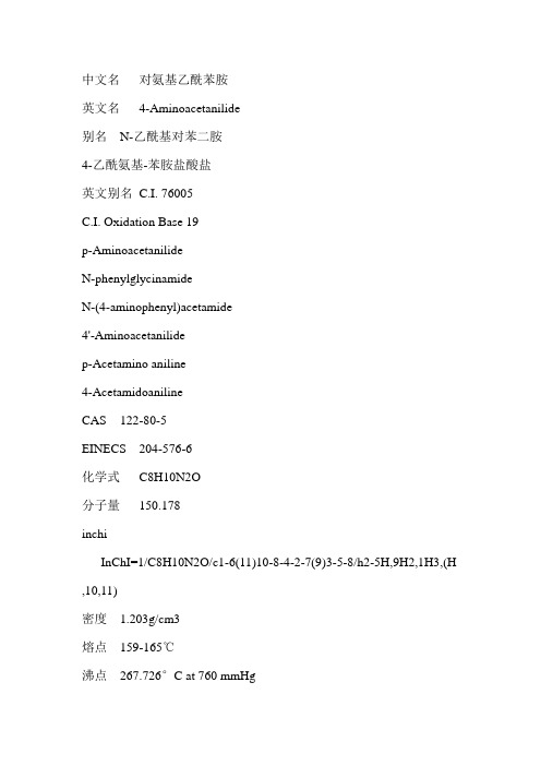
中文名对氨基乙酰苯胺英文名4-Aminoacetanilide别名N-乙酰基对苯二胺4-乙酰氨基-苯胺盐酸盐英文别名C.I. 76005C.I. Oxidation Base 19p-AminoacetanilideN-phenylglycinamideN-(4-aminophenyl)acetamide4'-Aminoacetanilidep-Acetamino aniline4-AcetamidoanilineCAS 122-80-5EINECS 204-576-6化学式C8H10N2O分子量150.178inchiInChI=1/C8H10N2O/c1-6(11)10-8-4-2-7(9)3-5-8/h2-5H,9H2,1H3,(H ,10,11)密度 1.203g/cm3熔点159-165℃沸点267.726°C at 760 mmHg闪点115.717°C水溶性0.1-1 g/100 mL at 25℃蒸汽压0.008mmHg at 25°C折射率 1.636物化性质沸点:267熔点:159 - 165产品用途用于制染料和医药中间体危险品标志Xi - 刺激性物品风险术语R36 - 刺激眼睛。
安全术语S26 - 不慎与眼睛接触后,请立即用大量清水冲洗并征求医生意见。
S39 - 戴护目镜或面具。
对氨基乙酰苯胺- 性质白色或微红色晶体。
在空气中颜色变深。
熔点165~168℃。
沸点267℃。
可燃。
微溶于水,溶于乙醇和乙醚。
对氨基乙酰苯胺- 制法N-乙酰苯胺经混酸硝化,生成对硝基-N-乙酰苯胺,然后再用铁粉还原制得对氨基-N-乙酰苯胺,反应液经中和、结晶、干燥后得成品。
对氨基乙酰苯胺- 用途制取分散黄G、直接耐酸朱红4BS、耐酸品红6B、活性蓝AG、黑色盐ANB和中性亮蓝GV等染料的中间体。
对氨基乙酰苯胺- 安全性有毒。
皮肤接触有明显的过敏现象。
应防止与皮肤直接接触及吸入粉尘。

二氨基二苯基甲烷详细信息本文详细介绍了二氨基二苯基甲烷的产品信息,包括中英文名称、别名、cas号、分子结构等基本信息,以及产品的物化性质、产品用途、产品上下游产品等综合信息,为广大化学品研究、化工产品生产制造从业者提供专业的产品信息。
本文所有信息来源化工字典。
二氨基二苯基甲烷
/detail-二氨基二苯基甲烷.html
产品介绍:
中文名称:二氨基二苯基甲烷
中文别名:
英文名称:Diaminodiphenylmethane;98%
英文别名:
CAS:19430-83-2
分子结构式:
EINECS:
分子式:C13H14N2
分子量:198.2637
风险术语:
安全术语:
物化性质:
熔点:
相对密度:1.143g/cm3 溶解性:
用途:
上游原料:
下游产品:。

n-羟乙基-2-咪唑烷酮质量标准概述说明1. 引言1.1 概述n-羟乙基-2-咪唑烷酮(简称HEMK)是一种重要的有机化合物,其分子式为C6H11NO2,具有较好的溶解性和稳定性。
作为一种重要的中间体,在许多领域中都有广泛的应用。
本文将对HEMK的质量标准进行概述和说明。
1.2 文章结构本文分为五个主要部分来探讨HEMK的质量标准。
引言部分介绍了文章的概述、目的和结构;正文部分会详细介绍HEMK的性质、用途以及质量标准制定的重要性;第三章节标题会提供进一步探讨HEMK质量标准相关内容;第四章节标题则将深入探讨更多与HEMK质量标准相关内容;最后在结论部分总结主要内容、发现结果,并对HEMK质量标准的重要性进行评价和展望未来研究方向。
1.3 目的本文旨在全面了解n-羟乙基-2-咪唑烷酮(HEMK)这种有机化合物,并对其质量标准制定进行说明。
通过本文,读者将对HEMK的性质、用途以及质量标准制定的重要性有一个整体的了解,并为未来的相关研究提供参考。
本文的目标是传达HEMK质量标准方面的核心概念和重要观点,使读者能够更好地理解和应用HEMK这一化合物。
2. 正文:2.1 n-羟乙基-2-咪唑烷酮的性质n-羟乙基-2-咪唑烷酮是一种有机化合物,其分子式为C5H9N3O2。
它是白色结晶固体,在常温下为固态形式。
该化合物的分子量为131.14 g/mol。
它在水中有良好的溶解度,并且可溶于许多有机溶剂如甲醇、乙醇和二氯甲烷等。
此外,n-羟乙基-2-咪唑烷酮具有较高的稳定性和低毒性。
2.2 n-羟乙基-2-咪唑烷酮的用途由于其特殊的化学性质和生物活性,n-羟乙基-2-咪唑烷酮在医药领域具有广泛应用。
首先,它被广泛用作药物包装材料和缓释剂。
将药物封装到n-羟乙基-2-咪唑烷酮颗粒中可以提高药物的稳定性和控制释放速率,从而增强药物治疗效果。
其次,n-羟乙基-2-咪唑烷酮还可用作药物载体和辅助溶剂,在药物传递和输送系统中发挥重要作用。

紫外线吸收剂UV-0化学名称 2,4-二羟基二苯甲酮CAS: 131-56-6分子式: C13H10O3分子量: 214规格指标及物理特性标准规格单位外观淡黄色结晶熔点℃142-147灰分%≤0.10%≤0.5挥发分透光率460nm%≥98.00500nm%≥99.00含量%≥99.00产品特点及应用紫外线吸收剂UV-0主要用于塑料等作为光稳定剂,能有效保护有机玻璃和布料,防止资料等因光照变质,也用作合成其它紫外线吸收剂的中间体储存于阴凉、干燥、通风处;避免阳光直射紫外线吸收剂UV-1化学名称:N-(乙氧基羰基苯基)-N'-甲基-N'-苯基甲脒CAS:57834-33-0英文名:Ethyl 4-[[(methylphenylamino)methylene]amino]benzoate分子式:C17H18N2O2分子量:282.34物理特性含量:≥98.5%外观:淡黄色液体水份:≤0.3%色度:≤2.5密度:1.05 g/cm沸点:416.9℃ at 760 mmHg闪光点:206℃产品应用紫外线吸收剂UV-1是一种能够有效防止双组分聚氨酯涂料、聚氨酯软泡、以及聚氨酯热塑性弹性体等高分子聚合物黄变高效的甲脒类高效紫外线吸收剂, 有明显的抗黄变作用。
其能有效吸收240~350nm的紫外光,几乎完全吸收300~330nm的紫外线,最大吸收峰为308nm,而在300-330nm这个区域聚氨酯易受到辐射而降解,所以紫外线吸收剂UV-1能有效抑制高分子聚合物的光催化降解,增强制品的色泽稳定性,延长使用寿命,尤其在聚氨酯制品如微孔泡沫、整皮泡沫、传统的硬泡、半硬泡、软泡、织物涂层、某些胶黏剂、密封胶和弹性体等聚合物中都具有优异的光稳定性能。
紫外线吸收剂UV-234化学名称:2-(2H-苯并三唑-2-基)-4,6-二(1-甲基-1-苯乙基)-苯酚CAS:70321-86-7分子式:C30H29N3O分子量:448规格指标及物理特性标准规格单位外观类白色粉末熔点℃137.0-141.0灰分%≤0.1%≤0.5挥发分透光率460nm%≥97.00500nm%≥98.00含量%≥99.00产品特点及应用紫外线吸收剂UV-234是羟基苯并三唑类紫外线吸收剂,通过把光化学作用把紫外线转化为热能。
报告摘要:甲基异丁基酮学名4-甲基-2-戊酮,简称MIBK。
MIBK是一种性能优良的中沸点溶剂,且无色、无毒,化学性能稳定,可用作硝化棉、乙基纤维素等纤维素型涂料和树脂性涂料的溶剂,也可用作其他很多领域的溶剂。
MIBK还可用作有色金属冶炼的选矿剂、炼油厂的石油脱蜡剂。
此外,MIBK还可用于生产甲基异丁基醇、偶氮二异庚腈、双-N,N-(甲基丁基甲叉)二乙撑三胺等多种下游产品。
如MIBK和4-氨基二苯胺经催化加氢缩合制得的抗氧剂4020,广泛应用于飞机、汽车及自行车轮胎、电缆生产等。
目前,世界MIBK总生产能力约34万吨/年,产量20万吨/年,主要分布在美国、西欧和日本。
1999年世界MIBK的需求量增长了1.4%。
美国MIBK的生产及消费均居世界第一位。
其具体消费构成为:约80%用作溶剂(其中85%用作涂料溶剂);15%用于橡胶抗氧剂,主要品种为N-(1,3-二甲基丁基)-N’-苯基对苯二胺(即:抗氧剂4020);2%作为专用表面活性剂用于墨水、农药配方中;其余主要用于萃取剂。
目前随着MIBK应用领域的不断扩大,其需求量也逐年增加,仅以涂料用量为例,美国涂料产量420万吨/年,耗用MIBK4.5万吨/年。
日本涂料产量220万吨/年,耗用MIBK2万吨/年。
从MIBK的应用以及环保要求来看,国际市场对MIBK的供求趋势大致处于平衡。
一方面,MIBK的应用在一些领域具有不可替代性,并且在橡胶抗氧剂上的应用有增长的势头;此外,由于高固性涂料使用量增长,需求更高溶解力的溶剂,将会刺激MIBK溶剂的需求。
另一方面,对美国MIBK市场近10年的分析表明,MIBK 的需求增长与美国的国民生产总值(GDP)的增长相当,这对生产小化工产品的企业显然不利;由于其被列入有害空气污染物(HAPS),对其应用范围还可能受到限制。
我国对MIBK的研究开发始于1964年,当时的上海溶剂厂采用丙酮三步法工艺,率先在国内建起1套200吨/年生产装置。
氨基丙氟灵质量标准
氨基丙氟灵(Prodiamine)是一种二硝基苯胺类除草剂,分子式为
C13H17F3N4O4,分子量为350.29。
其质量标准主要包括以下方面:
1.外观:氨基丙氟灵应为黄色结晶体,无其他杂质和颜色。
2.熔点:氨基丙氟灵的熔点范围应在124-125°C之间。
3.密度:氨基丙氟灵的密度应为1.4700g/cm³。
4.沸点:氨基丙氟灵的沸点应在433.0±4
5.0°C之间。
5.闪点:氨基丙氟灵的闪点应不低于-18°C。
6.分子量和化学式:氨基丙氟灵的分子量为350.29,化学式为
C13H17F3N4O4。
7.MSDS:应提供氨基丙氟灵的MSDS(物质安全数据表),包括成分、危险
性、使用注意事项等信息。
此外,氨基丙氟灵应无其他有害的物理或化学性质,如无腐蚀性、无刺激性等。
具体质量标准可能因产品用途、应用场景等因素而有所不同。