《环境微生物学》实验四 细菌的染色和细菌细胞构造的观察
- 格式:ppt
- 大小:883.50 KB
- 文档页数:18
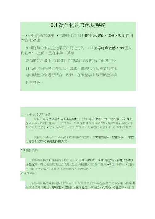
3、中性(复介)染料酸性染料与碱性染料的结合物叫做中性(复合)染料, 如瑞脱氏(Wright)染料和基姆萨氏(Gimsa)染料等,后者常川丁•细胞核的染色。
仁单纯染料这类染料的化学亲和力低,不能和被染的物质牛成盐, 英染色能力视K是否溶丁被染物而定,内为它们人多数都属于偶氮化合物,不溶于水,但溶于脂肪溶剂中,如紫丹类(Sudani))的染料。
甲染色法过程:(1)制片在于净的载玻片上漁上一小滴蒸饬I水,用接种环进行无歯操作,挑収培养物少许,概水滴中,打水混合做成想液并涂成直径约1厘米的薄层,为避免因菌数过多聚成集团,不利观察个体形态,町在载玻片■侧再加-滴水,从已涂布的菌液中再収■环于此水滴中进行稀释. 涂布成薄层,若材料为液体培养物或W体培屛物中洗卜-制备的I為液,则点接涂布丁•栽玻片上即可. >(2)自然「燥涂片最好在电温卜•使其n然干燥,为了干得快些,町将标本mi向I.. T持娥玻片一端小心在酒灯高处微微加热,便水分蒸发,但切勿紧靠火焰或加热时间过长,以防标木烤枯而变形。
(4) 染色:标本固定后•渝加染色液•约1・3分钟。
注:个涂片(或仃标本的部分)戒该浸染料Z 屮。
2、复染色法(革兰氏染色法)(1)革兰氏阳性凿和革兰氏阴性菌在化学组成和生理性质上仃很多 差别,染&反应不一样。
现在一般入为革兰氏阳性苗休内含有特殊的 核蛋白质镁盐与多糖的复合物,它与碘和结晶紫的复合物结合很牢, 不易脱色,阴性菌复合物结合程度底,吸附染料差,易脱色,这是染 色反应的主要依据。
(2) 阳杵菌菌体等电点较阴性菌为低,在相同PH 条件下进行染色, 阳竹:1為吸附«件染料}R^;. W 此不易脱去,阴件菌则相反。
所以染色 时的条件要严控制。
例如:在强碱的条件下进行染色,两类曲吸附碱性染料都多,都町 呈止反应;PH 很低时,则町都泉负反应。
(3) 两类菌的细胞瓏等对结品紫-碘复介物的通透件也不一致,阳性 闲透性小,故染色液 集色结果 特点右•碳酸复纣 菌体呈.红色 若色快,时间短美兰菌体呈兰色 若色慢,时间长,效果淸晰 堂酸铁酹体呈紫色染色迅速,着色深•水单染色过程:涂丿 ---- 固定 (5 )镜检11 12 13(3) 复染过程:(1) 制片——撫一定 (2) 脱色・初染(见询)腮蟄瞬臨•能霜淪豳瞬熾??结介的稳定与不被 革氏染&4)水洗掘療觀獄!嗪W 粼r<6)镜检:I :燥后的标木对用显微镜观察•细槽呈现第一次染色的效果紫色,< 兰氏阳性菊(Q±)oO<<呈现第二次染色的效果红色-称革兰 氏阴性菌(GOM •・,.7 :性厂;(b 用Attajt3用•霁耳《・■«*«»«««片上 M )冲洗(•革兰氏染色法■■色 nn■•«□ »»细菌呈现第一次染色的效Q 色,革兰氏阳性gf (G»•呈现第二次染色的效果红色,称革兰氏阴性菌(3'补V 'y z# ■s Jv • A 1 c。
Report of Microbiology ExperimentSmears, Simple Staining, Gram Staining and Structural StainsXXX 2012/3/25 Shandong UniversityPurpose1.Understand the rationale and procedure of all the staining methods.2.Perform and interpret all the stains.3.Learn the shapes and structures of bacteria in deep.Background1.Smear PreparationSmear is to place a thin film of bacterial cells on a slide. The smear must be fixed to kill the bacteria; coagulated proteins from the cells will cause cells to stick to the slide. Fixing denatures bacterial enzymes, preventing them from digesting cell parts, which cause the cell to break. Fixing also preserves microbes with minimal shrinkage or distortion when stained.2.Simple StainStaining procedures use only one stain are called simple stains. The dyes are usually salts composed of charged colored ions. If the chromophore is a positive ion, the stain is considered a basic stain; if it is a negative ion, it is an acidic stain.Most bacteria are stained when a basic stain permeates the cell wall and adheres by weak ionic bonds to the bacterial cell, which is slightly negatively charged.3.Gram StainGram stain is a stain that classifies bacteria as either gram-positive or gram-negative.Bacteria differ in their rate of decolorization. Those decolorize easily are referred to as gram-negative, whereas those that decolorize slowly and retain the primary stain are called gram-positive. This difference is because of the chemical and physical differences in bacteria’s cell wall. Gram-positive cell walls contain multiple layers of peptidoglycans which gram-negative cells only have a thin layer.When decolorized by alcohol, the crystal violet in gram-positive cells cannot be washed out because of the compact peptidoglycan. So there will be different colors for different bacteria. When bacteria die, their cell walls degrade and may not retain the primary stain, giving inaccurate results.4. Structural StainsStructural stains can be used to identify and study the structure of bacteria when there is no electron microscope.Endospores are “resting bodies” of several genera of bacteria. They do not metabolize and are resistant to heating, various chemicals, and many harsh environmental conditions. To know whether a bacterium is an endospore-former and also the position of the endospores is helpful. Endospores are impermeable to most stains, so heat is usually applied to drive the stain into the endospore.Once stained, the endospores do not readily decolorize.Flagella are thin proteinaceous structures that originate in the cytoplasm and project out from the cell wall. They are very frafgile (usually 20-50nm) and arenot visible with a light microscope. But when they are coated with a mordant, which increases their diameter, they can be seen with light microscopes.The presence and location of fragella are helpful in identifying and classifying bacteria. There are two main types of fragella: peritrichous and polar.MaterialsCompound light microscopesSlidesInoculating loopAlcohol burnerAbsorbent paperMethylene blue(simple stain)Gram-staining reagents(Crystal violet, Gram’s iodine, Ethanol, Safranin)Endospore stain reagents(Malachite green and Safranin)Flagella stain reagents(A and B)Distilled waterCulturesEscherichia coli slantStaphylococcus aureus slantBacillus subtilis slant(24h&72h)Clostridium sporogenes slant(24h&72h)Pseudomonas aeruginosa slantProcedureⅠPreparing smears1.Clean the slides.2.Put a drop of distilled water on a slide.3.Sterilize the inoculating loop.ing the cooled loop, scrape a small amount of the culture off the slant andemulsify the cells in the drop of water.5.Let the smears dry.6.Pass the slide quickly through the blue flame with smear side up two or threetimes.ⅡSimple Stain(E.coli,S.aureus and B.subtilis)1.Prepare smears.2.Cover the smears with Methylene blue and leave them for about 1min.3.Gently wash off the Methylene blue with water.4.Blot the slides dry and examine stained smears microscopically.ⅢGram Stain(E.coli,S.aureus and B.subtilis)1.Prepare fixed smears.2.Cover the smears with crystal violet for 1-2 min.3.Gently wash off the Crystal violet with water.4.Cover the smears with Gram’s iodine for 1 min.5.Gently wash off the Gram’s iodine with water.6.Decolorize the smears with ethanol.7.Gently wash off the ethanol.8.Cover the smears with safranin for 2 min.9.Gently wash off the safranin.10.Blot the slides dry and examine stained smears microscopically.ⅣStructural StainsThe endospore stain(B.subtilis and C.sporogenes(24h&72))1.Prepare fixed smears of bacteria.2.Place a piece of absorbent paper over the smears.3.Cover the paper with Malachite green.4.Steam the slides for 5 min.5.Wash the smears with water.6.Cover the smears with Safranin for 2 min.7.Wash the smears with water and blot them dry.8.Examine stained smears with a microscope.The flagella stain(B.subtilis, E.coli and P.aeruginosa)1. Place a drop of distilled water on a side of a slide.2. Lightly touch the drop of water with the bacteria without touching the slide.3. Tilt the slide so the drop will flow to the opposite side of the slide.4. Allow the slide to air dry.5. Cover the dried smear with dye liquor A for 5 min.6. Gently wash off the dye liquor A with distilled water.7. Gently wash the slide with dye liquor B until brown color appears.8. Wash the smears with water and blot them dry.9. Examine stained smears with a microscope.ResultsSimple stainE.coli 1000×S.aureus 1000×B.subtilis 1000×Gram stainE.coli and S.aureus 1000×Unknown bacteria 1000× Gram positive & gram negative bacillusUnknown bacteria 1000× Gram negative bacillusEndospore stainB.subtilis 1000× Central, swollen endosporesC.sporogenes 1000× 24h Terminal, swollen endosporesC.sporogenes 1000× 72h Free endosporesFlagella stainB.subtilis 1000× Peritrichous flagellaE.coli 1000× Peritrichous flagellaP.aeruginosa 1000× Monotrichous flagellumRethink1.The microscopes should have a blue optical filter in the condenser to make surethe blank filed is white. That will be helpful to recognize the color of different stains.2.Heat-fixing is important in smear preparation to keep the shape of cells. Whenperforming flagella stains, I found that many bacterial cells looks larger than their normal size in the field. It may because of autolysis when cells die.3.When performing a Gram stain, the time of decolorizing should be handle toprevent excessive decolorizing.4.When performing flagella stains, violent shaking should be avoided to prevent theflagella from coming off. According to my results, bacteria with few flagellas will easily lose their flagellas when being stained.此实验报告存在一部分小错误和疏漏,比如个别的语法错误、用词不当,裁剪的图片未标明比例尺等。
实验二细菌的染色方法和细胞形态结构的观察姓名:马天柔学号:120109112 日期:2014/03/30一·目的:1了解生物染色的一般知识。
2学习细菌的涂片,染色的基本技术,并掌握细菌的简单染色方法和革兰氏染色法。
3通过染色观察细菌细胞的形态和结构。
二·染色的一般知识:细菌个体不仅十分微小,而且是无色半透明的,在明视野显微镜下很难看的清楚。
因此,要借助不同的染色方法,使细菌细胞或某些结构着色,与背景形成明显反差。
染色的标本不仅在显微镜下易于观察,还有鉴别细菌不同的细胞结构的作用。
三·细菌的革兰氏染色法(一)1器材:显微镜,酒精灯,载玻片,镊子,接种耳,蒸馏水,香柏油,酒精,火柴,吸水纸,擦镜纸。
2结晶紫染色液,碘液,95%酒精,番红染色液。
3大肠杆菌,金黄色葡萄球菌。
(二)步骤:涂片-干燥-固定-结晶紫染色1min-水洗-碘液煤染1min-水洗-95%酒精脱色20-30s-番红染色4-5min-水洗-干燥-镜检1涂片:用镊子取浸泡在酒精中干净的载玻片,烧去酒精,在玻片中央加一滴清水,通过无菌操作方法从斜面上分别挑取大肠杆菌和金黄色葡萄球菌和清水滴混合均匀。
2干燥:涂片可让其在室温下干燥,必要时可将涂片朝上,小心的在酒精灯火焰高处稍烘,以助水分蒸发,快速干燥。
但切勿靠近火焰,将标本烤焦。
3固定:一般常采用加热固定法,即手持玻片一端,涂片朝上,在火焰外层通过3-4次,以玻片反面触及皮肤不觉得太烫为度。
待冷却后,在进行染色。
4染色:待固定涂片完全冷却后,在涂面上滴加染色液,染色液要布满整个涂面。
5水洗:染色时间到后,倾去染色液,斜置载玻片,用干净的细流水冲去多余的染料。
6干燥:用吸水纸吸取玻片上的水分,让其自然干燥。
7镜检:用油镜观察,并记录观察结果。
油镜下观察的大肠杆菌和金黄色葡萄球菌(100×10)四·心得体会:老师看了我染色后的标本后说班里的第一个模板出来了,心里是很高兴的,我按照步骤一步步染色,还算是染的均匀。


实训四细菌的染色与形态观察一、实验目的1、学习细菌涂片技术和无菌操作技术;2、掌握细菌的简单染色法,初步认识细菌的形态特征;3、了解革兰氏染色的原理,学习并掌握革兰氏染色的方法;4、进一步熟练显微镜的使用。
二、实验原理细菌个体微小,无色且较透明,折射率低,在普通光学显微镜下不易识别,因此必须借助染色法使菌体着色,将其折射率增大,使其与背景形成明显的色差,在显微镜下用油镜观察,可清楚观察到细菌的一般形态结构及特殊结构。
根据细菌个体形态观察的不同要求,可用简单染色法或革兰氏染色法进行染色观察。
(一)简单染色法简单染色法是最基本的染色方法,是利用单一染料进行细菌染色的方法。
此法操作简便,适于菌体一般形状和细菌排列的观察,但不能辨别细胞的构造。
常用碱性染料进行简单染色,因为在中性、碱性或弱酸性溶液中,细菌细胞通常带负电荷,而碱性染料在电离时,其分子的染色部分带正电荷(酸性染料电离时,其分子的染色部分带负电荷),因此碱性染料的染色部分很容易与细菌结合使其着色。
经染色后的细菌细胞与背景形成鲜明的对比,在显微镜下易于识别。
常用的简单染色染料有草酸铵结晶紫染液、吕氏碱性美蓝(亚甲基蓝)、石炭酸复红染液等。
(二)革兰氏染色法革兰氏染色法是细菌学上最重要的鉴别染色法。
由于细菌细胞壁的化学组成和结构不同,经革兰氏染色后,呈现不同的染色反应,可将所有细菌区分为两大类:革兰氏阳性细菌(染色反应呈蓝紫色),用G+表示;革兰氏阴性细菌(染色反应呈红色),用G-表示。
革兰氏阳性细菌的细胞壁主要是由肽聚糖形成的网状结构组成的,脂类含量低。
在染色过程中,用乙醇处理时,由于脱水而引起网状结构中的孔径变小,通透性降低,使结晶紫-碘复合物被保留在细胞内而不易脱色,因此呈现蓝紫色。
革兰氏阴性细菌的细胞壁中肽聚糖含量低,而脂类物质含量高,脂溶剂乙醇溶解脂类物质,使得细胞壁的通透性增加,结晶紫-碘复合物易被乙醇抽出而脱色,然后又被染上了复染液(番红)的颜色,因此呈现红色。


实验二细菌的染色和细菌细胞构造的观察染色是观察微生物的一种重要方法。
由于微生物细胞含有大量水分,一般在80%-90%以上,对光线的吸收和反射与水溶液的差别不大,与周围背景没有明显的明暗差。
所以,除了观察活体微生物细胞的运动性和直接计算菌数外,绝大多数情况下都必须经过染色后,才能在显微镜下进行观察。
一、目的要求1. 学习微生物涂片、染色的基本技术和无菌操作技术。
2. 掌握细菌的一般染色法和革兰氏染色法。
3. 进一步熟练显微镜油镜的使用技术。
4. 观察和识别细菌细胞的特殊构造。
二、染色剂和染色的原理微生物染色的基本原理,是借助物理因素和化学因素的作用而进行的。
物理因素如细胞及细胞物质对染料的毛细现象、渗透、吸附作用等。
细菌的等电点较低小pH值大约在2-5之间,故在中性、碱性或弱酸性溶液中,菌体蛋白质电离后带负电荷;而碱性染料电离时染料离子带正电荷。
因此,带负电荷的细菌常和带正电荷的碱性染料进行结合,所以在细菌学实验室中常用碱性染料进行染色。
染色剂是一种供染色用的有机化合物,当前普遍采用的染色剂是含有苯环的有机化合物,染料分子都由苯环、连接在苯环上的染色基团(或称呈色基团,色基和助色基因(或称作用基团三部分组成、助色基团具有电离特性,电离后带有正或负电荷的染料离子便能与细胞有较牢固地结合,使其呈现颜色。
染料可按其电离后染料离子所带电荷的性质分为酸性染料、碱性染料、中性(复合染料和单纯染料四大类。
(1酸性染料这类染料电离后染料离子带负电荷、如酸性复红、刚果红、伊红、藻红、苯胺黑等,可与碱性物质结合成盐。
(2碱性染料这类染料电离后染料离子带正电荷、可与酸性物质结合成盐。
微生物实验室一般常用的碱性染料有碱性复红、中性红、孔雀绿、番红、结晶紫、美兰、甲基紫等,在一般的情况下,细菌易被碱性染料染色。
(3中性(复合染料酸性染料与碱性的结合物叫做中性(复合染料,如伊红、美兰等,瑞脱氏(Wright 染料和基姆萨氏(Gimasa染料等,后者常用于细胞核的染色。

