
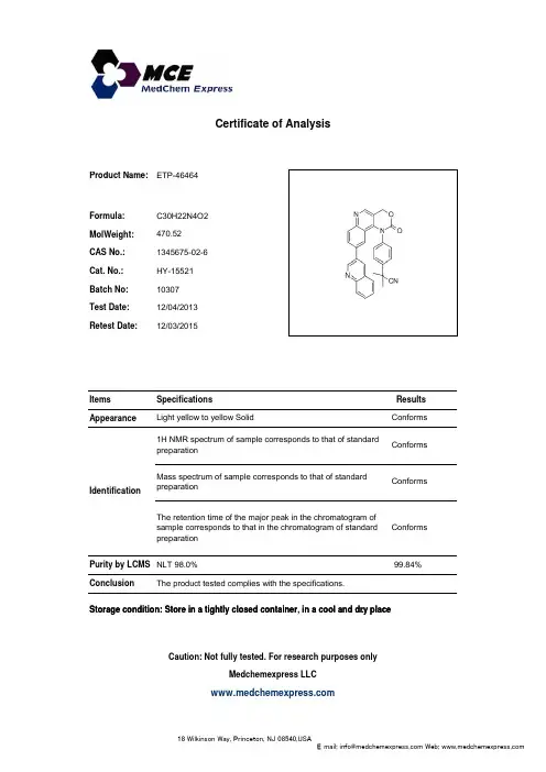
ETP-46464_1345675-02-6_CoA_MedChemExpress
- 格式:pdf
- 大小:52.18 KB
- 文档页数:1

Endotoxin Removal Solution Catalog Number E4274Product DescriptionEndotoxins are lipopolysaccharides (LPS), a major component of the Gram-negative bacterial cell wall, and are commonly found as contaminants in plasmid DNA preparations from E. coli. Endotoxins are large, negatively charged molecules that co-purify with DNA on ion exchange and size exclusion columns and in CsCl banding. Endotoxins are extremely potent stimulators of the mammalian immune system and are toxic to primary cells and to animals. The endotoxin toxicity is an obstacle to in vitro and in vivo transfection experiments.Non-ionic detergents, traditionally used for separation of integral membrane proteins,1 can be utilized for removal of endotoxins from DNA solutions by phase separation.2The solubility behavior of a detergent in a dilute, aqueous solution at physiological salt and pH conditions is strongly dependent upon the temperature of the solution. At low temperatures, the detergent forms a clear, micellar solution, but above the cloud point temperature, the micelles form larger, turbid aggregates and ultimately fuse to form a separate phase. The lower phase is detergent-enriched and the detergent-depleted upper phase contains detergent at a concentration slightly above the critical micellar concentration (CMC). Amphiphilic and hydrophobic molecules associated with the micelles of the detergent will aggregate within the detergent-enriched phase, while the soluble, hydrophilic molecules will remain in the detergent-depleted upper phase.Extraction of endotoxin contaminated DNA solutions with the appropriate non-ionic detergent will separate the hydrophilic DNA from the amphiphilic endotoxin. The amphiphilic endotoxin will associate with the lower phase, while the DNA will remain in the upper, detergent-depleted phase.2Reagents and equipment required, but not provided • Water, Molecular Biology Reagent, Catalog Number W4502• E-TOXATE® Water, Catalog Number 2107, or Tris-EDTA (TE) buffer 100×, Catalog NumberT9285• DNA solution (0.5 ml), ~ 1 mg/ml in E-TOXATE®Water or TE buffer• 3 M sodium acetate solution, pH 7.5.• 2-Propanol, Catalog Number I9516, or Ethanol, 190 proof, Catalog Number E7148; 200 proof, CatalogNumber E7023• 70% Ethanol• E-TOXATE®reagents Kits, Catalog Numbers 210A1, 210B1 or 210C1• Ice bucket• Heat block or incubator at 37 °C• Microcentrifuge at room temperature• 1.5 or 2 ml sterile microcentrifuge tubes• Endotoxin-free pipet tips (40-200 µl, 200-1000 µl) Precautions and DisclaimerThis product is for R&D use only, not for drug, household, or other uses. Please consult the Material Safety Data Sheet for information regarding hazards and safe handling practices.StorageStore at room temperature.Note: Removal of endotoxins from DNA preparations can be performed either during the final stage of DNApreparation, or during an earlier stage.Procedures for Endotoxin RemovalDuring the final stage of DNA preparationNote: The procedure described below was performed on plasmid DNA produced in E. coli DH5α cells.• Losses of up to 50% of the DNA are expected. • Use of a DNA concentration above therecommended 1 mg/ml reduces the efficiency ofthe procedure.1. Pipette 500 µl of the DNA solution into a sterilemicrocentrifuge tube.2. Add 50 µl of the 3 M sodium acetate solution to theDNA sample.3. Incubate on ice for 5 minutes.4. Add 100 µl of cold Endotoxin Removal Solution.5. Mix thoroughly and incubate on ice for 10 minutes.The solution should be light blue and clear.6. Incubate the tube at 37 °C for 20 to 30 minutes oruntil the phases separate.7. Spin for 5 minutes at 3000 x g in themicrocentrifuge. The upper phase is colorless and clear, while the lower phase is blue.8. Carefully transfer the upper phase containing theDNA to a clean microcentrifuge tube.9. Repeat steps 4 through 8 twice.10. Add 0.6× volume of 2-propanol. Mix by inversion atroom temperature and centrifuge at 15,000 x g for30 minutes at 4 °C. Alternatively, add2.5× volumes of ethanol. Incubate overnight at –20°C or 20 minutes at –70 °C and centrifuge at15,000 x g for 30 minutes at 4 °C.11. Carefully remove the supernatant12. Wash the DNA pellet twice with cold 70% ethanol.Remove the supernatant.13. Air-dry the pellet.14. Suspend the DNA in 100 µl of endotoxin free wateror TE buffer.15. Determine DNA concentration and endotoxin levelsusing endotoxin assay reagents and compare tothe starting material. During an earlier stage of DNA preparationThis procedure is based on the alkaline lysis of E. coli DH5α cells.3 The endotoxins are removed immediately after alkaline cell lysis, neutralization, and a clarification step. The resulting high salt solution is suitable for the endotoxin removal step. It is performed under “endotoxin free” conditions. The plasticware used is either sterile and disposable, or NaOH-treated. The buffers are prepared with endotoxin free water.1. Add the Endotoxin Removal Solution (0.2× volume)to the cold, crude DNA solution.2. Incubate on ice and mix occasionally by inversionto obtain a homogenous, clear blue solution3. Incubate at 37 °C for 20 to 30 minutes until thephase separation is obvious.4. Spin for 5 minutes at low speed (3000 x g) at roomtemperature.5. Transfer the upper aqueous phase to an endotoxinfree container.6. Proceed with the DNA purification by any method.Use endotoxin-free buffers and containers. References1. Bordier, C., J. Biol. Chem., 256, 1604-1607, (1981).2. Cotten, M. et al., Gene Therapy, 1, 239-246,(1994).3. Sambrook et al., Molecular Cloning, a LaboratoryManual, 2nd Ed. p. 1.38RK,PHC 09/05-1Sigma brand products are sold through Sigma-Aldrich, Inc.Sigma-Aldrich, Inc. warrants that its products conform to the information contained in this and other Sigma-Aldrich publications. Purchaser must determine the suitability of the product(s) for their particular use. Additional terms and conditions may apply. Please see reverse side ofthe invoice or packing slip.。

碧云天生物技术/Beyotime Biotechnology订货热线:400-168-3301或800-8283301订货e-mail:******************技术咨询:*****************网址:碧云天网站微信公众号生物素标记EMSA探针-β-Catenin/TCF (0.2μM)产品编号产品名称包装GS018B 生物素标记EMSA探针-β-Catenin/TCF (0.2µM) 200µl产品简介:生物素标记EMSA探针-β-Catenin/TCF是用于EMSA(也称gel shift)研究的生物素(Biotin)标记的β-Catenin/TCF consensus oligonucleotide。
这个生物素标记的双链寡核苷酸含有公认的β-Catenin/TCF结合位点,可以用作EMSA研究时的探针。
β-Catenin/TCF consensus oligo的序列如下:5'-CCC TTT GAT CTT ACC-3'3'-GGG AAA CTA GAA TGG-5'本生物素标记EMSA探针已经过纯化,可以直接用于EMSA结合反应。
本生物素标记EMSA探针可以和碧云天的化学发光法EMSA试剂盒(GS009)配套使用。
一个包装的生物素标记探针可以进行约200-400个样品的EMSA检测。
包装清单:产品编号产品名称包装GS018B 生物素标记EMSA探针-β-Catenin/TCF (0.2µM) 200µl—说明书1份保存条件:-20ºC保存,一年有效。
注意事项:避免加热到40ºC以上,温度过高会导致双链DNA探针解聚成单链。
而单链无法用于EMSA研究。
对于基于生物素标记的EMSA检测的详细操作可以参考碧云天的化学发光法EMSA试剂盒(GS009)的使用说明。
本产品仅限于专业人员的科学研究用,不得用于临床诊断或治疗,不得用于食品或药品,不得存放于普通住宅内。

液体样本游离胆固醇酶法测定试剂盒E1006描述:血浆总胆固醇包括2/3的胆固醇酯和1/3的游离胆固醇,是血脂的重要组分,二者与心脑血管疾病以及多种病理状态密切相关。
本试剂盒采用世界卫生组织(WHO)、美国FDA、中国《全国临床检验操作规程》推荐的总胆固醇和游离胆固醇检测方法,灵敏度高,检测范围20~5000 µmol/L。
可靠性和重复性俱佳。
原理:本试剂盒测定方法采用CHOD-PAP终点方法结合经典GPO Trinder酶学反应【1】,其原理为:(1) 胆固醇酯酶分解胆固醇酯为游离胆固醇。
(2) 胆固醇氧化酶将游离胆固醇氧化,产生过氧化氢。
(3) 在过氧化物酶催化下生色底物转化为苯醌亚胺,光密度值与胆固醇浓度成正比。
测定样本范围:血清、培养基组成:(1) R1试剂40 ml (2) R2试剂10 ml (3) 5 mmol/L胆固醇标准品0.5 ml储存条件:4ºC保存6个月有效所需设备:721、722型可见光分光光度计、酶标仪、生化分析仪。
最佳工作波长550-555nm,如仪器无此波长建议优先选用570 nm,次选530、490nm。
常规比色杯即可。
操作步骤:一、工作溶液配制: 按4:1比例,取4 ml试剂R1与1 ml试剂R2混合即可,立即使用或4ºC保存<1天,变色弃去。
二、样本处理:培养基:取无酚红无血清细胞培养液,4ºC,10,000g离心5 min,取上清测定。
新鲜培养基4ºC存放不宜超过1小时,若暂时无法测定可70ºC加热10 min后冻存,测定前离心。
血液:新鲜血液4ºC,2000 g离心5 min得到血浆,非抗凝血4ºC放置2小时得到血清。
可4ºC 存放数天或−20 ºC半年。
如超过线性范围用生理盐水1:1~1:5稀释后测定。
三、标准品稀释:5 mM胆固醇标准品用无水乙醇倍比稀释为2500、1250、625、312.5、156、78、39 µmol/L。

3种常用碳青霉烯类抗生素血药浓度UPLC-MS/MS检测方法的建立Δ秦怡1*,张瑞霞2,吕雅瑶2,翁莉莉1,张弋2 #(1.天津医科大学一中心临床学院,天津 300192;2.天津市第一中心医院药学部,天津 300192)中图分类号 R917;R978.1文献标志码 A 文章编号 1001-0408(2024)03-0343-05DOI 10.6039/j.issn.1001-0408.2024.03.14摘要目的建立3种临床常用碳青霉烯类抗生素——厄他培南(ETP)、亚胺培南(IPM)、美罗培南(MEM)血药浓度检测的超高效液相色谱-质谱联用(UPLC-MS/MS)法。
方法血浆样品经甲醇沉淀蛋白后,以3种抗生素的稳定性同位素(ETP-D4、IPM-D4、MEM-D6)为内标,采用ACQUITY UPLC BEH C18(2.1 mm×50 mm,1.7μm)色谱柱分离;流动相为98%乙腈+2%水+0.1%甲酸和98%水+2%乙腈+0.1%甲酸,梯度洗脱;流速为0.3 mL/min;柱温为40 ℃;采用正离子、多反应监测模式进行扫描分析。
结果该方法专属性良好,在ETP、IPM、MEM 0.2~200、0.1~100、0.1~100μg/mL范围内线性良好(r2≥0.993),批内、批间精密度和准确度良好(RE均≤5.14%,RSD均≤11.15%),基质效应、提取回收率较一致(RSD≤12.99%)。
结论本实验建立了一种可以同时定量ETP、IPM、MEM血药浓度的UPLC-MS/MS法,该方法样品前处理简单、检测时间短、所需样品量少,可满足临床需求。
关键词碳青霉烯类抗生素;超高效液相色谱-质谱联用;血药浓度;厄他培南;亚胺培南;美罗培南Establishment of UPLC-MS/MS method for the determination of plasma concentration of three common carbapenem antibioticsQIN Yi1,ZHANG Ruixia2,LYU Yayao2,WENG Lili1,ZHANG Yi2(1. First Central Clinical College of Tianjin Medical University,Tianjin 300192,China;2. Dept. of Pharmacy,Tianjin First Central Clinical Hospital,Tianjin 300192, China)ABSTRACT OBJECTIVE To establish a UPLC-MS/MS method for the determination of plasma concentration of three carbapenem antibiotics,i.e. ertapenem (ETP),imipenem (IPM)and meropenem (MEM).METHODS After protein precipitation with methanol,the plasma samples were separated by ACQUITY UPLC BEH C18column (2.1mm×50mm,1.7μm)using stable isotopes of three antibiotics (ETP-D4,IPM-D4,MEM-D6)as the internal standard. The mobile phases were 98%acetonitrile +2% water +0.1%formic acid and 98%water +2%acetonitrile +0.1%formic acid,by gradient elution. The flow rate was 0.3mL/min and the column temperature was 40 ℃. Scanning analysis was performed in the positive ion and multiple reaction monitoring mode. RESULTS The method had good specificity,good linearity (r2≥0.993)in the range of 0.2-200,0.1-100and 0.1-100μg/mL of ETP,IPM and MEM,and good intra-batch and inter-batch precision and accuracy (all RE≤5.14%,all RSD≤11.15%),the matrix effect and extraction recovery were consistent (RSD≤12.99%). CONCLUSIONS This study establishes the UPLC-MS/MS method to simultaneously quantify the plasma concentration of ETP,IPM and MEM. The method has the advantages of simple pretreatment, short detection time and small sample quantity to meet clinical requirement.KEYWORDS carbapenem antibiotics; UPLC-MS/MS; plasma concentration; ertapenem; imipenem; meropenem碳青霉烯类抗生素具有抗菌谱广、抗菌活性强、耐药率低的特点,已成为治疗重症感染的主要选择。