Qualitative Diagnosis of Systems With Models That Include a Class of Algebraic Loops
- 格式:pdf
- 大小:280.92 KB
- 文档页数:22

利用Petri网特征结构的故障诊断方法叶丹丹;罗继亮【摘要】For fault diagnosis in large complex systems,a on-line fault diagnose method is proposed to solve the problem of high computational complexity.First,modeled a Petri net model.Secondly,proposed the strict minimal place-invariant and the set of characteristic place-invariant,so that might describe the structure information of Petri net model.Finally, based on the set of characteristic place-invariants,the failure function for any current marking is proposed.And then,uti-lized this failure function to diagnose and locate the faults.The result shows that this fault diagnosis method with the structure information dose not need traverse all states space of system.Furthermore,this method is with the computa-tional complexity of polynomial,which makes this method meet the real time requirements.%为解决大规模复杂系统故障诊断中计算复杂性高的问题,提出一种基于 Petri网的在线故障诊断方法。

海绵窦海绵状血管瘤的CT、MRI诊断和鉴别诊断(作者:___________单位: ___________邮编: ___________)【摘要】目的探讨海绵窦海绵状血管瘤的CT和MRI表现及鉴别诊断。
方法搜集11例经手术病理证实的海绵窦海绵状血管瘤的CT和MRI影像资料,全部病例均行头颅MRI平扫及增强扫描,其中5例有CT检查。
结果 11例海绵状血管瘤均位于海绵窦区,呈单发病灶,右侧4例,左侧7例。
CT平扫为稍高密度影,增强扫描呈明显强化;MRI平扫表现为长T1均匀性或不均匀性低信号,明显长T2高信号,增强后病灶明显强化8例,中度强化3例,CT和MRI 定位诊断正确率为100%,CT定性诊断正确率为20%(1/5),MRI定性诊断正确率为54.5%(6/11)。
结论 CT表现与其它实体肿瘤相比无特异性,定性诊断困难;MRI表现呈底向外的哑铃型或类园型长 T1长T2信号改变较具特征性,增强表现为非常显著的强化,对定性具有决定诊断的作用。
【关键词】海绵窦;海绵状血管瘤;体层摄影术;X线计算机;磁共振成像[Abstract] Objective To study CT and MRI findings anddifferential diagnosis of cavernous sinus Cavernous angioma. Methods 11 cases with cavernous sinus cavernous angiomas were all proved by operation and pathology, 11 cases were examined all by MRI plan scan and enhancement scan, 5 cases were examined by CT. Results 11 cases cavernous angiomas all located in the cavernous sinus, The tumors were a little hyperdensity on CT plan scan. On MRI plan scan, the tumors displayed long T1 low-signal and high-signal intensity on T2WI. 8 cases intensified obviously and 3 cases intensified moderately after strengthening. The rate of CT and MRI in cavernous angiomas qua location was 100%, the rate of CT qua diagnosis was 20%(1 of 5), the rate of MRI qua diagnosis was 54.5%(6 of 11). Conclusion The CT resembles in carvenous angiomas has no particularity compared with the other entity tumors.The MRI image of the bottom outivardly of bottle gourd form or similar round and long T1 and long T2 signal changes was the characteristic of carverous angiomas. It enhances remarkably.MRI can make qualitative diagnosis of carverous angiomas.[Key words] cavernous sinus; cavernous angioma; tomography; X-ray computed; magnetic resonance imaging颅内海绵状血管瘤(cavernous angioma,CA)是一种血管畸形性良性肿瘤,分为脑内型和脑外型两种类型,脑外型海绵状血管瘤较少见,约占颅内海绵状血管瘤的0.4%~2.0%[1],常位于海绵窦,影像上常与此处的其它肿瘤混淆,导致临床的误诊和误治,本文总结笔者近5年来收集经手术病理证实的11例脑外海绵状血管瘤的CT和MRI资料,分析其CT和MRI表现,目的为提高该病的诊断和鉴别诊断。

专利名称:Diagnostic system发明人:Sampath, Meera,Godambe, Ashok,Jackson,Eric,Mallow, Edward W.申请号:EP00308026.4申请日:20000915公开号:EP1085416A2公开日:20010321专利内容由知识产权出版社提供专利附图:摘要:Through a combination of a hybrid diagnostic scheme based on qualitative and quantitative technologies, at least component level status information can be obtained about a machine, such as an electronic device. In particular, qualitative model baseddiagnostic technologies is used in conjunction with quantitative analysis techniques and signature analysis to achieve accurate and reliable diagnosis of components and systems down to the individual component level, for example, down to the consumer replaceable unit level. Thus, the hybrid diagnostic methodology exploits the diagnostic information content in already available system signals via intelligent processing and allows for diagnosis with minimal sensor requirements. Furthermore, the diagnostic methodology allows for a self-diagnosing machine having diagnostic intelligence which in turn can reduce service time, service costs, increased machine up time and improve customer satisfaction.申请人:Xerox Corporation地址:Xerox Square - 20A, 100 Clinton Avenue South Rochester, New York 14644 US 国籍:US代理机构:Skone James, Robert Edmund更多信息请下载全文后查看。

1、抗⽣素医嘱[Antibiotic order] Prophylaxis [预防性⽤药] Duration of oder[⽤药时间] 24hr Procedure[操作,⼿术] Empiric theraphy [经验性治疗] Suspected site and organism[怀疑感染的部位和致病菌] 72hr Cultures ordered[是否做培养] Documented infection[明确感染] Site and organism[部位和致病菌] 5days Other[其他] Explanation required [解释理由] 24hr Antibiotic allergies[何种抗⽣素过敏] No known allergy [⽆已知的过敏] Drug+dose+Route+frequency[药名+剂量+途径+次数] 2、医嘱⾸页[Admission / transfer] Admit / transfer to [收⼊或转⼊] Resident [住院医师] Attending[主治医师] Condition [病情] Diagnosis[诊断] Diet [饮⾷] Acitivity [活动] Vital signs[测⽣命体征] I / O [记进出量] Allergies[过敏] 3、住院病历[case history] Identification [病⼈⼀般情况] Name[性名] Sex[性别] Age [年龄] Marriage[婚姻] Person to notify and phone No.[联系⼈及电话] Race[民族] I.D.No.[⾝份证] Admission date[⼊院⽇期] Source of history[病史提供者] Reliability of history[可靠程度] Medical record No[病历号] Business phone No.[⼯作单位电话] Home address and phone No.[家庭住地及电话] Chief complaint[主诉] History of present illness[现病史] Past History[过去史] Surgical[外科] Medical[内科] Medications[⽤药] Allergies[过敏史] Social History[社会史] Habits[个⼈习惯] Smoking[吸烟] Family History[家族史] Ob/Gyn History[ 婚姻/⽣育史] Alcohol use[喝酒] Review of Aystems[系统回顾] General[概况] Eyes,Ears,Nose and throat[五官] Pulmonary[呼吸] Cardiovascular[⼼⾎管] GI[消化] GU[⽣殖、泌尿系统] Musculoskeletal[肌⾁⾻骼] Neurology[神经系统] Endocrinology[内分泌系统] Lymphatic/Hematologic[淋巴系统/⾎液系统] Physical Exam[体检] Vital Signs[⽣命体征] P[脉博] Bp[⾎压] R[呼吸] T[温度] Height[⾝⾼] Weight[体重] General[概况] HEENT[五官] Neck[颈部] Back/Chest[背部/胸部] Breast[*] Heart[⼼脏] Heart rate[⼼率] Heart rhythm[⼼律] Heart Border[⼼界] Murmur[杂⾳] Abdomen[腹部] Liver[肝] Spleen[脾] Rectal[直肠] Genitalia[⽣殖系统] Extremities[四肢] Neurology[神经系统] cranial nerves[颅神经] sensation[感觉] Motor[运动] *Special P.E. on diseased organ system[专科情况] *Radiographic Findings[放射] *Laboratory Findings[化验] *Assessment[初步诊断与诊断依据] *Summary[病史⼩结] *Treatment Plan[治疗计划] 4、输⾎申请单[Blood bank requisition form] (1)reason for infusion[输⾎原因] 红细胞[packed red cells, wshed RBCs]: *Hb20% blood volume lost [>20%⾎容量丢失] *cardio-pulmonary bypass with anticipated Hb 10 units[输⾎10单位以上者] *platelet count 15units[输⾎>15个单位] *warfarin or antifibrinolytic therapy with bleeding[华法令或溶栓治疗后出⾎] *DIC[⾎管内弥漫性凝⾎] *Antithrombin III dficiency[凝⾎酶III 缺乏] (2)输⾎要求[request for blood components] *patient blood group[⾎型] *Has the patient had transfusion or pregnancy in the past 3 months [近3个⽉, 病⼈是否输过⾎或怀孕过?] *Type and crossmatch[⾎型和⾎交叉] *Units or ml[单位或毫升] 5、出院⼩结[discharge summary] Patient Name[病⼈姓名] Medical Record No.[病历号] Attending Physician[主治医⽣] Date of Admission[⼊院⽇期] Date of Discharge[出院⽇期] Pirncipal Diagnosis[主要诊断] Secondary Diagnosis[次要诊断] Complications[并发症] Operation[⼿术名称] Reason for Admission[⼊院理由] Physical Findings[阳性体征] Lab/X-ray Findings[化验及放射报告] Hospital Course[住院诊治经过] Condition[出院状况] Disposition[出院去向] Medications[出院⽤药] Prognosis[预后] Special Instruction to the Patient(diet, physical activity)[出院指导(饮⾷,活动量)] Follow-up Care[随随访] 6、住院/出院病历⾸页[Admission/discharge record] Patient name[病⼈姓名] race[种族] address[地址] religion[宗教] medical service[科别] admit (discharge) date[⼊院(出院)⽇期] Length of stay [住院天数] guarantor name [担保⼈姓名] next of kin or person to notify[需通知的亲属姓名] relation to patient[与病⼈关系] previous admit date[上次住院⽇期] admitting physician [⼊院医⽣] attending phgsician[主治医⽣] admitting diagnosis[⼊院诊断] final (principal) diagnosis[最终(主要)诊断] secondary diagnosis[次要诊断] adverse reactions (complications)[副作⽤(合并症)] incision type[切⼝类型] healing course[愈合等级] operative (non-operative) procedures[⼿术(⾮⼿术)操作] nosocomial infection[院内感染] consutants[会诊] Critical-No. of times[抢救次数] recovered-No. of times[成功次数] Diagnosis qualitative analysis[诊断质量] OP.adm.and discharge Dx concur [门诊⼊院与出院诊断符合率] Clinical and pathological Dx concur[临床与病理诊断符合率] Pre- and post-operative Dx concur [术前术后诊断符合率] Dx determined with in 24 hours (3 days) after admission[⼊院后24⼩时(3 天)内确诊] Discharge status[出院状况] recovered[治愈] improved[好转] not improved[未愈] died [死亡] Dispositon[去向] home[家] against medical ad[⾃动出院] autosy[⼫检] transferred to[转院到]。

·临床研究·128层螺旋CT 结肠成像在结直肠癌诊断中的应用曹登攀李佩如柳泽华施向阳王骄阳徐芳陈涛徐升DOI :10.13558/ki.issn1672-3686.2021.002.009基金项目:金华市科学技术研究计划项目(2018-4-152)作者单位:321300浙江永康,永康市第一人民医院放射科(曹登攀、李佩如、柳泽华、施向阳、王骄阳),消化内科(徐芳、徐升),肛肠外科(陈涛)[摘要]目的探讨128层螺旋CT 结肠成像(CTC )在结直肠癌诊断中的临床应用价值。
方法选择82例疑似结直肠癌患者进行CTC 和结肠镜检查,以结肠镜病理结果为标准,分析CTC 诊断结直肠癌的灵敏度、特异度、阳性预测值、阴性预测值、定位、分型;其中16例手术的结直肠癌患者,以手术病理分期为标准,分析CTC 在结直肠癌T 分期中的准确度。
结果以结肠镜病理结果为标准,CTC 诊断结肠癌的灵敏度为100%,特异度90.91%,阳性预测值90.48%,阴性预测值100%,定位准确度100%,大体分型准确度为88.09%。
以手术病理分期为标准,CTC 结直肠癌T 分期准确度为87.00%。
结论128层螺旋CTC 在结直肠癌的定位、定性诊断及T 分期上具有较高的准确度,可作为结直肠癌筛查的有效手段和术前常规检查。
[关键词]结直肠癌;CT 结肠成像;结肠镜;体层摄影术;X 线计算机Application of 128-slice spiral CT colonography in diagnosing colorectal cancer CAO Dengpan ,LI Peiru ,LIU Ze-hua ,et al.Department of Radiology ,The First People ’s Hospital of Yongkang ,Yongkang 321300,China.[Abstract]ObjectiveTo explore the clinical value of 128-slice spiral CT colonography (CTC )in the diagnosis ofcolorectal cancer.MethodsTotally 82patients with suspected colorectal cancer who underwent CTC and colonoscopywere selected.The sensitivity ,specificity ,positive predictive value ,negative predictive value ,localization and classification of CTC in the diagnosis of colorectal cancer were analyzed according to the pathological results of colonoscopy.Taking the pathological staging as the gold standard ,the accuracy of CTC diagnosis in T staging of colon cancer was analyzed in 16patients with colorectal cancer who underwent surgery.ResultsThe diagnostic sensitivity ,specificity ,positive predictivevalue ,negative predictive value ,positioning accuracy and general classification accuracy of CTC for diagnosing colorectal cancer were 100%,90.91%,90.48%,100%,100%,88.09%,respectively.Taking pathological staging during operation as the gold standard ,the accuracy of CTC for diagnosing T staging of colorectal cancer was 87.00%.Conclusion 128-slicespiral CTC has high accuracy in the location ,qualitative diagnosis and T staging of colorectal cancer.It can be used forcolorectal cancer screening and routine examination before operation.[Key words]colorectal cancer ;computed tomography colonograpgy ;colonoscopy ;tomography ;X-ray computed结直肠癌是目前全球第三大常见男性肿瘤,第二大女性肿瘤,死亡率排第4位,国内结直肠癌的死亡率排第5位[1,2]。

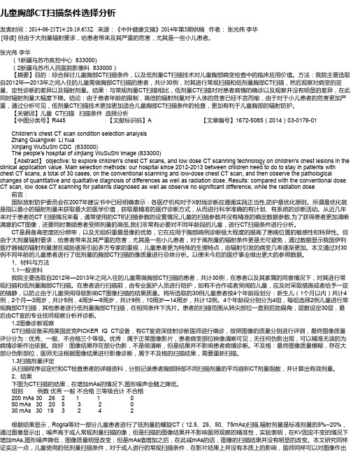
儿童胸部CT扫描条件选择分析发表时间:2014-06-25T14:20:19.653Z 来源:《中外健康文摘》2014年第3期供稿作者:张光伟李华[导读] 但由于大剂量辐射要求,给患者带来及其严重的危害,尤其是一些小儿患者。
张光伟李华(1新疆乌苏市疾控中心 833000)(2新疆乌苏市人民医院影像科 833000)【摘要】目的:综合探讨儿童胸部CT扫描条件,以及低剂量CT扫描技术对儿童胸部病变检查中的临床应用价值。
方法:我院主要选取自2012年—2013年之间入住的儿童需做胸部CT扫描的患者,共计30例,对其进行常规扫描和低剂量胸部CT扫描,然后观察对病变的定量、定性诊断的差异以及辐射剂量。
结果:与常规剂量CT扫描相比,低剂量CT扫描对对患者病情的确诊以及观察并没有明显的差异,在此同时辐射剂量大幅度下降。
结论:由于患者年龄的限制,高倍的辐射剂量对于人体的危害已经不言而喻,由于对于小儿患者的危害更加严重,通过分析可见,低剂量CT扫描技术更加更加适合儿童胸部CT扫描条件的检查,更加有利于儿童胸部的辐射防护。
【关键词】儿童 CT扫描扫描条件选择分析【中图分类号】R445 【文献标识码】A 【文章编号】1672-5085(2014)03-0176-01Children's chest CT scan condition selection analysisZhang Guangwei Li huaXinjiang WuSuShi CDC (833000)The people's hospital of xinjiang WuSuShi image (833000)【Abstract】 objective: to explore children's chest CT scans, and low dose CT scanning technology on children's chest lesions in the clinical application value. Main selection methods: our hospital since 2012-2013 between children need to do to stay in patients with chest CT scans, a total of 30 cases, on the conventional scanning and low-dose chest CT scan, and then observe the pathological changes of quantitative and qualitative diagnosis of differences as well as radiation dose. Results: compared with the conventional dose CT scan, low dose CT scanning for patients diagnosed as well as observe no significant difference, while the radiation dose 前言:国际放射防护委员会在2007年建议书中已经明确表示:各医疗机构对于X射线诊断应遵循实践正当性,防护最优化原则。
颅内畸胎瘤的CT与MRI表现姚丁华;韩福刚【摘要】Objective To explore the features and diagnostic value of X-ray computed tomography (CT) and magnetic resonance imaging (MRI) with intracranial teratomas. Methods The clinical and imaging data of 6 cases with intracranial teratomas who confirmed by pathology in the Affiliated Hospital of Southwest Medical University from Jan-uary 2014 to April 2016 were retrospectively analyzed. Results Among the 6 patients, there were 3 cases of mature ter-atoma, 2 cases of immature teratoma, and 1 case accompanied by malignant transformation of teratoma. Three cases were located in the central line area, and 3 were not. Four of the six cases of CT and MRI showed cyst-solidary mass, and 2 had special features (one case showed solidary mass on CT and mixed signal mass on MRI;the other of CT and MRI showed cyst-solidary mass associated with wall nudoul, liquid-liquid flat and slight peritumoral edema). The con-trast-enhanced scan of solid part and the wall nodule showed moderate and severe enhancement, and the cystic part was not enhanced. Only one case showed typical features (coexistence of fat and calcification) in 6 cases. All cases show no remarkable occupying effect. Conclusion The imaging features of the intracranial terotoma are related to the composi-tion of the tumors. It is easy to diagnose the typical cases and difficult for atypical ones. CT showed calcification sensitiv-ity, and MRI showed fat sensitivity. CT combined with MRI can improve the location and qualitative diagnosis ofintra-cranial terotoma.%目的探讨颅内畸胎瘤的X线计算机断层成像(CT)与磁共振成像(MRI)表现及其诊断价值.方法回顾性分析西南医科大学附属医院2014年1月至2016年4月经病理证实的6例颅内畸胎瘤的临床与影像学资料.结果本组患者中成熟型畸胎瘤3例,未成熟型畸胎瘤2例,伴有恶性转化的畸胎瘤1例;位于中线结构的3例,非中线结构3例.6例中4例CT与MRI均表现为囊实性肿块,另2例表现特殊;其中1例CT呈实性肿块,MRI呈混杂信号肿块;另1例CT与MRI均表现为囊性肿块伴壁结节,并见液液平,轻度瘤周水肿.增强扫描肿块实性部分及壁结节呈中重度强化,囊性部分未见强化.6例仅1例表现典型,脂肪与钙化并存.所有病例均无明显占位效应.结论颅内畸胎瘤的影像表现与肿瘤成分相关,典型者可做出明确诊断,不典型者诊断困难.CT显示钙化敏感,MRI显示脂肪敏感,CT联合MRI可以提高颅内畸胎瘤的定位及定性诊断.【期刊名称】《海南医学》【年(卷),期】2017(028)010【总页数】3页(P1635-1637)【关键词】畸胎瘤;颅内;计算机断层扫描;磁共振成像【作者】姚丁华;韩福刚【作者单位】西南医科大学附属医院放射科,四川泸州 646000;西南医科大学附属医院放射科,四川泸州 646000【正文语种】中文【中图分类】R739.41畸胎瘤属于生殖细胞类肿瘤,仅占颅内肿瘤的0.5%,常发生在脑中线部位,以松果体区和鞍区最常见。
智商即IQ,是一种数量化的、对智力的标准测量。
有两种个体施测的IQ测验至今还在广泛应用:斯坦福-比奈(Stanford-Binet)测验和韦克斯勒(Wechsler)测验。
斯坦福-比奈量表智商分布:140以上为非常优秀(天才);120-139为优秀;110-119为中上、聪慧;90-109为中等;80-89为中下;70-79为临界智能不足;69以下为智力缺陷。
本测试参照斯坦福-比奈量表智商制定,适用于11岁以上儿童智力测试及成人智力测试。
指导语:本测验共有60个题目,你应在45分钟内做完,不要超时。
1、五个答案中哪一个是最好的类比?工工人人人工人对于相当于工工人人工人人工对于1)2)3)4)5)2、找出与众不同的一个:①铝②锡③钢④铁⑤铜3、五个答案中哪一个是最好的类比?4、找出与众不同的一个:5、全班学生排成一行,从左数和从右数沃斯都是第15名,问全班共有学生多少人?①15 ②25 ③29 ④30 ⑤316、一个立方体的六面,分别写着A B C D E F 六个字母,根据以下四张图,推测B的对面是什么字母?7、找出与“确信”意思相同或意义最相近的词:①正确②明确③信心④肯定⑤真实8、五个答案中哪一个是最好的类比?脚对于手相当于腿对于___________①肘②膝③臂④手指⑤脚趾9、五个答案中哪一个是最好的类比?10、如果所有的甲是乙,没有一个乙是丙,那么,一定没有一个丙是甲。
这句话是:①对的②错的③既不对也不错11、找出下列数字中特殊的一个:1 3 5 7 11 13 15 1712、找出与众不同的一个:13、沃斯比乔丹大,麦瑞比沃斯小。
下列陈述中哪一句是正确的?1)、麦瑞比乔丹大2)、麦瑞比乔丹小3)、麦瑞与乔丹一样大4)、无法确定麦瑞与乔丹谁大14、找出与众不同的一个:15、五个答案中哪一个是最好的类比:“预杉”对于“须抒”相当于8326对于________.①2368 ②6238 ③2683 ④6328 ⑤362816、沃斯有12枚硬币,共3角6分钱。
MRCP在儿童胆总管囊肿诊断中的临床意义周琦芳① 盛茂① 郭万亮① 陈萌萌① 【摘要】 目的:探讨磁共振胆胰管成像(MRCP)诊断儿童胆总管囊肿的临床价值。
方法:选取2015年1月—2020年12月苏州大学附属儿童医院收治的100例胆总管囊肿患儿,所有研究对象均行磁共振成像(MRI)、MRCP检查,评价MRCP对儿童胆总管囊肿定性诊断的效果。
结果:100例胆总管囊肿患儿接受MRI检查,Todani Ⅰ型88例,胆总管全程呈囊性扩张并累及左右主肝管,其壁薄而均匀,肝内胆管无扩张,囊状扩张的胆管在T1WI和T2WI上呈水样信号;Ⅱ型3例,胆总管外侧壁囊性低密度影,胆总管侧壁与囊肿样扩张的短蒂或狭窄的基底连接;Ⅲ型2例,胆总管梭状扩张或囊性扩张;Ⅳ型2例,肝外胆管呈囊性扩张,且囊性扩张为多发性,伴或不伴肝内胆管囊性扩张;Ⅴ型5例,可见肝内以周围部分布为主的多发囊性高信号灶,与肝内胆管交通。
100例胆总管囊肿患儿接受MRCP检查,Todani分型Ⅰ型88例,肝内胆管无明显扩张,胆总管呈局限性的梭形或囊状扩张,胆总管壁有轻微均一增厚;Ⅱ型2例,胆总管明显扩张,肝管轻度扩张,胆囊下方有明显囊袋样改变,且与胆总管相连;Ⅲ型2例,胆总管末端囊状扩张,胰胆管合流异常;Ⅳ型3例,多个囊状或梭形扩张出现在肝内外胆管,扩张大小不一,胆总管远端有不同程度的狭窄;Ⅴ型5例,有多个串珠状和囊状扩张,扩张沿肝内胆管树分布,扩张囊腔与肝内胆管交通。
结论:MRCP利用重T2加权技术,使胆汁和胰液等水性结构呈现明显的高信号,而周围区域呈现低信号,诊断儿童胆总管囊肿的准确率较高。
【关键词】 磁共振成像 磁共振胆胰管成像 胆总管囊肿 Clinical Significance of MRCP in the Diagnosis of Choledochal Cyst in Children/ZHOU Qifang, SHENG Mao, GUO Wanliang, CHEN Mengmeng. //Medical Innovation of China, 2023, 20(23): 119-122 [Abstract] Objective: To investigate the clinical value of magnetic resonance cholangiopancreatography (MRCP) in the diagnosis of choledochal cyst in children. Method: A total of 100 children with choledochal cyst admitted to Children's Hospital of Soochow University from January 2015 to December 2020 were selected. All subjects underwent magnetic resonance imaging (MRI) and MRCP to evaluate the qualitative diagnosis effect of MRCP on choledochal cyst in children. Result: A total of 100 children with choledochal cyst were examined by MRI. Todani type Ⅰ 88 cases, the common bile duct cystic dilatation throughout the whole process and involved the left and right main hepatic ducts, the wall was thin and uniform, the intrahepatic bile duct no dilatation, and the cystic dilated bile duct showed water-like signals on T1WI and T2WI. Type Ⅱ 3 cases, the lateral wall of the common bile duct had a cystic low density shadow, and the lateral wall of the common bile duct was connected with a short pedicle or a narrow basal of cystic dilatation. Type Ⅲ 2 cases, common bile duct fusiform dilatation or cystic dilatation. Type Ⅳ 2 cases, extrahepatic bile duct cystic dilatation, and the cystic dilatation was multiple, with or without cystic dilatation of intrahepatic bile duct. Type Ⅴ 5 cases, multiple cystic hypersignal foci mainly distributed in the peripheral part of the liver, and communicated with the intrahepatic bile duct. A total of 100 children with choledochal cyst were examined by MRCP. Todani type Ⅰ 88 cases, intrahepatic bile duct dilatation was not obvious, and the common bile duct had localized fusiform or cystic dilatation, and the common bile duct wall was slightly uniform thickened. Type Ⅱ 2 cases, the common bile duct was obviously dilated, the hepatic duct was slightly dilated, and there were obvious bag-like changes under the gallbladder, which were connected to the common bile duct. Type Ⅲ 2 cases, the end of the common bile duct cystic dilatation, anomalous pancreaticobiliary ductal junctio. Type Ⅳ 3 cases, multiple cystic or fusiform dilatation appeared in the intrahepatic and extrahepatic bile duct, the dilatation size was different, and the distal common bile duct had different degrees of stenosis. Type Ⅴ 5 cases, there were multiple beading and cystic dilatations, which were distributed along the intrahepatic bile duct tree and communicated with the intrahepatic bile duct. Conclusion: MRCP uses heavy T2 weighting technology to①苏州大学附属儿童医院 江苏 苏州 215000通信作者:陈萌萌 儿童胆总管囊肿往往是由于小儿管壁存在先天性发育缺损,或存在异位胰腺组织,从而导致管壁处于低紧张状态,又或是先天性胆总管闭锁,导致管内压力增加,引起扩张所导致。