网状纤维染色液(改良Gordon-Sweets法)
- 格式:pdf
- 大小:245.10 KB
- 文档页数:2
二、网状纤维染色网状纤维是一种纤细的纤维。
它沿着网状细胞和突起分支,并互相交织成网,因而被称为网状纤维,又因这种纤维对银的浸染着色特别显著。
故又称为嗜银纤维。
它主要由III型胶原蛋白构成,也具有642nm周期性横纹[1]。
它主要分布于骨髓、脾、淋巴结、肝和肺等脏器。
癌组织中间未见有网状纤维,但在其边缘可见有大量的网状纤维,但在其边缘可见有大量的网状纤维围绕,俗称癌巢。
(1)网状纤维的形成它的形成主要是在纤维母细胞的粗面内浆网中,合成可溶性胶原。
在醛糖、类脂的作用下,形成可溶性胶原,随后它被分泌到细胞外,在基质中(或在细胞的表面)通过原胶原蛋白分子间的交联,聚合成为不溶性的胶原原纤维即网状纤维。
(2)网状纤维在组织化学上的反应网状纤维的主要成分是网硬蛋白,在组织化学上,网状纤维和胶原纤维是容易区别的。
网状纤维是分支纤细的纤维。
V an Gieson 氏染色不显色或微红色。
PAS反应呈现红紫色,银浸染为黑色。
胶原纤维用V an Gieson氏染色为红色,PAS反应呈微粉红色。
银浸染为黄色或棕黄色。
(3)状纤维的显示1.染色染料技术:从网状纤维的显示,区别和效果等方面,这种技术被认为是不可靠的,其原因是它只能跟胶原纤维区别开来。
2.金属浸染技术:这种技术对于显示网状纤维被认为是十分可靠和广泛使用的方法,它不但能够区别网状纤维和胶原纤维,而且可显示纤细的网状纤维。
嗜银反应(Argyrophil reaction)嗜银细胞与亲银细胞所不同的是需要一种附加的还原剂来还原,它们比亲银细胞数量多。
必须注意:亲银反应中将找不到任何的嗜银细胞。
亲银反应(Argentaffin reation)亲银细胞有能力,直接地,不靠外加试剂,将银液中的银还原为一种不溶性的黑色金属银沉淀的细胞称为亲银细胞。
(Cells that have the ability to directly reduce silver solution producing an insoluble black metallic silver precipitate are termed-angentaffin cells)。
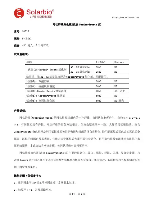
网状纤维染色液(改良Gordon-Sweets法)货号:G3525规格:6×50ml保存:4℃避光,3个月有效。
试剂盒组成:名称6×50ml Storage试剂(A):Gordon-Sweets氧化剂A1:GS氧化剂A25ml RT A2:GS氧化剂B25ml RT临用前,取A1、A2等量混合即为Gordon-Sweets氧化剂,即配即用。
试剂(B):草酸溶液50ml RT试剂(C):硫酸铁铵溶液50ml RT试剂(D):Gordon-Sweets银氨溶液50ml4℃避光试剂(E):Gordon-Sweets还原剂50ml RT试剂(F):核固红染色液50ml RT避光产品说明:网状纤维(Reticular fiber)是网状结缔组织内的一种纤维,由网状细胞所产生,直径多在0.2~1.0μm,有韧性而没有弹性。
网状纤维的染色方法很多,但染色原理基本一致,大都采用氨银浸法。
改良Gordon-Sweets染色原理是利用氨银液易被组织吸附与组织的蛋白质结合,经甲醛还原成黑色或棕黑色的金属银,沉积于组织内及其表面。
传统方法中还原后先采用氯化金调色,再用硫代硫酸钠溶液洗去组织上未还原的银盐,本改良法省略该步骤,使网状纤维对比得更清晰。
网状纤维染色液(改良Gordon-Sweets法)主要经过氧化、漂白、媒染、浸银、还原、复染等步骤,与改良Gomori法不同之处在于本法采用酸性氧化剂和核固红复染液。
冰冻切片、低温切片和火棉胶切片均可用于网状纤维染色。
操作步骤(仅供参考):1、组织固定于10%福尔马林固定液,常规脱水包埋。
2、切片厚4μm,常规脱蜡至水。
第1页,共2页3、把切片平置在染色架上,滴入配制好的Gordon-Sweets氧化剂氧化3min。
4、稍水洗。
5、草酸溶液漂白1min。
6、流水冲洗,蒸馏水稍洗。
7、硫酸铁铵溶液媒染10min。
8、稍水洗,蒸馏水稍洗。
9、滴加Gordon-Sweets银氨溶液染色11s。
特殊染色在肝脏活检病理诊断中的应用摘要:目的:对应用特殊染色后,肝脏活检组织的病理形态学变化予以探讨。
方法:以本院2012年6月-2016年6月间的62例肝病患者的肝组织活检标本作为研究对象,均对其予以铁染色、Masson染色、D-PAS染色与网状纤维Gordon-Sweets嗜银染色,对肝脏组织的各部位所显示的颜色变化进行观察。
结果:经铁染色后,肝脏部位的含铁血黄素会呈现出蓝色;经Masson染色后,胶原纤维组会呈现出蓝或绿色,而肌纤维呈现出红色,上皮组织与细胞核则分别呈现出淡红色与蓝黑色;经D-PAS染色后,胆汁、卟啉、脂褐素、铜与铜结合蛋白依次呈现出褐色、深红棕色、红色、紫色;网状纤维经Gordon-Sweets嗜银染色后,会呈现出黑色细丝网状。
结论:经特殊染色后,肝脏组织中的不同色素颗粒与组织会显示出不同颜色,对相关疾病与以判断具有重要的指导意义。
关键词:特殊染色;肝脏活检;病理诊断;应用价值在医学技术不断进步的今天,免疫组化与分子生物学技术在各类临床实践中都得以广泛应用,无论在病情诊断还是治疗过程中都有着不可或缺的作用,因而在临床上愈加受到医学人士的推崇。
但作为传统病研究手段之一的特殊染色,对病理诊断而言依旧有着重要的应用价值,其重要性从病理检验类论著中的相关介绍可见一斑。
本文以肝组织活检标本作为研究对象,就特殊染色在肝脏活检病理诊断中的应用予以探讨,现简述如下。
1 材料和方法1.1 材料取本院2012年6月-2016年6月间收治的62例肝病患者的肝组织活检标本为研究对象,经浓度为4%的甲醛固定液予以固定,采用常规石蜡进行制片,切片厚度在4-6μm之间。
1.2 方法按照相应求配置染色液,对前述所制切片分别进行铁染色、Masson染色、D-PAS染色与网状纤维Gordon-Sweets嗜银染色,并通过显微镜对相应组织的染色结果予以观察。
2 结果观察结果显示,经铁染色后,肝脏组织中的含铁血黄素显示为蓝色;经Masson染色后,胶原纤维组织会呈现出蓝或绿色,而肌纤维呈现出红色,上皮组织与细胞核则分别呈现出淡红色与蓝黑色;经D-PAS染色后,胆汁、卟啉、脂褐素、铜与铜结合蛋白依次呈现出褐色、深红棕色、红色、紫色;网状纤维经Gordon-Sweets嗜银染色后,会呈现出黑色细丝网状。
常用特殊染色结果速记注:为了方便广大病理技术人员熟悉和掌握常用病理技术特殊染色的结果,现将作者摘录整理的“常用特殊染色结果速记”呈现给大家,以便使用者在日常工作和学习中记忆。
内容不一定全面,如有错误请以人民卫生出版社出版的《2016年全国卫生技术资格考试指导-病理学技术》一书为准。
结缔组织染色:1.Mallory三色染色结果:胶原和网状纤维蓝色,软骨、黏液、淀粉样变物资呈淡蓝色,神经胶质纤维、肌纤维和酸性颗粒红色,髓鞘和红细胞橘红色。
2.Masson三色染色结果:胶原纤维绿色,肌纤维红色,红细胞橘红色。
胶原纤维染色:1.Van Gieson(V G)苦味酸-酸性品红染色结果:胶原纤维鲜红色,肌纤维、细胞质和红细胞黄色,细胞核蓝褐色。
2.天狼星红苦味酸染色结果:胶原纤维红色,细胞核绿色,其他黄色。
网状纤维染色:1.Gordon-Sweets银氨染色结果:网状纤维黑色,细胞核红色(核固红复染),胶原纤维黄棕色,细胞质淡红(红液复染)。
弹性纤维染色:1.弹性、胶原纤维的双重组合染色法:弹性纤维蓝绿色,胶原纤维红色,背景淡黄色。
2.Gomori醛品红染色法:弹性纤维呈紫红色,背景橘红色。
横纹肌组织染色:1.Mallory磷坞酸苏木素(PTAH)染色结果:细胞核、纤维、肌肉、神经胶质纤维、纤维蛋白、横纹肌均为蓝色;胶原纤维、网状纤维软骨基质及骨呈黄色或玫瑰红色;粗弹性纤维有时为微紫色;有缺氧早期病变的心肌为紫蓝色或棕黄色。
早期心肌病变组织染色:1.Nagar-Olsen染色结果:缺氧心肌、红细胞红色,正常心肌黄色或黄棕色,细胞核蓝色。
2.Poley显示缺氧心肌染色结果:缺氧心肌红色,细胞核紫色,其他组织呈绿色。
糖类染色:1.过碘酸-Schiff(PAS)染色结果:糖原红色,细胞核蓝色。
黏液物质染色:1.Mowry阿尔辛蓝过碘酸雪夫AB/PAS染色结果:中性黏液物资红色,酸性黏液物资蓝色,混合性黏液物资紫红色。
网状纤维染色在临床病理诊断中的应用及技术网状纤维染色法是病理学传统的特殊染色方法之一,在临床病理学诊断及鉴别诊断中应用广泛。
病理学诊断至今仍为临床多种肿瘤性疾病明确诊断及分型等的金标准,网状纤维染色法在多种肿瘤病理诊断中发挥重要的作用。
网状纤维的组织改变在临床疾病病理诊断上有重要意义,如血液系统肿瘤、乳腺纤维上皮肿瘤、胃肠道间皮瘤、神经系统肿瘤等,但网状纤维在染色过程中易出现问题,如纤维着色不良、纤维退色、染色背景过深等。
本文就网状纤维染色在临床病理诊断中的应用及技术作一综述。
[Abstract] Reticular fiber staining is one of the classical method for special staining in pathology and has been wildly used in diagnosis and differential diagnosis of various diseases. Pathological diagnosis has been regarded as the golden standard in diagnosis and classification of many tumors,in which reticular fiber staining plays important roles. The changes of reticular fiber have important implications for clinical pathological diagnosis including hematological malignancies,fibro-epithelioma of breast,gastrointestinal mesothelioma,nervous system tumors,etc. However,in the process of reticular fiber staining,there may be some problems such as fiber coloring bad,fibrous degeneration and calcification,etc. This article reviews application of reticular fiber staining and common problems in the process of staining.[Key words] Reticular fiber;Dyeing;Tumor;Pathological diagnosis網状纤维是网状结缔组织内的一种纤维,由网状细胞所产生。
网状纤维染色在临床病理诊断中的应用及技术作者:王晓燕高俊岩王艳霞来源:《中国医药导报》2016年第09期[摘要] 网状纤维染色法是病理学传统的特殊染色方法之一,在临床病理学诊断及鉴别诊断中应用广泛。
病理学诊断至今仍为临床多种肿瘤性疾病明确诊断及分型等的金标准,网状纤维染色法在多种肿瘤病理诊断中发挥重要的作用。
网状纤维的组织改变在临床疾病病理诊断上有重要意义,如血液系统肿瘤、乳腺纤维上皮肿瘤、胃肠道间皮瘤、神经系统肿瘤等,但网状纤维在染色过程中易出现问题,如纤维着色不良、纤维退色、染色背景过深等。
本文就网状纤维染色在临床病理诊断中的应用及技术作一综述。
[关键词] 网状纤维;染色;肿瘤;病理诊断[中图分类号] R817.4 [文献标识码] A [文章编号] 1673-7210(2016)03(c)-0072-04[Abstract] Reticular fiber staining is one of the classical method for special staining in pathology and has been wildly used in diagnosis and differential diagnosis of various diseases. Pathological diagnosis has been regarded as the golden standard in diagnosis and classification of many tumors,in which reticular fiber staining plays important roles. The changes of reticular fiber have important implications for clinical pathological diagnosis including hematological malignancies, fibro-epithelioma of breast, gastrointestinal mesothelioma, nervous system tumors, etc. However, in the process of reticular fiber staining, there may be some problems such as fiber coloring bad,fibrous degeneration and calcification, etc. This article reviews application of reticular fiber staining and common problems in the process of staining.[Key words] Reticular fiber; Dyeing; Tumor; Pathological diagnosis网状纤维是网状结缔组织内的一种纤维,由网状细胞所产生。
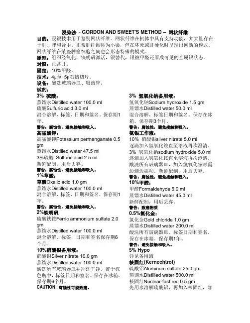
浸染法- GORDON AND SWEET'S METHOD –网状纤维目的:浸银技术用于鉴别网状纤维。
网状纤维在机体中具有支持功能,并大量存在于肝、脾和肾中。
正常肝纤维称为小梁,但在坏死或肝硬化时呈现出间断的模式。
网状纤维在某些肿瘤细胞之间也会形态特殊的模式。
原理:组织经氧化,铁明矾激活,银替代,银被甲醛还原成可见的金属银状态。
对照:正常肝。
固定:10%甲醛。
技术:4μ至5μ石蜡切片。
设备:酸洗玻璃器皿、吸液管。
试剂:3% 硫酸:蒸馏水Distilled water 100.0 ml硫酸Sulfuric acid 3.0 ml混合溶解,标签,日期和签名。
保存期1年。
警告:腐蚀性,避免接触和吸入。
高猛酸钾:高猛酸钾Potassium permanganate 0.5 gm蒸馏水Distilled water 47.5 ml3%硫酸Sulfuric acid 2.5 ml新鲜配制,用后丢弃。
警告:腐蚀性,避免接触和吸入。
1%草酸:草酸Oxalic acid 1.0 gm蒸馏水Distilled water 100.0 ml混合溶解,标签,日期和签名,保存期1年。
警告:腐蚀性,避免接触和吸入。
2%铁明矾硫酸铁铵Ferric ammonium sulfate 2.0 gm蒸馏水Distilled water 100.0 ml混合溶解,标签,日期和签名保存期6个月。
10%硝酸银备用液:硝酸银Silver nitrate 10.0 gm蒸馏水Distilled water 100.0 ml酸洗所有玻璃器皿并冲洗干净。
置于棕色瓶中,标签日期和签名。
保存在冰箱。
保存期6个月。
CAUTION: 腐蚀性可能致癌。
3% 氢氧化钠备用液:氢氧化钠Sodium hydroxide 1.5 gm蒸馏水Distilled water 50.0 ml混合溶解,标签日期和签名。
保存在冰箱。
保存期3个月。
警告:腐蚀性,避免接触和吸入。

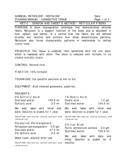
SURGICAL PATHOLOGY - HISTOLOGY Date: STAINING MANUAL - CONNECTIVE TISSUE Page: 1 of 4 PURPOSE: A silver impregnation technique that demonstrates reticular fibers. Reticulum is a support function of the body and is abundant in liver, spleen, and kidney. In a normal liver the fibers are will defined strands, but necrotic and cirrhotic liver show discontinuous patterns. Reticulum also forms characteristic patterns in relationship to certain tumor cells.PRINCIPLE: The tissue is oxidized, then sensitized with the iron alum, which is replaced with silver. The silver is reduced with formalin to its visible metallic state.CONTROL: Normal liver.FIXATIVE: 10% formalinTECHNIQUE: Cut paraffin sections at 4m to 5m.EQUIPMENT: Acid cleaned glassware, pipettes.REAGENTS:3% Sulfuric Acid:Distilled water100.0 ml Sulfuric acid 3.0 ml Mix well, label with initial and date. Solution is stable for 1 year. CAUTION: Corrosive, avoid contact and inhalation.Potassium Permanganate: Potassium permanganate0.5 gm Distilled water47.5 ml 3% Sulfuric acid 2.5 ml Make fresh, discard after use. CAUTION: Corrosive, avoid contact and inhalation.1% Oxalic Acid:Oxalic acid 1.0 gm Distilled water100.0 ml Mix well, label with initial and date. Solution is stable for 1 year. CAUTION: Corrosive, avoid contact and inhalation.2% Iron AlumFerric ammonium sulfate 2.0 gm Distilled water100.0 ml Mix well, label with date and initial. Solution is stable for 6 months.RETIC P age: 2 of 410% Silver Nitrate StockSilver nitrate10.0 gm Distilled water100.0 ml Acid clean all glassware and rinse well. Store in a brown bottle, keep in the refrigerator. Label with initial and date. Stable for 6 months.CAUTION: Corrosive, possible carcinogen.3% Sodium Hydroxide Stock: Sodium hydroxide 1.5 gm Distilled water50.0 ml Mix well, label with date and initials. Store in the refrigerator, stable for 3 months.CAUTION: Corrosive, avoid contact and inhalation.Ammoniacal SilverWorking Solution:10% silver nitrate 5.0 ml Add ammonium hydroxide drop by drop until clear again.3% sodium hydroxide 5.0 ml Add ammonium hydroxide drop by drop until clear again.Distilled water40.0 ml Using acid clean glassware, agitate solution continually while adding ammonium hydroxide. Make right before use, discard.CAUTION: Corrosive, avoid contact and inhalation.10% Formaldehyde: Formaldehyde 5.0 ml Distilled water45.0 ml Make fresh, discard after use. CAUTION: Carcinogen.0.5% Gold Chloride:Gold chloride 1.0 gm Distilled water200.0 ml Use acid clean glassware, label with date and initials. Store in the refrigerator. Stable for 1 year. CAUTION: Avoid contact and inhalation.5% HypoSee Stock SolutionNuclear-Fast Red (Kernechtrot) Aluminum sulfate25.0 gm Distilled water500.0 ml Nuclear-fast red0.5 gm Dissolve the aluminum sulfate in distilled water, then the nuclear-fast red, using heat. Cool, filter, and add a few grains of thymol as a preservative. Label with date and initials. Stable for 1 year. CAUTION: Irritant, avoid contact and inhalation.RETIC Page: 3 of 4 inhalation.Potassium permanganate: Skin and eye irritant, ingestion will lead to severe gastrointestinal distress. Strong oxidant.Sulfuric acid: Strong irritant to skin, eyes and respiratory system. Inhalation can produce target organ effects on skin, respiratory, reproductive and fetal systems. Corrosive.can cause severe burns of the eyes, skin or mucous membranes. Toxic by inhalation and ingestion. Target organ effects on kidneys and cardiovascular system, repeated exposure can cause dermatitis. Corrosive. Silver nitrate: severe skin and eye irritant. Oxidizer. Ingestion will produce violent gastrointestinal discomfort. Possible carcinogen: equivocal tumorigenic agent.Ammoniacal silver solution can be explosive, discard down the drain followed by copious amounts of running water.Ammonium hydroxide: severe eye irritant, irritating to respiratory system. Target organ effects on respiratory system. Corrosive. Work under hood.Sodium hydroxide: severe skin and eye irritant. Corrosive. Formaldehyde: known carcinogen.Sodium thiosulfate: Toxic on ingestion. Can irritate the stomach. Irritant to skin, eyes and respiratory tract.PROCEDURE:1.Deparaffinize and hydrate to distilled water.2.Potassium permanganate solution, 5 minutes.3.Wash in water.4.5% oxalic acid until clear.5.Wash in distilled water.6.Iron alum solution, 10 minutes.7.Wash in running tap water, rinse in distilled, 3 changes.8.Silver solution, 7 dips, shake excess solution off slides.9.Distilled water, 2 changes, 3 quick dips each.RETIC Page: 4 of 411.Wash in distilled water.12.0.5% Gold chloride, 1 minute.13.Rinse in distilled water.14.5% hypo, 1 minute.15.Wash in tap water.16.Nuclear-fast red solution, 5 minutes.17.Wash in running tap water.18.Dehydrate, clear, and coverslip.RESULTS:Reticular fibers blackNuclei redNOTES:e acid clean glassware, or rinse 5x with distilled water.2.When making working silver solution, if over 30 drops of ammoniumhydroxide are used to turn the solution, then the ammonium hydroxide is too old. Start over with fresh ammonium hydroxide.3.When adding the 3% sodium hydroxide solution to the silver solution itshould turn black, if not make fresh sodium hydroxide.4.Because of the alkalinity of the solution, it may cause some tissues tofall off the slides, celloidinize (see Stock Solutions).5.Change distilled water after every slide , step '9'.REFERENCE:Bancroft J,Stevens A, Theory and Practice of Histological Techniques, 2nd Ed, 1982, pp142-143, Churchill Livingstone, NYCarson F, Histotechnology: A Self-Instructional Text,1990,pp154-155,ASCP,IllCrookham,J, Dapson,R, Hazardous Chemicals in the Histopathology Laboratory, 2nd ED, 1991, AnatechPrepared: By:Approved: By:Downloaded from WebPath: Internet Pathology Laboratory/WebPath/webpath.htmlPROCEDURE CARDRETIC - RETICULAR FIBERS - GORDON AND SWEET'S Page 1 of 21.Deparaffinize and hydrate to distilled water.2.Potassium permanganate solution, 5 minutes.3.Wash in water.4.5% oxalic acid until clear.5.Wash in distilled water.6.Iron alum solution, 10 minutes.7.Wash in running tap water, rinse in distilled, 3 changes.8.Silver solution, 7 dips, shake excess solution off slides.9.Distilled water, 2 changes, 3 quick dips each.10.10% formaldehyde solution until gray black, 30 seconds.11.Wash in distilled water.12.0.5% Gold chloride, 1 minute.13.Rinse in distilled water.14.5% hypo, 1 minute.15.Wash in tap water.16.Nuclear-fast red solution, 5 minutes.17.Wash in running tap water.18.Dehydrate, clear, and coverslip.RESULTS:Reticular fibers blackNuclei red3% Sulfuric Acid:Solution is stabel for 1 year.CAUTION: Corrosive acid.Potassium Permanganate:Potassium permanganate0.5 gm Distilled water47.5 ml 3% Sulfuric acid 2.5 ml Make fresh, discard after use.1% Oxalic Acid:Solution is stable for 1 year.2% Iron AlumSolution is stable for 6 months.10% Silver Nitrate StockStore in the refrigerator. Label with initial and date. Stable for 6 months.CAUTION: Corrosive.3% Sodium Hydroxide Stock:Store in the refrigerator, stable for 3 months.CAUTION: Corrosive.Working Ammoniacal Silver Solution: 10% silver nitrate 5.0 ml Add ammonium hydroxide drop by drop until clear again3% sodium hydroxide 5.0 ml Add ammonium hydroxide drop by drop until clear againDistilled water40.0 ml Using acid clean glassware, agitate solution continually while adding ammonium hydroxide. Make right before use, discard.PROCEDURE CARDRETIC - RETICULAR FIBERS - GORDON AND SWEET'S Page 2 of 210% Formaldehyde:Formaldehyde 5.0 ml Distilled water45.0 ml Make fresh, discard after use.0.5% Gold Chloride:Store in the refrigerator. Stable for 1 year.5% Hypo:See Stock Solutions Nuclear-Fast Red (Kernechtrot) Aluminum sulfate25.0 gm Distilled water500.0 ml Nuclear-fast red0.5 gm Dissolve the aluminum sulfate in distilled water, then the nuclear-fast red, using heat. Cool, filter, and add a few grains of thymol as a preservative. Label with date and initials. Stable for 1 year.CAUTION: Irritant, avoid contact and inhalation.SODIUM HYDROXIDE STOCK: Sodium hydroxide 1.5 gm Distilled water50.0 ml Mix well, label with date and initals. Store in the refrigerator, stable for 3 months.CAUTION: Corrosive.DATE:TECH:EXPIRATION:3% SULFURIC ACID:Distilled water100.0 ml Sulfuric acid 3.0 ml Mix well, label with initial and date. Solution is stabel for 1 year. CAUTION: Corrosive acid.DATE:TECH:EXPIRATION:0.2% GOLD CHLORIDEDATE: TECH: NUCLEAR-FAST REDDATE: TECH: 2% IRON ALUMFerric ammonium sulfate 2.0 gmDistilled water100.0 ml Mix well, label with date and initial. Solution is stable for 6 months. Discard when iron alum precipitates.DATE:TECH:EXPIRATION:2% IRON ALUMDATE:TECH:10% SILVER NITRATESilver nitrate40.0 gm Distilled water400.0 ml Mix well. Pour into an acid cleaned brown bottle. Refrigerate, stable 1 yr.CAUTION: Corrosive, irritant, possible cacinogen.DATE: TECH: EXPIRATION:1% OXALIC ACIDOxalic acid 4.0 gm Distilled water400.0 ml Solution is stable for 1 year. CAUTION: Avoid contact and inhalation.TECH:DATE EXPIRATION: 1% OXALIC ACIDDATE: TECH:。
第一节结缔组织和肌纤维染色Van Gieson苦味酸酸性复红法(VG法)用途:VG法用于区分胶原纤维和肌纤维。
原理:酸性复红和苦味酸对这两种纤维都具有较强的亲合力,结果胶原纤维被酸性复红染成红色,肌肉被苦味酸染成黄色。
此方法是一种优良的传统方法。
固定:10%福尔马林切片:4-6微米试剂:Weigert铁苏木精液:A液:苏木精1g,无水酒精100 ml。
一般配制后需要数周或数月自然氧化,成熟后使用。
B液:29%三氯化铁水溶液4 ml,蒸馏水95 ml,盐酸1 ml。
两液分别配制,临用时A、B液等量混合,过滤后使用。
24h后失去染色能力。
Van Gieson液:1%酸性复红水溶液 10 ml苦味酸饱和水溶液 90 ml临用时1:9混合过虑后使用。
Van Gieson苦味酸酸性复红法(VG法)步骤:1.石蜡切片脱蜡至水。
2. Weigert苏木精液5-10 min。
3.充分水洗。
4. Van Gieson液1-5 min。
5. 95%酒精迅速分化数秒。
6.无水酒精脱水、二甲苯透明、中性树胶封固。
结果:胶原纤维呈红色、肌肉、神经胶质、胞浆、红血球呈黄色、细胞核呈黑色或棕蓝色。
体会与说明:由于酸性复红退色较快,染色结果不能长期保存,一般只能保存3~6个月。
二 Masson三色法试剂:Regaud 氏苏木精:苏木精1g,95%酒精10ml,甘油10ml,蒸馏水80ml。
将苏木精加入蒸馏水内加温溶解,冷却后加入酒精和甘油,放数日后即可应用。
Masson丽春红酸性复红液:丽春红0.7g,酸性复红0.3g,蒸馏水99ml,冰醋酸1ml。
0.2%冰醋酸水溶液:冰醋酸0.2 ml,蒸馏水100 ml。
1%磷钼酸水溶液:磷钼酸1g,蒸馏水100 ml。
苯胺蓝水溶液:苯胺蓝2g,蒸馏水98 ml,冰醋酸2 ml。
1%光绿水溶液:光绿 1g,蒸馏水100 ml。
Masson三色法步骤1.石蜡切片脱蜡至水。
2.铬化处理或去汞盐沉淀(甲醛固定的组织此步可略)。
结缔组织染色法1.1 Mallory三色染色法蓝色:胶原和网状纤维淡蓝色:软骨、粘液、淀粉样变物质红色:神经胶原纤维、肌纤维、酸性颗粒橘红色:髓鞘、红细胞图表A 1.1.Mallory染色,显示胶原纤维,A组排列规那么1.2.Masson三色染色法绿色:胶原纤维红色:肌纤维橘红色:红细胞图表B 1.2 Mssson三色法图表C 1.2.Masson三色染色胃癌组织中血管平滑肌1.3.显示胶原、网状和弹性纤维的三联染色法红色:胶原纤维黑色:网状纤维绿色:弹性纤维淡黄色:肌肉、红细胞图表D 4.Weigert间苯二酚法二、胶原纤维染色法2.2.Van Gieson〔V.G〕苦味酸-酸性品红法黄色:肌纤维、细胞质、红细胞蓝褐色:胞核图表E 2.胶原纤维,Van Gieson(V.G.)苦味酸-酸性品红法图心肌堵塞myocardial infarction:心肌堵塞后2个月,van Gieson 染色, 坏死心肌被染成红色的纤维组织所代替,黄色区域为残留的心肌纤维。
2.1 天狼星红〔Sirius red〕苦味酸染色法(参照上图)绿色:细胞核黄色:其他三、网状纤维染色3.1 Gordon-Sweets银氨染色法〔梅花开枝图,金色伴树枝〕黑色:网状纤维红色:胞核〔核固红复染〕黄棕色:胶原纤维淡红色:细胞质〔红液复染〕图表F 3.Gordon-Sweets氢氧化银氨液浸染法3.2 Gomori氏银氨液配制法图表GGomori氏银氨液配制法四、弹性纤维染色Gomori醛复红染色法*甲醛生理盐水液固定的染色效果最正确图表H4.GOMORI醛复红染色法五、显示弹性、胶原纤维的双重组合染色法蓝绿色:弹性纤维红色:胶原纤维黄色:背景图表I 4.Weigert间苯二酚法六、肌肉组织染色△横纹肌组织染色Mallory磷钨酸木精染色法〔PTAH〕蓝色:胞核、纤维、肌肉、神经胶质纤维、纤维蛋白、横纹肌黄色或枚红色:胶原纤维、网状纤维软骨基质、骨微紫色:粗弹性纤维〔有时〕紫蓝色或棕黄色:缺血缺氧早期病变的心肌图表J 6.1.磷钨酸木素法图表K 6.1.磷钨酸木素染色液△早期心肌病变组织染色1.Nagar-Olsen染色法〔1974年〕红色:缺氧心肌、红细胞黄色或黄棕色:正常心肌蓝色:细胞核图各组小鼠心肌组织Nagar-Olsen染色〔光学显微镜, ×200〕2.Poley显示缺氧心肌染色法〔1964年〕红色:缺氧心肌紫色:胞核七、糖类染色过碘酸-Schiff〔PAS〕染色法红色:糖原及其他PAS反响阳性物质蓝色:细胞核图表L 7.胃贲门腺体胞浆呈PAS阳性八、黏液物质〔黏多糖〕染色1.Mowry阿尔辛蓝过碘酸雪夫〔ABPAS〕染色法〔1956〕红色:中性黏液物质蓝色:酸性黏液物质紫红色:混合性黏液物质图表M 8.1.AB-PAS染色结肠粘膜图表N 8.1.胃粘膜组织AB-PAS染色40×:2.爱先蓝〔PH2.5〕法蓝色:唾液酸、弱硫酸化黏液物质、一般粘液红色:胞核不着色:强硫酸化黏液物质图表O 8.2.爱先蓝〔PH2.5〕染色液图表P 8.2.爱先蓝法图表Q 8.2.爱先蓝法—3、爱先蓝〔PH1.0〕法蓝色:含硫酸黏液物质不着色:非硫酸化酸性黏液物质红色:复染后的胞核九、黑色素染色1.Masson-Fontana黑色素银浸染色法黑色:黑色素及嗜银细胞颗粒红色:胶原纤维浅黄色:背景图表R9.1.Melanin pigment in cells of9.1. malignant melanoma, Fontana-Masson stain.2.Lillie亚铁染色法暗绿色:黑色素浅绿或不着色:背景黄色:肌纤维和背景十、含铁血黄素染色Perls blue〔普鲁士蓝〕反响显示三价铁蓝色:含铁血黄素浅红色:其他组织图表S 10.普鲁士蓝染色肝脏普鲁士蓝染色呈蓝色的含铁血黄素颗粒大量沉着在肝实质细胞和库普弗细胞。
经典和改良胺银染液配制方法比较解立武常芳DOI :10.3969/j.issn.0253⁃9926.2020.17.045作者单位:030013太原,山西省肿瘤医院病理科通信作者:常芳,Email ****************网状纤维染色在病理诊断中应用非常广泛。
网状纤维在组织内形态以及分布特征,预示着不同的病理诊断结果,尤其在鉴别间叶组织和上皮组织来源的恶性肿瘤具有重要意义[1]。
网状纤维的染色方法较多,Gomori 银氨染色法是最常用的染色方法。
其中的氨银染色液是网状纤维染色的关键染液,高质量的胺银染液是染色成败的关键。
我们对经典的配制方法进行了改良,结果是网状纤维清晰,和背景对比分明,配制方法简单有效,而且保存期大大延长。
1材料与方法1.1材料硝酸银、氢氧化钠、氢氧化钾、氨水。
1.2仪器磁力搅拌器、滴定管、烧杯。
1.3配制方法1.3.1经典的配制方法[1]:①10%的硝酸银3m L ,加入10%的氢氧化钾3m L ,立即生成棕黑色颗粒沉淀。
②加入10倍以上的蒸馏水洗涤沉淀物,倾去上清液,再加入蒸馏水,反复洗涤3次。
③然后逐滴滴入氨水,并轻轻不断摇晃,直至沉淀物恰好完全溶解。
④再加入10%硝酸银数滴至溶液稍变浑浊,再小心加入氨水使溶液恰好又变清。
⑤按照原来总量蒸馏水稀释10倍。
配制好的氨银溶液装入棕色试剂瓶中,放入4°C 冰箱保存,可以使用4~8周。
1.3.2改良的配制方法:①10%的硝酸银10m L ,加入10%的氢氧化钠10m L 的烧杯中,立即生成棕黑色颗粒沉淀。
②将烧杯置于磁力搅拌器上,磁力棒放入杯内溶液中,开启旋转。
转速调至低速转动溶液。
③将注入了氨水的滴定管加盖,滴速调为每分钟3滴,形成最小液滴自行滴入转动的溶液中,棕黑色慢慢溶解,滴加至所形成的沉淀物恰好溶解为清亮液为止,用蒸馏水补足200m L 。
配制好的氨银溶液装入棕色试剂瓶中放置4°C 冰箱保存,可以使用5~6个月。
特殊染色法一,抗酸杆菌染色1,试剂配制:(1)碱性品红乙醇液:碱性品红5g,无水乙醇100ml(2)5%苯酚水溶液:苯酚(稍加温使共溶解)5ml,蒸馏水95ml(3)苯酚碱性品红液:碱性品红乙醇液1ml,5%苯酚水溶液9ml(4)20%硫酸水溶液:浓硫酸20ml,蒸馏水80ml(5)汽油松节油液:航空汽油(也可为普通汽油)1份,松节油1份2,染色程序:(1)切片用汽油松节油脱蜡2次,每次5-10min。
(2)不经乙醇洗,只用纱布将切片周围余液擦干。
(3)流水稍洗。
(4)滴入苯酚碱性品红液,于室温下染15—30min。
(5)流水去多余液体。
(6)用20%硫酸水溶液分化至适当为止(一般需1—5min)(7)流水充分冲洗。
(8)用Mayer苏木精液浅染细胞核。
(9)洗流水冲洗10—15min。
(10)用风扇把切片吹干,随即二甲苯透明,中性树胶封固。
3,结果:抗酸杆菌(包括麻风杆菌和结核杆菌)呈鲜红色,胞核呈蓝色。
二,奥新兰过碘酸雪夫代染色简称(AB—PAS染色)(溶液配制)1,0.3%的奥辛兰醋酸液:奥新兰0.3g,蒸馏水97ml,冰醋酸3ml2,1%过碘酸水溶液,schitf代试剂及亚硫酸冲洗液配发同。
(染色方法)1,切片按常规脱蜡水洗,再入蒸馏水洗。
2,染于0.3%或1%奥新兰醋酸液中10—30分钟。
3,用蒸馏水充分洗,至少洗4次。
4,1%过碘酸水溶液氧化5—10分钟。
5,用蒸馏水从分洗,至少3—4次。
6,Schiff代试剂染10—30分钟。
7,直接用亚硫酸冲洗3次,每次2分钟。
(有固定的作用,把多余的schiff试液洗净)8,流动自来水洗5分钟后蒸馏水洗。
9,苏木素染核。
必要时盐酸究竟稍分化后充分用自来水洗。
10,95%酒精,无水酒精脱水,二甲苯透明,树胶封固。
(结果)酸性粘多糖呈靛兰色,中性粘旦白呈鲜紫红色或品红色,中性及酸性粘蛋白混合物呈紫兰色。
软骨紫蓝核呈蓝色。
三,改良Hale胶状铁染色液的配制和应用(溶液配制)29%氯化铁4.4ml,蒸馏水250ml将蒸馏水置于烧杯内煮沸,然后滴加氯化铁不断搅动直至溶液变成暗红色。
Bielschowsky氏染色改良法显示网状纤维
杨壮来
【期刊名称】《解剖学杂志》
【年(卷),期】2011(034)001
【摘要】@@ 显示网状纤维最常用的方法是银浸染法[1].银浸染技术早在1904年由Bielschowsky氏用于神经原纤维的研究,后经多次改良,发展为各种氨银液浸染法.例如:改良Bielschowsky氏法,Perdrau氏法,Foot氏法,Gomori氏法,Wilder氏法,Gorden-Sweets氏法,James氏法等十多种.
【总页数】2页(P135-136)
【作者】杨壮来
【作者单位】江汉大学卫生技术学院解剖学与组织学胚胎学教研室,武汉430016【正文语种】中文
【相关文献】
1.一种改良Bielschowsky染色法显示触觉小体和游离神经末梢的方法 [J], 杨壮来
2.介绍一种网状纤维染色改良法 [J], 杨世峰;程国平
3.Foot氏网状纤维染色改良法 [J],
4.改进的Bielschowsky氏法对神经末梢的显示 [J], 王京景
5.Bielschowsky氏改良法(摘要) [J], 孟群
因版权原因,仅展示原文概要,查看原文内容请购买。