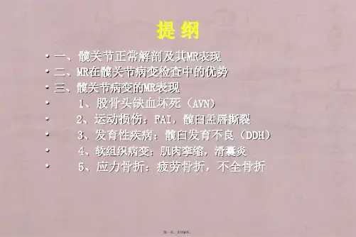
髋关节常见病变MR诊断
- 格式:pdf
- 大小:12.41 MB
- 文档页数:113
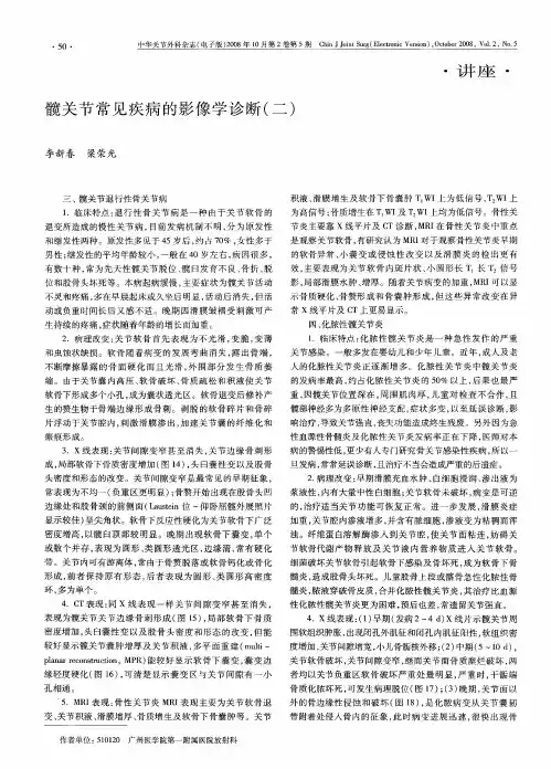
正常髋关节的MRI表现髋关节的主要组织成分为以下四个部分:(一)骨性部分包括髋臼与股骨头、颈。
髋臼为髂、耻、坐骨之膨大体部所构成。
儿童发育期间,髋臼部为Y型骨骺。
股骨头为球形,上覆软骨,表面光滑,对向髋臼。
股骨头中心有一浅窝,为圆韧带窝。
在磁共振图像上骨皮质呈半环形低信号区,内为高信号松质骨。
头颈骨小梁束呈向心状排列,形成粗大放射状低信号区。
干骺端愈合的骨板仍清晰可见,呈波浪状低信号线。
由于股骨头的松质骨被皮质及致密的骨小梁所包绕,形成相对闭合的系统。
髓腔内处于正压状态,骨血运与髓腔内压力关系密切,动脉血流减少时压力下降,而静脉血流减少时压力上升。
反之,压力的升降又可以直接影响血运。
磁共振T1与T2加权像可直接反映股骨头血运的变化。
血管充血、静脉滞流可引起长T1与长T2信号改变,细胞外液的增加也使T2弛豫延长。
(二)软骨部分包括软骨关节面和关节盂唇。
软骨关节面由透明软骨构成,被覆于股骨头和髋臼表面,在髋臼前、上、后壁形成半月状关节面,髋臼内面中心凹陷为镜臼窝。
透明软骨约2mm厚,在MRI所有序列上均为中等信号强度。
透明软骨含水量高,弹性强,为关节内的承重结构,其T2值较纤维软骨高,但低于关节内滑液。
因此,透明软骨在信号上易与纤维软骨及关节液相鉴别。
关节盂唇由纤维软骨构成,附着于关节盂边缘,使髋臼加深,髋臼唇缘及下缘的软骨唇最宽,约10mm。
纤维软骨内所含蛋白成分及水分均与透明软骨不同。
纤维软骨含水量低,致密胶原组织的T2弛豫时间短,因而在T2加权像上其信号低于透明软骨。
(三)韧带与肌腱部分髋关节包括3个纵行韧带、1个圆韧带和2个附属韧带,其中以髋臼横韧带、股骨头圆韧带在髋关节病变的诊断中较为重要。
韧带与肌腱属于纤维结构,在MRI所有扫描序列上均呈低信号或无信号。
(四)关节囊关节囊与髋关节纵行韧带交织在一起。
关节囊起于髋臼的骨缘及横韧带,包括软骨唇、股骨头、颈。
关节囊在MRI所有序列上均为低信号,囊内少量滑液呈条形高信号。
* 器材应用与技术研究 *MR、CT检查对强直性脊柱炎髋关节病变的诊断价值比较邓洪波,,曾宪春刘宗才刘宗才,,邓洪波贵州省人民医院医学影像科,贵州贵阳550001摘要目的比较核磁共振(magnetic resonance, MR)、电子计算机断层摄影(computer Tomography, CT)检查对强直性脊柱炎髋关节病变的诊断价值。
方法以贵州省人民医院于2017年10月—2022年10月收治的95例强直性脊柱炎疑似髋关节病变患者作为研究对象,收集患者MR、CT检查的影像资料进行分析,并以临床综合诊断作为“金标准”比较两种检查方式的诊断效能,同时观察两种检查方式对髋关节病变征像的检出率。
结果 80例患者出院诊断为强直性脊柱炎髋关节病变。
MR检查对强直性脊柱炎髋关节病变的诊断符合率、特异度、灵敏度、阴性预测值以及阳性预测值分别为96.84%、93.33%、97.50%、87.50%、98.73%,较CT检查的81.05%、60.00%、85.00%、42.86%、91.89%高,差异有统计学意义(χ2=12.046、4.658、7.828、7.695、4.097,P< 0.05),MR检查的误诊率、漏诊率分别为6.67%、2.50%,较CT检查的40.00%、15.00%低,差异有统计学意义(P<0.05)。
MR检查对关节强直、关节面侵蚀、关节间隙狭窄、关节软组织肿胀、关节面下骨质囊变、关节面下骨质硬化的检出率分别为88.57%、88.24%、95.38%、94.87%、93.44%、93.75%,高于CT检查的64.29%、64.71%、81.54%、75.64%、75.41%、77.08%,差异有统计学意义(P<0.05)。
结论 MR检查可有效检出强直性脊柱炎髋关节病变,诊断效能高于CT检查,而且,该检查方式对病变征象的检出率较高,因此,可作为临床诊断该疾病的优选方案。
关键词MR;CT;强直性脊柱炎髋关节病变;诊断效能;诊断符合率;病变征象816..8文献标志码A doi10.11966/j.issn.2095-994X.2023.09.04.28中图分类号R816Comparison of Diagnostic Value of MR and CT Examinations in Ankylosing Spondy⁃litis Hip Joint LesionsLIU Zongcai, DENG Hongbo, ZENG XianchunDepartment of Medical Imaging, Guizhou Provincial People's Hospital, Guiyang, Guizhou Province, 550001 ChinaAbstract Objective To compare the diagnostic value of nuclear magnetic resonance (MR) and computerized tomography (CT) in ankylosing spondylitis hip joint lesions. Methods A total of 95 patients with ankylosing spondylitis suspected of hip joint lesions admitted to Guizhou Pro⁃vincial People's Hospital from October 2017 to October 2022 were selected as the research objects. The imaging data of MR and CT examina⁃tions of patients were collected and analyzed, and the diagnostic efficacy of the two examination methods was compared using comprehensive clinical diagnosis as the "gold standard". At the same time, the detection rate of the two inspection methods on the signs of hip joint disease was observed. Results Eighty patients were discharged and diagnosed as ankylosing spondylitis hip joint lesions. The diagnostic coincidence rate, specificity, sensitivity, negative predictive value, and positive predictive value of MR examination for ankylosing spondylitis hip joint le⁃sions were 96.84%, 93.33%, 97.50%, 87.50%, and 98.73%, respectively, which were higher than those of CT examination 81.05%, 60.00%, 85.00%, 42.86%, 91.89%, the difference was statistically significant (χ2=12.046, 4.658, 7.828, 7.695, 4.097, P<0.05), the misdiagnosis rate and missed diagnosis rate of MR examination were 6.67% and 2.50%, respectively, which were lower than those of CT examination (40.00% and 15.00%), the difference was statistically significant (P<0.05). The detection rates of MR examination for joint ankylosis, articular surface erosion, joint space narrowing, joint soft tissue swelling, subarticular bone cystic degeneration, and subarticular bone sclerosis were 88.57%, 88.24%, 95.38%, 94.87%, 93.44%, 93.75%, higher than CT examination 64.29%, 64.71%, 81.54%, 75.64%, 75.41%, 77.08%, the differ⁃ence was statistically significant (P<0.05). Conclusion MR examination can effectively detect hip joint lesions in ankylosing spondylitis, with a higher diagnostic efficiency than CT examination. Moreover, this examination method has a higher detection rate of lesion signs, making it a preferred option for clinical diagnosis of the disease.Key words MR; CT; Ankylosing spondylitis hip joint lesion; Diagnostic efficiency; Diagnostic coincidence rate; Lesion signs收稿日期:2023-02-06;修回日期:2023-02-27作者简介:刘宗才(1974-),男,硕士,副主任医师,研究方向为影像学。
3种髋关节常见疾病的影像学诊断来源:骨今中外01髋关节退行性骨关节病临床特点:退行性骨关节病是一种由于关节软骨的退变所造成的慢性关节病,目前发病机制不明,分为原发性和继发性两种。
原发性多见于45岁后,约占70%,女性多于男性;继发性的平均年龄较小,一般在40岁左右,病因很多,有数十种,常为先天性髋关节脱位、髋臼发育不良、骨折、脱位和股骨头坏死等。
本病起病缓慢,主要症状为髋关节活动不灵和疼痛,多在早晨起床或久坐后明显,活动后消失,但活动或负重时间长后又感不适。
晚期因滑膜皱褶受刺激可产生持续的疼痛,症状随着年龄的增长而加重。
病理改变:关节软骨首先表现为不光滑,变脆,变薄和虫蚀状缺损。
软骨随着病变的发展弯曲消失,露出骨端,不断摩擦暴露的骨面硬化而且光滑,外围部分发生骨质萎缩。
由于关节囊内高压、软骨破坏、骨质疏松和积液使关节软骨下形成多个小孔,成为囊状透光区。
软骨退变后修补产生的赘生物于骨端边缘形成骨刺。
剥脱的软骨碎片和骨碎片浮动于关节腔内,刺激滑膜渗出,加速关节囊的纤维化和瘢痕形成。
X线表现:关节间隙变窄甚至消失,关节边缘骨刺形成,局部软骨下骨质密度增加 (图14 ),头臼囊性变以及股骨头密度和形态的改变。
关节间隙变窄是最常见的早期征象,常表现为不均一(负重区更明显 ) ;骨赘开始出现在股骨头凹边缘处和股骨颈的前侧面 (Laustein 位 - 仰卧屈髋外展照片显示较佳 )呈尖角状。
软骨下反应性硬化为关节软骨下广泛密度增高,以髋臼顶部较明显。
晚期出现软骨下囊变,单个或数个并存,表现为圆形、类圆形透光区,边缘清,常有硬化带。
关节内可有游离体,常由于骨赘脱落或软骨钙化或骨化形成,前者保持原有形态,后者表现为圆形、类圆形高密度环,多为单个。
CT表现:同X线表现一样关节间隙变窄甚至消失,表现为髋关节关节边缘骨刺形成 (图2 ),局部软骨下骨质密度增加,头臼囊性变以及股骨头密度和形态的改变,但能较好显示髋关节囊肿增厚及关节积液图15 双髋退行性骨关节病CT扫描VR冠状位重建示双侧髋臼骨质增生,骨性关节面硬化,关节间隙轻度变窄多平面重建(multi-planarreconstruction,MPR)能较好显示软骨下囊变,囊变边缘轻度硬化(图16),可清楚显示囊变区与关节间隙有一小孔相通。
髋关节撞击综合征影像诊断标准
髋关节撞击综合征是一种常见的髋部疾病,通常由于髋关节周
围软组织的摩擦和压力引起。
影像学诊断在确认和评估髋关节撞击
综合征方面起着关键作用。
以下是髋关节撞击综合征的影像诊断标准:
1. X线检查,X线检查是最常用的影像学诊断方法之一。
在髋
关节撞击综合征的X线影像中,可以观察到髋臼形态异常、髋臼过
深或过浅、髋臼前倾角增大、股骨头形态异常等特征。
2. 磁共振成像(MRI),MRI能够提供更为详细的软组织结构
信息,对于评估髋关节周围软组织的损伤和炎症有很高的诊断价值。
在髋关节撞击综合征的MRI影像中,可以观察到髋臼软骨损伤、股
骨头软骨损伤、股骨颈骨刺等特征。
3. 计算机断层扫描(CT),CT扫描可以提供关于骨结构的高
分辨率图像,对于评估髋臼形态和骨质异常有很高的诊断准确性。
在髋关节撞击综合征的CT影像中,可以观察到髋臼形态异常、髋臼
前倾角增大、股骨头形态异常等特征。
4. 超声检查,超声检查对于评估髋关节周围软组织的情况有一定的帮助,特别是对于股腱炎、髌股关节囊肿块等情况有较高的诊断准确性。
综上所述,髋关节撞击综合征的影像诊断标准主要包括X线检查、MRI、CT和超声检查。
结合临床症状和体征,可以更准确地诊断和评估髋关节撞击综合征。
希望以上信息能够对你有所帮助。
《中青年髋关节疼痛的影像鉴别诊疗》xx年xx月xx日CATALOGUE 目录•概述•髋关节影像学检查•中青年髋关节疼痛的常见疾病鉴别诊断•髋关节疼痛的诊疗流程•病例讨论•总结与展望01概述髋关节疼痛是指由多种病因引发的髋关节局部或周围疼痛的一种症状。
定义根据病因可分为原发性髋关节疼痛和继发性髋关节疼痛;根据疼痛部位可分为髋关节前方疼痛、侧方疼痛和后侧方疼痛。
分类定义与分类发病率髋关节疼痛在各年龄段均可发病,但以中青年为主,40-60岁人群发病率较高。
年龄分布髋关节疼痛好发于30-50岁人群,男女比例相近,但女性患者略多。
发病率与年龄分布常见病因髋关节疼痛的常见病因包括髋关节骨关节炎、股骨头坏死、髋关节发育不良、强直性脊柱炎等。
病理机制髋关节骨关节炎主要是由于软骨磨损、骨质增生、骨赘形成等引起;股骨头坏死主要是由于股骨头血供中断或受损引起;髋关节发育不良主要是由于遗传和环境因素影响引起;强直性脊柱炎则是一种自身免疫性疾病。
常见病因与病理机制02髋关节影像学检查X线平片X线平片可以显示髋关节的形态、结构、骨质的改变以及关节间隙的变化等。
X线平片具有操作简便、费用低廉等优点,但成像质量受限于体位、曝光参数等因素。
X线平片是髋关节影像学检查的基本方法,适用于大多数髋关节病变的检查。
计算机断层扫描(CT)CT检查可以提供髋关节的横断面图像,对骨质的细微结构和病变的显示更为准确。
CT检查对于判断髋关节骨折、骨坏死及骨髓炎等病变具有较高的敏感性和特异性。
CT检查的辐射较大,价格相对较高,但成像质量稳定可靠,对细节的显示优于X线平片。
磁共振成像(MRI)MRI检查可以提供髋关节的多种成像序列,对软组织、骨髓及关节软骨的显示效果较好。
MRI检查对于诊断髋关节炎症、肿瘤及结核等病变具有较高的敏感性和特异性。
MRI检查的价格较高,成像时间较长,但无需使用放射性物质,对人体无损伤。
这些方法一般不作为髋关节病变的首选检查方法,但在特定情况下可以辅助诊断。