
临床髓质海绵肾影像诊断
- 格式:pptx
- 大小:8.02 MB
- 文档页数:31


【双语病例】髓质海绵肾(IVP、CT)鉴别诊断
往期相关链接:
【书摘】“肾皮/髓质钙质沉着症”的鉴别诊断
History: A 75-year-old man with a chronic history of
microhematuria was referred for a CT urogram by his
urologist.
病史:75岁男性,长期镜下血尿,泌尿外科医生建议其行CTU检查。
Scout, coronal precontrast, axial nephrographic/excretory
phase images, and volume-rendered coronal reconstructions of
the left and right kidneys are shown below.
定位像、冠状平扫、轴位排泄期图像及左、右肾冠状VR重建如下所示。 Additional history: The patient has a prior history of
microhematuria and kidney stones. Review of the PACS for
prior imaging reveals an intravenous pyelogram (IVP) from
2005. Scout and postcontrast exposures are shown below.
病史补充:患者之前患有镜下血尿及肾结石。回顾PACS中2005年的静脉尿路造影(IVP)图像,腹部平片及造影后图像图像所示。 Findings 影像表现
CT urogram (2017): Noncontrast images demonstrate
multiple bilateral nonobstructing renal calculi, the largest

髓质海绵肾
髓质海绵肾是一种先天性的肾髓质囊性病变,可能具有遗传倾向,多为双侧。肾髓质锥体部乳头管及集合管呈梭形或囊状扩张,并伴发感染和尿路结石形成。标本切面上,可见髓质中呈海绵状改变。
尿路平片肾锥体部多发性阳性结石影,结石大小及形态不规则,小如细砂粒,大至横径可达0.5厘米。结石呈扇形或密集簇状排列,结石也可不规则散在分布于各锥体部。
静脉尿路造影典型病例在静脉尿路造影时,首先见到肾锥体部囊腔充盈,在输尿管加压时显影更清楚,压力解除后肾盏肾盂已排空,而囊腔内仍残留造影剂。静脉尿路造影片上常见到的肾锥体或肾盏周围的阴影为:造影剂在扩张的集合管和乳头管内呈扇形;充盈小囊腔呈葡萄串样;或与结石影重叠形成密度不均的斑片影;肾小盏增宽,杯口大而变平。
CT平扫显示肾锥体内多发小斑点状高密度影,散在或呈簇状,花瓣样。增强扫描后扩张肾集合管内可见造影剂聚集,伴有结石时可见条纹状或小囊状造影剂聚集,伴有结石时可造成结石影覆盖或结石影增大的假象,扩张的集合管内造影剂排空较为延迟。
鉴别诊断
肾钙质沉着症:如甲状旁腺机能亢进、肾小管酸中毒、维生素D过多症、慢性肾小球肾炎等,一般无肾集合管扩张和乳头囊腔形成,钙化较弥漫并涉及肾皮质。
肾小盏内散发小结石:位于肾盏,可并发肾盂肾盏轻度积水,位置可变。
肾结核:病变较广泛,不局限于髓质,常见钙化为弧线状、斑点状、囊腔不规则,常伴有输尿管膀胱结核病变。
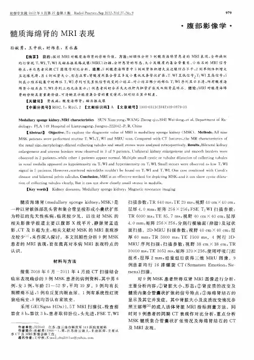
放射学实践2012年9月第27卷第9期Radiol Practice,Sep 2012,Vol 27,No.9
髓质海绵肾的MRI表现 979
腹部影像学
孙献勇,王中秋,时维东,-g永鑫
【摘要】 目的:探讨MR1对髓质海绵肾的诊断价值。方法:回顾性分析9例髓质海绵肾患者的MRI表现,全部病例 均行常规T wI、T wI及磁共振尿路成像(MRU)扫描,分析患肾的形态、大小及髓质内集合管囊变、小结石的MR1信号
特点,并与患者同期CT图像作对比分析。结果:9例髓质海绵肾中3例双肾体积增大且边缘凹凸不平,2例单侧体积增大
且边缘光滑,另4例双肾大小、形态正常;肾髓质内集合管呈多发小囊状及条管状扩张,T wI呈低信号,TzwI呈高信号;5
例在小结石较集中的部位T。wI序列可见呈低信号改变的小结石,对小结石稀少的部位T。wI序列显示不清,所有髓质海
绵肾小结石在T wI序列上均无法显示;1例患者同时合并先天性肝内胆管扩张及双侧肾盂结石。结论:MRI对髓质海绵
肾的诊断具有重要价值,可清晰显示髓质集合管的囊变情况,但对结石显示较差。
【关键词】 肾疾病;髓质海绵’g-;磁共振成像
【中图分类号1 R692.1;R445.2【文献标识码】A【文章编号1 1000—0313(2012)09—0979—03
Medullary sponge kidney:MRI characteristics SUN Xian—yong,WANG Zhong—qiu,SHI Wei dong,et al
diology,PLA 149 Hospital of Lianyungang,Jiangsu,222042,P.R.China [Abstract]Objective: Fo explore the diagnostic value of MRI in medullary sponge kidney(MSK).Methods:All nine

髓质海绵肾超声诊断标准
英文回答:
The diagnostic criteria for medullary sponge kidney
(MSK) on ultrasound include the presence of multiple small
cysts in the medullary pyramids, dilatation of the
collecting ducts, and the absence of other renal
abnormalities. The cysts appear as hypoechoic structures
with well-defined borders. The dilated collecting ducts can
be visualized as anechoic tubular structures extending from
the cysts towards the renal pelvis. The absence of other
renal abnormalities, such as hydronephrosis or renal
calculi, is important in confirming the diagnosis of MSK.
In addition to the ultrasound findings, clinical
symptoms and laboratory tests can also aid in the diagnosis
of MSK. Patients with MSK often present with recurrent
urinary tract infections, kidney stones, and hematuria.
Laboratory tests may show hypercalciuria, hypocitraturia,