颞下颌关节的解剖生理.
- 格式:ppt
- 大小:887.50 KB
- 文档页数:15
颞下颌关节构成颞下颌关节是人体中一个重要的关节结构,位于颞骨和下颌骨之间。
它的主要功能是连接颞骨和下颌骨,使得我们能够进行咀嚼、开口和闭口等动作。
下面将详细介绍颞下颌关节的构成和功能。
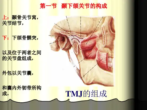
颞下颌关节由颞骨和下颌骨之间的关节窝和骨头组成。
颞骨上的关节窝是一个凹陷的部分,而下颌骨上的骨头是一个凸起的部分。
这种凹凸结构使得颞下颌关节能够相互咬合并保持稳定。
在颞下颌关节中,有一个软骨盘位于关节窝和骨头之间,起到缓冲和减少摩擦的作用。
这个软骨盘可以随着下颌骨的运动而移动,使得颞下颌关节能够灵活地进行开口和闭口的动作。
颞下颌关节还有一系列的韧带和肌肉组成。
这些韧带和肌肉的作用是支持和稳定颞下颌关节的运动。
它们能够控制下颌骨的运动范围,使得我们能够进行正常的咀嚼和开口动作。
颞下颌关节的功能非常重要。
它不仅能够使我们进行咀嚼和开口动作,还能够保护脑部和面部结构。
当我们进行咀嚼动作时,颞下颌关节能够使下颌骨向下移动并咬合食物,从而使得我们能够正常地消化食物。
颞下颌关节还与语言和面部表情有关。
它的灵活性和稳定性能够使我们进行各种表情,并参与到语言的发音中。
当我们说话时,颞下颌关节能够使下颌骨进行适当的运动,从而产生清晰的语音。
然而,颞下颌关节也存在一些问题。
颞下颌关节紊乱是一种常见的颞下颌关节疾病,常常导致下颌骨运动异常和疼痛。
这种紊乱可能是由于韧带和肌肉的损伤、软骨盘的移位或关节窝和骨头之间的不匹配引起的。
颞下颌关节紊乱的症状包括下颌骨疼痛、咀嚼困难、面部肌肉痉挛等。
对于这种疾病,我们可以通过物理治疗、药物治疗和手术治疗来缓解症状和恢复颞下颌关节的功能。
颞下颌关节是人体中一个重要的关节结构,它连接颞骨和下颌骨,使得我们能够进行咀嚼、开口和闭口等动作。
它由颞骨和下颌骨之间的关节窝和骨头、软骨盘以及韧带和肌肉组成。
颞下颌关节的功能非常重要,它不仅能够使我们进行咀嚼和开口动作,还能够参与到语言和面部表情中。
然而,颞下颌关节也可能出现紊乱等问题,需要及时治疗。
颞下颌关节功能解剖特点
颞下颌关节是指头骨与下颌骨之间的关节,也称为颞下颌关节。
它是人体唯一的可活动关节,能够承受大量的力量和压力。
颞下颌关节的解剖特点主要包括以下几个方面:
1.关节结构
颞下颌关节由上下两个部分组成,包括颞骨和下颌骨。
颞骨包括颞骨突和颞骨关节窝,下颌骨包括下颌支和下颌骨头。
两者之间的关节面是由软骨组织构成的,它们能够缓冲和吸收关节运动时产生的冲击和压力。
2.关节囊
颞下颌关节囊是连接颞骨和下颌骨的纤维性袋状结构。
它包围着整个关节,保护关节组织不受损伤。
颞下颌关节囊内有关节液,它能够润滑和减少关节的磨损。
3.关节韧带
颞下颌关节韧带是连接颞骨和下颌骨的强韧结构。
韧带的主要作用是固定和稳定颞下颌关节,防止关节脱位和损伤。
4.肌肉组织
颞下颌关节周围有多个肌肉组织,包括咀嚼肌、颞肌和颈肌等。
这
些肌肉组织的协同作用能够使颞下颌关节在咀嚼和说话等活动中保持稳定和灵活。
颞下颌关节的解剖特点非常复杂和精细。
它是人体唯一一个可以自由活动的关节,对于人体的正常生理功能有着至关重要的作用。
在医学领域,颞下颌关节也是研究口腔颌面疾病和颞下颌关节紊乱等问题的重要研究对象。

动物解剖生理颞下颌关节的组成英文回答:The temporomandibular joint (TMJ) is a synovial joint that connects the mandible (lower jaw) to the temporal bone of the skull. It is responsible for the movement of the jaw, allowing for actions such as opening and closing the mouth, chewing, and speaking.The TMJ is composed of several components. The mandibular condyle, which is the rounded portion of the mandible, fits into the mandibular fossa of the temporal bone. This forms the hinge-like joint that allows for the opening and closing of the mouth. The articular disc, a fibrous cartilage structure, is located between the condyle and the fossa. It acts as a cushion and helps to distribute the forces during jaw movement.The TMJ also has ligaments that provide stability tothe joint. The lateral ligament, also known as thetemporomandibular ligament, connects the condyle to the zygomatic arch. It limits excessive movement of the jaw and helps to maintain proper alignment. The stylomandibular ligament, on the other hand, connects the styloid process of the temporal bone to the angle of the mandible. It provides additional support to the joint.Furthermore, the TMJ is surrounded by a joint capsule, which is a fibrous structure that encloses the joint and helps to maintain its integrity. The capsule is lined with a synovial membrane that produces synovial fluid, which lubricates the joint and reduces friction during movement.In addition to these components, there are also several muscles involved in the movement of the TMJ. The muscles of mastication, such as the temporalis, masseter, and medial pterygoid, are responsible for the opening and closing of the jaw. They work together to provide the necessary force for chewing and biting.中文回答:颞下颌关节(TMJ)是一种滑膜关节,连接下颌骨(下颚)和颞骨。