医学-细胞外基质作用机理共41页文档
- 格式:ppt
- 大小:4.84 MB
- 文档页数:41


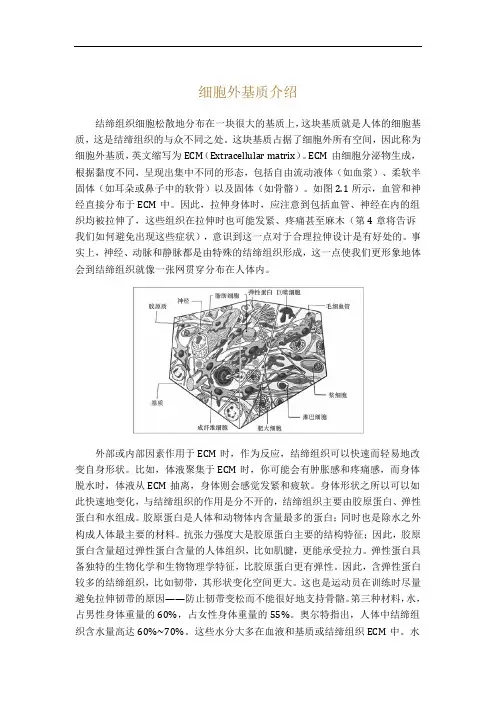
细胞外基质介绍结缔组织细胞松散地分布在一块很大的基质上,这块基质就是人体的细胞基质,这是结缔组织的与众不同之处。
这块基质占据了细胞外所有空间,因此称为细胞外基质,英文缩写为ECM(Extracellular matrix)。
ECM由细胞分泌物生成,根据黏度不同,呈现出集中不同的形态,包括自由流动液体(如血浆)、柔软半固体(如耳朵或鼻子中的软骨)以及固体(如骨骼)。
如图2.1所示,血管和神经直接分布于ECM中。
因此,拉伸身体时,应注意到包括血管、神经在内的组织均被拉伸了,这些组织在拉伸时也可能发紧、疼痛甚至麻木(第4章将告诉我们如何避免出现这些症状),意识到这一点对于合理拉伸设计是有好处的。
事实上,神经、动脉和静脉都是由特殊的结缔组织形成,这一点使我们更形象地体会到结缔组织就像一张网贯穿分布在人体内。
外部或内部因素作用于ECM时,作为反应,结缔组织可以快速而轻易地改变自身形状。
比如,体液聚集于ECM时,你可能会有肿胀感和疼痛感,而身体脱水时,体液从ECM抽离,身体则会感觉发紧和疲软。
身体形状之所以可以如此快速地变化,与结缔组织的作用是分不开的,结缔组织主要由胶原蛋白、弹性蛋白和水组成。
胶原蛋白是人体和动物体内含量最多的蛋白;同时也是除水之外构成人体最主要的材料。
抗张力强度大是胶原蛋白主要的结构特征;因此,胶原蛋白含量超过弹性蛋白含量的人体组织,比如肌腱,更能承受拉力。
弹性蛋白具备独特的生物化学和生物物理学特征,比胶原蛋白更有弹性。
因此,含弹性蛋白较多的结缔组织,比如韧带,其形状变化空间更大。
这也是运动员在训练时尽量避免拉伸韧带的原因——防止韧带变松而不能很好地支持骨骼。
第三种材料,水,占男性身体重量的60%,占女性身体重量的55%。
奥尔特指出,人体中结缔组织含水量高达60%~70%。
这些水分大多在血液和基质或结缔组织ECM中。
水的表面张力很大,在血管和结缔组织中,水表面就像动力弹性片一样活动。
我们滑倒、摔倒或与其他人相撞却没有产生外伤,原因就在这里。
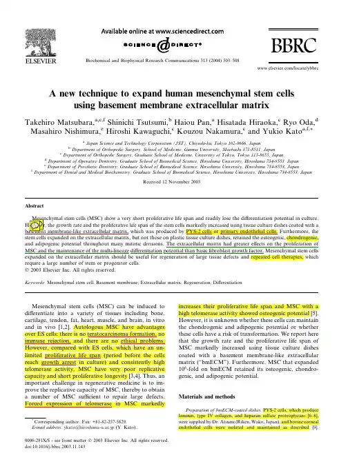
A new technique to expand human mesenchymal stem cellsusing basement membrane extracellular matrixTakehiro Matsubara,a,c,f Shinichi Tsutsumi,b Haiou Pan,a Hisatada Hiraoka,c Ryo Oda,d Masahiro Nishimura,e Hiroshi Kawaguchi,c Kouzou Nakamura,c and Yukio Kato a,f,*aJapan Science and Technology Corporation (JST),Chiyoda-ku,Tokyo 102-8666,JapanbDepartment of Orthopedic Surgery,School of Medicine,Gunma University,Maebashi 371-8511,Japan cDepartment of Orthopedic Surgery,Graduate School of Medicine,University of Tokyo,Tokyo 113-8655,JapandDepartment of Operative Dentistry,Graduate School of Biomedical Science,Hiroshima University,Hiroshima 734-8553,Japan eDepartment of Prosthetic Dentistry,Graduate School of Biomedical Science,Hiroshima University,Hiroshima 734-8553,JapanfDepartment of Dental and Medical Biochemistry,Graduate School of Biomedical Science,Hiroshima University,Hiroshima 734-8553,JapanReceived 12November 2003Abstractshort proliferative life span and readily lose the differentiation potential in culture.span of the stem cells markedly increased using tissue culture dishes coated with a which was produced by PYS-2cells or primary endothelial cells.Furthermore,the stem cells expanded on the extracellular matrix,but not those on plastic tissue culture dishes,retained the osteogenic,chondrogenic,and adipogenic potential throughout many mitotic divisions.The extracellular matrix had greater effects on the proliferation of MSC and the maintenance of the multi-lineage differentiation potential than basic fibroblast growth factor.Mesenchymal stem cells expanded on the extracellular matrix should be useful for regeneration of large tissue defects and repeated cell therapies,which require a large number of stem or progenitor cells.Ó2003Elsevier Inc.All rights reserved.Keywords:Mesenchymal stem cell;Basement membrane;Extracellular matrix;Regeneration;DifferentiationMesenchymal stem cells (MSC)can be induced to differentiate into a variety of tissues including bone,cartilage,tendon,fat,heart,muscle,and brain,in vitro and in vivo [1,2].Autologous MSC have advantages over ES cells:there is no teratocarcinoma formation,no immune rejection,and there are no ethical problems.However,compared with ES cells,which have an un-limited proliferative life span (period before the cells reach growth arrest in culture)and consistently high telomerase activity,MSC have very poor replicative capacity and short proliferative longevity [3,4].Thus,an important challenge in regenerative medicine is to im-prove the replicative capacity of MSC,thereby to obtain a number of MSC sufficient to repair large defects.Forced expression of telomerase in MSC markedlyincreases their proliferative life span and MSC with a high telomerase activity showed osteogenic potential [5].However,it is unknown whether these cells can maintain the chondrogenic and adipogenic potential or whether these cells have a risk of transformation.We report here that the growth rate and the proliferative life span of MSC markedly increased using tissue culture dishes coated with a basement membrane-like extracellular matrix (“bmECM”).Furthermore,MSC that expanded 106-fold on bmECM retained its osteogenic,chondro-genic,and adipogenic potential.Materials and methodsPreparation of bmECM-coated dishes .PYS-2cells,which produce laminin,type IV collagen,and heparan sulfate proteoglycans [6–8],were supplied by Dr.Atsumi (Riken,Wako,Japan),and bovine corneal endothelial cells were isolated and maintained as described [9].*Corresponding author.Fax:+81-82-257-5629.E-mail address:ykato@hiroshima-u.ac.jp (Y.Kato).0006-291X/$-see front matter Ó2003Elsevier Inc.All rights reserved.doi:10.1016/j.bbrc.2003.11.143Biochemical and Biophysical Research Communications 313(2004)503–508BBRC/locate/ybbrcBmECM-coated dishes were prepared according to the method of Dr. Gospodarowicz[10].The cells were seeded at2Â104cells/cm2on60-mm tissue culture dishes(Corning,Corning,NY)and maintained in 4ml of Dulbecco’s modified Eagle’s medium(DMEM)-Ham’s F12 medium(1:1)(Sigma,St.Louis,MO)in the presence of10%fetal bo-vine serum(Hyclone,Logan,Utah)and antibiotics(100U/ml penicillin G and100l g/ml streptomycin)(medium-A).Medium was changed every other day.Once the cultures became confluent,the media were renewed by4ml of medium-A supplemented with5%dextran (200,000Da,Wako,Osaka,Japan)and the cultures were further in-cubated for7days.Treatment of the cultures with20mM NH3resulted in cell lysis,exposing the extracellular matrix adhering to the substrata of tissue culture dishes.The substratum was washedfive times with PBS.Previous studies have shown that bmECM is composed of lami-nin,heparan sulfate,entactin,and type IV collagen[11,12].Preparation of laminin-coated dishes,type IV collagen-coated dishes, and ECM gel-coated dishes.Three milliliters of10mM NaHCO3 containing30l g/ml type IV collagen or30l g/ml laminin(Koken, Tokyo,Japan)was incubated in60-mm plastic tissue culture dishes at 4°C for12h.Two hundred micrograms per milliliter ECM gel solution (Sigma)was made by diluting with DMEM-high glucose.Three mil-liliters of the ECM gel solution was incubated in60-mm tissue culture dishes at4°C for12h.These concentrations of laminin,type IV col-lagen,and ECM gel were optimal for proliferation of MSC(data not shown).MSC culture.Human MSC were obtained from the ilium or the alveolar bone according to a protocol approved by ethical authorities at Hiroshima University.Cells in marrow aspirates(1ml/100-mm dish) were seeded on plastic tissue culture dishes.Passages were performed when cells were approaching confluence.Unless otherwise specified, MSC obtained from the primary cultures were seeded at1Â103cells/ cm2on60-mm of laminin-,type IV collagen-or ECM gel-coated dishes,on bmECM-coated dishes or on plastic tissue culture dishes, and cells were fed with DMEM-low glucose supplemented with10% fetal bovine serum and antibiotics(medium-B)every3days.Sub-sequent passages were performed on the appropriate substrata.In these studies,we seeded MSC at a low density(1Â103cells/cm2)to avoid frequent passages and the risk of contamination considering clinical application,although MSC showed a higher growth rate and a longer proliferative life span at a high seeding cell density(5Â103cells/ cm2)(data not shown).Differentiation.Chondrogenic,osteogenic or adipogenic conversion of MSC was determined according to the procedures reported by Pittenger et al.[1],with some modifications.For chondrogenic differ-entiation,cells were seeded at2.5Â105cells per15ml plastic centrifuge tube and maintained in0.5ml of serum-free a-MEM supplemented with3500mg/ml glucose, 6.25l g/ml insulin, 6.25l g/ml transferrin, 6.25ng/ml selenite,5.33l g/ml linolate,1.25mg/ml bovine serum al-bumin,10ng/ml transforming growth factor-b3,100nM dexametha-sone,and50l g/ml ascorbic acid-2-phosphate.The cultures were fed with0.5ml of the medium until3days after seeding.Thereafter,the cultures were fed with1ml of the medium every other day.Cells were cultured under the chondrogenic status for28days.For osteogenic differentiation,cells were seeded at4Â104cells per16-mm dish and maintained for21–28days in DMEM-low glucose supplemented with 10l g/ml insulin,10mM b-glycerophosphate,100nM dexamethasone, and50l g/ml ascorbic acid-2-phosphate.For adipogenic differentia-tion,cells were seeded at2Â105cells per35-mm dish and grown to confluence in medium-B.Thereafter,adipogenic differentiation was induced by subjecting confluent monolayers to3–4rounds of adipo-genic treatments.Each round had two steps;incubation with adipo-genic medium(DMEM-high glucose,10%fetal bovine serum,0.2mM indomethacin,1l M dexamethasone,0.5mM methyl-isobutylxanthine, and10l g/ml insulin)for72–96h and incubation with maintenance medium(DMEM-high glucose,10%fetal bovine serum,and10l g/ml insulin)for72–96h.Cells were cultured under the adipogenic status for 28days.Glycosaminoglycan content,alkaline phosphatase activity,calcium level,glycerol-3-phosphate dehydrogenase activity,and DNA content. The glycosaminoglycan(GAG)content was determined using a sul-fated GAG assay kit(Biocolor,Newtownabbey,UK)[13].The alka-line phosphatase(ALP)activity was determined by the method of Bessey[14].The calcium level was determined by the method of Git-elman[15].The glycerol-3-phosphate dehydrogenase activity was de-termined using an assay kit(Hokudo,Sapporo,Japan)[16].The DNA content was determined using afluorescent DNA quantification kit (Bio-Rad,Chicago,IL).RT-PCR.Total RNA was extracted using Isogen(Nippon Gene, Tokyo,Japan).Thefirst-strand cDNA was synthesized from1l g of total RNA using the SUPERSCRIPT II RNase HÀreverse trans-criptase(Life Technologies,Rockville,MD).Using the cDNAs as a template,PCR was carried out under the following conditions:dena-turation at94°C for30s and primer extension at65°C for1.5min in 27cycles.Pairs of nucleotides,50-TGGTGGAGCAGCAAGAGCAA-30and50-TGCCCAGTTCAGGTCTCTTA-30for type II collagen,50-CCCAACACCAAGACACAGTT-30and50C-ATCACCTTTGATG CCTGGCT-30for type X collagen,and50-GTCAAGGCCGAGAAT GGGAA-30and50-GCTTCACCACCTTCTTGATG-30for GAPDH, 50-CATTTTGGGAATGGCCTGTG-30and50-ATTGTCTCCTCCG CTGCTGC-30for bone sialoprotein,50-CTAGGCATCACCTGTGC CATACC-30and50-CAGTG ACCAGTTCATCAGATTCATC-30for osteopontin,50-CCACCGAGACACCATGAGAG-30and50-CCATA GGGCTGGGAGGTCAG-30for osteocalcin,and50-CATTCTGGC CCACCAACTT-30and50-CCTTGCA TCCTTCACAAGCA-30for PPAR-c2were used as primers for RT-PCR.Obtained PCR products were separated on1%agarose gels and stained with ethidium bromide.Statistical analysis.Student’s t test was used.ResultsThe extracellular matrix produced by PYS-2cells or endothelial cells adhered to the substratum of plastic tissue culture dishes and could be easily cut with a needle and turned over like a sheet of paper(Fig.1A).When cells in marrow aspirates were seeded on plastic culture dishes,adherent cells—MSC—proliferated in the pres-ence of10%fetal bovine serum at a high growth rate in primary cultures[3],but their growth rate rapidly de-creased in secondary and tertiary cultures(Figs.1B–D). In cultures on plastic tissue culture dishes,non-adherent cells were removed completely by thefirst passage. However,when cells in marrow aspirates were seeded directly on bmECM,both MSC and many other cells adhered to the substratum and these adherent cells were not removed by changing the medium.Accordingly,we harvested MSC when the cells were approaching con-fluence in primary cultures on plastic tissue culture dishes,and seeded the isolated MSC on bmECM or plastic tissue culture dishes without the matrix (“plastic”)to examine the effects of bmECM on the proliferation of MSC.The growth rate of human MSC isolated from the ilium(Fig.1B)or the alveolar bone(Fig.1C)on bmECM was much higher than that on plastic,and thus the cumulative cell number in the cultures on bmECM was105-fold greater than that on plastic on day50. After MSC obtained from primary cultures were seeded504T.Matsubara et al./Biochemical and Biophysical Research Communications313(2004)503–508on bmECM or plastic,the proliferative life span of MSC on bmECM(50.3Æ1.5days)was also significantly (p<0:0001)longer than that of MSC on plastic (29.2Æ4.4days)(Figs.1B and C).The effect of bmECM produced by PYS-2was similar to that of endothelial cell bmECM(Fig.1).MSC seeded at a low density and grown on plastic lost their spindle-like appearance,be-comingflat with an increase in the passage number (Fig.1E).Theflat appearance is characteristic of se-nescent cells.However,most MSC grown on bmECM maintained the spindle-like appearance until the5th passage culture on day45(Fig.1F),suggesting that bmECM suppressed cell senescence.Laminin and type IV collagen are the major com-ponents of bmECM,but MSC on laminin-or type IV collagen-coated dishes showed lower growth rates than on bmECM(Fig.1F).The ECM gel isolated from Engelbreth–Holm–Swarm murine sarcoma also showed less growth stimulation than bmECM(Fig.1F),sug-gesting a loss of active substances during isolation of the extracellular matrix components or the necessity of an intact structure for growth stimulation.The chondrogenic potential of MSC was examined as a function of the passage number.MSC obtained from the2nd and the5th passage cultures on day15and day 51(Fig.1B)were maintained in pellet cultures for28 days(Fig.2A).The amount of cartilage proteoglycan stained with toluidine blue was greater in the pellets obtained from the5th passage cultures grown on bmECM than in the pellets from the2nd and the5th passage cultures grown on plastic.The expressions of type II collagen and type X collagen mRNAs were higher in pellets from the5th passage cultures on bmECM than in pellets from the5th passage cultures on plastic(Fig.2B).GAG content and ALP activity in the pellets decreased with the increase in the passage num-ber,irrespective of the presence or absence of bmECM. However,at each passage number,the GAG content (Fig.2C)and ALP activity(Fig.2D)were higher with MSC from cultures on bmECM than with MSC from cultures on plastic.Next,MSC from the2nd and the5th passage cultures on bmECM or plastic were incubated under the osteo-genic status on plastic tissue culture dishes.DuringT.Matsubara et al./Biochemical and Biophysical Research Communications313(2004)503–508505osteogenesis,we did not use bmECM to discriminate the effect of the extracellular matrix on proliferation from its direct effect on differentiation.MSC from cultures on bmECM became stained with alizarin red more in-tensely than MSC from cultures on plastic on day 21(Fig.2E).The expressions of bone sialoprotein,osteo-pontin,and osteocalcin mRNAs on day 28were also higher in cultures of MSC from cultures on bmECM than in cultures of MSC from cultures on plastic at the 5th passage (Fig.2F).ALP activity and calcium level on day 28decreased with the increase in the passage num-ber.However,MSC from cultures on bmECM showed a higher ALP activity (Fig.2G)and a higher calcium level (Fig.2H)than MSC from cultures on plastic at the 2nd and the 5th passages.To examine the adipogenic potential,MSC from the 2nd and the 5th passage cultures on bmECM or plastic were incubated under the adipogenic status on plastic tissue culture dishes for 28days.MSC from cultures on bmECM showed higher adipogenic differentiation,which was indicated by more intense staining with oil-red O (Fig.2I),higher PPAR-c 2mRNA expression (Fig.2J),and higher glycerol-3-phosphate dehydroge-nase activity (Fig.2K).Next,human MSC were grown on bmECM or plastic with 10%human serum,since human serum may be safer than fetal bovine serum for clinical use.Under these conditions,MSC proliferated more rapidly and showed a longer proliferative life span on bmECM than on plastic (Fig.3A).Furthermore,this effect of bmECM was greater than that of basic fibroblast growth factor (FGF).The MSC grown on bmECM for 66days de-veloped into a cartilage-like tissue (Fig.3B),even though these MSC on bmECM had lost proliferation capability.In contrast,scarcely any cartilage-like tissue was formed with MSC obtained from 66-day-old cul-tures on plastic (Fig.3C).DiscussionThe extracellular matrix (ECM)plays a vital role in organ morphogenesis,maintenance,and reconstruction following injury,and actions of ECM can be attributed to its effect on proliferation of stem cells,since stem cells reside on the basement membrane in the epithelium and some other tissues [17].In muscle,satellite cells (stem cells)—which can be induced to differentiate intomuscle,Fig.2.Retention of chondrogenic,osteogenic,and adipogenic potential on bmECM.The MSC obtained from the 2nd and the 5th passage cultures on plastic or bmECM produced by PYS-2cells (Fig.1B)were transferred into the chondrogenic status in pellet cultures for 28days (A–D)and stained with toluidine blue (A).(B)The mRNA levels of type II collagen (type II)and type X collagen (type X)were analyzed by RT-PCR.GAG content (C)and ALP activity (D)were determined on day 28.The MSC from the 2nd and the 5th passage cultures on plastic or bmECM were transferred into the osteogenic status (E–H).(E)The cell layers were stained with alizarin red on day 21.(F)The mRNA levels of bone sialoprotein (BSP),osteopontin (OP),and osteocalcin (OC)were analyzed by RT-PCR on day 28.The ALP activity (G)and calcium level (H)of the cell-matrix layers were determined on day 28.The MSC obtained from the 2nd and the 5th passage cultures on plastic or bmECM were cultured under the adipogenic status for 28days (I–K).(I)The cell layers were stained with oil-red O.(J)The mRNA level of PPAR-c 2was analyzed by RT-PCR.(K)Glycerol-3-phosphate dehydrogenase activity was determined.(C,D,G,H,and K)“)”and “+”represent MSC from culture on plastic and bmECM,respectively.Values are averages þ=ÀSD for four cultures.*p <0:05,**p <0:01,and ***p <0:001vs plastic.506T.Matsubara et al./Biochemical and Biophysical Research Communications 313(2004)503–508bone,cartilage,and fat —are also in close contact with the basement membrane;satellite cells became myo-blasts after detachment from the basal membrane [18].These observations suggest that the basement membrane and/or some other ECMs play a crucial role in the proliferation of stem cells and the maintenance of their undifferentiated state.In this study,we showed that bmECM markedly increased both the growth rate and the proliferative life span of MSC.Furthermore,MSC that had expanded 106-fold on bmECM maintained its multi-lineage differentiation potential.The mechanism by which bmECM stimulates MSC proliferation and maintains their differentiation potential is not known,but even MSC transfected with telomerase gradually decreased its osteogenic potential with the increase in the passage number [5],although their replicativecapacity was maintained.In contrast,MSC on bmECM maintained the differentiation potential even after they lost their replicative capacity.Thus,the extracellular matrix and telomerase may have complementary effects on the proliferation of MSC and their differentiation potential.In any case,the remarkable effects of bmECM in MSC cultures demonstrated here meet the expecta-tions of doctors eager to expand MSC extensively in vitro from a small volume of marrow aspirates before transplantation.Henceforth,bmECM will be prepared using human ES or human cell lines,and in the near future bmECM-coated dishes will probably have a great use in MSC studies and regeneration medicine.References[1]M.F.Pittenger, A.M.Mackay,S.C.Beck,R.K.Jaiswal,R.Douglas,J.D.Mosca,M.A.Moorman,D.W.Simonetti,S.Craig,D.R.Marshak,Multilineage potential of adult human mesenchy-mal stem cells,Science 284(1999)143–147.[2]R.J.Deans,A.B.Moseley,Mesenchymal stem cells:biology and potential clinical uses,Exp.Hematol.28(2000)875–884.[3]S.Tsutsumi,A.Shimazu,K.Miyazaki,H.Pan,C.Koike,E.Yoshida,K.Takagishi,Y.Kato,Retention of multilineage differentiation potential of mesenchymal cells during proliferation in response to FGF,mun.288(2001)413–419.[4]A.Muraglia,R.Cancedda,R.Quarto,Clonal mesenchymal progenitors from human bone marrow differentiate in vitro according to a hierarchical model,J.Cell Sci.113(2000)1161–1166.[5]S.Shi,S.Gronthos,S.Chen,A.Reddi,C.M.Counter,P.G.Robey,C.Y.Wang,Bone formation by human postnatal bone marrow stromal stem cells is enhanced by telomerase expression,Nat.Biotechnol.20(2002)587–591.[6]A.Oohira,T.N.Wight,J.McPherson,P.Bornstein,Biochemical and ultrastructural studies of proteoheparan sulfates synthesized by PYS-2,a basement membrane-producing cell line,J.Cell Biol.92(1982)357–367.[7]B.Tyree,E.A.Horigan,D.L.Klippenstein,J.R.Hassell,Heter-ogeneity of heparan sulfate proteoglycans synthesized by PYS-2cells,Arch.Biochem.Biophys.231(1984)328–335.[8]J.R.Couchman,A.Woods,M.Hook,J.E.Christner,Character-ization of a dermatan sulfate proteoglycan synthesized by murine parietal yolk sac (PYS-2)cells,J.Biol.Chem.260(1985)13755–13762.[9]D.Gospodawowicz,A.L.Mesher,C.R.Birdwell,Stimulation of corneal endothelial cell proliferation in vitro by fibroblast and epidermal growth factors,Exp.Eye Res.137(1977)15–23.[10]D.Gospodarowicz,R.Gonzalez,D.K.Fujii,Are factors origi-nating from serum,plasma,or cultured cells involved in the growth-promoting effect of the extracellular matrix produced by cultured bovine corneal endothelial cells?J.Cell.Physiol.114(1983)191–202.[11]A.Martinez-Hernandez,A.E.Chung,The ultrastructural local-ization of two basement membrane components:entactin and laminin in rat tissues,J.Histochem.Cytochem.32(1984)289–298.[12]E.Hahn,G.Wick,D.Pencev,R.Timpl,Distribution of basementmembrane proteins in normal and fibrotic human liver:collagen type IV,laminin,and fibronectin,Gut 21(1980)63–71.Fig.3.Effects of bmECM on proliferation of MSC and their chon-drogenic potential in cultures with human serum.(A)Human MSC,in the presence of 10%human serum,showed faster proliferation and a longer proliferative life span on bmECM than on plastic.Cells in marrow aspirates (1ml/100-mm dish)from the ilium were seeded and maintained on plastic tissue culture dishes,as described in the legend of Fig.1.The first passage was performed on plastic at a seeding density of 5000cells/cm 2.In this study,MSC obtained from the 1st passage cultures on plastic were used for experimentation.The isolated cells were seeded on bmECM (endothelial)or on plastic as described in Materials and methods.FGF at 1ng/ml was added to some cultures on plastic.*;**Differs significantly from the cell number in cultures on plastic dishes with FGF (“FGF”)or without FGF (“plastic”)on days 66–87(*p <0:05,**p <0:01).The MSC from 66-day-old cultures on bmECM (B)or plastic (C)were transferred into the chondrogenic status in pellet cultures for 28days and stained with toluidine blue.T.Matsubara et al./Biochemical and Biophysical Research Communications 313(2004)503–508507[13]R.W.Farndale,C.A.Sayersm,A.J.Barrett,A direct spectropho-tometric microassay for sulfated glycosaminoglycans in cartilage cultures,Connect.Tissue Res.9(1982)247–248.[14]O.A.Bessey,O.H.Lowry,O.H.Brock,A method for the rapiddetermination of alkaline phosphatase withfive cubic millimeters of serum,J.Biol.Chem.164(1946)321–329.[15]H.J.Gitelman,An improved automated procedure for thedetemination of calcium in biological specimens,Anal.Biochem.18(1967)521–531.[16]W.Guo,J.K.Choi,J.L.Kirkland,B.E.Corkey,J.A.Hamilton,Esterification of free fatty acids in adipocytes:a comparison between octanoate and oleate,Biochem.J.349(2000)463–471.[17]P.Kaur,A.Li,Adhesive properties of human basal epidermalcells:an analysis of keratinocyte stem cells,transit amplifying cells,and postmitotic differentiating cells,J.Invest.Dermatol.114 (2000)413–420.[18]D.R.Campion,The muscle satellite cell:a review,Int.Rev.Cytol.87(1984)225–251.508T.Matsubara et al./Biochemical and Biophysical Research Communications313(2004)503–508。


细胞外基质的分解与合成机制细胞外基质是由生物体细胞分泌出来的一种复杂的非细胞化物质,它包含多种不同的分子,如胶原蛋白、纤维连接蛋白、弹性纤维等,这些分子具有不同的功能和作用。
细胞外基质不仅在组织的形态结构上起到支持作用,还对细胞的生长、分化和迁移等生命过程发挥了重要的调节作用。
在正常生理状态下,细胞外基质的合成和分解处于平衡状态,但在疾病状态下,这种平衡会被破坏,从而导致细胞外基质积累或降解过度,从而影响细胞的功能和生命活动。
一、细胞外基质的组成与功能1、细胞外基质的主要成分细胞外基质由一系列分子组成,包括基质蛋白、蛋白聚糖、蛋白酶和生长因子等。
其中最主要的成分是基质蛋白,包括胶原蛋白、纤维连接蛋白和弹性纤维等。
这些基质蛋白具有不同的结构和功能,如胶原蛋白在皮肤和骨骼中起着结构支持作用,纤维连接蛋白在肌肉和心脏中作为收缩力的传输介质,弹性纤维在大血管中起着拉伸和回弹作用。
2、细胞外基质的生理功能细胞外基质不仅在组织的形态上起着支持作用,还在多种生理过程中发挥重要的调节作用。
例如,在细胞增殖和分化过程中,细胞外基质通过调节细胞信号传递,影响细胞的命运决定和功能表达。
在细胞迁移和侵袭过程中,细胞外基质通过提供支持性结构和调节细胞-基质粘附等机制,影响细胞的运动和定位。
二、细胞外基质的分解机制细胞外基质的分解主要是通过蛋白酶介导的酶解过程完成的。
蛋白酶包括基质金属蛋白酶和血管生成抑制素家族的蛋白酶等,它们分别在不同的生理情况下起作用,从而导致不同类型的细胞外基质分解。
1、基质金属蛋白酶的作用基质金属蛋白酶是一类依靠金属离子作为催化中心并介导蛋白分解的酶。
它们作用于不同类型的细胞外基质蛋白,如胶原蛋白、纤维连接蛋白和基质聚糖等,从而导致这些蛋白的降解和组织的形态和功能的改变。
基质金属蛋白酶在多种生理和病理情况下都起到重要的作用,如在发育、创伤愈合和炎症等过程中发挥着重要的调节作用。
2、血管生成抑制素家族的作用血管生成抑制素家族是一类不含金属离子的蛋白酶,其中主要的代表包括组织抑制素和天然抑制素。
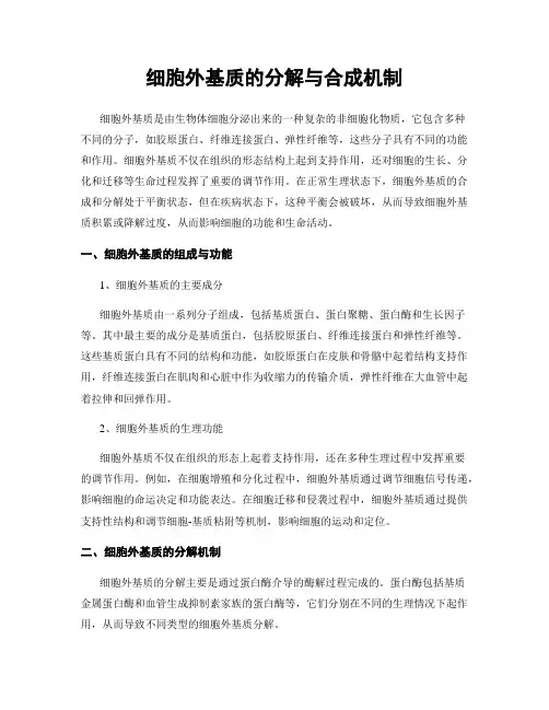
细胞外基质细胞外基质(extracellular matrix,ECM)是由大分子构成的错综复杂的网络。
为细胞的生存及活动提供适宜的场所,并通过信号转导系统影响细胞的形状、代谢、功能、迁移、增殖和分化。
细胞外基质的成分构成细胞外基质的大分子种类繁多,可大致归纳为四大类:胶原、非胶原糖蛋白、氨基聚糖与蛋白聚糖、以及弹性蛋白(图10-1,2)。
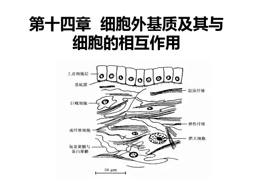
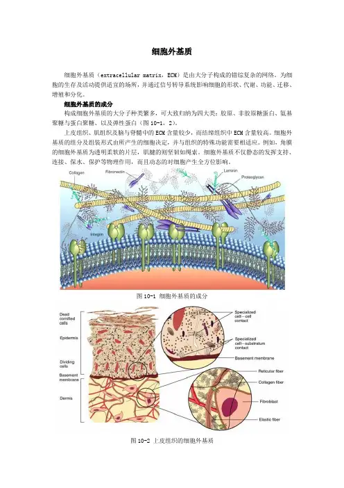
上皮组织、肌组织及脑与脊髓中的ECM含量较少,而结缔组织中ECM含量较高。
细胞外基质的组分及组装形式由所产生的细胞决定,并与组织的特殊功能需要相适应。
例如,角膜的细胞外基质为透明柔软的片层,肌腱的则坚韧如绳索。
细胞外基质不仅静态的发挥支持、连接、保水、保护等物理作用,而且动态的对细胞产生全方位影响。
图10-1 细胞外基质的成分图10-2 上皮组织的细胞外基质一、胶原(collagen)胶原是动物体内含量最丰富的蛋白质,约占人体蛋白质总量的30%以上。
它遍布于体内各种器官和组织,是细胞外基质中的框架结构,可由成纤维细胞(图10-3)、软骨细胞、成骨细胞及某些上皮细胞合成并分泌到细胞外。
图10-3 成纤维细胞周围的胶原纤维目前已发现的胶原至少有19种(表10-1),由不同的结构基因编码,具有不同的化学结构及免疫学特性。
Ⅰ、Ⅱ、Ⅲ、Ⅴ及Ⅺ型胶原为有横纹的纤维形胶原。
各型胶原都是由三条相同或不同的肽链形成三股螺旋,含有三种结构:螺旋区,非螺旋区及球形结构域。
其中Ⅰ型胶原的结构最为典型。
表10-1 胶原的类型图10-4 胶原的结构(左模式图,右电镜照片)Ⅰ型胶原的原纤维平行排列成较粗大的束,成为光镜下可见的胶原纤维,抗张强度超过钢筋。
其三股螺旋由二条α1(Ⅰ)链及一条α2(Ⅰ)链构成。
每条α链约含1050个氨基酸残基,由重复的Gly-X-Y序列构成。
X常为Pro(脯氨酸),Y常为羟脯氨酸或羟赖氨酸残基。
重复的Gly-X-Y序列使α链卷曲为左手螺旋,每圈含3个氨基酸残基。
一.细胞外基质的定义细胞外基质是指分布于细胞外空间的蛋白质和多糖纤维等交错形成的网络胶状结构体系,或简言之为细胞成分之外的组织成分的总称。
二.细胞外基质的生物学作用细胞外基质不仅将细胞整合在一起并决定其物理性质,而且对细胞的存活、形态、功能、增殖、分化、迁移及死亡等各种生物学行为加以调节。
细胞与细胞外基质是相辅相成、互相联系的。
一方面,细胞外基质的结构和功能的异常可作为细胞组织病理改变的重要生理指标;另一方面,结构和功能异常的细胞外基质也会作用于周围的细胞及组织器官,进而促使和导致相关病理改变的发生。
三.细胞外基质的主要组分可分为三类:①氨基聚糖与蛋白聚糖--凝胶样基质;②胶原和弹性蛋白等--纤维网架, 结构蛋白;③非胶原性黏合蛋白,包括纤连蛋白和层粘连蛋白--粘附成分1.氨基聚糖和蛋白聚糖1)氨基聚糖(1)结构:重复的二糖单位聚合而成的无分支直链多糖(2)分类:(3)重要特征:1.与蛋白质链不同,该碳水化合物链不会折叠成致密结构,因此氨基聚糖在基质中占据很大的空间2.氨基聚糖带负电荷,具有强烈的亲水性和吸附阳离子能力。
氨基聚糖可与水分子结合形成凝胶,结果产生膨胀压可抵抗外界压力。
透明质酸:结构:最简单,无硫酸基团,含有大量亲水性的负电荷基团COO-,全部是由单纯的葡萄糖醛基和乙酰氨基葡萄糖二糖结构单位重复排列聚合而成。
形态:呈无规则卷曲状功能:赋予组织弹性、抗压性,并具有润滑剂的作用,促进细胞迁移、增殖降解:透明质酸酶2)蛋白聚糖结构:是由一条称之为核心蛋白质的多肽链与硫酸氨基聚糖共价结合的高分子量复合物,是一种含糖量极高的糖蛋白。
核心蛋白为单链多肽,在同一个核心蛋白上可同时结合一个到上百个同一种类或不同种类的氨基聚糖链,形成大小不等的蛋白聚糖单体,若干个蛋白聚糖单体又能通过连接蛋白与透明质酸以非共价键结合形成蛋白聚糖多聚体。
2.胶原和弹性蛋白1)胶原胶原是细胞外基质中的一个纤维蛋白家族,是动物体内含量最多的蛋白质。