纳米纤维支架
- 格式:pdf
- 大小:1.15 MB
- 文档页数:6
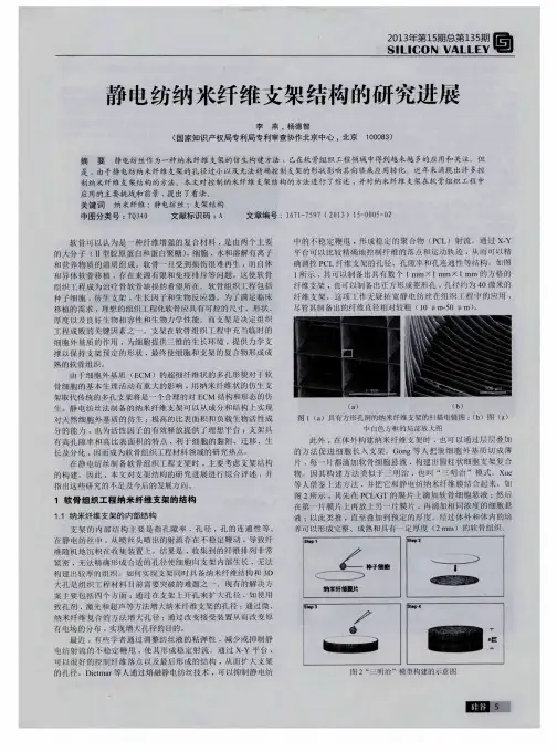

生物医用纳米纤维材料的制备及应用一、生物医用纳米纤维材料概述生物医用纳米纤维材料是一种新型的生物医用材料,它具有独特的物理和化学性质,在生物医学领域具有广泛的应用前景。
纳米纤维材料的直径通常在1 - 1000纳米之间,其比表面积大、孔隙率高、机械性能良好等特点使其在生物医用方面表现出独特的优势。
1.1纳米纤维材料的分类生物医用纳米纤维材料可以根据其组成成分进行分类。
主要包括有机纳米纤维材料和无机纳米纤维材料。
有机纳米纤维材料如天然高分子纳米纤维材料(如纤维素纳米纤维、壳聚糖纳米纤维等)和合成高分子纳米纤维材料(如聚酯纳米纤维、聚酰胺纳米纤维等)。
无机纳米纤维材料包括金属氧化物纳米纤维(如二氧化钛纳米纤维、氧化锌纳米纤维等)和陶瓷纳米纤维(如羟基磷灰石纳米纤维等)。
1.2纳米纤维材料的特性(1)高比表面积:纳米纤维材料的直径很小,这使得其比表面积非常大。
高比表面积有利于细胞的附着和生长,同时也能增加材料与生物分子之间的相互作用。
(2)良好的孔隙率:纳米纤维材料具有较高的孔隙率,能够为细胞的生长和营养物质的传输提供良好的空间环境。
(3)可调节的机械性能:通过改变纳米纤维材料的组成和制备工艺,可以调节其机械性能,使其能够适应不同的生物医用需求。
(4)生物相容性:许多纳米纤维材料具有良好的生物相容性,能够与生物组织和细胞良好地相互作用,减少免疫反应和炎症反应。
二、生物医用纳米纤维材料的制备方法2.1静电纺丝法静电纺丝法是制备纳米纤维材料最常用的方法之一。
该方法基于静电作用,将聚合物溶液或熔体在高压电场下拉伸成纳米纤维。
静电纺丝法具有操作简单、可制备多种材料、纤维直径可控等优点。
(1)静电纺丝的基本原理:在静电纺丝过程中,聚合物溶液或熔体在喷头处形成液滴,当施加高压电场时,液滴表面的电荷聚集,产生静电斥力,使液滴克服表面张力形成泰勒锥,并进一步拉伸成纳米纤维。
(2)影响静电纺丝的因素:包括聚合物溶液的浓度、粘度、表面张力,电场强度、喷头到接收屏的距离等。
纳米科技在骨科医学中的实际应用方法和临床经验引言:随着纳米科技的快速发展,其在医学领域的应用越来越广泛,尤其是在骨科医学领域。
纳米科技在骨科医学中的应用方法和临床经验,不仅为骨科医学带来了革命性的变革,更为患者的治疗和康复提供了更好的解决方案。
本文将介绍纳米科技在骨科医学中的实际应用方法和临床经验,旨在为相关专业人士提供参考和启示。
一、纳米材料在骨替代和修复中的应用1. 纳米生物活性材料的制备及应用纳米材料因其独特的性质,可用于制备具有生物活性的骨替代材料。
例如,通过纳米尺度下的表面改性,可以在材料表面引入生物活性物质,如生长因子、细胞诱导剂等。
这些纳米生物活性材料能够促进骨细胞的生长和骨组织的再生,被广泛应用于骨替代和修复领域。
2. 纳米纤维支架在骨组织工程中的应用纳米纤维支架是一种新型的骨组织工程材料,其具有高度的孔隙率和大表面积,能够提供良好的细胞附着和生长环境。
通过纳米纤维支架的植入,可以促进骨细胞的迁移和增殖,刺激新骨的形成和修复。
研究表明,纳米纤维支架在骨组织工程中的应用有望成为一种有效的治疗方法。
二、纳米药物在骨疾病治疗中的应用1. 纳米药物的制备和输送系统纳米药物是将药物包裹在纳米载体中,通过特定的输送系统将药物传递到病灶部位。
在骨科医学中,纳米药物可以被设计成靶向骨组织的输送系统,提高药物的局部浓度,减少对其他组织的不良影响。
2. 纳米药物在骨疾病治疗中的临床应用针对不同的骨疾病,纳米药物的应用方法也不同。
例如,在骨骼肿瘤治疗中,可以使用纳米药物来实现肿瘤的靶向治疗,提高药物的疗效同时减少副作用。
在骨质疏松治疗中,可以利用纳米药物来增加骨密度和骨强度,预防和治疗骨折。
三、纳米工具在骨科手术中的应用1. 纳米手术器械的研发和应用纳米手术器械是指尺寸在纳米级别的手术工具,具有更好的精确性和操作性。
这些纳米手术器械可以被用于微创手术、精细手术等骨科手术中,提高手术的精确性和安全性。
2. 纳米影像技术在骨科手术中的应用纳米影像技术可以提供更高分辨率的骨骼图像,帮助骨科医生更好地了解患者的病情。
PLGA、PLGAβ-TCP纳米纤维支架的制备、体外降解及细胞相容性研究的开题报告1. 研究背景纳米材料在医学领域中有着广泛的应用。
PLGA(聚乳酸-羟基乙酸共聚物)是一种常用的生物降解材料,具有良好的生物相容性和生物降解性,广泛应用于组织工程、药物传输等领域。
PLGA材料能够制备成纳米纤维支架,以其高比表面积和良好的光学、机械性能被广泛地研究和应用。
同时,PLGAβ-TCP(聚乳酸-co-羟基乙酸/β-三钙磷酸共聚物)是一种新型的生物降解材料,具有良好的生物相容性和生物降解性,在组织工程领域中具有广泛的应用前景。
因此,研究PLGA、PLGAβ-TCP纳米纤维支架的制备、体外降解和细胞相容性,对于在组织工程领域中广泛应用PLGA、PLGAβ-TCP纳米纤维支架具有重要意义。
2. 研究内容(1)PLGA、PLGAβ-TCP纳米纤维支架的制备以静电纺丝技术为基础,利用PLGA、PLGAβ-TCP材料制备纳米纤维支架。
探究纤维支架的制备条件对纤维形态、直径大小等的影响。
(2)PLGA、PLGAβ-TCP纳米纤维支架的体外降解采用模拟体液,分别研究PLGA、PLGAβ-TCP纳米纤维支架的体外降解行为。
通过评估降解速率、降解产物及其对细胞的毒性等指标,比较纳米纤维支架的体外降解行为。
(3)PLGA、PLGAβ-TCP纳米纤维支架的细胞相容性研究利用体外细胞培养模型,分别研究PLGA、PLGAβ-TCP纳米纤维支架对细胞增殖、分化等方面的影响。
通过评估细胞存活率、细胞形态等指标,探究纳米纤维支架材料的细胞相容性。
3. 研究意义(1)以静电纺丝技术为基础,制备PLGA、PLGAβ-TCP纳米纤维支架;(2)通过模拟体液方式,研究PLGA、PLGAβ-TCP纳米纤维支架的体外降解行为;(3)评估PLGA、PLGAβ-TCP纳米纤维支架的细胞相容性,为其在组织工程领域中的应用提供科学依据。
蚕丝蛋白纳米纤维生物体外支架材料的制备
和修饰
生物体外支架材料作为一种新型医疗器械,近几年发展迅速,具有体内外应用
广泛、环境适应性好、材料可降解和生物相容性高等优点。
其中,蚕丝蛋白纳米纤维生物体外支架材料成为近年来的研究热点,其制备和修饰技术也逐渐成熟。
1.蚕丝蛋白纳米纤维的制备
蚕丝蛋白作为一种天然的高分子材料,具有优异的生物相容性和生物可降解性,被广泛应用于生物医学领域。
制备蚕丝蛋白纳米纤维的方法主要有静电纺丝法、模板法、相分离法等多种。
其中,静电纺丝法是制备蚕丝蛋白纳米纤维的主流方法。
其基本原理为将高分子液体通过电场引力拉伸成纤维。
2.生物体外支架材料的制备
将蚕丝蛋白纳米纤维制备成为生物体外支架材料的方法主要有两种,一种是直
接将纳米纤维交织成为三维网络结构;另一种是将纳米纤维形成薄膜、海绵状或三维印刷等结构,再通过堆叠、卷曲等方式构建成为三维网络结构。
3.生物体外支架材料的修饰
生物体外支架材料的性能和功能可以通过修饰来改变。
目前,主要的修饰手段
有表面处理、功能小分子修饰、共价交联修饰等。
表面处理包括化学修饰、物理修饰、微生物修饰等;功能小分子修饰可以通过掺杂功能小分子进入材料中来实现材料的多样化;共价交联修饰可以通过芳香化反应、氧化反应等方式实现材料的化学交联。
总之,蚕丝蛋白纳米纤维生物体外支架材料具有广泛的应用前景。
其制备和修
饰技术的不断改进和优化,为其在生物医学领域的应用提供了更多的可能性。
未来,
蚕丝蛋白纳米纤维生物体外支架材料还有望在再生医学、组织工程、医学检测等领域发挥重要作用。
纳米纤维及其在生物医学领域中的应用纳米纤维是指纤维的直径在100纳米及以下的物质,具有高比表面积、多孔性、柔韧性以及生物相容性等特性。
因此,纳米纤维在生物医学领域中得到了广泛的应用。
一、纳米纤维制备技术1. 静电纺丝法静电纺丝法是一种制备纳米纤维的有效方法,其原理是通过高压电场将溶液中的聚合物分子从微孔的电极中“吹”出来,形成纳米级别的纤维。
静电纺丝法可以制备出具有不同形貌、直径和孔径的纳米纤维,并且可以在制备过程中添加药物或生物分子,用于生物医学领域的检测和治疗。
2. 纺丝-电极位置调整法纺丝-电极位置调整法也是制备纳米纤维的一种方法,其原理是通过控制电极之间的距离和位置,调整电极表面的电势差,使聚合物分子从其中一个电极表面“吹”出来,在将纤维收集在另一个电极上。
3. 模板法模板法制备纳米纤维是利用模板表面的孔道,通过聚合物的沉淀和交联形成纤维。
该方法制备的纤维形貌较为单一,但可以通过调整模板孔的大小和表面化学性质来调节纤维的直径和物理性质。
二、纳米纤维在生物医学领域中的应用1. 组织工程纳米纤维支架可以模拟生物组织的结构和功能,并且具有适宜的孔径和孔隙度,可以提供细胞生长和分化所需的微环境。
利用纳米纤维支架,可以制备出高度仿真人体组织的体外模型,用于生理学、毒理学、药物筛选等研究。
2. 纳米药物传递系统纳米纤维可以作为载体,包裹药物或生物分子,用于治疗疾病。
纳米纤维载体具有较大的比表面积和孔隙度,可以增加药物的负载量和释放速率。
而且,纳米纤维支架具有生物相容性和生物可降解性,可以降低药物副作用和提高治疗效果。
3. 纳米纤维传感器利用纳米纤维的传感性能,可以制备出高灵敏度的生物传感器。
纳米纤维传感器可以用于检测生物分子、重金属离子、细菌等微生物,具有较高的灵敏度、特异性和快速响应。
4. 人工血管和人工心脏瓣膜纳米纤维可以模拟天然血管的形态和性质,用于制备人工血管和心脏瓣膜。
纤维支架材料可以与人体细胞进行良好的相互作用,大大降低了人工器官与人体组织之间的排异反应,提高了植入物的生物相容性和耐久性。
International Journal of Biological Macromolecules 48 (2011) 571–576Contents lists available at ScienceDirectInternational Journal of BiologicalMacromoleculesj o u r n a l h o m e p a g e :w w w.e l s e v i e r.c o m /l o c a t e /i j b i o m acFabrication of chitosan/poly(caprolactone)nanofibrous scaffold for bone and skin tissue engineeringK.T.Shalumon a ,K.H.Anulekha a ,K.P.Chennazhi a ,H.Tamura b ,S.V.Nair a ,∗,R.Jayakumar a ,∗a Amrita Center for Nanosciences and Molecular Medicine,Amrita Institute of Medical Sciences and Research Centre,Amrita Viswa Vidyapeetham,Kochi 682041,India bFaculty of Chemistry,Materials and Bioengineering and High Technology Research Centre,Kansai University,Osaka 564-8680,Japana r t i c l e i n f o Article history:Received 15December 2010Received in revised form 18January 2011Accepted 24January 2011Available online 1 February 2011Key words:ChitosanTissue engineering Poly(caprolactone)Nanofibers Contact anglea b s t r a c tChitosan/poly(caprolactone)(CS/PCL)nanofibrous scaffold was prepared by a single step electrospinning technique.The presence of CS in CS/PCL scaffold aided a significant improvement in the hydrophilicity of the scaffold as confirmed by a decrease in contact angle,which thereby enhanced bioactivity and protein adsorption on the scaffold.The cyto-compatibility of the CS/PCL scaffold was examined using human osteoscarcoma cells (MG63)and found to be non toxic.Moreover,CS/PCL scaffold was found to support the attachment and proliferation of various cell lines such as mouse embryo fibroblasts (NIH3T3),murine aneuploid fibro sarcoma (L929),and MG63cells.Cell attachment and proliferation was further confirmed by nuclear staining using 4 ,6-diamidino-2-phenylindole (DAPI).All these results indicate that CS/PCL nanofibrous scaffold would be an excellent system for bone and skin tissue engineering.© 2011 Elsevier B.V. All rights reserved.1.IntroductionTissue engineering,a combination of principles of engineering and life sciences to improve tissue function has evolved decades ago [1].The main aspect of tissue engineering is the develop-ment of a suitable scaffold which can mimic the extra cellular matrix.Natural extra cellular matrix is a combination of proteo-glycans (glycosaminoglycans)and fibrous proteins.Certain specific requirements of the scaffolds for tissue reconstruction are ade-quate pore size for cell seeding,diffusability throughout the matrix,and biodegradability.The design of a scaffold involves the selec-tion of a suitable material which is biodegradable,biocompatible as well as non toxic to the cells,selection of a suitable method/type of scaffold which can provide better surface for cell attachment,proliferation and differentiation.The extra cellular environment formed on nanofibers compared to that on solid-walled surfaces has led to the report of increased cellular attachment with several cell lines including osteoblastic cells [2,3],fibroblasts [4],normal rat kidney cells,smooth muscle cells [5],neural stem cells [6],and embryonic stem cells [7].This increased attachment across various cell types provides tissue engineers,a potential tool to generate functional tissues in shorter time frames than would be possible on more traditional scaffolds.As of now so many nat-∗Corresponding authors.Tel.:+914842801234;fax:+914842802020.E-mail addresses:nairshanti@ (S.V.Nair),rjayakumar@ ,jayakumar77@ (R.Jayakumar).ural and synthetic polymers as well as their blends have been tried in this case.A wide variety of polymers are used in fabri-cating scaffolds viz-poly(lactic acid)[6],poly(glycolic acid)[8,9],poly(lactic-co-glycolic acid)[10],poly(caprolactone)[11],or natu-ral ones such as collagen [12],gelatin [13],silk [14]and chitosan [15,16].Recently there is seen a growing interest in the produc-tion of scaffolds by using natural polymers like chitin [17,18],chitosan [19,20],alginate [21],collagen [22],gelatin [23–25]etc.,due to their non-toxicity,enhanced biocompatibility,cell adhe-sion and proliferation.Since the use of natural polymers have certain disadvantages like low stability,toxic degradation products which can be harmful to the cells,the natural polymers are often blended with synthetic polymers [26,27].Also this have enhanced mechanical properties,degradation stability and enhanced affin-ity to the cellular components.Chitin and chitosan have been used as scaffolds due to their biodegradability,hydrophilicity,non-antigenicity,non-toxicity,antimicrobial activity,bio adherence and cell affinity,which make chitosan the ideal candidate for uses in a wide range of applications [28–31].The scaffold material in our study is a blend of chitosan and polycaprolactone nanofibers obtained by single step electrospinning.In this technique the poly-mer solution is pumped through a syringe,forms fibers when high electric field is applied.When the applied field overcomes the sur-face tension of the polymer solution,the polymer forms continuous filaments and can be collected in a collector which is grounded.Many properties of PCL such as thermal degradation,hydrophilic-ity,biodegradability and mechanical properties can be improved by incorporation of CS in PCL [32].The blend of the both polymers0141-8130/$–see front matter © 2011 Elsevier B.V. All rights reserved.doi:10.1016/j.ijbiomac.2011.01.020572K.T.Shalumon et al./International Journal of Biological Macromolecules48 (2011) 571–576can render good environment for cell attachment,proliferation and differentiation.In the present work,nanofibrous scaffold was prepared by a single step electrospinning technique using a new solvent mixture of formic acid and acetone as reported in our pre-vious study[32].Bioactivity and cytocompatibility of the CS/PCL nanofibrous scaffold was evaluated by in vitro biomineralisation, contact angle measurements,cell adhesion,cytotoxicity and pro-tein adsorption studies.2.Materials and methods2.1.MaterialsChitosan(CS)with degree of deacetylation80–85%and MW: 100–150kDa was purchased from Koyo chemicals Co.Ltd.,Japan. Poly(caprolactone)(PCL)(M W43,000–50,000)was purchased from Polysciences Inc.,USA.Formic acid and acetone purchased from RFCL Ltd.and Qualigens Fine Chemicals Ltd.,India,respectively. Syringes and needles were purchased from BD Sciences Ltd.,Spain.2.2.Preparation of polymer solutionsThe polymer solution for electrospinning was prepared by opti-mizing the CS and PCL concentrations in the solvent mixture,formic acid/acetone(70:30).CS and PCL solutions were then blended in dif-ferent compositions and concentrations to achieve the optimum range for obtaining nanofibers as reported in our previous study [32].From the combination,CS1%and PCL8%in the blending ratio 1:3were selected for further studies.2.3.Electrospinning of polymer solutionCS/PCL nanofibrous scaffold was prepared by elecrospinning with selected combination of CS/PCL solution to a static target using a high voltage DC power supply connected to a multi terminal dis-tribution box(Model RR30P,0–30kV,Gamma High Voltage Inc., and USA).An infusion syringe pump(KDS220,kdScientific Inc., USA)with10ml syringe having blunt end needles of21gauge was used to deliver the polymer solutions.A distance of4cm was main-tained between the aluminium target and spinneret.All the sample solutions were spun at a rate of0.5ml/h to a grounded aluminium target.The aluminium targets werefixed on a wooden stand from the tip of the needle by applying a voltage of10kV.2.4.CharacterizationThe SEM images of the CS/PCL nanofibrous scaffold were taken using JEOL JSM6490LS operating at15kV.The samples werefixed on aluminium stubs and the stubs were coated with platinum using JEOL JFC1600for2min at10mA before imaging.The hydrophilicity of the CS/PCL scaffold was qualitatively determined by measuring the contact angle of the material with different media;distilled water and MEM using KRUSS drop shape analyzer(DSA100).The CS/PCL scaffold was cut into12mm2size pieces andfixed into the custom made sample holder of the drop shape analyzer.The analyzing media were taken in a2ml leur lock syringefitted with blunt edge needle.A single drop of volume∼2l was poured on the scaffold.The drop shape on the scaffold surface was recorded using the camera attached with the system.Initial contact angle values on the scaffolds were measured from each frame of the recorded videofiles.The contact angle was measured by the sessile drop approximation of the inbuilt software in the machine.2.5.Biomineralisation studiesCS/PCL nanofibrous scaffold with identical weight and shape were immersed in1.5×simulated bodyfluid(SBF)solution and were then incubated at37◦C in a closed Falcon tube for7and 14days.The SBF solution was prepared according to the litera-ture reported by Kukubo and Takadama[33].After prescribed time, samples were taken out of the medium and washed three times with deionised water to remove the impurities.CS/PCL nanofibrous scaffold was then lyophilized and examined in SEM for mineraliza-tion.The adsorbed minerals in the scaffold were also quantified using EDS attachment of the SEM.2.6.Protein absorption studiesThe protein absorption studies were carried out for the nanofi-brous scaffold for1,4and10h in MEM.The samples were cut in equal weight and size and kept immersed in MEM with serum proteins and growth factors for the respective time peri-ods.The samples were then given PBS washing once and were kept immersed in the elution buffer(1ml)at37◦C in the incu-bator for1h.200l of elution buffer was then mixed with25l of BCA/CuSO4solution in a96well plate;incubated at37◦C for 30min.The protein absorption was then quantified by measuring the absorbance of the eluted buffer/BCA/CuSO4at562nm in a plate reader(BioTEK2000).2.7.Cell culture studiesThree different cell lines such as NIH3T3,MG63and L929were obtained from NCCS,Pune and used for cell attachment studies on the CS/PCL nanofibrous scaffold.The cells were routinely cul-tured in MEM,supplemented with10%Fetal Bovine Serum(Sigma) and antibiotics(Penicillin,Streptomycin,Amphotericin and Gen-tamycin)(Invitrogen,USA)under standard conditions of37◦C and 5%CO2.The culture medium was changed twice a week and the cells were harvested prior to culture on the scaffold,by treating with0.25%trypsin-EDTA.2.8.Cytotoxicity studiesThe cell viability was measured using Alamar Blue Assay(Invit-rogen,USA).Alamar blue(Resazurin)is a nonfluorescent dye, which works based on the reduction of mitochondria of the living cells which converts Rezasurin to bright red-fluorescent resorufin of metabolically active cells.The amount offluorescence produced is proportional to the number of living cells.Initially,fibrous scaf-fold was incubated in culture medium for10min to wet the surface of the scaffold,prior to cell seeding.MG63cells were then seeded on the scaffold surface as per ISO standard10993-5for three time intervals viz;24,48and96h.After each specified culture period, culture medium with10%alamar blue was added to each well and incubated for5h.The absorbance of the solution was measured using the plate reader.The cell viability was measured in triplicate and plotted in terms of percentage optical density.2.9.Cell attachment and proliferation studiesCS/PCL scaffold was cut into12mm2size samples and steril-ized by immersion in70%ethanol for30min.Then the scaffold was washed thrice with sterilized PBS and transferred to24-well non-treated polystyrene cell culture plates to increase cell-matrix interactions and to reduce cell attachment on the plates.The scaf-fold was incubated for30min in1ml of the cell culture medium at37◦C before cell seeding to saturate the scaffold with growth medium and improve cell attachment.Three different cell linesK.T.Shalumon et al./International Journal of Biological Macromolecules 48 (2011) 571–576573Fig.1.SEM image of electrospun CS/PCL fiber in 1:3composition.such as NIH3T3,MG63and L929were suspended in MEM and seeded with a density of 5×10³cells per each well.The cell seeded scaffold sections were then maintained at 37◦C with humidified 5%CO 2atmosphere for cell adhesion in the CO 2incubator for 48and 96h.The culture medium was changed every alternate day for the entire culture period.The cell attachment was then observed by taking the SEM image of the scaffold after 48and 96h.The attached cells on the scaf-fold were treated with 2.5%glutyraldehyde followed by thorough washing in PBS for two times.Then the cells adhered to the scaf-fold was dehydrated in an ethanol-graded series (50,60,70,80,90and 100%)for 10min each and allowed to dry on a clean petridish at room temperature.The surface of cell-adhered scaffold sections was observed by SEM after platinum coating.After culturing the cells on the scaffold for 48and 96h,the culture media of samples were washed with PBS twice and the cells on the scaffolds were then fixed in 4%phosphate-buffered paraformaldehyde.The samples were washed with PBS and per-meablized with 0.1%Triton-X100in PBS at room temperature for 5min.In order to reduce nonspecific background staining,samples were blocked with 1%Fetal Bovine Serum (FBS)for about 20min,followed by PBS washing.The cells were then mounted using DAPInuclear stain for 5min and stored at 4◦C.All incubations were con-ducted at room temperature.Nuclear staining images of the cells on scaffolds were recorded using fluorescent microscope (Olympus BX 51).3.Results and discussion3.1.Preparation and characterization of electrospun CS/PCL scaffoldsCS (1%)/PCL (8%)solution with 1:3composition was selected and electrospun to very fine nanofibers [32].The SEM images show that the fibers had approximately 102±24nm diameter and smooth morphology.As reported in our early study,the characteristic of the solvent system which used for the fabrication of fibers is the reason for obtaining fibers in very smaller diameter.Representative SEM image of electrospun fiber is shown in Fig.1.3.2.Contact angle measurementsIn contact angle measurements,the effect of incorporation of CS in PCL scaffolds was examined using two media viz;distilled water and MEM.Generally,hydrophilic surfaces would give a contact angle between 0◦and 30◦and less hydrophilic surfaces show a contact angle up to 90◦.Materials which show a con-tact angle more than 90◦is usually termed as hydrophobic.PCL scaffolds without CS show a Âvalue of 130.1±2and 129.8±3,respectively,for water and MEM,which in turn points towards the hydrophobic behavior of PCL scaffolds.When CS was incor-porated,a decrease in the contact angle values was observed both for water and MEM.CS/PCL scaffold show a contact angle 95.3±6for water and 94.9±5for MEM.The video contact angle images of CS/PCL scaffolds are shown in Fig.2A.In the figures,(a)&(b)represent the contact angle values of PCL scaffold alone and (c)&(d)represent contact angle values of CS/PCL scaffold for water and MEM.Blending of CS in PCL not only increases the hydrophilicity but also enhances the degradation possibility of thescaffold.Fig.2.(A)The contact angle images on PCL (a and b)and CS/PCL (c and d)scaffolds using distilled water and MEM,respectively,and (B)and (C)represents the SEM images of mineralized CS/PCL scaffolds after 7and 14days of SBF immersion.(D)Represents the Ca/P ratio of in biomineralized scaffold.574K.T.Shalumon et al./International Journal of Biological Macromolecules48 (2011) 571–576Fig.3.Protein adsorption studies with CS/PCL nanofibrousscaffolds.Fig.4.Cytotoxicity studies with MG63cells.3.3.Biomineralization studiesThe SEM images of the biomineralisation studies showed the mineral deposition over CS/PCL nanofibrous scaffolds.From the SEM images,it was found that the calcium phosphate deposition is increasing with time.The amount of minerals deposited on the sur-face of the scaffolds after 7and 14days are shown in Fig.2B and C.After 14days,scaffold shows a higher mineral deposition with sur-face breakage.As the calcium phosphate formation is important for the bone regeneration in the body,a scaffold which can provide immediate mineralization of hydroxyapatite would support for the formation of natural bone [34].The nanofibrous nature of the scaf-folds also increases the surface bioactivity of the scaffolds leading to the faster mineral deposition due to this reason.The EDS spectra of the apatite formation in scaffold having Ca/P ratio 1.69is given in Fig.2D,which is slightly higher than the original value.3.4.Protein adsorption studiesProtein adsorption was done using Bi cinchoninic Acid (BCA)assay.The principle of BCA assay relies on the reduction of Cu 2+to Cu 1+.The amount of reduction is proportional to the protein present.It has been shown that amino acids like cisterns,cystine,tryptophan,tyrosine and the peptide bond are able to reduce Cu 2+to Cu 1+.BCA forms a purple–blue complex with Cu 1+in alkaline environments,thus the reduction of alkaline Cu 2+by proteins can be determined.Protein absorption studies were carried out with the CS/PCL nanofibrous scaffolds for a period of 1,4,and 10h,respectively.As the incubation period increases,an increase in adsorption of proteins on the nanofibrous scaffolds was observed (Fig.3).The increases in protein deposition on the scaffolds are essential for the cell growth and differentiation.Also it has been already proved that the protein deposition on the fibrous scaffolds increases as the fiber diameter decreases.Also,in our previous study,PCL nanofibers exhibited higher protein adsorption rate on it compared to microfibers [35].So it is clear that CS/PCL nanofibers with much smaller diameter are expected to adsorb more amounts of proteins as a function of time.More studies are underway to eval-uate the long term protein adsorption as well as specific protein adsorption.3.5.Cytotoxicity studiesCytotoxicity of the prepared CS/PCL scaffold was analyzed using Alamar Blue Assay.Cell viability on the CS/PCL nanofibrous scaffold was examined for 24,48and 96h using MG63cell line (Fig.4).A time depended increase in optical density was observed for CS/PCL scaffold,when it was analyzed for three different time points.After 96h,the OD values show comparatively higher value with respect to negative control,which clearly points towards the nontoxicFig.5.SEM images of cell attachment on CS/PCL scaffold using MG63cells after 48and 96h (1A and 1B)and nuclear staining images on the same scaffold for same time using MG63(2A and 2B).Index image in 2B is the DAPI stained fibers without cells (2C).K.T.Shalumon et al./International Journal of Biological Macromolecules 48 (2011) 571–576575Fig.6.Represents the SEM images of cell attachment on CS/PCL scaffold using L929cells afetr 48and 96h (1A and 1B)and nuclear staining images on the same scaffold for same time using L929(2A and 2B).behavior of CS/PCL scaffold.Since the increase in OD values is the direct measure of number of living cells,our results clearly suggest that cell proliferation is taking place.3.6.Cell attachment studiesCell attachment studies were carried out using MG63,L929and NIH3T3cells.MG63cells show good attachment and spread-ing to CS/PCL fibers after 46and 96h,respectively,and it is shown in Fig.5(1A and 1B).The cells showed spread morphol-ogy at higher incubation periods (96h).The result was again confirmed with nuclear staining method using DAPI for 48and96h.The corresponding microscopic images for both the time points are given in Fig.5(2A and 2B).In the case of L929and NIH3T3,the scaffold showed spreading and proliferation similar to MG63(Figs.6and 7).The SEM image of morphology of L929and NIH3T3at 48and 96h are shown in Figs.6and 7(1A and 1B)and the corresponding nuclear staining images are shown in Figs.6and 7(2A and 2B).In the case of fibers without cells,no nuclear images were observed in DAPI staining and the correspond-ing image is shown as 2C in Fig.5.Since all the cells showed good attachment and spreading on the nanofibrous scaffold of CS/PCL,it can be used for bone and skin tissue engineering appli-cations.Fig.7.Represents the SEM images of cell attachment on CS/PCL scaffold using NIH3T3cells afetr 48and 96h (1A and 1B)and nuclear staining images on the same scaffold for same time using NIH3T3(2A and 2B).576K.T.Shalumon et al./International Journal of Biological Macromolecules48 (2011) 571–5764.ConclusionsThe CS/PCL nanofibrous scaffold was prepared andfiber diam-eter was confirmed by SEM.The contact angle measurements showed that the hydrophobic character of poly(caprolactone)has been reduced by CS blending.Bioactivity of the CS/PCL scaffold was confirmed by enhanced biomineralization and protein adsorption. The nanofibrous scaffolds show good cell attachment and spread-ing with MG63,L929,and NIH3T3.The cyto-compatibility studies using MG63cells showed increase in the cell viability with respect to time.These results suggest that CS/PCL hybrid scaffold could be one of the ideal candidates for skin and bone tissue engineering.AcknowledgementsThe authors are thankful to the Nanomission,Department of Science and Technology(DST),Govt.of India for supporting this work.The author Shalumon K.T.is grateful to Council of Scientific and Industrial Research(CSIR)for awarding Senior Research Fel-lowship(SRF).Thanks are also to Koyo chemicals Co.Ltd,Japan for providing Chitosan.We are also grateful to Mr.Sajin P.Ravi,Ms. Soumya and Ms.Chandini for their assistance in characterization and cellular studies.References[1]nger,J.P.Vacanti,Science260(1993)920–926.[2]K.M.Woo,V.J.Chen,P.X.Ma,J.Biomed.Mater.Res.A67(2003)531–537.[3]K.M.Woo,J.H.Jun,V.J.Chen,J.Seo,J.H.Baek,H.M.Ryoo,G.S.Kim,M.J.Somer-man,P.X.Ma,Biomaterials28(2007)335–343.[4]M.Schindler,I.Ahmed,J.Kamal,A.Nur-E-Kamal,T.H.Grafe,H.Y.Chung,S.A.Meiners,Biomaterials26(2005)5624–5631.[5]C.Y.Xu,R.Inai,M.Kotaki,S.Ramakrishna,Biomaterials25(2004)877–886.[6]F.Yang,C.Y.Xu,M.Kotaki,S.Wang,S.Ramakrishna,J.Biomater.Sci.15(2004)1483–1497.[7]Nur-E-Kamal,I.Ahmed,J.Kamal,M.Schindler,S.Meiners,Stem Cells24(2006)426–433.[8]E.D.Boland,G.E.Wnek,D.G.Simpson,K.J.Pawlowski,G.L.Bowlin,J.Macromol.Sci.:Pure Appl.Chem.38(2001)1231–1243.[9]W.J.Li,urencin,F.K.Ko,J.Biomed.Mater.Res.60(2002)613–621.[10]H.Yoshimoto,Y.M.Shin,H.Terai,J.P.Vacanti,Biomaterials24(2003)2077–2082.[11]W.J.Li,R.Tuli,C.Okafor,A.Derfoul,K.G.Danielson,D.J.Hall,R.S.Tuan,Bioma-terials26(2005)599–609.[12]J.A.Matthews,G.E.Wnek,D.G.Simpson,G.L.Bowlin,Biomacromolecules3(2002)232–238.[13]Y.Z.Zhang,H.W.Ouyang,C.T.Lim,S.Ramakrishna,Z.M.Huang,J.Biomed.Mater.Res.B:Appl.Biomater.B72(2005)156–165.[14]K.Ohgo,C.Zhao,M.Kobayashi,T.Asakura,Polymer44(2003)841–846.[15]B.M.Min,G.Lee,S.H.Kim,Y.S.Nam,T.S.Lee,W.H.Park,Biomaterials25(2004)1289–1297.[16]P.Sangsanoh,P.Supapho,Biomacromolecules7(2006)2710–2714.[17]K.T.Shalumon,N.S.Binulal,N.Selvamurugan,S.V.Nair,T.Deepthy Menon,H.Furuike,R.Tamura,Jayakumar,Carbohydr.Polym.77(2009)863–869.[18]M.Peter,P.T.Sudheesh kumar,N.S.Binulal,S.V.Nair,H.Tamura,R.Jayakumar,Carbohydr.Polym.78(2009)926–931.[19]R.Jayakumar,M.Prabaharan,S.V.Nair,H.Tamura,Biotechnol.Adv.28(2010)142–150.[20]R.Jayakumar,M.Prabaharan,S.V.Nair,S.Tokura,H.Tamura,N.Selvamurugan,Prog.Mater.Sci.55(2010)675–709.[21]V.V.Divya Rani,R.Ramachandran,K.P.Chennazhi,H.Tamura,S.V.Nair,R.Jayakumar,Carbohydr.Polym.83(2011)858–864.[22]Q.Lu,K.Ganesan,D.T.Simionescu,N.R.Vyavahare,Biomaterials25(2004)5227–5237.[23]M.Peter,N.Ganesh,N.Selvamurugan,S.V.Nair,T.Furuike,H.Tamura,R.Jayaku-mar,Carbohydr.Polym.80(2010)687–694.[24]M.Peter,N.S.Binulal,S.V.Nair,N.Selvamurugan,H.Tamura,R.Jayakumar,Carbohydr.Polym.158(2010)353–361.[25]L.Xiaohua,ura,H.Jiang,X.M.Peter,Biomaterials30(2009)2252–2258.[26]Z.Yingshan,Y.Dongzhi,C.Xiangmei,X.Qiang,L.Fengmin,N.Jun,Biomacro-molecules9(2008)349–354.[27]Ying,H.W.Wun, C.Xiaoying, D.Siqin,Polym.Degrad.Stabil.93(2008)1736–1741.[28]R.Jayakumar,N.Nwe,S.Tokura,H.Tamura,Int.J.Biol.Macromol.40(2007)175–181.[29]R.Jayakumar,M.Prabaharan,R.L.Reis,J.F.Mano,Carbohydr.Polym.62(2005)142–158.[30]E.Brunner,H.Ehrlich,P.Schupp,R.Hedrich,S.Hunoldt,M.Kammer,S.Machill,S.Paasch,V.V.Bazhenov,D.V.Kurek,T.Arnold,S.Brockmann,M.Ruhnow,R.Born,J.Struct.Biol.168(2009)539–547.[31]H.Ehlrich,E.Steck,M.Ilan,M.Maldonado,G.Muricy,G.Bavestrello,Z.Klja-jic,J.L.Carballo,S.Schiaparelli,A.Ereskovsky,P.Schupp,R.Born,H.Worch, V.V.Bazhenov,D.Kurek,V.Varlamov,D.Vyalikh,K.Kummer,V.V.Sivkov, S.L.Molodtsov,H.Meissner,G.Richter,S.Hunoldt,M.Kammer,S.Paasch,V.Krasokhin,G.Patzke,E.Brunner,Int.J.Biol.Macromol.47(2010)141–145. [32]K.T.Shalumon,K.H.Anulekha,C.M.Girish,R.Prasanth,S.V.Nair,R.Jayakumar,Carbohydr.Polym.80(2010)413–419.[33]T.Kukubo,H.Takadama,Biomaterials27(2006)2907–2915.[34]T.Kokubo,Mater.Sci.Eng.25(2005)97–104.[35]N.S.Binulal,M.Deepthy,N.Selvamurugan,K.T.Shalumon,S.Suja,U.Mony,R.Jayakumar,S.V.Nair,Tissue Eng.A16(2010)393–404.。