肝血管瘤的CT影像诊断
- 格式:ppt
- 大小:3.16 MB
- 文档页数:21

CT和B超诊断肝血管瘤的影像学征象分析发表时间:2015-09-10T13:40:24.257Z 来源:《健康世界》2015年1期作者:董少军李炳淑[导读] 山东省寿光市人民医院 262700 探讨、分析肝血管瘤的多层螺旋CT和B超影像学表现.山东省寿光市人民医院 262700摘要:目的探讨、分析肝血管瘤的多层螺旋CT和B超影像学表现;方法选择、收集我院经CT和B超检查诊断为肝血管瘤的患者96例,并对两种检查影像学征象进行对比分析;结果肝血管瘤CT平扫为境界清楚,密度均匀的圆形或卵圆形低密度影,部分较大病灶中央可见星形或不规则更低密度区;增强扫描动脉期边缘结节样或中心强化,强化区进行性向中央扩展,延迟扫描病灶呈等密度填充,较大病灶中央可见始终不填充区同平扫;B超检查:病灶直径<3.0cm的肝血管瘤,声像图表现为均匀强回声,边界清晰、锐利,呈浮雕样,周边无声晕,后方无声影;病灶>3.0cm的肝血管瘤,多数呈强回声,表现为形态不规整、边界清晰、无声影及声晕;少数呈低回声;结论应用多层螺旋CT诊断肝血管瘤可发现动脉期的典型征象并不受呼吸影响,且不遗漏病灶,诊断准确率很高,是最佳、最有效、可确诊的方法;而肝血管瘤行B超检查,则具有无伤、无痛、价廉、快速等优点,并且正确率极高,应为诊断肝血管瘤的首选检查方法。
关键词:肝血管瘤;CT扫描;B超检查;影像学征象【Summary】purpose study,analyze super image of many spiral CTs and B of liver afferent lump to learn performance;Method choice,collect my hospital through the CT and the B the super check diagnosis be 96 for the sufferer of liver afferent lump,and learn to advertise for elephant to carry on contrast to two kinds of check images analysis;Result liver the afferent lump CT Be even to sweep to know for state,density even circular or egg circular the low density copy,the part bigger focus the center it is thus clear that star polygon or irregular and much lower density area;Strengthen to scan artery to expect the edge knot stanza kind or center to enhance,enhance area to carry on aptitude the center to expand,delay to scan a focus to present to wait density to fill,bigger focus the center it is thus clear that always don't fill area together even sweep;B super check:Focus diameter<the liver of the 3.0 cms afferent lump,voice the being like chart copy for even strong echo,boundary being clear,sharp-edged,presenting ectype kind,periphery having no voice dizzy,rear having no voice now;Focus>the liver of the 3.0 cms afferent lump,majority present strong echo,the performance is an appearance the rules isn't whole,clear boundary,have no voice the shadow and voice is dizzy;The minority present to loiter a voice;The conclusion applies many spiral CT diagnosis liver the afferent lump can discover that the typical model that the artery expects advertises for elephant and be free from breath influence,and don't leave out a focus,diagnose accuracy very high,is the best,most effective,the method for canning confirm;But liver the afferent lump go a B super check and then haven't hurt,have no pain,low price,rapid etc.advantage,and the correctness is the highest,should for diagnose the head of liver afferent lump to choose check method.【Keyword】liver the afferent lump CT scanThe B super check image learns to advertise for elephant1、资料和方法选择、收集我院2010年01月—2012年01月均经多层螺旋CT和B超检查诊为肝脏血管瘤的患者96例,其中男62例,女34例;年龄最大者75岁,最小者30岁;无临床症状者88例,具有腹部饱胀、不适者8例。


肝血管瘤45例CT诊断目的:探讨肝血管瘤的CT征象及诊断价值。
方法:45例患者行CT平扫加增强扫描,对其影像进行观察分析。
结果:所有病灶平扫表现为低密度,增强扫描表现为早期强化,随时间进展呈向心性强化,部分直至完全充填,然后强化逐步减退,表现为快进慢出,即“早出晚归”特征。
结论:CT平扫及增强扫描对诊断肝血管瘤意义极大,是诊断肝血管瘤的一种有效方法。
标签:肝血管瘤;增强扫描;CT诊断肝血管瘤是肝脏最常见的良性肿瘤。
尸检发现率为7.3%[1],女性居多,一般多无临床症状,常在腹部影像学检查中偶然发现。
随着多层螺旋CT多期动态增强扫描技术在肝脏中的应用,肝血管瘤的正确检出率明显提高,其重要性在于与恶性肿瘤的鉴别。
现将我院2005年6月~2009年6月行螺旋CT平扫加三期增强扫描诊断,临床最后确诊为肝血管瘤的45例患者的CT资料进行回顾性分析,并总结报道如下:1 资料与方法1.1 一般资料本组45例中,女性37例,男性8例;年龄22~65岁,平均39岁。
全部病例无肝硬化病史,甲胎蛋白定性、定量均为阴性,31例行B超检查,其中,23例B超诊断为肝血管瘤,8例B超未能确诊,诊断为肝内占位病变;14例因右上腹不适直接行CT 检查。
1.2 方法检查前,患者空腹喝水800~1 200 ml,以充盈胃肠道,使用Siemens SOMATOM欢悦6多层螺旋CT扫描机,先平扫后增强扫描。
采用仰卧位,扫描范围上至膈肌,下至肝脏最下缘,全部显示至满意为止。
层厚6 mm,层距6 mm,用高压注射器经肘静脉注射碘海醇100 ml行增强扫描,流速2.5 ml/s,在降主动脉段进行预监测,当降主动脉内造影剂达到一定浓度时自动进入动脉期扫描,时间为注射碘海醇后10~20 s,后在45~60 s进行门静脉扫描,延迟期为2~10 min,对小病灶进行薄层重建。
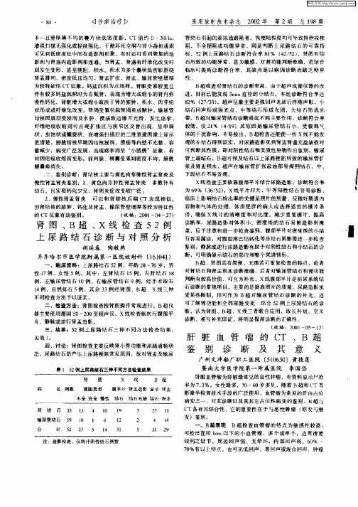
2 结果2.1 平扫表现累计发现53个病灶,病灶分布为肝右叶32个,肝左叶18个,肝尾叶3个。
肝癌和肝血管瘤的影像学鉴别要点下载提示:该文档是本店铺精心编制而成的,希望大家下载后,能够帮助大家解决实际问题。
文档下载后可定制修改,请根据实际需要进行调整和使用,谢谢!本店铺为大家提供各种类型的实用资料,如教育随笔、日记赏析、句子摘抄、古诗大全、经典美文、话题作文、工作总结、词语解析、文案摘录、其他资料等等,想了解不同资料格式和写法,敬请关注!Download tips: This document is carefully compiled by this editor. I hope that after you download it, it can help you solve practical problems. The document can be customized and modified after downloading, please adjust and use it according to actual needs, thank you! In addition, this shop provides you with various types of practical materials, such as educational essays, diary appreciation, sentence excerpts, ancient poems, classic articles, topic composition, work summary, word parsing, copy excerpts, other materials and so on, want to know different data formats and writing methods, please pay attention!一、肝癌和肝血管瘤的定义和病因。
肝癌是一种恶性肿瘤,通常起源于肝细胞或胆管细胞,主要的病因包括病毒性肝炎、酗酒、非酒精性脂肪性肝病等。
肝血管瘤的CT诊断与鉴别诊断【摘要】目的探讨肝血管瘤的ct诊断标准以及与常见其它疾病的鉴别。
方法 2010年5月以来,我院ct室确诊的肝血管瘤患者120例,搜集其ct诊断方法。
结果 120例患者均为肝血管瘤,其影像学表现为典型的在造影剂强化时的“慢进慢出”改变。
结论肝脏血管瘤很多无明显临床症状,查体时才被发现,ct强化是确定肝血管瘤的金标准。
【关键词】肝血管瘤;ct强化;诊断及鉴别诊断肝血管瘤是一种较为常见的良性病变,临床上以海绵状血管瘤最多见,自然人群尸检发现率为0.35-7.3%[1],占肝良性肿瘤的5-20%[2]。
近年来,随着人们健康意识提高及各种影像诊断技术的进步,无症状的血管瘤发现率明显升高。
多数病例临床无症状或症状轻微,病程长、生长缓慢,预后良好。
2010年5月以来,我院ct室确诊的肝血管瘤患者120例,现就对其临床ct诊断及鉴别诊断进行论述。
1 资料与方法1.1 一般资料 2010年5月以来,我院ct室确诊的肝血管瘤患者120例,其中男80例,女40例,年龄25-65岁,平均年龄45岁。
其中85例患者是经临床查体发现,35例患者因血管瘤较大,出现腹痛腹胀的临床症状就诊,b超可疑血管瘤,后经ct强化扫描确诊,其中最大的直径为10cm,最小的直径为0.5cm。
1.2 方法采用simens公司16层螺旋ct扫描,扫描参数为管电压130kv,管电流80ma,扫描层厚3mm,造影剂为扬子江制药生产的碘海醇100ml,注射速度为3ml/s,扫描前一天嘱咐患者禁食进水,扫描时嘱咐患者饮水800ml-1200ml。
扫描采用三期扫描:平扫、动脉期、静脉期、延迟期。
扫描范围为由膈肌顶部至肝脏下缘。
2 结果2.1 血管瘤的大小以及数目肝血管瘤最大的为10.0cmx8.0cm,最小的0.5cmx0.5cm,其中肝右叶大约140个,肝左叶120个,其中边缘规则的155例,边缘模糊105例,病灶呈现圆形的160例,椭圆形的60例,不规则的40例。
肝海绵状血管瘤(典型与不典型)肝脏最常见的良性肿瘤,约占良性肿瘤的84%好发于女性,男女比例约1:4-6;30-60岁多见生长缓慢,病程长达数年以上;90%为单发,10%多发直径从2mm-20cm不等,超过5cm称巨大海绵状血管瘤临床上可无任何症状,多为偶然在体检中发现,巨大肿瘤可出现上腹部胀痛不适,肿瘤破裂可引起出血肝动脉供血;门静脉和肝静脉周围支引流肝脏海绵状血管瘤-概述组织病理学分型◆海绵状血管瘤(最常见):由扩张的异常血窦组成,内衬单层的血管内皮细胞,血窦间由纤维组织不完全分隔形成海绵状结构,血管间隔大,纤维结缔组织成分少,典型小于3cm◆毛细血管瘤(16%): 血管间隔小,纤维结缔组织多,常小于1cm◆硬化性血管瘤(少见):病变中心血管腔闭塞,大量纤维组织增生,系肝血管瘤退变的结果,也被称为血栓形成或玻璃化血管瘤,平均3.7cm海绵状血管瘤肝海绵状血管瘤-典型影像表现肝海绵状血管瘤-典型影像表现CT:边界清楚,等/低密度,类似血管MRI:长T1长T2,类似脑脊液,且随着回波时间的延长T2信号也随之增高,形成所谓“灯泡征”增强扫描:动脉期周边结节状强化,后期缓慢渐进性向心性填充,延迟期等/稍高信号(与肝实质相比);“早出晚归”女,62岁,体检发现肝肿物2年肝血管瘤-不典型影像表现肝血管瘤-不典型影像表现不典型血管瘤占20%-40%原因:✓Altered morphology or structure:血管瘤伴中央瘢痕;血管瘤伴内部分隔;巨大血管瘤;带蒂血管瘤;血管瘤伴胆管扩张;血管瘤伴钙化;囊性或多房性血管瘤;血管瘤伴液液平面✓Unusual flow pattern:快速填充血管瘤;硬化性血管瘤✓Associated liver abnormalities:血管瘤伴包膜皱缩;脂肪肝背景血管瘤;肝硬化背景血管瘤;血管瘤合并动脉-门脉瘘;血管瘤合并FNH形态或结构改变-血管瘤伴中央瘢痕巨大海绵状血管瘤通常不均匀,可能伴有中央瘢痕,增强无强化 在病理上,疤痕是由粘液瘤样变性、血栓形成、纤维化或坏死组成形态或结构改变-血管瘤伴内部分隔内部分隔在T1、T2均为低信号,与存在纤维化成分有关形态或结构改变-巨大海绵状血管瘤巨大海绵状血管瘤:直径>5m,密度/信号不均匀可出现肝脏肿大,腹部不适CT:病灶呈不均匀低密度,中心密度更低MRI:长T1长T2,内不信号不均,可见裂隙状低信号增强:典型的早期周边结节状强化,静脉期、延迟期出现渐进性向心性填充,但不会完全填充形态或结构改变-带蒂血管瘤罕见,可以无症状,合并亚急性扭转、梗死可出现相应症状强化方式同典型海绵状血管瘤病灶通过一个细蒂附着在肝脏上,有时定位困难,需要与胃及肾上腺去肿瘤鉴别,多平面重组可以帮助确定病灶来源,强化方式有助于鉴别诊断形态或结构改变-血管瘤伴胆管扩张位于肝脏中央的巨大血管瘤可导致胆管扩张虽然肝内胆管细胞癌较常发生胆管扩张,但是所有肝内外占位性病变均可引起胆管扩张,因此血管瘤不能排除血管瘤的诊断形态或结构改变-囊性、多发性血管瘤囊性和多房性血管瘤含有一个大的中心腔是非常罕见的在MRI上,这种不典型的特征是一个或多个肿瘤内囊腔,呈长T1、长T2信号与血栓形成和陈旧性出血有关;周边典型强化方式仍然存在形态或结构改变-血管瘤伴钙化血管瘤的钙化可以在中心或周边,多发为静脉石引起,但也可能是纤维化和营养不良性变化的最终结果女,28岁,体检发现肝血管瘤形态或结构改变-血管瘤伴液液平面很少见血管瘤内可见叶液平面,代表停滞或缓慢流动的血液随着红细胞在相关部位的沉淀,上层由血清组成,下层含有未凝结的沉积红细胞✓上层CT低密度,T1等信号(与肌肉相比),T2明显高信号✓下层CT高密度,T1高信号(与肌肉相比),T2轻度高信号肝脏内液平面是非特异性征象,良恶性肿瘤均可见部分学者认为CT/MRI可见液平面,超声未见,提示血管瘤不典型强化方式-快速填充的血管瘤快速填充血管瘤并不常见,占16%对应的病理为毛细血管瘤,小的血管间隔、大量的结缔组织快速填充型血管瘤较常见于2cm以下的血管瘤,表现为动脉期明显均匀强化这些血流动力学的不同可能与构成血管瘤的血管通道的大小有关不典型强化方式-硬化性血管瘤罕见,被认为是血管瘤退化的终末阶段血管间隙被大量胶原纤维组织所代替,使其失去典型的影像特征 需要病理确诊CT平扫病灶相对于肝脏呈高密度或等密度(易漏诊)增强周边结节状强化,后期渐进性填充,部分病灶在动脉期可能为相对低密度 有时CT可以在血管瘤周围看到致密的晕,对应无脂肪浸润的肝组织可能是由于压迫而导致病灶周围动脉优先流入和/或由于门脉血流减少而引起的MRI检查具有优势,尤其是同反相位肝硬化背景下血管瘤少见,CT占0.6%,明显低于无肝硬化背景的血管瘤尽管肝实质变硬,血管瘤通常保留典型的影像特征在少数肝硬化晚期,由于肝脏纤维化,血管瘤会失去典型强化方式,病灶体积缩小,T2信号减低女,53岁,右上腹痛2天血管瘤合并肝动脉-门静脉瘘肝动脉-门静脉瘘通常与肝脏恶性肿瘤相关,尤其是HCC原因尚不明确,可能因血管瘤有粗大的动脉供应,呈高压力状态,因此需要建立宽大而快速的引流通道,血管瘤与门静脉形成瘘或瘤周间隙扩大而使门脉早显,即动静脉瘘可能是肿瘤适应高流入、快流出的血流动力学状态的结果表现为动脉期病变的早期强化伴门静脉早显女,61岁,体检发现肝多发占位半年余血管瘤合并FNH较常见,约20%肝脏血管瘤合并FNH目前认为二者伴生并不是偶然,有一定的联系可能的病理生理机制:FNH为 肝脏局部动脉血流增加引起增生反应,类似血管瘤,均为血管源性病变女,72岁,右上腹痛10天男,50岁,体检发现“右肝占位”5年80%30%50%35%鉴别诊断◆肝内胆管细胞癌◆肝细胞癌◆FNH◆肝细胞腺瘤65%鉴别诊断-胆管细胞癌多发生于50-70岁男性,边界不清,CA199升高增强动脉期边缘轻-中度不均匀强化,门脉期持续渐进性强化内部或周围可见胆管扩张,常伴有胆管结石;远端肝叶萎缩,肝包膜皱缩凹陷 肝门及腹腔内、腹膜后淋巴结转移鉴别诊断-肝细胞癌多有乙肝、肝硬化病史,可有AFP升高边界清楚,可见假包膜(TIWI、T2WI均呈低信号,延迟强化) 增强呈“快进快出”;常有血管侵犯(癌栓形成)鉴别诊断-FNH年轻女性多见CT:等或稍低密度;MRI:T1等/稍低信号,T2等/稍高信号,中央纤维瘢痕呈长T1长T2信号增强扫描:动脉期呈显著均匀强化,中央瘢痕无强化;门脉期及延迟期呈等、稍高密度(信号),中央瘢痕延迟强化肝胆期呈等、稍高信号鉴别诊断-肝细胞腺瘤年轻女性多与口服避孕药有关,男性与糖尿病、使用类固醇激素有关 边界清楚,有完整包膜密度/信号多不均匀,出血、坏死、脂肪变性常见肝海绵状血管瘤-小结肝脏最常见良性肿瘤,通常无症状,30-60岁女性多见典型影像表现:T2明显高信号(灯泡征);增强动脉期周边结节状强化,门脉期、延迟期渐进性、向心性填充不典型影像表现:✓形态或结构改变:血管瘤伴中央瘢痕;血管瘤伴内部分隔;巨大血管瘤;带蒂血管瘤;血管瘤伴胆管扩张;血管瘤伴钙化;囊性或多房性血管瘤;血管瘤伴液液平面✓Unusual flow pattern:快速填充血管瘤;硬化性血管瘤✓伴有肝实质异常:血管瘤伴包膜皱缩;脂肪肝浸润性血管瘤;肝硬化背景血管瘤;血管瘤合并动脉-门脉瘘;血管瘤合并FNH谢谢!。
简述肝血管瘤与肝细胞肝癌的影像学鉴别要点1.引言1.1 概述肝血管瘤和肝细胞肝癌是两种常见的肝脏疾病,其影像学鉴别是诊断和治疗的关键。
在肝血管瘤中,血管瘤是最常见类型,占所有肝血管瘤的大部分,而肝细胞肝癌是最常见的原发性肝癌。
虽然它们在影像学上有一些相似之处,但也存在一些显著的差异。
肝血管瘤往往是良性病变,主要由扩张的血管组成。
它们通常呈圆形或椭圆形,具有清晰的边界,可以单个或多个。
在血供方面,肝血管瘤常常呈一种动脉和门静脉双供血的模式,而门静脉成分可能更具有支配性。
在增强扫描中,肝血管瘤往往显示早期均匀强化,且延迟期仍保持明显强化。
此外,肝血管瘤还可以表现为多发性或巨大血管瘤,这些特点在影像学上有助于与肝细胞肝癌相鉴别。
与此相反,肝细胞肝癌是一种恶性病变,即癌细胞来源于肝细胞。
在影像学上,肝细胞肝癌常常呈现为不规则的结节性病灶,边界模糊,形状多样。
血供方面,肝细胞肝癌主要通过肝动脉供应血液。
在增强扫描中,肝细胞肝癌通常呈动脉早期强化,但延迟期常表现为强化减退。
此外,肝细胞肝癌在影像学上可表现为肿块内坏死、出血、包膜侵犯等特点。
为了准确鉴别肝血管瘤和肝细胞肝癌,我们需要综合分析影像学表现。
除了形态学特征外,我们还需注意其病灶血供情况、强化模式和延迟期的变化。
此外,结合患者的临床病史、肝功能等方面的信息,可以提高鉴别的准确性。
总之,了解肝血管瘤和肝细胞肝癌的影像学特点对于正确诊断和治疗非常重要。
进一步研究这两种疾病的影像学鉴别要点有助于临床医生更好地评估和管理肝脏疾病患者。
1.2文章结构文章结构的设计是为了确保文章有条理、易读和易理解。
本文主要介绍了肝血管瘤与肝细胞肝癌在影像学上的鉴别要点。
为了更好地呈现这些内容,本文分为引言、正文和结论三个部分。
引言部分首先对文章的主题进行了概述,简要介绍了肝血管瘤和肝细胞肝癌的概念和重要性。
同时,文章还介绍了整篇文章的结构,并说明了各个部分的内容和目的。
正文部分是整篇文章的核心部分,主要分为两个小节:肝血管瘤的影像学特点和肝细胞肝癌的影像学特点。