(PLoS ONE |August 2011 | Volume 6 | Issue 8 | e22962)
Methods 1.Cell culture
Norden laboratory feline kidney (NLFK) and HeLa cells were grown in Dulbecco’s modified Eagle medium (DMEM)supplemented with 10% fetal bovine serum (Gibco, Paisley, UK)at 37℃ in the presence of 5% CO2.
(1)注意实验温度的控制。 (2)漂白区域大小的选择和荧光恢复检测时间的长短要根据具体情况而 定。 (3)激发光的波长和强度应不会使细胞严重损伤;尽量减少漂白前和漂 白后的荧光淬灭。
Page 7
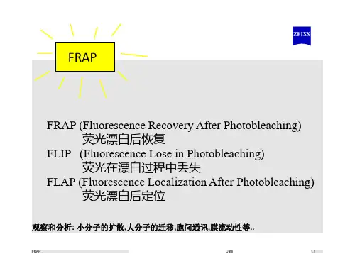
FRAP技术的不足之处
第一,它只能检测膜蛋白的群体移动,而不能 观察单个蛋白的移动。 其次,它不能证明膜蛋白在移动时是否受局部 条件的限制。
2. FRAP experiments
The FRAP experiments were performed on a laser scanning confocal microscope FV1000 with an IX-81 microscope frame(Olympus, Tokyo, Japan) using an Olympus UPLSAPO 606 (NA= 1.2) water immersion objective. The sample stage was heated to 37uC prior the experiments. To image the cell geometry, a confocal stack was acquired before and after the FRAP experiment. The voxel size was adjusted to (200 nm)3or(150 nm)3 .The pinhole size was adjusted to 1 Airy unit. The 514 nm laser line was used for EYFP excitation and the emitted fluorescence was detected using a 530–600 nm band pass filter.Imaging was performed with a laser intensity of 0.1–2 For bleaching a circular (r = 1.85 mm and 2.83 mm) region of interest(ROI) was defined in the middle of the cytoplasm. As bleaching times in FRAP are usually rather large compared to the time scales of the measured diffusion processes, the region of the cell, which is actually bleached, is usually larger than the defined ROI. The size of the actually bleached region and its intensity distribution were measured by bleaching fixed cells (Fig. 1). ImageJ [32] was then used to construct an average shape and intensity profile of that region.