Multiplex Amplicon Genotyping by High-Resolution Melting
- 格式:pdf
- 大小:310.89 KB
- 文档页数:5
特种经济动植物2020年第2期一例英短猫次氯酸钠中毒的诊治●相启安原英梅葛春艳(山东省威海市文登区畜牧兽医技术服务中心山东威海264400)摘要:次氯酸钠中毒是宠物猫的常见病,快速准确地诊断和治疗是救治的关键。
本文接诊了一例宠物猫次氯酸钠中毒的病例,通过发病症状及时确诊,并采取抢救措施,使宠物猫最终得以康复,可为宠物猫养殖户提供参考。
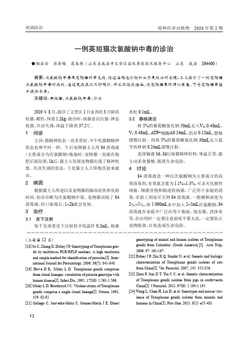
关键词:英短猫;次氯酸钠中毒;诊治2019年8月,接诊了文登区1只家养的3月龄英短猫,雌性,体重1.2kg,就诊时,病猫意识沉郁,神态恍惚,共济失调,体温下降到37.2℃。
1问诊主诉:猫精神状态一直非常好,中午吃猫粮精神状态也和平时一样,午后宠物猫主人用84消毒液(主要成分为次氯酸钠)拖地时,宠物猫一直跟在拖把后面玩耍,1h后,猫主人发现宠物猫出现了精神恍惚、共济失调的状态,于是猫主人立即抱其前来就诊。
2病因根据猫主人所述以及宠物猫的临床症状和发病时间,初步诊断为次氯酸钠中毒。
宠物猫误舔了84消毒液,经口染毒后,1~2h就会发病。
3治疗3.1皮下注射每千克体重皮下注射桂辛低温针0.2mL、地塞米松0.1mL。
3.2静脉滴注将5%的葡萄糖氯化钠50mL兑入V B10.48mL、V C0.48mL、ATP+辅酶A0.24mL、肌苷0.12mL,静脉缓慢注射。
再将5%的葡萄糖氯化钠30mL兑入氨苄西林钠0.24mL缓慢注射。
连续输液3d,3d后病猫精神好转,体温正常,能主动采食猫粮,脱离生命危险。
4讨论84消毒液是一种以次氯酸钠为主要成分的高效消毒剂,有效氯含量为1.1%~1.3%,可杀灭化脓性球菌、细菌芽孢和肠道致病菌,广泛用于家庭的消毒,市面上到处可买到84消毒液。
一般稀释浓度为2‰~5‰,即1000mL水中加入2~5mL次氯酸钠。
84消毒液在家庭中广泛应用于拖地、泡衣服、洗抹布等,在应用时一定要注意浓度不要太高,一定要防止宠物舔食,以免造成生命危险。
(上接第12页)[18]Su C,Zhang X,Dubey J P.Genotyping of Toxoplasma gon-dii by multilocus PCR-RFLP markers:A high resolution and simple method for identification of parasites[J].Inter-national Journal for Parasitology,2006,36(7):841-848.[19]Howe D K,Sibley L D.Toxoplasma gondii comprisesthree clonal lineages:correlation of parasite genotype with human disease[J].Infect.Dis,1995,172(6):1561-1566.[20]Sibley L D,Boothroyd J C.Virulent strains of Toxoplasmagondii comprise a single clonal lineage[J].Nature,1992, 359:82-85.[21]Gallego C,Saavedra-Matiz C,Gómez-Marín J E.Directgenotyping of animal and human isolates of Toxoplasma gondii from Colombia(South America)[J].Acta Trop, 2006,97:161-167.[22]Dubey J P,Zhu X Q,Sundar N,et al.Genetic and biologiccharacterization of Toxoplasma gondii isolates of cats from China[J].Vet.Parasitol,2007,145:352-356. [23]Zhou P,Sun X T,Yin C C,et al.Genetic characterizationof Toxoplasma gondii isolates from pigs in southwestrn China[J].J Parasitol,2011,97(6):1193-1195.[24]Wang L,Chen H,Liu D,et al.Genotypes and mouse viru-lence of Toxoplasma gondii isolates from animals and humans in China[J].Plos One,2013,8(1):e53483.疾病防治。


高分辨率熔解曲线技术常用的仪器和荧光染料郭心灵;尤崇革【摘要】高分辨率熔解曲线技术(HRM)是近年来发展迅速的一项用于基因突变扫描与检测的新技术.由于具备快速、简便、价廉、高通量和闭管均相检测等优点,HRM已被广泛地应用于突变基因的扫描、基因分型、遗传配型以及法医学鉴定等领域,并受到越来越多的关注且发展迅速.本文就该技术常规应用时的相关仪器与荧光染料的发展及应用进行分析总结.【期刊名称】《分子诊断与治疗杂志》【年(卷),期】2012(004)001【总页数】4页(P50-53)【关键词】高分辨率熔解曲线技术;聚合酶链反应;荧光染料;PCR仪【作者】郭心灵;尤崇革【作者单位】兰州大学第二医院中心实验室,甘肃,兰州730030;兰州大学第二医院中心实验室,甘肃,兰州730030【正文语种】中文高分辨率熔解曲线技术(high resolution melting,HRM)是美国Utah大学Wittwer实验室在2003年首次提出的基于新型饱和荧光染料LC Green的发明而进行基因突变检测的新技术[1]。
其基本原理就是利用与荧光染料结合的双链DNA 在温度升高的过程中会发生减色效应的物理性质,通过检测双链DNA在熔解过程中释放的染料荧光信号所形成的特征熔解曲线,进行PCR产物中核苷酸差异的鉴别。
该技术的应用不仅需要饱和荧光染料,而且更需要能进行高分辨检测的定量PCR仪。
近年来用于HRM技术的仪器和染料都在不断更新,朝着操作简便化、试剂专业化的方向发展,在常规检测、疾病诊断、实验室研究、前沿问题研究等方面发挥着重要作用[2,3]。
目前HRM技术可用于基因突变扫描、遗传配型、细菌分型和多重基因分型等领域[4,5]。
本文就HRM技术的常规开展所需要的相关仪器与荧光染料进行综述。
1 HRM仪器常规定量PCR仪由于温控不够灵敏、分辨率不够高及其熔解曲线无法区分熔解温度上差异很小的序列(如单核苷酸多态性、单碱基插入/缺失等),所以不能用于HRM分析。

人类白细胞抗原(human leukocyte antigen ,HLA )是由Dausset 在1958年首先发现,HLA 基因位于人类第6号染色体短臂6p21.31区域,全长3600kb ,占人类基因组的1/3000,分为HLA-Ⅰ、Ⅱ、Ⅲ类基因。
HLA 是人类主要组织相容性复合物抗原,经典的HLA-Ⅰ、Ⅱ基因包含三个主要位点(HLA-Ⅰ:A 、B 、C ;HLA-Ⅱ:DP 、DQ 、DR ),并具有高度的多态性[1]。
在再生移植医学领域,HLA 与同种异体器官移植的排斥反应密切相关,故又被称为移植抗原。
同种异体组织移植时,若供受体移植抗原不同,尤其是主要组织相容性抗原不匹配,将会诱发受体产生明显的移植排斥反应。
这就需要应用HLA 分型技术进行供受者之间的组织配型。
最近Kongtim P 等[2]研究了在单倍体中供体特异性HLA 抗体与造血干细胞结合移植失败的风险。
进一步证实了HLA 在再生移植医学中的作用,为更好地改进异体反应控制、选择需要移植的患者及合理地安排治疗措施和降低移植的失败率提供了经验。
在自身免疫疾病医学领域,Cavalli G 等[3]研究了HLA-DR 和HLA-DQ 分子对白癜风自身免疫的影响。
研究表明,在自身免疫性白癜风中,HLA-Ⅱ分子的表达水平比特异性抗原更重要。
在法医学领域,目前主要采用基因分型方法分析HLA 基因序列多态性,进行法医亲子鉴定和个体识别[4]。
随着等位基因数的不断增多,要对一个样本进行尽可能确切的等位基因型分析是一个非常大的工作。
根据Robinson J 等[5]研究,目前已知的、不同的HLA-Ⅰ等位基因超过7300种,HLA-Ⅱ的等位基因超过2200种。
除了大量的等位基因变异,HLA 的等位基因在不同的位点显示出了高度的序列相似性,这就增加了HLA 分析技术难度。
目前HLA 分型方法随着测序技术的不断发展,已经实现了从传统的血清学和细胞学分型方法到DNA 水平的飞跃。

Axiom ® Apple Genotyping Array (Axiom_Apple480) was designed through the Expert Design Program at Affymetrix in collaboration with the FruitBreedomics consortium (). The sequencing and marker selection was conducted by experts from Fondazione Edmund Mach, INRA, Dalhousie University, Wageningen UR, University Di Bologna, and Universita’ Degli Studi Di Milano.Apple (Malus domestica ) is one of the most cultivated plants in the world. The apple genome is an ancient tetraploid, with some varieties being either allotetraploid or triploid. The apple genome is highly polymorphic with approximately 1 single-nucleotide polymorphism (SNP) per 50 bp and has a rapid linkage disequilibrium (LD) decay (20–55 kb). Axiom Apple Genotyping Array includes 480,000 markers and together with Axiom™ Analysis Suite software overcomes the genotyping challenges associated with polyploidy, rapid LD decay, and high polymorphism observed in the apple genome.The 96-format array includes markers identified using whole-genome sequence data from 63 Malus domestica cultivars and two double haploid accessions. T able 1 lists the number of cultivars and corresponding country of origin used in the SNP discovery process. The names of these cultivars are provided in T able 2.Array highlightsnVery high diversitynIncludes markers discovered in 63 worldwide M. domestica cultivars nHigh resolution to address rapid decay of LDn487,249 markers on the arraynBias towards common variants with minor allele frequency (MAF) >0.05nIncludes 21,463 previously validated markers: 19,990 markers from an existing in-market 20K Fruitbreedomics apple array and 1,473 markers identified using genotyping-by-sequencing (GBS)nAbsence of paralogous variants through the use of double haploid accessions in SNP discoveryApplicationsn Construction of high-resolution genetic maps n Fine mapping of quantitative trait loci n Genome-wide association studies nSelection sweep analysis1Axiom ® Apple Genotyping ArrayThe most comprehensive high-density apple genotyping arrayData SheetComprehensive coverage of world-wide diversity in applesThe 63 cultivars used in sequencing represent diverse apple germplasm and include some of the core European apple breeding founder varieties. These cultivars were chosen to maximize the genetic diversity in the SNP discovery phase. Two double haploid (DH) accessions, `X9273’ and `X9748’ derived from Golden Delicious, were included to identify pseudo-SNPs created from the erroneous assembly of paralogous regions of the apple genome.Markers that have been previously validated and associated with desirable traits are very important in maintaining and breeding elite commercial populations. The inclusion of 19,990 markers from the existing in-market 20K Fruitbreedomics genotyping array ensures that the new Axiom® array can be used for comparison with data generated by previous studies. The data analysis with Axiom Analysis Suite overcomes the limitations associated with poor coverage and the challenging analysis1 of genotype data observed in the 20K in-market apple genotyping array. The backwards compatibility also provides the ability to continue existing projects, while making use of the latest and most informative content, to extend the usefulness of the study. The performance of the existing in-market array markers is less than ideal because the markers represent a small set of core founder lines, the arrays have a very low density making it difficult to work with the rapid LD decay in the apple genome, and data analysis software is not suitable for polyploid analysis.A key benefit of the Axiom array is the capability to genotypeSNPs that may have neighboring markers as few as 20 bp away.This design feature is important in genotyping the highlypolymorphic structure of apple. The array manufacturingtechnology from Affymetrix also guarantees 100% fidelity andensures all markers are present on every manufacturing batch,unlike other technologies that experience batch-to-batchvariability and SNP dropouts.Genotyping is performed using Axiom Analysis Suite in aconvenient 96 format. With one-click analysis, hands-on time forgenotyping is reduced, minimizing costs and time to results.Axiom Analysis Suite genotypes and classifies the markers into sixeasy-to-visualize categories. The AxiomGT1 algorithm is the onlyalgorithm that adapts to shifted clusters and cluster compressionthat is typically observed in polyploid species, eliminating theneed for manual editing of the clusters and manual assignmentof genotypes.Array designThe markers on the array were identified from whole-genomedata from 63 cultivars. The average number of reads for eachcultivar was 95.2 million, which represents a mean sequencingdepth of 25X. Sequencing reads were mapped as single ends onthe reference genome.2 A total of 15.5 million markers wasidentified from the whole-genome sequencing data. A putativelist of 12,701,549 markers was submitted to Affymetrix tocalculate in silico design scores. The putative list was generatedby removing (i) markers with a low-quality phred score (<20), ahigh combined read depth (>4,000), and a low single-cultivarread depth (<8) in more than 50% of the sequenced cultivars; (ii)heterozygous markers identified in the DH cultivars because theseare evidence of paralogous sequences; and (iii) insertion ordeletions. The in silico design pipeline developed by Affymetrixidentified the following additional markers that were then alsoremoved: (i) markers with low in silico design score (<0.6), (ii)markers with 16-mer count >300 in the genome, (iii) multi-allelicmarkers, (iv) A/T or C/G transversions, and (v) markers with a SNP35 bases up/downstream.The remaining 2.8 million markers were used for choosingmarkers for array synthesis. The following criteria were applied tochoose all the tag markers within a ±10 kbp window: (i) markers2in genic regions with high MAF ≥0.1 and a Hardy-Weinberg Fisher’s test p-value >10-8 with less than 32 missing genotypes, (ii) markers in intergenic regions with MAF >0.1 and Hardy-Weinberg Fisher’s test p-value >10-8 with less than 14 missing genotypes, and (iii) genomic markers with 0.05 ≤ MAF <0.1 with Hardy-Weinberg Fisher’s test p-value >10-8 with less than 14 missing genotypes. The 465,786 markers identified using this method were then combined with 21,463 previously validated markers from the in-market 20K Fruitbreedomics array and GBS data. The resulting 487,249 markers selected for array synthesis represented 40,192 sequence contigs and 562 Mb of the apple genome.Automated genotyping and classificationAxiom Apple Genotyping Array was evaluated with a diverse set of cultivars to demonstrate the array’s performance. A total of 1,200 samples were processed and analyzed using Axiom Analysis Suite, as per the Axiom ® Genotyping Solution Data Analysis Guide (PN 702961 Rev. 3). Approximately 360,565 or 74% of the markers were automatically identified as high-quality markers under the polymorphic high-resolution (PolyHighResolution) category. The call rate of markers in this category was greater than 99%. The data was automatically clustered, assigned genotypes, and classified into six categories for easy visualization. SNP concordance with sequencing was carried out by genotyping 42 of the 63 accessions used in SNP discovery. A total of 347,805 markers in the PolyHighResolution category had 96% concordance, demonstrating the success of the array and the appropriate selection of the cultivars for SNP discovery.3Table 3: Axiom ® Apple Genotyping Array results assigned into six categories. The third column displays the classification of the markers that are available on the legacy apple array with 20,000 markers. The markers in the recommended categories include: 1) PolyHighResolution markers: markers demonstrating three clusters with good cluster resolution and at least two examples of the minorallele; 2) NoMinorHomozygous markers: markers exhibiting two clusters with no examples of the minor allele; 3) MonoHighResolution markers: markers demonstrating a single cluster; 4) OffT argetVariant markers: reproducible yet uncharacterized variants caused by double deletion, sequence non-homology, or DNA secondary structure.SNP classification Percentage of all markers in the different SNP categories (%)Percentage of markers that were previously validated on 20K in-market array and GBS (%)All markers100%100%Recommended markers nPolyHighResolution 7457 nNoMinorHomozygous 25 nMonoHighResolution 14nOffTargetVariant13Unexpected heterozygosity 29High variance62References1. Bianco L., et al. Development and validation of a 20K single nucleotide polymorphism (SNP) whole genome genotyping array for apple (Malus ×domestica Borkh). PLoS ONE9(10): e110377 (2014).2. Velasco R., et al. The genome of the domesticated apple (Malus domestica Borkh.) Nature Genetics42(10):833{9} (2010).Affymetrix, Inc: (US) +1-888-362-2447, +1-408-731-5000n(EU) +44-(0)1628-552550n(JP) +81-(0)3-6430-4020n(CN) +86-21-63915511 eBioscience Products: (US) +1-888-999-1371, +1-858-642-2058n(EU) +43 1 796 40 40 305n(JP) +81-(0)3-6430-4020USB Products: (US) +1-800-321-9322, +1-216-765-5000n(EU) +44-(0)1628-552600 Please visit our website for international distributor contact information.For Research Use Only. Not for use in diagnostic procedures.P/N GGNO06531 Rev. 1© 2015 Affymetrix, Inc. All rights reserved. Affymetrix®, Axiom®, Command Console®, CytoScan®, DMET™, GeneAtlas®, GeneChip®, GeneChip-compatible™, GeneTitan®, Genotyping Console™, myDesign™, NetAffx®, OncoScan®, Powered by Affymetrix™, PrimeView®, Procarta®, and QuantiGene® are trademarks or registered trademarks of Affymetrix, Inc. All other trademarks are the property of their respective owners.4。
多重pcr nc 算法
多重PCR(Multiplex PCR)是一种在同一个PCR反应体系中加入多对引物,同时扩增出多个核酸片段的PCR反应。
其主要用于多种病原微生物的同时检测或鉴定某些病原微生物、某些遗传病及癌基因的分型鉴定。
多重PCR的生信算法主要包括:
1. MultiPLX:用于计算现有PCR引物的相容性评分,并对多重PCR引物进行分组。
2. Oli2go:将多个序列作为输入,为所有序列设计多重引物和探针,其特异性检查不仅限于单个物种,而且可以在多种物种上进行。
3. MPprimer和PrimerStation:设计多重引物组,其扩增子的大小不同,以便通过电泳分离。
4. MCMC-ODPR:采用Markov chain Monte Carlo优化方法,围绕单核苷酸多态性设计多重简并引物,注重引物的可重复使用性,以降低成本。
5. SADDLE:一种二聚体似然估计的模拟退火设计算法。
在大型扩增子法测序panel中实现了超低的引物二聚体水平。
请注意,对于生信算法的选择和使用,应依据实际研究需求和目标来选择最合适的方法。
如果需要更具体的信息或对某一算法有进一步的了解,建议查阅相关的专业文献或咨询专业人士。
高分辨熔解技术常用荧光染料的SNP基因分型能力比较尤崇革;李晓军;郜莉娜;李菲菲【摘要】目的评价LC Green、Syto 9和Eva Green 3种高分辨熔解(HRM)染料对单核苷酸多态性(SNP)基因分型的能力.方法以肿瘤坏死因子(TNF)基因启动子-857 C >T(rs1799724)位点为例,分别用上述3种荧光染料进行PCR-HRM检测,依据特征熔解曲线进行基因分型并测序验证.基因分型能力评价以野生型与纯合突变型PCR产物Tm值之差(△Tm)来表示.结果PCR-HRM检测到4种特征熔解曲线,经测序后发现第4条为双突变杂合,包含-863C>A(rs1800630)位点.HRM染料LC Green,Syto 9,EVa Green的基因分型能力分别为(0.52±0.030)℃、(0.51±0.066)℃和(0.39±0.152)℃.其中Syto 9野生型(CC)和突变型(TT)的变异系数(CV)均为0.03%,为3种染料中最低.结论3种染料均能用于HRM技术进行SNP基因分型,Syto 9和LC Green分辨率优于Eva Green,其中Syto 9易用性最好、Eva Green性价比最高.【期刊名称】《临床检验杂志》【年(卷),期】2011(029)008【总页数】3页(P575-577)【关键词】LC Green;Syto 9;Eva Green;高分辨熔解技术;单核苷酸多态性【作者】尤崇革;李晓军;郜莉娜;李菲菲【作者单位】南京军区南京总医院解放军临床检验医学研究所,南京210002;兰州大学第二医院中心实验室,兰州730030;南京军区南京总医院解放军临床检验医学研究所,南京210002;兰州大学第二医院中心实验室,兰州730030;兰州大学第二医院中心实验室,兰州730030【正文语种】中文【中图分类】Q75单核苷酸多态性(single nucleotide polymorphisms,SNPs)作为第三代遗传标记被广泛用于疾病基因组学和药物基因组学以及分子诊断研究。
Research ReportsO. Henegariu, N.A. Heerema, S.R. Dlouhy, G.H. Vance and P.H. Vogt1Indiana University, Indianapo-lis, IN, USA and 1Heidelberg University, Heidelberg, GermanyABSTRACTBy simultaneously amplifying more than one locus in the same reaction, multiplex PCR is becoming a rapid and convenient screening assay in both the clinical and the research laboratory. While numerous pa-pers and manuals discuss in detail condi-tions influencing the quality of PCR in gen-eral, relatively little has been published about the important experimental factors and the common difficulties frequently en-countered with multiplex PCR. We have ex-amined various conditions of the multiplex PCR, using a large number of primer pairs. Especially important for a successful multi-plex PCR assay are the relative concentra-tions of the primers at the various loci, the concentration of the PCR buffer, the cycling temperatures and the balance between the magnesium chloride and deoxynucleotide concentrations. Based on our experience, we propose a protocol for developing a mul-tiplex PCR assay and suggest ways to over-come commonly encountered problems. INTRODUCTIONMultiplex polymerase chain reac-tion (PCR) is a variant of PCR in whichtwo or more loci are simultaneouslyamplified in the same reaction. Sinceits first description in 1988 (6), thismethod has been successfully appliedin many areas of DNA testing, includ-ing analyses of deletions (2,8), muta-tions (14) and polymorphisms (11), orquantitative assays (10) and reverse-transcription PCR (7).The role of various reagents in PCRhas been discussed (3,9,12,13), andprotocols for multiplex PCR have beendescribed by a number of groups. How-ever, few studies (5,15) have presentedan extensive discussion of some of thefactors (e.g., primer concentration, cy-cling profile) that can influence the re-sults of multiplex analysis. In thisstudy, over 50 loci were amplified invarious combinations in multiplexPCRs using a common, KCl-containingPCR buffer. Because of specific prob-lems associated with multiplex PCR,including uneven or lack of amplifica-tion of some loci and difficulties in re-producing some results, a study of theparameters influencing the amplifica-tion was initiated. Based on this experi-ence, a step-by-step multiplex PCRprotocol was designed (Figure 1), withpractical solutions to many of the prob-lems encountered. This protocol shouldbe useful to those using PCR technolo-gy in both the research and the clinicallaboratories.MATERIALS AND METHODSStandard Solutions and Reagents forthe PCRNucleotides (dNTP) (PharmaciaBiotech [Piscataway, NJ, USA] orBoehringer Mannheim [Indianapolis,IN, USA]) were stored as a 100 mMstock solution (25 mM each dA TP,dCTP, dGTP and dTTP). The standard10×PCR buffer was made as described(Perkin-Elmer, Norwalk, CT, USA) andcontained: 500 mM KCl, 100 mM Tris-HCl, pH 8.3 (at 24°C) and 15 mMMgCl2. Taq DNA Polymerase was pur-chased from Life Technologies(Gaithersburg, MD, USA) or fromPerkin-Elmer. Dimethyl sulfoxide(DMSO), bovine serum albumin (BSA)and glycerol were purchased from Sig-ma Chemical (St. Louis, MO, USA).Primers were either commercially ob-tained (Genosys [The Woodlands, TX,USA] or Research Genetics [Hunts-ville, AL, USA]) or synthesized locallyand were used in a final concentrationof 10–25 pmol/µL each. One set ofprimer pairs (sY) was used to map dele-tions on the human Y chromosome(8,16). Another 10–15 primer pairswere for the Duchenne muscular dys-trophy (DMD) gene on human chromo-some X (4). Other primers representvarious polymorphic loci (microsatel-lites) on human chromosome 12 (Re-search Genetics). Primers were com-bined in multiplex mixtures asdescribed in Table 1 and Figures 2b, 3bMultiplex PCR: Critical Parameters and Step-by-Step ProtocolBioTechniques 23:504-511 (September 1997)and 5e. Genomic DNA was preparedusing a standard sodium dodecyl sul-fate (SDS)/proteinase K protocol(Boehringer Mannheim).Basic PCR ProtocolThe basic PCR (25 µL vol) includ-ed: autoclaved ultra-filtered water; PCR buffer (1×); dNTP mixture (200µM each); primer(s) (0.04–0.6 µM each); DMSO, glycerol or BSA (5% - if used); Taq DNA polymerase (1–2 U/25µL) and genomic DNA template (150 ng/25 µL). The components of the re-action can be added in any order, pro-vided that water is added first. Pipetting was done on ice, and the vials were placed from ice directly into the pre-heated metal block or water bath (94°C) of the thermal cycler. For ra-dioactive labeling, 1 µCi [32P]dCTP (Amersham, Arlington Heights, IL,USA) was added to a 100 µL mastermixture immediately before setting upthe reaction. Results of PCR were thesame when 100- or 25- or 6.2-µL reac-tion volumes were used. With smallervolumes, pipetting is critical, especial-ly for dNTP. Various thermal cyclerswere used during these studies and,with minor cycling adjustments, allperformed well.Gel Analysis of PCR ProductsThe PCR products of non-polymor-phic loci (chromosomes X and Y) wereseparated by electrophoresis on 3%SeaKem®LE or NuSieve®(3:1)Agarose Gels (FMC BioProducts,Rockland, ME, USA) in 1×TAE [0.04M Tris-acetate; 0.001 M EDTA (pH8.0)] or 1×TBE [0.09 M Tris-borate;0.002 M EDTA (pH 8.0)] buffer, re-spectively, at room temperature usingvoltage gradients of 7–10 V/cm. Forany given gel analysis, the same vol-ume of PCR products was loaded ineach gel slot. Results were visualizedafter staining the gels in 0.5–1 µg/mLethidium bromide. Sequencing gels(6% polyacrylamide [PAA]/7 M urea)were used for separation of the PCRproducts when the loci tested werepolymorphic or a higher resolution wasrequired. The equivalent of about 0.2µL radioactively labeled PCR productwas loaded in each gel lane, after mix-ing it in loading buffer. These gels wererun in 0.6×TBE at 1800–2000 V (60A) for about 2 h. Autoradiographs wereobtained after overnight exposure.RESULTS AND DISCUSSIONBased on many experiments, a pro-tocol for establishing a multiplex PCRhas been designed (Figure 1), includinga number of practical solutions to someof the most commonly encounteredproblems. For convenience and ease ofuse, the words in italic characters linkthe scheme with various points present-ed in Materials and Methods and thefollowing subsections.Basic Principles of the MultiplexPCRDNA primers (Steps 1 and 2).Primer selection followed simple rules:primer length of 18–24 bp or higherand a GC content of 35%–60%, thushaving an annealing temperature of55°-58°C or higher. Longer primers(DMD primers, 28-30 bp) allowed thereaction to be performed at a higher an-nealing temperature and yielded lessunspecific products. To calculate themelting point and test for possibleprimer-primer interactions, “Primers1.2” (a freeware that can be down-loaded from ) wasused. To test for possible repetitive se-quences, many of the primers usedwere aligned with the sequence data-bases at the National Center forBiotechnology Information (NCBI) us-ing the Basic Local Alignment SearchTool (BLAST) family of programs.Single locus PCR (Step 3).A PCRFigure 1. Step-by-step protocol for the multiplex PCR.Research Reports program to amplify all loci individuallywas designed. Reaction mixture includ-ed 1×PCR buffer, 0.4 µM each primer,5% DMSO and 1 U Taq DNA poly-merase/25 µL reaction volume. Resultsof PCR were compared when the reac-tions were done consecutively in thesame thermal cycler, or in parallel, inmachines of the same model and in ma-chines of different models or manufac-turers. Results were very reproduciblewhen the same machine or same ma-chine model was used but couldmarkedly differ when the same exactPCR program was used on thermal cy-clers from different manufacturers.However, with adjustments in only thecycling conditions, results became re-producible even in different types ofmachines. We have observed that forthe loci tested (100–300-bp long), yieldof some products was increased by de-creasing the extension temperature. Forindividually amplified loci, the anneal-ing time (from 30–120 s) and the exten-sion time (from 30–150 s) did not visi-bly influence the results, but thespecificity and yield of PCR productwere increased or decreased bychanges in annealing temperature. Toamplify the 22 Y-specific loci (Figure2a), PCR program A gave best results(Table 2).Multiplex PCR: equimolar primermixture (Step 4).Combining the pri-mers in various mixtures and amplify-ing many loci simultaneously (Table 1and Figure 2b), required alteration/opti-mization of some of the parameters ofthe reaction. When the multiplex reac-tion is performed for the first time, it isuseful to add the primers in equimolaramounts. The results will suggest howthe individual primer concentration andother parameters need to be changed.Examples of some useful changes areillustrated and discussed below; howev-er, these examples do not necessarilyfollow the exact order as listed in theprotocol (Figure 1) since a number of parameters (e.g., extension tempera-ture) are referred to more than once.Optimization of Multiplex PCR Cycling ConditionsExtension temperature (Step 5, A–C).Figure 2c illustrates the results obtained when four different amplifica-tion mixtures containing equal amounts(0.4 µM each) of different Y-chromo-some primers were subjected to multi-plex PCR with program A and programB (Table 2); the latter program had ahigher extension temperature (72°C)and longer annealing and extensiontimes. In general, there was a visiblyhigher yield of PCR products for mix-tures Y-1, Y-3* and Y-4 with programA. In addition, with program B, someproducts are missing (in Y-1 and Y-2)and some unspecific products appear(in Y-1 and Y-3*). The results withprogram B were considered less desir-able overall and suggested that thehigher extension temperature in pro-gram B decreased the amplification of Name Size Name Size Name Size Name Size (locus)(bp)(locus)(bp)(locus)(bp)(locus)(bp) Y-1Y-2Y-3Y-4sY84 326sY143311sY86320sY14472 DYS273DYS231DYS148SRYsY134 301sY157285sY105301sY95303 DYS224DYS240DYS201DYS280sY117262sY81209sY82264sY127274DS209DYS271DYS272DYS218sY102 218sY182125Y6HP35226sY109233 DYS198KAL Y DYS274DYF43S1sY151 183sY147100Y6PHc54166sY149132 KAL Y DYS232n.a.DYS1sY94 150sY153139DYS279DYS237sY88 123sY97104DYS276DYS281DMD exon Size DMD exon Size Name Size No.(bp)No. (bp)(locus)(bp) X-1X-312-1 No. 45547PM535AFM263zd1 317-341D12S332PM535No. 3410AFM205ve5 271-291D12S93No. 19459No. 50271AFM205xg3 243-253D12S310No. 17416No. 6202AFM211wb6 228-238D12S98No. 51388No. 60139AFM206ze5 183-201D12S94No. 8360AFM299zd5 165-181D12S349No. 12331AFM135xe3 142-168D12S87No. 44268AFM122xf6 105-125D12S85No. 4196n.a. = locus not assigned.PM = promoter regionTable 1. List of Primers Used in the Multiplex Mixturessome loci, even though we tried to compensate using a longer annealing time and slightly longer extension time.Extension time (Step 5, A, B and D).In multiplex PCR, as more loci are simultaneously amplified, the pool of enzyme and nucleotides becomes a limiting factor and more time is neces-sary for the polymerase molecules to complete synthesis of all the products.Two experiments illustrated the influ-ence of the extension time. In one ex-periment, a Y -chromosome primer pair (Y6BaH34pr, 910bp) was added to a X-chromosome primer mixture (X-3).The results (Figure 3b) showed that in-creasing the extension time in the mul-tiplex PCR (program A vs. program D)increased the amount of longer prod-ucts. In another experiment, four Y multiplex mixtures were amplified us-ing PCR programs C and A (Figure 3a and Table 2). Visibly higher yields of PCR products were obtained for all Y mixtures when a longer extension time was used.Annealing time and temperature (Step 5, A–D; Figure 1).Modification of the annealing time from 20 s to 2min did not alter the amplification effi-ciency (not shown), but the annealing temperature was one of the most im-portant parameters. Although many in-dividual loci could be specifically am-plified at 56°–60°C, our experience showed that lowering the annealing temperature by 4°–6°C was required for the same loci to be co-amplified in multiplex mixtures. This is demonstrat-ed in Figure 3, d–f, which depict an op-timal multiplex annealing temperature of 54°C for primers individually usable at 60°C. At 54°C, although unspecific amplification probably occurs (e.g.,Figure 3c), it is overcome by the con-current amplification of an increased number of specific loci in the multiplex reaction and thus remains invisible.Similarly, when many specific loci are simultaneously amplified, the more ef-ficiently amplified loci will negatively influence the yield of product from the less efficient loci. This is due to the fact that PCR has a limited supply of en-zyme and nucleotides, and all products compete for the same pool of supplies. Number of PCR cycles. Primer mixture Y -3* was used to amplify two different genomic DNA samples, stop-Figure 2.(a) Single-locus PCR. Amplification of the sY loci using 1×PCR buffer and program A. On the gel, the products are arranged in increasing order of sY number (1=sY14, 2=sY81, 3=sY82, 4=sY84,5=sY86, 6=sY88, 7=sY94, 8=sY95, 9=sY97, 10=sY102, 11=sY105, 12=sY109, 13=sY117, 14=sY127,15=sY134, 16=sY143, 17=sY147, 18=sY149, 19=sY151, 20=sY153, 21=sY157 and 22=sY182). All products had the expected length, and there was no visible unspecific amplification. In all gels, lanes without a label show the size marker (1-kb ladder; Life Technologies). (b) Optimized multiplex reactions.Multiplex PCR with primer mixtures Y-1 (sY84, sY134, sY117, sY102, sY151, sY94 and sY88), Y-2(sY143, sY157, sY81, sY182 and sY147), Y-3 (sY86, sY105, sY82, Y6HP35, Y6Phc54, sY153 and sY97) and Y-4 (sY14, sY95, sY127, sY109 and sY149) in 1.6×PCR buffer (PCR program E). Mix Y-3*is mixture Y-3 without primers Y6HP35 and Y6Phc54. Arrows indicate the expected amplification prod-ucts. (c) Extension temperature. Multiplex PCR with mixtures Y-1 to Y-4 with PCR programs A and B (Table 2). All amplification products are visible in the first four lanes (extension at 65°C). In the last four lanes (extension at 72°C), bands are missing in Y-1 and Y-2, and unspecific products appear in Y-1 and Y-3*. Length marker in all figures = 1-kb ladder. In all images, electrophoresis was conducted from top to bottom. Program AProgram B Program C First Denaturing 94°C, 4 min 94°C, 4 min 94°C, 4 min Denature 94°C, 30 s 94°C, 30 s 94°C, 30 s Anneal 54°-56°C, 30 s*54°C, 1 min54°C, 45 sExtend 65°C, 1 min72°C, 1 min, 20 s 65°C, 2 min 32 cycles 32 cycles 32 cycles Final Extension65°C, 3 min 72°C, 3 min 65°C, 3 min Program DProgram E Program F First Denaturing 94°C, 4 min 94°C, 4 min none Denature 94°C, 30 s 94°C, 30 s 94°C, 30-45 s Anneal 55°C, 30 s 54°C, 45 s 56°-58°C, 45 s Extend 65°C, 4 min 65°C, 2 min 68°C, 2 min, 30 s 32 cycles 45 cycles 35 cycles Final Extension65°C, 3 min65°C, 5 minnoneBold characters show most important modifications when programs are com-pared.*Program A was used with two different annealing temperatures, according to the type of PCR amplification (see Results and Discussion).Table 2. Cycling Conditions/PCR ProgramsResearch Reportsping the reaction after increasing num-bers of cycles (Figure 4a). One of the two genomic DNAs was a better tem-plate, possibly due to the higher quality and/or amount of DNA. Both of them,however, show a gradual increase in the yield of all bands with the number of cycles. The most obvious variation in the amount of products was around 25cycles (for ethidium bromide-stained gels). Twenty-eight to thirty cycles are usually sufficient for a reaction; little is gained by increasing cycle number up to 60.Optimization of Multiplex Reaction ComponentsInitially, there was some variation from test to test when the same PCR program was used (e.g., Figures 2c and 3a). Solving this reproducibility prob-lem required adjustments of PCR com-ponents.Amount of primer (Step 5, B and C). Initially, equimolar primer concen-trations of 0.2–0.4µM each were used in the multiplex PCR (Figure 3c), but there was uneven amplification, with some of the products barely visible even after the reaction was optimized for the cycling conditions. Overcoming this problem required changing the pro-portions of various primers in the reac-tion, with an increase in the amount of primers for the “weak” loci and a de-crease in the amount for the “strong”loci. The final concentration of theprimers (0.04–0.6µM) varied consider-ably among the loci and was estab-lished empirically.dNTP and MgCl 2concentrations (Step 5D).dNTP .The significance of the dNTP concentration was tested in a multiplex PCR test with primer mixture Y-4.Magnesium chloride concentration was kept constant (3 mM), while the dNTP concentration was increased stepwisefrom 50–1200µM each (Figure 4b).The best results were at 200 and 400µM each dNTP, values above which the amplification was rapidly inhibited.Lower dNTP concentration (50µM) al-lowed PCR amplification but with visi-bly lower amounts of products. dNTP stocks are sensitive to thawing/freezing cycles. After 3–5 such cycles, multi-plex PCRs often did not work well;products became almost completely in-visible. To avoid such problems, small aliquots (2–4 µL, 10–20 reactions) of dNTP (25 mM each) can be made and kept frozen at -20°C and centrifuged before use. This “low stability” of dNTP is not so obvious when single loci are amplified.MgCl 2. A recommended magne-sium chloride concentration in a stan-dard PCR is 1.5 mM at dNTP con-centrations of around 200µM each. To test the influence of magnesium chlo-ride, a multiplex PCR (mixture Y -3)was performed, keeping dNTP concen-tration at 200 µM and gradually in-creasing magnesium chloride from 1.8–10.8 mM (Figure 4c). Amplifica-tion became more specific (unspecific bands disappeared), and the products acquired comparable intensities (at 10.8 mM). In PCRs with up to 20 mM MgCl 2, products became barely visible,as if the reactions were inhibited (not shown).dNTP/MgCl 2balance.To work properly, Taq DNA polymerase re-quires free magnesium (besides theFigure 3.(a) Extension time. Multiplex PCR of mixtures Y-1 to Y-4, comparing PCR programs C (2-minextension time) and A (1-min extension time, 54°C annealing temperature). Comparison of equivalent lanes shows an improvement in yield when extension time is 2 min. Some faint unspecific bands appear,possibly due to the low buffer concentration (1×). (b) Extension time. Multiplex PCR with mixture X-3(primers for DMD gene exons Nos. PM, 3, 50, 6, 60) and primer pair Y6BaH34 (910-bp product, upper arrow). Primers giving shorter amplification products are preferentially amplified with short extension times (1-min, program A). (c) Equimolar primer mixture. PCR with individual primer pairs of mixture 12–1 (separate and multiplex), using program F. Products are arranged on the gel according to their de-creasing length. Individual products have comparable intensities. When equimolar amounts of primers were mixed for the multiplex reaction (first lane), some products were not efficiently amplified but un-specific products disappeared. (d–f) Annealing temperature, buffer concentration and number of primers.Multiplex amplification of mixture Y-3* (first three lanes in each gel), primer pair sY 153 (lanes 4–6)and mixture Y-3 (lanes 7–12 in 1×or 2×PCR buffer) on three different template DNAs using three PCR programs differing in annealing temperature (48°, 54°or 59°C). Lanes 1–9 on each gel show reactions in 1×PCR buffer. Lanes 10–12 on each gel show reactions in 2×PCR buffer. Lanes 7–12 on each gel (under 1×PCR and 2×PCR) were with primer set Y-3. The very last lane in Figure 3, d and f is the marker (1-kb ladder). Small horizontal arrows indicate the expected products of mixture Y-3* (five products) including the longest specific product on the gel. Oblique arrow (3e) indicates a strong unspecific product. Solid arrowheads indicate the two extra products expected in mixture Y-3 (total of seven products) compared with Y-3*. Arrowhead outlines show positions of some missing products (e.g., 3e, first lane). With mul-tiplex amplification at 48°C, many unspecific bands appear. In 1×PCR buffer, the sY153 product is stronger when amplified in mixture Y-3* (5 primer pairs) than in mixture Y-3 (7 primer pairs), which shows that at least for some products, an increased number of simultaneously amplified loci can influ-ence the yield at some specific loci. Raising the PCR buffer concentration from 1×to 2×allows a more even amplification of all specific products and helps to decrease the intensity of many longer unspecific products (compare lanes 7–9 vs. 10–12). The strong 470–480-bp unspecific band (oblique arrow) seen with 2×buffer was eliminated by varying the proportion of different primers in the reaction (compare with Y-3, Figure 2b). At 59°C the sY153 product can be seen only when 2×buffer is used or when the lo-cus is amplified alone.magnesium bound by the dNTP and theDNA) (9). This is probably why in-creases in the dNTP concentrations(Figure 4b) can rapidly inhibit the PCR,whereas increases in magnesium con-centration often have positive effects (Figure 4c). By combining various amounts of dNTP and MgCl2, it was found that 200 µM each dNTP work well in 1.5–2 mM MgCl2, whereas 800µM dNTP require at least 6–7 mM MgCl2. The threshold for the reaction was roughly 1 mM MgCl2when 200µM dNTP was used, with reduced PCR amplification below this MgCl2con-centration.PCR buffer (KCl) concentration.Comparison of PCR buffers (Step 5, B–D).KCl or PCR buffer concentration.Raising the buffer concentration to 2×(or only the KCl concentration to 100mM) improved the efficiency of the multiplex reaction (Figure 4d and alsoFigure 3, d–f), this effect being moreimportant than using any of the adju-vants tested (DMSO, glycerol or BSA). Generally, primer pairs with longer am-plification products worked better at lower salt concentrations, whereas primer pairs with short amplification products worked better at higher salt concentrations, where longer products become harder to denature (compare 0.4×with 2.8×in Figure 4d). For exam-ple, pair sY94 (melting point ca. 58°C) is favored over both sY88 (melting point ca. 58°C) and sY151 (melting point ca. 52°C) at 0.8×buffer but not at higher salt concentrations. The proper buffer concentration may help over-come other factors (product size, GCFigure 4.(a) Number of cycles. Amplification with two different DNA templates using primer mixture Y-3* in 1.4×PCR buffer, with increasing numbers of cycles by units of three. (b) dNTP concentration. PCR amplification using mixture Y-4 in 2×PCR buffer (3 mM MgCl2) and increasing concentrations of dNTP (50, 100, 200, 400, 600 and 1200 µM). Most efficient amplification is seen at concentrations of 200–400 µM dNTP. Further increase in the dNTP concentration inhibits the reaction when MgCl2 concentration is kept constant. (c) MgCl2concentration. Multiplex PCR was performed with mixture Y-3 in 1.4×PCR buffer, using PCR program E and gradually raising the concentration of MgCl2. (d) PCR buffer concentration. Amplification products of mixture X-1 (DMD gene exons Nos. 45, PM, 19, 17, 51, 8, 12, 44 and 4) using increasing concentrations of PCR buffer and program E. As the stringency in the reaction mixture decreases, shorter products are amplified more efficiently, whereas the intensity of longer products gradually decreases. For this particular primer mixture, the optimal buffer concentration was 1.2×–1.6×. (e) Comparison of PCR buffers. Comparison of multiplex PCR of mixture X-1 in the DMD buffer and the 1.6×KCl-based PCR buffer, using the same proportion of ingredients (DNA, Taq DNA polymerase, primer amount) and PCR program E. For every DNA sample tested, the amounts of products were increased when 1.6×PCR buffer was used. Only four lanes are shown, although the gel had more samples loaded, and identical results were observed. (f) Amount of template DNA. Various amounts of template DNA were amplified with primer sY153 and mixture Y-3* in 2×PCR buffer with program E. Reaction volumes were 25 µL. There were no major differences using 500 or 30 ng DNA; however, some bands became weaker as the DNA amount was further decreased to 0.5 ng/25 µL reac-tion. No major differences due to the DNA template concentration were seen when primer pair sY153 was used alone.Research Reportscontent, etc).Comparison of PCR buffers.We have compared a previously described multiplex PCR buffer (6), called “DMD” for the purpose of this paper,with the less complex, KCl-based buffer in the multiplex reaction. The 5דDMD” buffer contains 83 mM (NH 4)2SO 4, 335 mM Tris-HCl (pH8.8), 33.5 mM MgCl 2, 50 mMβ-mer-captoethanol, 850µg/mL BSA, and it is used at 1×final concentration together with 10% DMSO and 1.5 mM each dNTP (1,2,4). When tested with the DMD gene primers (mixture X-1) the regular KCl-based PCR buffer at 1.6×worked better than the “DMD” buffer (visibly higher yield of products) (Fig-ure 4e). Results were reproducible indozens of patient DNA samples tested.The KCl-based buffer is less complex and easier to adjust and optimize. Also,since the fidelity of the Taq DNA poly-merase is higher at lower dNTP con-centrations (9), using the KCl-based buffer (which requires much less dNTP) can be beneficial when the PCR products need to be further analyzed for mutations.Amount of template DNA and Taq DNA polymerase (Step 5, A and D).At DNA template quantities between 30 and 500 ng/25 µL reaction, mixture Y-3* showed no significant differences (Figure 4f); however, below 30 ng the amount of some of the products de-creased. When the amount of template DNA is very low (pg of DNA), efficientand specific amplification can be ob-tained by further lowering the anneal-ing temperature, sometimes by as much as 10°–12°C (data not shown).Different concentrations of Taq DNA Polymerase (Perkin-Elmer) were tested using primer mixture Y -3 (Figure 5a). The most efficient enzyme concen-tration seemed to be around 0.4µL or 2U/25µL reaction volume. Too much enzyme, possibly because of the high glycerol concentration in the stock so-lution, resulted in an unbalanced ampli-fication of various loci and a slight in-crease in the background. Five native Taq DNA polymerases, from five dif-ferent sources, performed similarly on mixture Y -4 in 1.6×PCR buffer using 2U/25µL (Figure 5b).Use of adjuvants: DMSO, glyc-erol, BSA (Step 5E). Various authors recommend DMSO and glycerol to im-prove amplification efficiency (higher amount of product) and specificity (no unspecific products) of PCR, when used in concentrations varying between 5%–10% (vol/vol) (9). However, in the multiplex reaction, these adjuvants gave conflicting results. For example,5% DMSO improved the amplification of some products and decreased the amount of others, whereas some loci were not influenced at all (Figure 5c).Similar results were obtained with 5%glycerol (data not shown). Therefore,the usefulness of these adjuvants needs to be tested in each case. BSA, in con-centrations up to 0.8µg/µL (higher than previously described) increased the efficiency of the PCR much more than either DMSO or glycerol. BSA did not have an inhibitory effect on any of the loci amplified (data not shown).Agarose vs. Polyacrylamide Gels Agarose.Multiplex PCR products,differing from each other by 30–40 bp in length could be conveniently sepa-rated on 3% gels of commonly used agaroses, such as SeaKem or NuSieve (FMC BioProducts). Overnight separa-tion of products at lower voltage gradi-ents notably decreased the sharpness of individual PCR bands, especially when the products were smaller than 400–500 bp.Polyacrylamide (PAA) gels.To separate PCR products differing in onlyFigure 5.(a) Amount of enzyme. Amplification products of mixture Y-3, after using 0.5, 1, 2, 4 and 8U/25µL reaction volume are shown. Arrows indicate the expected positions of the amplification prod-ucts. The most appropriate enzyme concentration was between 1–2 U/25µL. (b) Source of enzyme. Mul-tiplex PCR of mixture Y-4 in 1.6×PCR buffer usesTaq DNA polymerases from five sources. Lane 4*shows the products obtained when the enzyme from lane 4 was used in the buffer provided by the vendor.An unspecific product appeared. (c) Use of adjuvants. Comparative multiplex PCR using the Y-specific mixtures with 5% DMSO (superscript D) and without DMSO, in 1×buffer. Loci sY151 and sY88 from mixture Y-1D (oblique arrows) are stronger when no DMSO is used. However, DMSO helps amplify (vertical arrows) locus sY81 in mixture Y-2 and locus sY95 in mixture Y-4. (d) Nondenaturing PAA gel separation. Simultaneous PCR amplification of loci D12S93 and D12S349 performed on genomic DNA from two human-rodent cell lines, GM 10868 (A) and GM 12072 (B), each containing a different copy of human chromosome 12, and their combination (A+B). Although in lanes A and B each locus should have yielded only one allele (i.e., one band), on a nondenaturing polyacrylamide gel, each of the two expected products (arrows) was accompanied by another one running slower on the gel (oblique lines). A similar aspect persisted in lane A+B. Lanes labeled 1 and 2 show separation of amplification products of mixture 12-1 (including eight D12S polymorphic loci, the numbers of which are indicated to the left side of Pan-el e) on two different genomic template DNAs. (e) Denaturing PAA gels. Sequencing gel separation of the same multiplex products as in Figure 4e, after “hot” PCR. Lanes A and B show mono-allelic amplifi-cation of the respective polymorphic loci (D12S93 and D12S349). Lane A+B shows simultaneous am-plification of both alleles at each locus. Lanes 1 and 2 show results using primer mixture 12-1 on two dif-ferent human genomic DNAs, with polymorphisms detected at some loci. Lane 3 shows results after multiplex PCR with mixture 12-1 on DNA from hybridoma cell line GM 10868 yielding homozygous amplification of all loci tested. Numbers to the left of the figure indicate the D12S loci tested.。
多轴差分吸收光谱法英文Multi-axis differential absorption spectroscopy (MAD) is a technique used to measure the absorption of light by a sample at different angles and wavelengths. This method provides detailed information about the molecular structure and composition of the sample, making it a valuable tool in various fields such as environmental monitoring, atmospheric science, and materials analysis.In MAD, multiple light beams are directed at the sample from different angles, and the absorption of light at each angle and wavelength is measured. By analyzing the changes in absorption as a function of angle and wavelength, researchers can obtain a wealth of information about the sample, including the concentration of different molecules, their orientation, and their interactions with other substances.One of the key advantages of MAD is its ability to provide spatially resolved information about the sample. By measuring absorption at different angles, researchers can obtain a 3D map of the sample's molecular composition, allowing them to identify different components and theirspatial distribution. This makes MAD particularly usefulfor studying complex mixtures or heterogeneous samples.Another important feature of MAD is its high sensitivity. By measuring absorption at multiple angles and wavelengths, researchers can enhance the signal-to-noise ratio anddetect subtle changes in the sample's composition. This makes MAD suitable for studying trace components or low-concentration substances, which may be challenging todetect using traditional spectroscopic techniques.Furthermore, MAD can be used to study dynamic processesin real time. By continuously measuring absorption at multiple angles and wavelengths, researchers can track changes in the sample's composition as a function of time, providing valuable insights into reaction kinetics,diffusion processes, and other dynamic phenomena.In summary, multi-axis differential absorption spectroscopy is a powerful technique for studying the molecular composition and structure of samples. Its ability to provide spatially resolved, sensitive, and real-time information makes it a valuable tool for a wide range ofapplications, from environmental monitoring to materials analysis.多轴差分吸收光谱法(MAD)是一种用于测量样品在不同角度和波长下光吸收的技术。
COMMUNICATIONMultiplex Amplicon Genotyping by High-Resolution MeltingMichael T.Seipp,1Jacob D.Durtschi,1Karl V.Voelkerding,1,2and Carl T.Wittwer1,2,3 1ARUP Institute for Clinical and Experimental Pathology and3ARUP Laboratories,Salt Lake City,Utah84108;and2Department of Pathology,University of Utah Medical School,Salt Lake City,Utah84132High-resolution amplicon melting is a simple method for genotyping that uses only generic PCR primers anda saturating DNA dye.Multiplex amplicon genotyping has previously been reported in a single color,but twoinstruments were required:a carousel-based rapid cycler and a high-resolution melting instrument forcapillaries.Manual transfer of capillaries between instruments and sequential melting of each capillary at0.1°C/s seriously limited the throughput.In this report,a single instrument that combines rapid-cyclereal-time PCR with high-resolution melting[LightScanner-32(LS-32),Idaho Technology,Salt Lake City,UT]was used for multiplex amplicon genotyping.The four most common mutations associated with thrombo-philia,F5(factor V Leiden1691GϾA),F2(prothrombin20210GϾA),and methylenetetrahydrofolate reduc-tase(MTHFR;1298AϾC and677CϾT)were genotyped in a single homogeneous assay with internal controlsto adjust for minor chemistry and instrument variation.Forty temperature cycles required9.2min,and eachcapillary required2.2min by melting at0.3°C/s,3ϫthe prior rate.Sample volume was reduced from20lto10l.In a blinded study of109samples(436genotypes),complete concordance with standard assays wasobtained.In addition,the rare variant MTHFR1317TϾC was genotyped correctly when present.The LS-32simplifies more complex high-resolution melting assays by reducing hands-on manipulation,total time ofanalysis,and reagent cost while maintaining the resolution necessary for multiplex amplicon genotyping.K EY W ORDS:Thrombophilia,F5,F2,MTHFR,PCR,SNP,LightScanner-32INTRODUCTIONGenotyping by high-resolution amplicon melting uses only two PCR primers/locus and a generic,saturating DNA dye that detects heteroduplexes as well as homoduplexes.Het-erozygous genotypes have a characteristic melting curve shape and a broader width than homozygous genotypes, which are assigned based on small melting temperature (T m)differences that are usually around1.0°C but can be less depending on the base change and the length of the amplicon.1,2Genotyping accuracy depends on the resolu-tion of the melting instrument and appropriate software for analysis.3–5The discrimination of different homozygous genotypes by T m is affected by chemistry and instrument variance and can be corrected by the inclusion of tempera-ture correction controls within each PCR.6,7Multiplex genotyping by amplicon melting is also pos-sible.Multiple amplicons targeting different loci may be separated naturally in T m based on their guanine-cytosine content and sequence.If the T m s of different loci are not separated naturally,adequate separation can often be achieved by modifying the length of the amplicons or by selectively adding T m-shifting primer tails.8,9The ability to genotype up to four loci in the same reaction using ampli-con melting has been reported using rapid-cycle PCR for amplification in one instrument and high-resolution melt-ing in separate instruments.9However,individual capillary samples had to be transferred manually between instru-ments,and the melting rates were slow(0.1°C/s),resulting in limited throughput.Here,we report use of a new instrument that com-bines rapid-cycle PCR with high-resolution melting for multiplex amplicon genotyping to eliminate manual sample handling.Associated advantages include a three-fold increase in melting rate to0.3°C/s,which triples throughput,and a twofold reduction in reaction volume that halves reagent costs.Genotyping accuracy is dem-onstrated using a four-plex thrombophilia amplicon melting assay for the F5(factor V Leiden,1691GϾA), F2(prothrombin20210GϾA),and methylenetetrahy-drofolate reductase(MTHFR;1298AϾC and677CϾT) mutations and also discriminates MTHFR1317TϾC successfully.A DDRESS CORRESPONDENCE TO:Michael T.Seipp,ARUP Institute for Clini-cal and Experimental Pathology,500Chipeta Way,Salt Lake City,UT 84108(Phone:801-583-2787x2679;E-mail:seippmt@) Statement of Competing Interests:Aspects of high-resolution melt-ing and rapid-cycle PCR are licensed by the University of Utah to Idaho Technology and from Idaho Technology to Roche Applied Systems.C.T.W.holds equity interest in Idaho Technology.MATERIALS AND METHODSLightScanner-32(LS-32)LS-32(Idaho Technology,Salt Lake City,UT)is a new instrument that combines LightCycler(Roche Applied Systems,Indianapolis,IN)and HR-1™(Idaho Technol-ogy)technologies(Fig.1).The LS-32eliminates the man-ual transfer of individual capillaries from the LightCyclerto the HR-1™.This transfer is replaced by the automatic rotation of the LS-32carousel into position over the high-resolution melting ingot(Fig.1B).The ingot is elevated to enclose each capillary in turn to perform the high-resolu-tion melting analysis.When one tube is finished,the ingot drops,and the carousel rotates the next sample into posi-tion.This greatly reduces the hands-on time associated with the original method.The LS-32can perform rapid-cycle PCR,acquire real-time fluorescence,and provide low-and high-resolution melting analysis on10l sam-ples.Study Samples and DNA ExtractionWhole blood samples were submitted to ARUP(Salt Lake City,UT)for F5(1691GϾA),MTHFR(1298AϾC and 677CϾT),or F2(20210GϾA)genotyping.DNA was extracted with the Roche Applied Systems MagNA Pure LC system(Roche Applied Systems),resulting in concen-trations of10–67ng/l by absorbance at260nm,which were diluted to a uniform concentration of10ng/l prior to four-plex amplification.Samples were genotyped at all loci using HybProbe(MTHFR)7or unlabeled probe(F5 and F2)assays.10A total of109samples was selected to enrich rare genotypes at each locus.The genotypes of these samples were89wild-type,10heterozygous,and10ho-mozygous at F5Leiden;70wild-type,24heterozygous, and15homozygous at MTHFR1298;59wild-type,33 heterozygous,and17homozygous at MTHFR677;and90 wild-type,15heterozygous,and4homozygous at F2 20210.At MTHFR1317,103of these samples were wild-type,five heterozygous,and one homozygous.The samples were de-identified according to a global ARUP protocol (IRB#7275)after blinding and analyzed by the thrombo-philia multiplex amplicon melting assay.Thrombophilia Multiplex High-Resolution AmpliconMelting Assay Oligonucleotide sequences for the primers as well as the inter-nal controls have been published previously.9PCR was per-formed in10l vol with1ϫLightCyclerFastStart DNAMaster HybProbe(Roche Applied Systems),0.5M each F5 primers,0.15M each MTHFR1298and677primers,0.16M each F2primers,0.06M low-temperature correctioncontrol and0.08M high-temperature correction control, 3.5mM MgCl2(including1mM MgCl2contributed by the LightCyclerFastStart DNA Master HybProbe solution), 0.01U/reaction heat-labile uracil-DNA glycosylase(Roche Applied Systems),1ϫLCGreenPlus(Idaho Technology), and20ng template DNA.All oligonucleotides were mixed together and stored as a20ϫstocksolution.FIGURE1Photo(A)and schematic(B)of LS-32.(A)The instrument.(B)The internal design of LS-32showing the32-sample carousel and the high-resolution melting ingot with light source/fluorescence-detec-tion housing.PCR and high-resolution melting were done on the LS-32(Idaho Technology).PCR included an initial hold of 95°C for 10min,followed by 15cycles of 95°C for 2s,56°C for 1s,and 72°C for 1s,and 25cycles of 95°C for 2s,58°C for 1s,and 72°C for 4s.During amplification,no fluorescence acquisition was performed to avoid prolong-ing the temperature cycles.All heating and cooling steps during PCR were done with ramp rates programmed at 20°C/s.After PCR,samples were cooled (10°C/s)from 95°C to 40°C and melting curves generated with continu-ous fluorescence acquisition from 55°C to 95°C at 0.3°C/s.Data processing included normalization of fluorescence,exponential background removal,11and display of deriva-tive melting curves,which were adjusted by identifying the maxima of the temperature correction control peaks and aligning curves by shifting and linear-scaling using custom software.7Heterozygotes were identified by melting-peak width and shape.Homozygotes were assigned genotypes by visual inspection based on T m (melting-peak maxima).Predicted T m s based on nearest-neighbor parameters were calculated as described previously.1,2RESULTSRepresentative multiplex genotyping results are shown in Figure 2A as derivative melting curves after normalization,exponential background subtraction,and correction with internal controls.The melting transitions for genotyping are spread over an 18°C temperature range with the con-trols bracketing this range by an additional 5°C.For each locus,heterozygous genotypes result in an altered melting-curve shape with a wider transition,and homozygous ge-notypes can be distinguished by T m .All 109blinded F5,MTHFR 677C ϾT,MTHFR 1298A ϾC,and F2variants were genotyped correctly.Furthermore,variation at the 1317T ϾC locus that is internal to the MTHFR 1298amplicon was also detected and genotyped correctly in six samples,which included four 1298AA,1317TC heterozygotes,one 1298AC,1317TC double heterozygote,and one 1298AA,1317CC homozygote.The 1298AA,1317CC homozy-gote had a T m of 81.6°C between the 1298AA,1317TT (81.1°C)and 1298CC,1317TT (81.9°C)homozygotes (Fig.2B).Nearest-neighbor modeling predicted the same order of homozygote stabilities with separations of 0.4°C between 1298AA,1317TT and 1298AA,1317CC and 0.3°C between 1298AA,1317CC and 1298CC,1317TT.Four 1298AA,1317TC heterozy-gotes followed a melting-curve cluster distinctly differ-ent from 1298AC,1317TT heterozygotes (Fig.2C).The double heterozygote (1298AC and 1317TC)cor-FIGURE 2Derivative melting plots of the multiplex thrombo-philia melting assay.(A)Four representative melt-ing profiles are shown,which in combination,con-tain examples of all genotypes at each locus.The melting plots are shown as a solid black line (F51691GA,MTHFR 1298AC and 677CT,and F220210GG),solid gray line (F51691GG,MTHFR 1298CC and 677CC,and F220210AA),dotted black line (F51691GG,MTHFR 1298CC and 677CC,and F220210GA),and dotted gray line (F51691AA,MTHFR 1298AA and 677TT,and F220210GG).The derivative melting plot includes all four thrombophilia loci and high and low 50bp complimentary oligonucleotide temperature cor-rection controls.(B and C)Atypical derivative melting plots from the MTHFR 1298A ϾC ampli-con.The gray lines represent characteristic MTHFR 1298melting curves for AA (solid line),CC (dotted line),and AC (dashed line)genotypes.(B)Derivative melting curves of a MTHFR 1298AA sample that was homozygous MTHFR 1317T ϾC (solid black).(C)A representative deriv-ative melting curve of a MTHFR 1298AA sample that was heterozygous MTHFR 1317T ϾC (solid black).The dotted black line represents the deriv-ative melting curve of a sample heterozygous for MTHFR 1298A ϾC and 1317T ϾC.related with a distinct heteroduplex peak between79°C and80°C(Fig.2C).The T m s and SDs of all homozygotes,with and with-out correction by internal controls,are shown in Table1. The T m s of alternative homozygotes were separated by 0.60°C(F5),0.76°C(MTHFR1298),0.39°C(MTHFR 677),and0.10°C(F2)without correction and0.80°C (F5),0.73°C(MTHFR1298),0.40°C(MTHFR677),and 0.30°C(F2)after correction.Internal controls decreased the T m SD by an average of67%.Internal controls de-creased the average homozygous T m SD from0.13°C to 0.04°C,a variation low enough to allow accurate genotyp-ing of all homozygotes.Without internal control correc-tion,14of the possible436genotypes were assigned incor-rectly.Six of the14incorrectly assigned genotypes were MTHFR1298samples confounded by the presence of the 1317variant.The other samples included one F51691GG assigned as1691AA,one F220210GG and two20210AA called20210AA and GG,respectively,and one MTHFR 677CC assigned as677TT.DISCUSSIONDeep venous thrombosis is dependent on many factors including heredity and acquired lifestyle risk factors.12 Mutations at several well-defined loci in genes coding for proteins involved in coagulation,fibrinolysis,and homo-cysteine metabolism contribute to the development of deep venous thrombosis.13The potential cooperative interac-tion of these mutations in the development of thrombo-philia suggests multiplex genotyping.High-resolution melting of amplicons is an attractive method for genotyp-ing because of its simplicity and has been reviewed recent-ly.14–16Up to four different single-base variants were amplified and genotyped previously by high-resolution melting in one reaction using a single fluorescence color.9However, two different instruments,one for rapid-cycle PCR and the other for high-resolution melting,were required.9Further-more,manual transfer of individual sample capillaries was necessary.Each20l sample was melted at0.1°C/s requir-ing6.7min on an instrument that only melted one sample at a time.Although the data quality was excellent,the throughput was low and required significant hands-on time.Internal controls decreased the SD within a genotype by38%to an average weighted SD of0.06°C.In this prior study,internal controls were not necessary for correct geno-typing.With minimal adjustments of oligonucleotide concen-trations,the same quadraplex assay was adapted to the LS-32platform.The LS-32integrates rapid-cycle PCR and high-resolution melting on one instrument so that no manual transfers are necessary.Temperature cycling for PCR required9.2min,similar to the time of10.1min for the LightCycler1.5.We found that the LS-32generated high-resolution melting profiles similar to the HR-1™at 3ϫthe rate(0.3°C/s),lowering the analysis time of each sample to2.2min.In addition,the sample volume was reduced from20l to10l.Internal controls were re-quired for correct genotyping.When internal control ad-justments were not made,14of the possible336genotypes were assigned incorrectly.The T m SD after internal control correction was decreased by67%to an average weighted SD of0.04°C,even lower than similar data reported on the HR-1™.The MTHFR1298primers bracket position1317, where a known variant is observed inϽ5%of Caucasians andϾ40%of Blacks.17In our prior study,the1317 variants were unexpected and resulted in atypical MTHFR 1298melting profiles that could not be assigned a geno-type.Sequencing confirmed that the variations were corre-lated with1317variants.In the current study,similar variants also occurred,but the patterns were expected,and by prior experience,all melting curves were genotyped correctly at1298and1317.The1317variant can con-found MTHFR1298genotyping with hydrolysis probe and restriction fragment-length polymorphism assays.18In contrast,all common genotypes with1317variants (1298AC/1317TC,1298AA/1317TC,and1298AA/ 1317CC)were clearly distinct by melting analysis(Fig.2BT A B L E1.Tms and SDs of Homozygous GenotypesWild-type a Wild-type b Variant a Variant b T m SD T m SD T m SD T m SD F577.200.12177.270.05276.600.06776.470.048 MTHFR129881.090.12181.160.05081.850.15581.890.026 MTHFR67785.030.13385.100.01884.640.12384.700.039 F288.280.14688.330.04788.180.15088.030.050 a Data without internal controls;b Internal control temperature adjusted data.and C).Furthermore,we expect the more rare genotypes 1298CC/1317CC,1298AC/1317CC,and1298CC/ 1317TC to be distinct also.For example,by nearest-neigh-bor predictions,the T m of the1298CC/1317CC homozy-gote should be0.4°C higher than the1298CC/1317TT homozygote and should be easily identifed.In addition, different single heterozygotes within the same amplicon are usually distinct.19,20These studies were performed to evaluate the LS-32as a rapid-cycle real-time PCR instrument with high-resolu-tion melting.The LS-32required9.2min for40cycles compared with10.1min on a carousel LightCycler. High-resolution melting on the LS-32easily discriminated homozygous from heterozygous melting profiles as well as different heterozygous profiles from each other.The T m precision of the LS-32with internal controls appears supe-rior to the HR-1™,previously reported as the instrument with the highest melting precision.3The LS-32is more convenient than the LightCycler/HR-1™pair by elimi-nating manual transfer of capillaries between instruments. The LS-32should be able to run all carousel LightCyclerassays with the added advantage of high-resolution melt-ing.Rapid cycling and high-resolution melting appear nec-essary for multiplex amplicon genotyping.9We have not been able to reproduce this work on instruments other than the LS-32or the carousel LightCycler/HR-1™combina-tion,presumably because of limits in cycling speed and/or melting resolution of other instruments.Access to expo-nential background subtraction methods21may be another reason why multiplex amplicon genotyping has only ap-peared on these platforms.Further work will focus on automatic genotype analysis in software to provide objec-tive results not requiring expert evaluation. REFERENCES1.Liew M,Pryor R,Palais R,et al.Genotyping of single-nucleotidepolymorphisms by high-resolution melting of small amplicons.Clin Chem2004;50:1156–1164.2.Palais RA,Liew MA,Wittwer CT.Quantitative heteroduplexanalysis for single nucleotide polymorphism genotyping.Anal Biochem2005;346:167–175.3.Herrmann MG,Durtschi JD,Bromley LK,Wittwer CT,Voel-kerding KV.Amplicon DNA melting analysis for mutation scan-ning and genotyping:cross-platform comparison of instruments and dyes.Clin Chem2006;52:494–503.4.Herrmann MG,Durtschi JD,Bromley LK,Wittwer CT,Voel-kerding KV.Instrument comparison for heterozygote scanning of single and double heterozygotes:a correction and extension of Herrmann et al.,Clin Chem2006;52:494–503.Clin Chem2007;53:150–152.5.Herrmann MG,Durtschi JD,Wittwer CT,Voelkerding KV.Expanded instrument comparison of amplicon DNA melting analysis for mutation scanning and genotyping.Clin Chem2007;53:1544–1548.6.Liew M,Seipp M,Durtschi J,et al.Closed-tube SNP genotypingwithout labeled probes/a comparison between unlabeled probe and amplicon melting.Am J Clin Pathol2007;127:341–348. 7.Seipp MT,Durtschi JD,Liew MA,et al.Unlabeled oligonucleo-tides as internal temperature controls for genotyping by amplicon melting.J Mol Diagn2007;9:284–289.8.Erali M,Pounder JI,Woods GL,Petti CA,Wittwer CT.Multi-plex single-color PCR with amplicon melting analysis for identi-fication of Aspergillus species.Clin Chem2006;52:1443–1445.9.Seipp MT,Pattison D,Durtschi JD,Jama M,Voelkerding KV,Wittwer CT.Quadruplex genotyping of F5,F2,and MTHFR variants in a single closed tube by high-resolution amplicon melting.Clin Chem2008;54:108–115.10.Chou LS,Meadows C,Wittwer CT,Lyon E.Unlabeled oligonu-cleotide probes modified with locked nucleic acids for improved mismatch discrimination in genotyping by melting analysis.Bio-techniques2005;39:644,646,648.11.Erali M,Palais R,Wittwer CT.SNP genotyping by unlabeledprobe melting analysis.In Seitz O,Marx A(eds.):Methods in Molecular Biology(Clifton,N.J.)2008;429;199–206.12.Dahlback B.Blood ncet2000;355:1627–1632.13.Key NS,McGlennen RC.Hyperhomocyst(e)inemia and throm-bophilia.Arch Pathol Lab Med2002;126:1367–1375.14.Erali M,Voelkerding KV,Wittwer CT.High resolution meltingapplications for clinical laboratory medicine.Exp Mol Pathol 2008;85:50–58.15.Farrar JS,Reed GH,Wittwer CT.High Resolution Melting CurveAnalysis for Molecular Diagnostics,2nd ed.Burlington,MA: Elsevier,2009.16.Reed GH,Kent JO,Wittwer CT.High-resolution DNA meltinganalysis for simple and efficient molecular diagnostics.Pharma-cogenomics2007;8:597–608.17.Pegoraro RJ,Chikosi A,Rom L,Roberts C,Moodley J.Methyl-enetetrahydrofolate reductase gene polymorphisms in black South Africans and the association with preeclampsia.Acta Obstet Gynecol Scand2004;83:449–454.18.Allen RA,Gatalica Z,Knezetic J,Hatcher L,Vogel JS,Dunn ST.A common1317TC polymorphism in MTHFR can lead toerroneous1298AC genotyping by PCR-RE and TaqMan probe assays.Genet Test2007;11:167–173.19.Graham R,Liew M,Meadows C,Lyon E,Wittwer CT.Distin-guishing different DNA heterozygotes by high-resolution melt-ing.Clin Chem2005;51:1295–1298.20.Montgomery J,Wittwer CT,Kent JO,Zhou L.Scanning thecystic fibrosis transmembrane conductance regulator gene using high-resolution DNA melting analysis.Clin Chem 2007;53:1891–1898.21.Erali M,Palais R,Wittwer C.SNP genotyping by unlabeledprobe melting analysis.Methods Mol Biol2008;429:199–206.。