Use of UV Absorbance To Monitor Furans in Dilute Acid Hydrolysates of Biomass
- 格式:pdf
- 大小:259.34 KB
- 文档页数:5
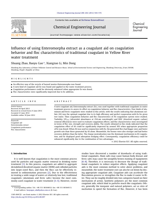
Influence of using Enteromorpha extract as a coagulant aid on coagulation behavior and floc characteristics of traditional coagulant in Yellow River water treatmentShuang Zhao,Baoyu Gao ⇑,Xiangzuo Li,Min DongShandong Key Laboratory of Water Pollution Control and Resource Reuse,School of Environmental Science and Engineering,Shandong University,Ji’nan 250100,Shandong,People’s Republic of Chinah i g h l i g h t s"An effective way of the recycle of hazard wastes-Enteromorpha was found."A new kind of coagulant aid-Ee was found and applied in the water treatment process."Coagulation performance could be obviously enhanced when appropriate Ee was dosed."Floc characteristics were significantly improved due to Ee addition.a r t i c l e i n f o Article history:Received 24April 2012Received in revised form 21June 2012Accepted 21June 2012Available online 30June 2012Keywords:Coagulant aidEnteromorpha extract Coagulation performance Floc characteristica b s t r a c tA new coagulant aid,Enteromorpha extract (Ee),was used together with traditional coagulants in water treatment process to assess its effect on coagulation behavior and floc characteristics.Four kinds of alu-minum and ferric coagulants were studied in this article,with the results indicating that aluminum sul-fate (AS)was the optimal coagulant due to its high efficiency and perfect cooperation with Ee for given test water.Then coagulation behavior and floc characteristics of AS coagulation system were studied.Turbidity,UV 254(ultraviolet absorbance at 254nm wavelength)and DOC (dissolved organic carbon)removal efficiencies were used to evaluate coagulation effects;and floc characteristics were investigated in terms of floc size,strength and recovery ability.The results obtained in this study indicated that the coagulation effect of AS could be significantly improved by around 30%when appropriate proportion of Ee was dosed.When AS was used in conjunction with Ee,the generated flocs had bigger sizes and faster growth rate than those generated by AS alone.Meanwhile,the former were also stronger and had better recovery abilities than the latter.The charge neutralization was the dominant mechanism in AS coagula-tion,and Ee displayed good adsorption bridging effect.Consequently,coagulation efficiency could be enhanced significantly due to the combination of two advantages.Ó2012Elsevier B.V.All rights reserved.1.IntroductionIt is well known that coagulation is the most common process used for particles and organic matter removal in drinking water treatment [1].In this process,coagulants are added to aggregate destabilized dissolved organic matter (DOM)and colloidal particles into larger-sized flocs,and then the flocs can be effectively re-moved in sedimentation processes [2].Due to the effectiveness in treating a wide range of waters at relatively low lost,traditional coagulants (aluminum and ferric salts)become the most com-monly used coagulant in water treatment [3–5].However,recent studies have discovered a number of drawbacks of using tradi-tional coagulants.Alum salts may cause human body disease and ferric salts may cause the unsightly brown staining of equipments [6–8].Therefore,it is necessary to decrease the dosage of tradi-tional coagulants to reduce negative effects.Applying coagulant aids is the most common method to solve above problems,and meanwhile,the water treatment cost can also be cut down by dos-ing appropriate coagulant aids.Coagulant aids can accelerate the flocculation process or strengthen the floc to make it easier to fil-ter.They can be roughly divided into two categories based on their mechanism of action.Synthetic polymers are the most common coagulant aids,which bind to particles much like coagulants.Oth-ers,generally the inorganic and natural polymers,act as sites of nucleation to speed the formation of floc.However,it has been1385-8947/$-see front matter Ó2012Elsevier B.V.All rights reserved./10.1016/j.cej.2012.06.097Corresponding author.Tel.:+8653188364832;fax:+8653188364513.E-mail address:baoyugao_sdu@ (B.Gao).reported that synthetic polymers contain contaminants from the manufacturing process that may threaten human health[8,9]. Meanwhile,Synthetic polymers can also react with other chemi-cals added to the water treatment process to form undesirable sec-ondary products[10–12].Natural polymers,which are extracted from plant or animal life,can be workable alternatives to synthetic polymers[13–16].They are biodegradable and also have a wider effective dosage range offlocculation for various colloidal suspen-sions.Therefore,it is indispensable to develop high efficient and low cost natural polymers which were used as coagulant aids for water and wastewater treatment[17–19].Enteromorpha is a kind of green algae,which is known as a dominant species in saline coastal wetlands with high nitrogen levels[20].Enteromorpha can tolerate salinities varying from freshwater to seawater.As a fast growing and opportunistic macro-algae,this species can proliferate in a wide range of abiotic and biotic conditions[21–23].In the summer of2008,Enteromorpha broke out in Yellow Sea and East Sea on a large scale,which posed a threat to the Sailing Competition of the2008Beijing Olympics. The local government had to spend much manpower andfinancial resources to carry out the emergency treatment measures against the disaster.But Enteromorpha management is supposed to be a long termfight rather than an episode during the29th Olympic Games for China,since it has been gaining in scale during the past 3decades in both marine and estuary environment all over the world.As we all know that waste is a kind of resource that was put in the wrong place.The comprehensive utilization of Enteromorpha is the ultimate way to settle the problem rather than piled them on the beach.But there has not been too much literature about the recycle used method of Enteromorpha at pres-ent since most researches focused on the Enteromorpha biological characteristics[24,25].It was once reported that Enteromorpha can be successfully transformed into biomass oil under appropriate conditions[26],but the cost was higher than the low-level fuel oil existing on market.The higher production cost limits its application and this technology is not suitable for the actual mass production.Enteromorpha is rich in macromolecular substances which can play an positive role in water supply and wastewater treatment,and also it has advantages of containing many active groups,various natural polymers and non-toxic character. Considering all the advantages of Enteromorpha,it is worth trying to apply the extracting solution of Enteromorpha(compound of many kinds of natural polymers)as a coagulant aid in coagulation process of water treatment.It is a tentative try since there is no relevant literature so far.By doing this,we attempt to provide a new solution to the Enteromorpha disaster,on the other hand,a new-style,cheap and non-toxic coagulant aid may be found.In this research,several kinds of aluminum and ferric salts were selected to use in conjunction with Ee in the treatment process of test water.Traditional coagulants were addedfirstly at the start of rapid mixing in coagulation process,and then Ee was dosed after 30s.That was denoted as dual-coagulant,which can be displayed in form of coagulant–coagulant aid.The coagulation aid perfor-mance of Enteromorpha extract(Ee)forflocculation–sedimenta-tion process was studied through a series of contrast test. Coagulation effects of traditional coagulants and dual-coagulants were comparatively investigated in terms of turbidity,UV254 (ultraviolet absorbance at254nm wavelength)and DOC(dissolved organic carbon)removal efficiency.Zeta potential was also researched in order to study the coagulation mechanism. Furthermore,the growth,breakage and re-growth properties of flocs produced in optimal condition were conducted.Based on the coagulation performance andfloc properties,the coagulation aid effect of Ee can be found,meanwhile,the coagulation aid mechanism can be discussed.2.Materials and methods2.1.Chemicals and raw waterThe chemicals used in this study including FeCl3Á6H2O,FeS-O4Á7H2O,AlCl3Á6H2O and Al2(SO4)3Á18H2O,which were all pur-chased from Sinopharm Chemical Reagent Co.,Ltd.,Beijing.All reagents used were of analytical grade.Deionized water was used to prepare all solutions.The raw water investigated in the study was sampled from Quehua water works of Jinan city in China in October2011.The properties of the test water were:temperature=19.5–22.5°C, pH=8.17–8.45,turbidity=2.18–2.76NTU,UV254=0.038–0.045cmÀ1,DOC=3.636–4.284mg/L.2.2.Preparation of coagulantsFresh Enteromorpha were gathered fromfirst bathing beach of Qingdao,China.They were put into the vacuum drying oven for5h at the constant temperature of70°C after being washed.The dry sample were shattered into powder by magnetic crusher and then passed through100mesh sieve.The screening Enteromorpha and deionized water were mixed at the mass ratio of1:75and then that compound was heated in water bath for4h at the constant tem-perature of90°C[27].The extract of Enteromorpha were centrifu-galized for30min at the speed of5000r/min.Supernatant were collected to measure volume and then diluted with equal volume of deionized water.That was used as the coagulant aid and the dos-age of Ee was metering by volume.Traditional coagulants and dual-coagulants,were compara-tively investigated in this study.The conventional coagulants, FeCl3Á6H2O and FeSO4Á7H2O,were used to prepare iron solutions with deionized water at a concentration of1.0g/L as Fe.Similarly, AlCl3Á6H2O and Al2(SO4)3Á18H2O were used to prepare alum solu-tions at a concentration of1.0g/L as Al.2.3.Jar-testAt a room temperature of20±2°C,Coagulation experiments were conducted by a jar-test apparatus(ZR4–6,Zhongrun Water Industry Technology Development Co.Ltd.,China).The test water of1000mL was poured into each of the1400mL plexiglass beakers and a six-paddled stirrer was used for mixing.The coagulation pro-cedure was as follow:predetermined amount of coagulant or dual-coagulant was dosed after30s rapid stirring of200rpm;after 1.5min,the stirring speed was changed to40rpm with a duration of15min;then after30min of quiescent settling,sample was col-lected from1cm below the surface for measuring.Part of sample was directly used for measuring residual turbidity and zeta poten-tial by using a2100P turbidimeter(Hach,USA)and zeta sizer 3000HSa(Malvern Instruments,UK),respectively.The remaining samples wasfiltrated by0.45l mfiber membrane and then was used to test UV254absorbance and DOC.UV254absorbance was analyzed with an UV-754UV/VIS spectrophotometer(Precision Scientific Instrument Co.Ltd.,Shanghai,China),and DOC was mea-sured by a TOC analyzer(TOC-VCPH,Shimadzu,Japan).2.4.On-line monitoring offloc formation,breakage and re-growth measurementA laser diffraction instrument Mastersizer2000(Malvern,UK) was used to monitor the evolution offloc size in coagulation pro-cess.The suspendedfloc was monitored through the sample cell of the Mastersizer and then transferred back into the jar by a peri-staltic pump(LEAD-1,Longer Precision Pump,China)using a5mm570S.Zhao et al./Chemical Engineering Journal200–202(2012)569–576internal diameter tube at aflow rate of2.0L/h.The pump was de-signed to be located at the downstream of Mastersizer to prevent disturbing theflocs prior.The inflow and outflow tubes were posi-tioned opposite each other at a depth just above the impeller in the holding ports.Size measurements of thefloc were taken every half minute in the process of coagulation and the results were recorded automatically by computer.Floc characteristic experiments were carried out under different dosage of(aluminum sulfate)AS and Ee.The coagulation procedure were conducted as follows:(1)a rapid mix step at200rpm for 1.5min;(2)a slow stir step at40rpm for15min;(3)a breakage step for5min200rpm;and(4)a slow stirring at40rpm for a fur-ther15min forflocs re-growth.2.5.Characteristic representation offlocIn this study,the median volumetric diameter(d50)was used to denote thefloc size.The growth rate was an important representa-tion offloc characteristic.It was denoted as the slope of rapid growth region.The larger the growth rate,the fasterfloc generated. It was calculated as follow[28,29]:growth rate¼D sizeð1ÞFor comparing the strength and re-growth ability offlocs in differ-entflocculated systems,floc strength factor(S f)and recovery factor (R f)were induced.They could be calculated by the following equa-tions[30,31]:S f¼d2d1Â100ð2ÞR f¼d3Àd212Â100ð3Þwhere d1(l m)and d2(l m)represent the stablefloc size before and after breakage,while d3represents thefloc size after re-growth to the new steady step.The strength factor value indicates the ability offlocs to withstand shear,such that a higher value suggests that theflocs are stronger.Meanwhile,the recovery factor is an indica-tive offloc recoverability,such that a higher value implies that theflocs have better re-growth ability after breakage.All the exper-iments were carried out three times and very little variation was observed.3.Results and discussion3.1.Coagulation effect of EeAlthough the Enteromorpha was non-toxic,but the outbreaks of it could block sunshine just as the red tides,and meanwhile the death Enteromorpha would consume oxygen in the sea,so the floating Enteromorpha may affect the algae growth in seabed.Fur-thermore,the outbreak of Enteromorpha will seriously affect land-scape and the proceeding of water sports.That is the biggest disadvantage that the people want to try to eliminate.Therefore, it is of great significance to explore an effective way for compre-hensive utilization of Enteromorpha,as well as for the restoration and improvement of the ecological environment and development of a recycling economy.In this section,Ee was used as a kind of coagulant.Fig.1showed the variation of turbidity,UV254and DOC removal efficiency with various Ee dosages.Fig.1showed that the turbidity and UV254removal gradually increased with the increasing Ee dosage,while the removal trend of DOC showed a parabola shape.The optimum point was at the dosage of0.1mL/L,in which condition the DOC removal rate was 20.5%.Zeta potential decreased with the increasing Ee dosage and below zero all the time within the whole dose ranges investigated. This indicated that charge neutralization is not the dominate mechanism of Ee.Due to the repulsive force between negative col-loids,thefloc was quiet small and hardly to settle down.Therefore, the removal of organic matter was in low levels.Overall,Ee exhib-ited some potential in removing organics,but good coagulation efficiency could not be achieved when Ee was used as a coagulant. So the performance of using Ee as a coagulant aid in coagulation process was studied as follow.3.2.Determination of the optimal coagulantAppropriate coagulant will not only improve the effluent water characteristics,but also can decrease the cost of water treatment. In order to ascertain the optimal coagulant for given test water, experiments were performed with afixed coagulant aid(Ee)in conjunction with four traditional coagulants(FeCl3,FeSO4,AlCl3 and Al2(SO4)3).Coagulants doses varied within a range of 2–12mg/L while coagulant aid dose was constant at0.1mL/L. Turbidity,UV254and DOC removal efficiency were taken to esti-mate coagulation performance.Results were shown in Fig.2.It can be seen from thefigure that the coagulation performance of four coagulants showed significant pared with iron salts coagulants,aluminum salts had better coagulation ef-fects with fasterflocs growth rates and largerflocs sizes.For DOC removal:the removal rates were rather low when iron salts were used and the maximal value was21%,while the removal of DOC can reach34%when aluminum salts were used.The conditions of turbidity and UV254removal efficiencies are similar to that of DOC.For aluminum salts coagulants,both of them could provide relatively high performances even at low dosages.Meanwhile the efficiencies of coagulation increased with coagulants doses increasing.In case of AlCl3,the application of coagulant aid could improved the performance,but only slightly effect could be ob-served,while the addition of Ee led to a significant increase of coagulation performance in Al2(SO4)3system.Consequently, Al2(SO4)3was chosen to carry out the following experiments as the optimal coagulant for the test water.3.3.Determination of the optimal dose of coagulant aidIn this section,the tests were performed with afixed coagulant (AS)based on Section3.2.Optimization tests were conducted to ascertain the optimum Ee dosage for particles and organic matterS.Zhao et al./Chemical Engineering Journal200–202(2012)569–576571removal under the raw water pH conditions.Traditional coagulant (AS)and dual-coagulant(aluminum sulfate–Enteromorpha extract (AS–Ee))were comparatively evaluated in terms of coagulation performance.The variation of coagulation efficiencies and zeta po-tential were presented in Fig.3with the coagulant dosages2–12mg/L as Al.When AS was used alone,Fig.3showed that UV254and DOC re-moval efficiency increased with alum dosage increasing,but the turbidity removal increased rapidly at a low alum dosage and then decreased slightly as the alum dose further increased.The coagulation with AS has been studied for decades and the coagula-tion mechanisms of it include charge neutralization,precipitation, bridge-aggregation,adsorption and sweep-flocculation[32,33].As shown in Fig.3,the organic matter removal efficiency increased as AS dosage increasing,which was in accordance with the increas-ing zeta potential(Fig.3d).The highest organics removal was achieved at the dosage of12mg/L with zeta potential of0.3mV, which indicated that charge neutralization was the dominant mechanism of AS coagulation system.The alkaline test water could produce precipitation of amorphous hydroxide,so the charge neu-tralization was likely to be achieved by adsorbed precipitate. Therefore,efficient turbidity removal still occurred at low dosages, even though the zeta potentials were below zero.When adequate Ee(0.1mL/L,0.3mL/L)was used in conjunction with AS,the turbidity,UV254and DOC removal were higher than that of AS used alone.Especially when Ee dosage was0.3mL/L,the organics removal efficiency was significantly improved:the UV254and DOC removal rate exceeded47%and44%at the alum dosage of12mg/L,while the removal obtained by AS alone were only37%and35%in the same test condition.Even though higher organic matter removal was obtained,zeta potential was still be-low zero(Fig.3d).That suggested that charge neutralization may be not the dominant mechanism for organics removal by AS–Ee. When AS was dosedfirstly,it quickly adsorbed on the surface of the microflocs and then neutralized the negative charge on it.So repulsion forces between the colloids became very weak.When Ee was dosed30s after AS,the adsorption bridging ability of it could play a significantly positive effect on theflocculation process, which could generate largerflocs those had preferably settling ability.Therefore,better coagulation efficiency could be achieved. But further increase in the dosage of Ee did not cause further ele-vation in the removals of organics:when Ee dosage was0.5mL/L, the coagulation performance was poor and even showed lower le-vel than that of AS used alone.The turbidity removal was about8% lower than that of AS.Since Ee itself was a complicated organic matter,the overdosed Ee in the coagulation system caused the de-crease of organics removal.Meanwhile,in such a situation,zeta potential was underÀ10mV due to larger dose of negatively charged Ee(À35.0mV).Therefore,the stronger inter-particle repulsion and the restrainedflocs growth resulted in poor coagula-tion efficiency.Consequently,based on thefigure and discussion above,it is easy for us to draw the conclusion that the optimal572S.Zhao et al./Chemical Engineering Journal200–202(2012)569–576coagulant aid dosage was 0.3mL/L.Considering the coagulants cost and coagulation performance,the dosage of Ee was constant at 8mg/L for the next floc characteristic experiments.3.4.Effect of coagulant aid on floc size and growth rateThe growth profiles of flocs were investigated and the results were shown in Fig.4,where the floc size was represented by med-ian equivalent diameter.Floc growth rate in steady state were adopted to examine the floc formation process,and the growth rates were shown in Table 1.The dosages of AS were constant at 8mg/L based on Section 3.3,and all the floc characteristic tests were taken in situation of raw water pH.Fig.4showed that the floc in the AS flocculation process dis-played a sharp increase in size during the first 5min,achieving the largest floc size of 338.1l m,followed by a steady-state period during the next 15min.The sharp growth of floc size in the first 5min is likely due to the aggregation of particles and the stable phase indicated that the floc growth and floc breakage reachedTable 1Flocs growth rates for various doses.Ee dosage (mL/L)0.00.10.30.5Growth rate (l m/min)45.8787.78122.20123.54S.Zhao et al./Chemical Engineering Journal 200–202(2012)569–576573appropriate balance.The sizes of flocs generated by AS–Ee and AS showed the similar variation tendency but the former were larger than the latter.As shown in Fig.4:the floc sizes at the steady stagewere 334.7l m,570.4l m,717.3l m,743.3l m for 0.0mL/L,0.1mL/L,0.3mL/L,0.5mL/L Ee dosage,respectively.Meanwhile,Table 1indicated that flocs produced by AS–Ee gave faster aggrega-tion than that of AS:at constant alum dose of 8mg/L,the flocs growth rate could achieve 122.20l m /min when 0.3mL/L Ee was used,while the growth rate of flocs produced by AS was only 45.87l m/min.The phenomenon mentioned above may be related to the dom-inate coagulation mechanism.The prime mechanism of AS was charge neutralization [2]while extra adsorption bridge were dom-inating when AS–Ee was used.Ee could promote the growth of flocs by adsorption bridge ability due to natural polymers con-tained in it.This result was in agreement with the observation of Ray and Hogg [33],who had reported that flocs produced by bridging flocculation and charge neutralization can be much largerTable 2Strength and recovery factors of flocs for different Ee dosages.ParameterEe dose (mL/L)0.00.10.30.5Strength factor 37.0840.6469.3975.33Recovery factor19.2625.7346.2255.63574S.Zhao et al./Chemical Engineering Journal 200–202(2012)569–576than those formed simply by charge neutralization.As we can see, when0.3mL/L Ee was used,the generatedflocs sizes and growth rate were much lager than those of0.1mL/L Ee applied,but there was no sharp improvement when Ee dosage further increased.The growth curves were almost overlapping when Ee dosage larger than0.5mL/L.This can be explained as follow:The bridge role of Ee can led to the increase of aggregation at the initial aggregation. There were not enough macromolecule matter particles when Ee dosage was small,so the adsorption bridge effect would be enhanced as the increase dosage of Ee.But all particles were positively aggregated and precipitated when Ee dosage larger than 0.5mL/L.Therefore,the due role of extra Ee couldn’t play out. Accordingly,thefloc sizes were hardly to further increased.3.5.Effect of coagulant aid onfloc breakage and re-growthThe effect of Ee onfloc breakage and re-growth was investi-gated in this section.Fig.5showed that in all cases,theflocs sizes immediately decreased when the shear was introduced by increas-ing the mixing speed up to200rpm.After the shear period of 5min,d50offlocs was about131l m for AS.When AS–Ee were used,theflocs sizes were230l m,493l m,591l m for0.1mL/L, 0.3mL/L,0.5mL/L Ee dosage,respectively.As the shear was re-duced again,theflocs began to re-grow.However,irreversible breakage was usually observed.The initialflocs sizes were not recovered after breakage:flocs formed by AS re-grew up to 172l m size and when Ee was used besides AS,the corresponding floc sizes after the re-growth period was312l m,579l m and 682l m for0.1mL/L,0.3mL/L and0.5mL/L Ee dosage,respectively. In order to investigatefloc breakage and re-growth in detail, the strength and recovery factors were used to interpret thefloc strength and recoverability,and the results were shown in Table2.In parallel,theflocs particle size distributions were also analyzed in this section,and the results were shown in Fig.6.Table2indicated that when AS–Ee was used in the coagulation process,the strength of generatedflocs were much larger than those of AS used alone:when0.5mL/L Ee was applied in conjunc-tion with AS,thefloc strength factor achieved75.33,which is twice larger than that of AS used alone(37.08).Meanwhile,For Fig.6,an apparent shift in the major peak after breakage could be seen for AS,while,comparatively,small extent of shift in the major peak was found of AS–Ee.Since the shift degree represented the ability of resisting shear,large excursion indicated poorflocs strength. Therefore,the results of Fig.6was in accordance with the conclu-sion of Table2obtained above.This results agreed with the conclu-sion by Li et al.[30]who found thatflocs generated by bridging were stronger than those generated simply by charge neutraliza-tion.Table2also showed that the strength factors increased as the rising dose of Ee with the constant AS dosage.Since there were not enough polymers between the particles when Ee dosage was low,the producedflocs were weak.But when the dose of Ee was large,the polymers were adequate to generate strongerflocs,so the strength factors became larger.For recovery ability,Table2 showed a gradual uptrend as the Ee dosage increased.The result was consist with the conclusion of Fig.6,which showed that:for AS–Ee,the small peak showed a comparatively apparent shift to the right of the major peak,while minor change could be observed between thefloc particle size distributions before and after re-growth of AS.Thefloc recovery factors of AS–Ee were larger than that of AS,which may be the result from Ee,which could undergo scission under high shear rate.Meanwhile,the renewed adsorbent polymer could reform on the particle surface whileflocs generated by AS were immediately broken when the high shear rate was introduced and then could not recover[33].4.ConclusionIn this study,the coagulation aid effect of Ee was investigated and it was found that Ee could be used as a new kind of coagulant aid due to its notable aid effects.Meanwhile,applying Ee in the water treatmentfield can be a new recycle way of disaster wastes–Enteromorpha.In this paper,AS was selected as the opti-mum coagulant from different traditional coagulants due to its high efficiency and perfect cooperation with Ee.The coagulation ef-fect of AS could be improved by around30%when0.3mL/L Ee was dosed.It was also found thatflocs generated by AS–Ee had bigger sizes and faster growth rates than that of AS.Meanwhile,the for-mer were also stronger and had better recovery ability than the lat-ter.The results of coagulation andflocs characteristic test showed that:the main coagulation mechanism of AS was precipitate charge neutralization,and Ee can play a positive aid role by adsorp-tion bridging in water treatment process.Therefore,better coagu-lation performance of AS–Ee could be achieved by the combination of two advantages.AcknowledgmentsThis study is supported by The Scientific Technology Research and Development Program of Shandong,China(No. 2010GZX20605)and a grant from the National High Technology Research and Development Program of China(863Program)(No. SQ2009AA06XK1482412).The kind suggestions from the anony-mous reviewers are highly appreciated.References[1]P.Jarvis,B.Jefferson,S.A.Parsons,Breakage,re-growth and fractal nature ofnatural organic matterflocs,Environ.Sci.Technol.39(7)(2005)2307–2314.[2]C.Hu,H.Liu,J.Qu,D.Wang,J.Ru,Coagulation behavior of aluminum salts ineutrophic water:significance of Al13species and pH control,Environ.Sci.Technol.40(2006)325–331.[3]B.Dempsey,W.J.De,M.Taylor,J.W.Potter,Guidance Manual for CoagulantChangeover,American Water Works Association Press,Denver,2006.pp.5–6.[4]I.L.Shih,Y.T.Van,L.C.Yeh,H.G.Lin,Y.N.Chang,Production of a biopolymerflocclant from Bacillus licheniformis and itsflocculation properties,Bioresour.Technol.78(2006)267–272.[5]Z.Li,S.Zhong,H.Y.Lei,R.W.Chen,Q.Yu,H.L.Li,Production of a novelbioflocculant by Bacillus licheniformis X14and its application to low temperature drinking water treatment,Bioresour.Technol.100(2009)3650–3656.[6]R.Divakaran,N.S.Pillai,Flocculation of kaolinite suspension in water bychitosan,Water Res.35(2001)3904–3908.[7]R.J.Pan,C.Huang,S.Chen,Y.C.Chung,Colloids Surf A:Physicochem.Eng.Aspects147(2009)359–364.[8]M.Ozacar,I.A.Sengil,The use of tannins from Turkish,J.Eng.Environ.Sci.2(2006)255–263.[9]G.Ruiz,D.Jeison,R.Chamy,Nitrification with high nitrite accumulation for thetreatment of wastewater with ammonia concentration,Water Res.37(2003) 1371–1377.[10]B.A.Bolto,Soluble polymers in water purification,Prog.Polym.Sci.20(1995)987–1041.[11]M.I.Aguilar,J.Saez,M.Llorens,A.Soler,J.F.Ortuno,Nutrient removal andsludge production in the coagulation–flocculation process,Water Res.36 (2006)2910–2919.[12]M.I.Aguilar,J.Saez,M.Llorens,A.Soler,J.F.Ortuno,V.Meseguer,A.Fuentes,Improvement of coagulation–flocculation process using anionic polyacrylamide as coagulant aid,Chemosphere58(2005)47–56.[13]S.Kawamura,Effectiveness of natural polyelectrolyte in water treatment.J.AWWA(1991)89–91.[14]J.Roussy,M.V.Voopren, E.Guibal,Chitosan for the coagulation andflocculation of mineral colloids,J.Disper.Sci.Technol.25(5)(2004)663–677.[15]M.Ashmore,J.Hearn,Flocculation of model latex particles by chitosans ofvarying degrees of acetylation,Langmuir16(11)(2002)4906–4911.[16]R.Divakaran,N.S.Pillai,Flocculation of river silt using chitosan,Water Res.36(9)(2002)2414–2418.[17]M.Ozacar,I.A.Sengil,Effect of tannin on phosphate removal using alum,Turkish J.Eng.Environ.Sci.27(2003)227–236.[18]M.Ozacar,I.A.Sengil,Evaluation of tannin biopolymer as a coagulant aid forcoagulation of colloidal particles,Colloids Surface A:Physicochem.Eng.Aspects229(2002)85–96.S.Zhao et al./Chemical Engineering Journal200–202(2012)569–576575。

UV Protection Test Method1. IntroductionUV protection has be a major concern for many industries, especially in the field of sunscreens, eyewear, textiles, and outdoor gear. The ability to effectively block and absorb ultraviolet (UV) radiation is crucial for the safety and well-being of consumers. Therefore, reliable and standardized test methods are essential for evaluating the UV protection performance of various products.2. PurposeThe purpose of this article is to provide an overview of themonly used test methods for UV protection, including their principles, procedures, and limitations. By understanding these test methods, manufacturers, researchers, and consumers can make informed decisions and ensure the quality and effectiveness of UV protection products.3. Spectrophotometric MethodSpectrophotometric method is one of the most widely used techniques for measuring the UV protection of materials. This method involves the use of a spectrophotometer to analyze thetransmittance and absorbance of UV radiation through a sample. The higher the transmittance, the lower the UV protection, while the higher the absorbance, the better the UV protection. This method is simple, quick, and accurate, making it suitable for routine quality control and research purposes.4. SPF TestingSPF (Sun Protection Factor) testing is specifically designed for sunscreen products. It measures the ability of a sunscreen to protect the skin from UVB radiation, which is responsible for sunburn and skin cancer. The test involves applying a specific amount of sunscreen on the skin and exposing it to UV radiation. The SPF value is calculated based on the difference in the time it takes for the skin to burn with and without the sunscreen. While SPF testing is essential for sunscreen products, it does not account for UVA protection, which is also important for overall UV protection.5. UPF TestingUPF (Ultraviolet Protection Factor) testing is similar to SPF testing but is intended for textiles and clothing. It evaluates the ability of a fabric to block both UVA and UVB radiation. The test involves exposing the fabric to UV radiation and measuring theamount of radiation that passes through. The UPF value is calculated based on the percentage of UV radiation that is blocked. UPF testing is crucial for assessing the UV protection of clothing and ensuring that consumers are adequately protected from the sun.6. Weathering TestIn addition to laboratory testing, weathering test is also important for evaluating the long-term UV protection performance of materials. This test simulates the effects of sunlight, temperature, and moisture on the material over an extended period. By subjecting the material to accelerated aging conditions, manufacturers can assess its durability, colorfastness, and UV protection stability. Weathering test helps ensure that UV protection products maintain their effectiveness and quality under real-world conditions.7. LimitationsWhile the aforementioned test methods are valuable for assessing UV protection, it is important to acknowledge their limitations. For instance, spectrophotometric method may not account for the effects of sweat, water, and friction on sunscreen performance. SPF and UPF testing may not fullyreflect the variability of human skin and behaviors, leading to potential discrepancies between laboratory results and real-world protection. Weathering test, while informative, may not perfectly replicate theplex environmental factors that materials encounter in outdoor settings.8. ConclusionIn conclusion, the test methods for UV protection play a critical role in ensuring the quality, safety, and performance of various products. By utilizing spectrophotometric method, SPF testing, UPF testing, and weathering test, manufacturers can accurately evaluate the UV protection properties of sunscreens, textiles, eyewear, and outdoor gear. It is essential for researchers and industry professionals to stay informed about the latest developments in UV protection testing and to continuously improve the reliability and relevance of these test methods. Ultimately, aprehensive understanding of UV protection test methods contributes to the advancement of UV protection technology and the empowerment of consumers to make informed choices for sun safety.。

Analog/photoelectric BOD 63M Time-of-Flight Distance SensorDesigned for the most demanding applications, the Balluff BOD 63M combines precision measurement and discrete sensing in one unit using time of flight technology.The BOD 63M's rugged metal housing has a working range of 500...6000 mm and is an ideal supplement to the BOD 26K(45...85 mm) and BOD 66M (200...2000 mm) families. It features adjustable background suppression and an analog output of 0...10V or 4...20 mA.A multi-turn potentiometer sets the discrete output. Its highlyfocused, easily visible red light enables long range sensing with very low gray shift value changes and hysteresis.Features– Measuring the propagation time of light allows for greater ranges than with sensors using triangulation or energetic diffuse technologies– Small laser spot for detecting small objects over large distances – Virtually unaffected by the reflective properties of the object within a particular sensing range– Background suppression is adjustable over the entire working range– Discrete sensing and alarm outputs Applications– Precise detection over large distances (e. g., installation constraints or when local temperatures are very high)– Detecting objects with changing colors, reflective surfaces or at unfavorable angles to the light beam– Flexible solutions for position sensing, level control, distance or height measurement, quality assuranceTime-of-Flight Priciplereceiver. This principle is extremely insensitive to ambient light.C o u r t e s y o f C M A /F l o d y n e /H y d r a d y n e ▪ M o t i o n C o n t r o l ▪ H y d r a u l i c ▪ P n e u m a t i c ▪ E l e c t r i c a l ▪ M e c h a n i c a l ▪ (800) 426-5480 ▪ w w w .c m a f h .c o mPhotoelectric SensorsAnalog2.102Large Block200...2000 or 200…6000 mm<1 mm15…30 Vdc ± 2.5 V 250 Vac ± 75 mA 4 - 20 mA 3 PNP or NPN NO200 mA Diffuse, Light-on Programmable setpointLaser Red LED 650 nm 10 mm @ 6 m -10 … +55 °C 1.5mm/°C 1%± 1.5%100 Hz ± 5 ms Green LED Yellow LEDM12 8-pin connector C04 ANT -00-PB-050MS Anodized AluminumGlass 260 g IP67Yes YesLarge Block200...2000 or 200…6000 mm<1 mm15…30 Vdc ± 2.5 V 250 Vac ± 75 mA 0 - 10 Vdc 3 PNP or NPN NO200 mA Diffuse, Light-on Programmable setpointLaser Red LED 650 nm 10 mm @ 6 m -10 … +55 °C 1.5 mm/°C1%± 1.5%100 Hz ± 5 ms Green LED Yellow LEDM12 8-pin connector C04 ANT -00-PB-050MS Anodized AluminumGlass 260 g IP67Yes YesBOD 63M SeriesClass II LaserBody Style Sensing range ResolutionMeasurement Sensor2 m Range TOF PNP 2 m Range TOF NPN 6 m Range TOF PNP 6 m Range TOF NPNSupply VoltageVoltage Drop Ud at Ie Rated Isolation VoltageCurrent Consumption Io (No Load)Analog Output T ype Discrete Output T ypeDiscrete Output Current (max.)Sensing Mode Additional Features Emitter Light Source Wavelength Spot SizeOperating Temperature Range Temperature Drift LinearityGray Shift Value (90%/18% reflectivity)Switching Frequency Response TimeSupply Voltage Indicator Output Function Indicator ConnectionWire InformationRecommended Connector Housing MaterialSensing Face Material WeightDegree of Protection per IEC 60529Short Circuit Protection Reverse Polarity Protection q w q w * Availability is September 2008.q = Number indicates wiring diagram A = Letter indicates detection diagram See pages 2.103 for diagramsC o u r t e s y o f C M A /F l o d y n e /H y d r a d y n e ▪ M o t i o n C o n t r o l ▪ H y d r a u l i c ▪ P n e u m a t i c ▪ E l e c t r i c a l ▪ M e c h a n i c a l ▪ (800) 426-5480 ▪ w w w .c m a f h .c o mConnectorC04 ANT-00-PB-050MS ACourtesyofCMA/Flodyne/Hydradyne▪MotionControl▪Hydraulic▪Pneumatic▪Electrical▪Mechanical▪(8)426-548▪www.cmafh.co m。

紫外可见吸收光谱微反应器英文回答:Ultraviolet-visible (UV-Vis) absorption spectroscopy is a commonly used technique in analytical chemistry to determine the presence and concentration of various compounds in a sample. This spectroscopic method involves the measurement of the absorption of light in the UV-Vis range (typically 190-900 nm) by a sample. The absorption spectrum obtained provides valuable information about the electronic transitions occurring in the molecules presentin the sample.UV-Vis absorption spectroscopy relies on the fact that molecules absorb light in the UV-Vis region when their electrons undergo transitions from lower energy levels to higher energy levels. The absorption of light causes the electrons to move from the ground state to an excited state. The energy difference between these two states correspondsto a specific wavelength of light, which can be detectedand measured using a spectrophotometer.The absorption spectrum obtained from UV-Vis spectroscopy consists of a series of peaks and valleys, each corresponding to a specific electronic transition. The position and intensity of these peaks provide information about the nature and concentration of the compounds present in the sample. For example, a compound with a strong absorption peak at a certain wavelength indicates the presence of chromophores that absorb light at that specific wavelength.UV-Vis absorption spectroscopy has a wide range of applications in various fields, including pharmaceuticals, environmental analysis, and biochemistry. It is commonly used in the quantification of analytes in solution, such as determining the concentration of a drug in a pharmaceutical formulation or the amount of a pollutant in an environmental sample. UV-Vis spectroscopy is also used to study the kinetics of chemical reactions, as the absorption of light can be directly related to the concentration of reactants or products.In recent years, there has been an increasing interest in the development of microreactors for performing chemical reactions. Microreactors, also known as microfluidic devices, are miniaturized reaction systems that enable the efficient and controlled synthesis of various compounds. These devices offer advantages such as improved heat and mass transfer, enhanced reaction selectivity, and reduced reaction times.Microreactors can be integrated with UV-Vis spectroscopy to monitor and analyze chemical reactions in real-time. By coupling a microreactor with a spectrophotometer, it is possible to continuously measure the absorption spectrum of the reaction mixture as the reaction progresses. This provides valuable information about the reaction kinetics, intermediate species, and reaction pathways.For example, consider a reaction where a starting material undergoes a series of transformations to form a final product. By monitoring the UV-Vis absorption spectrumof the reaction mixture at different time points, one can observe the disappearance of the starting material's absorption peak and the appearance of new peaks corresponding to intermediate species and the final product. This allows for the determination of reaction rates,reaction mechanisms, and the identification of reaction intermediates.In conclusion, UV-Vis absorption spectroscopy is a powerful analytical technique that provides valuable information about the electronic transitions occurring in molecules. It is widely used in various fields for the quantification of analytes and the study of chemical reactions. When coupled with microreactors, UV-Vis spectroscopy allows for real-time monitoring and analysisof chemical reactions, providing insights into reaction kinetics and mechanisms.中文回答:紫外可见(UV-Vis)吸收光谱是在分析化学中常用的一种技术,用于确定样品中各种化合物的存在和浓度。

紫外可见光谱仪操作流程英文回答:UV-Vis spectrophotometer is a commonly used instrument in analytical chemistry to measure the absorption and transmission of light by a sample in the ultraviolet and visible regions of the electromagnetic spectrum. The instrument is widely used in various fields such as pharmaceuticals, environmental monitoring, and biological research.To operate a UV-Vis spectrophotometer, the following steps are generally followed:1. Preparation: Before starting the measurement, it is important to ensure that the instrument is clean and in proper working condition. This involves cleaning the cuvettes, checking the lamp intensity, and calibrating the instrument if necessary.2. Sample preparation: The sample to be analyzed needs to be prepared appropriately. This may involve dilution, filtration, or any other necessary treatment to obtain a homogeneous solution. It is important to note that the sample should be transparent and free from any particulate matter that may interfere with the measurement.3. Setting up the instrument: Once the sample is prepared, it is placed in a cuvette and inserted into the spectrophotometer. The instrument is then turned on and allowed to warm up for a few minutes to stabilize the lamp intensity.4. Wavelength selection: The desired wavelength for measurement needs to be selected. This can be done using the instrument's control panel or software. The wavelength is usually chosen based on the absorption characteristics of the sample under investigation.5. Baseline correction: Before measuring the sample, a baseline correction is performed. This involves measuring the absorbance of a blank solution (usually the solventused for sample preparation) and subtracting it from the absorbance of the sample. This step helps to eliminate any background absorption and provides more accurate results.6. Measurement: Once the baseline correction is done, the instrument is ready to measure the absorbance of the sample. The absorbance is recorded and can be used to determine the concentration or other properties of the analyte.7. Data analysis: After the measurement is complete, the obtained data can be analyzed using appropriate software. This may involve plotting a calibration curve, calculating the concentration of the analyte, or comparing the absorbance values with known standards.Overall, operating a UV-Vis spectrophotometer requires careful preparation, accurate measurement, and proper data analysis. It is important to follow the manufacturer's instructions and guidelines for specific instrument models.中文回答:紫外可见光谱仪是分析化学中常用的仪器,用于测量样品在紫外和可见光区域的吸收和透射。
[Article]物理化学学报(Wuli Huaxue Xuebao )Acta Phys.-Chim.Sin .,2009,25(11):2285-2290November Received:March 20,2009;Revised:June 22,2009;Published on Web:September 3,2009.*Corresponding authors.Email:jiangjq@,lxy@;Tel:+86-133********.The project was supported by the National Natural Science Foundation of China (20704017,20374025),Qing Lan Project of Jiangsu Province,China and Scientific and Technological Iinnovation Team Project of Jiangsu Province,China (2007-5).国家自然科学基金(20704017,20374025)、江苏省高校“青蓝工程”和江苏省高等学校优秀科技创新团队计划(2007-5)资助项目鬁Editorial office of Acta Physico -Chimica Sinica含叔胺结构高光响应性香豆素衍生物的设计合成江金强*代鹏宗奕吾刘晓亚*张胜文陈明清(江南大学化学与材料工程学院,江苏无锡214122)摘要:以环氧氯丙烷为桥接,将具有较强给电子能力的芳叔胺(ATA)结构引入香豆素衍生物C 分子中,通过芳叔胺结构的光化学促进作用加速香豆素基元的光二聚反应.用紫外(UV)和荧光(FL)光谱等手段对该香豆素衍生物进行表征.结果表明,芳叔胺结构的引入可有效增强香豆素基元在260-400nm 之间的吸收.紫外点光源光二聚反应实验表明,芳叔胺结构的引入使香豆素基元的光响应性能得以大幅增强,在相同光照条件下,其在匀速反应区间内对光照时间的依赖性的斜率高达6.47,光二聚反应程度达到80%时所需光照时间仅为29s.关键词:香豆素;环氧氯丙烷;芳叔胺;光诱导;光二聚反应中图分类号:O648Design and Synthesis of Highly Photo -Crosslinkable CoumarinDerivatives Containing Aromatic Tertiary AmineJIANG Jin -Qiang *DAI PengZONG Yi -Wu LIU Xiao -Ya *ZHANG Sheng -WenCHEN Ming -Qing(School of Chemical and Material Engineering,Jiangnan University,Wuxi 214122,Jiangsu Province,P.R.China )Abstract :Using epichlorohydrin as bridged unit,the aromatic tertiary amine (ATA)was introduced to prepare the highly photo -crosslinkable coumarin compound C and to accelerate the photo -dimerization of coumarin unit.Ultraviolet (UV)and fluorescence (FL)spectra were used to evaluate the coumarin C.It was found that the introduced ATA could enhance the absorption between 260-400nm.The spot UV irradiation experiments showed that,after being bridged with ATA,the coumarin C would gain a highly photo -dimerization with a high slope of 6.47in the uniform photo -reaction,and a short time of 29s to gain 80%photo -dimerization.Key Words :Coumarin;Epichlorohydrin;Aromatic tertiary amine;Photo -induced;Photo -dimerization香豆素类化合物广泛分布于植物中,特别是在被子植物中多见,如伞形科、芸香科、豆科、菊科、瑞香科等,因其具有优异的抗艾滋病、降血压、抗辐射及光二聚等性能,被广泛应用于生物、医药、染料、聚合物科学等领域.特别是利用其对外界光刺激(如紫外、近红外等)的快速光反应性能,可设计合成“光响应型”智能聚合物[1],从而通过对光照频率、光照位置、光照强度及光照时间长短的选择,“智能”调控其物理化学性能.自1902年Ciamician 等[2]首次发现香豆素化合物可在太阳光照射下进行光化学反应以来,化学家们从光化学反应时的剂量、浓度和溶剂几方面对其光二聚反应进行了广泛深入的研究.Delzenne [3]在1960年将香豆素基元引入聚合物中,从而得到可进行光二聚反应的光响应聚合物,并研究了香豆素接入率对光交联网络的影响.近年来,有文献[3-6]报道将香豆素引入液晶聚合物中,可以通过2285Acta Phys.-Chim.Sin.,2009Vol.25香豆素基元的光二聚反应控制液晶材料的取向等;还有将其引入双亲聚合物中,可以通过香豆素的光化学反应控制被包覆在纳米微胶囊中的小分子客体的释放[7-9].但是,由于长时间的紫外光照射会使香豆素内酯键发生不可逆的开环反应,同时也会使聚合物发生氧化和降解,因此如何设计并合成出具有高效、快速的光响应香豆素衍生物,对感光高分子的发展将起到重要的作用.光诱导电荷转移聚合反应是利用正性乙烯基电子受体(如乙烯基咔唑VCZ)与各种强给电子化合物(如叔胺)形成电荷转移复合物(CTC),从而既能进行热聚合也能通过光诱导聚合.曹维孝[10-12]、丘坤元[13,14]等研究发现,即使是使用诸如丙烯腈、丙烯酸甲酯、丙烯酰胺、甲基丙烯酸甲酯、甲基丙烯酸-2-甲氧基乙酯或甲基丙烯酸羟基丙酯等吸电子能力不是很强的丙烯酰基单体作为电子受体,虽然不足以与叔胺形成稳定的CTC,但是仍能进行光聚合反应.这说明叔胺对丙烯酰基的光化学反应有一定的促进作用.本文设计并合成了一种含芳叔胺结构的香豆素衍生物,将供电子的芳叔胺引入香豆素基元的7-位上,通过芳叔胺结构的光化学促进作用加速香豆素基元的光二聚反应,并通过紫外光谱、荧光光谱、光二聚实验等验证该设计.1实验部分1.1仪器与试剂1.1.1试剂N-甲基苯胺、环氧氯丙烷、N,N-二甲基苯胺(DMA)、无水乙醇、N,N-二甲基甲酰胺(DMF)、碳酸钾、碘化钾、2-溴乙醇、三氯甲烷、丙酮等皆为分析纯,中国医药集团上海试剂公司产品.7-羟基-4-甲基香豆素(香豆素A)为本实验室参照文献[8]所述合成;7-羟乙醚-4-甲基香豆素(香豆素B)为本实验室参照文献[7]合成.1.1.2仪器TU-1901型紫外-可见光分光光度计,北京普析通用公司;Bruker-DMX500型核磁共振仪(NMR),德国Bruker公司;RF5301PC型荧光分光光度计,日本岛津公司;POWER ARC高强度点电光源紫外灯,光谱范围在300-500nm之间,主波峰为365nm,光强为3000mJ·cm-2,水银短弧灯泡103W,蓝天特灯发展有限公司.1.2含芳叔胺结构香豆素衍生物(香豆素C)的合成将32.1g(0.30mol)N-甲基苯胺和80mL无水乙醇置于250mL三口瓶中,取37.0g(0.4mol)环氧氯丙烷置于恒压滴液漏斗中,N2气保护下升温至回流,搅拌下缓慢滴加环氧氯丙烷,滴加完后继续回流12h,减压除去乙醇溶剂和过量的环氧氯丙烷得到淡黄色粘稠液体化合物(I),直接用于下一步Williamson醚化反应.将52.80g(0.30mol)香豆素A、41.37g(0.30 mol)K2CO3和200mL丙酮置于500mL三口瓶中,将39.94g(0.20mol)化合物(I)置于恒压滴液漏斗中,以少量KI为催化剂,N2气保护下升温至回流,待香豆素A完全溶解后,搅拌下缓慢往烧瓶中滴加,滴加完后继续回流24h,减压除去溶剂乙醇.所得固体用碳酸钾饱和溶液洗涤多次除去未反应的香豆素A,然后再用无水乙醇重结晶两次,得白色固体.纯化产物经30℃真空干燥至恒重,得到56.50g含叔胺结构香豆素衍生物(香豆素C),放入棕色瓶中保存,产率为83.3%.核磁共振(1H-NMR)测定使用氘代二甲基亚砜(d-DMSO)为溶剂,四甲基硅烷(TMS)为内标,频率500MHz所得数据:7.65-7.70(3H,ArH),6.94-7.05 (5H,ArH),6.22(1H,CH襒),4.10-4.28(3H,O—CH2—CH—OH), 2.9(3H,CH3—N), 2.73(2H, CH2—N), 2.37-2.4(3H,CH3—Ar).元素分析(C20H21NO4)测定值:C70.63%,H6.35%,N4.10%,理论值:C70.78%,H6.24%,N4.13%.1.3不同结构香豆素衍生物的光二聚性能比较以氯仿为溶剂,分别准确称量26.4mg(1.50×10⁻⁴ mol)香豆素A,33.0mg(1.50×10⁻⁴mol)香豆素B, 50.8mg(15.0×10⁻⁴mol)香豆素C,20μL DMA,分别定容为25mL摩尔浓度为4×10⁻⁵mol·L-1的香豆素(A,B,C)和DMA四种溶液.以溶液香豆素A+DMA为例,分别用移液枪准确移取20μL已配制好的香豆素A和DMA溶液,加入3mL氯仿于石英比色皿中,加入小型磁力搅拌子,密闭该系统.搅拌下利用高强度点电光源紫外灯进行光二聚反应,点光源从比色皿的透光侧垂直射入,光束与入射面的距离为1cm,以时间为参数,用紫外-可见光光谱扫描对香豆素A的光二聚进程进行跟踪.扫描范围为220-420nm,间隔为1nm,溶液浓度为4×10⁻⁵mol·L-1;荧光光谱的扫描范围设定为320-500nm,光栅激发和发射的狭缝均为3 nm,溶液浓度为2×10⁻⁵mol·L-1,荧光最佳激发波长2286No.11江金强等:含叔胺结构高光响应性香豆素衍生物的设计合成为334nm,其确定方法是以香豆素A在紫外光下的最强吸收峰对应的波长320nm激发,得到发射谱图,再用最强发射峰对应的波长去扫描得到激发谱图,其最强激发峰对应的波长是334nm,故将其定为荧光最佳激发波长.按如上操作进行香豆素A、香豆素B、香豆素C及香豆素B+DMA溶液的光二聚性能检测.2结果与讨论2.1含芳叔胺结构香豆素衍生物(香豆素C)的设计与合成香豆素化合物作为一种分子内共轭的电荷转移化合物,其发光能力与7-位处基团的推电子能力以及3,4-位处双键的电荷密度大小密切相关[15,16].吴飞鹏[17],杨永源[18]等通过在香豆素3-位上引入与香豆素共轭的二苯乙烯,7-位上引入电子给体二乙氨基,发现该化合物在光作用下很容易发生分子内电荷转移(intramolecular charge transfer,ICT),最大吸收波长比没有引入共轭基团的香豆素红移约20nm.如图1所示,香豆素化合物可在紫外光照射下进行光二聚反应[19,20],吴世康等[21-23]在香豆素基元上引入长链,改变香豆素化合物溶解性能,探究其链长、盐浓度、溶剂极性、溶剂中的聚集行为等因素对香豆素光二聚反应的影响和机理.本文的工作将根据前人的研究结果,在香豆素基元的7-位上引入供电子的芳叔胺,通过芳叔胺结构的光化学促进作用加速香豆素基元的光二聚反应.图2为含叔胺结构香豆素衍生物(香豆素C)的合成路线.如图所示,香豆素基元和芳叔胺结构通过与环氧氯丙烷反应得以桥接,从而使香豆素C分子结构中具备三种基元,即丙烯酰基光二聚基元,使香豆素具有光化学活性;芳叔胺光促进基元可以加速香豆素母体上丙烯酰基基元的光二聚反应;羟基可反应活性基元,可进行酯化反应,从而接入可聚合双键(例如和丙烯酸进行酯化)或合成为聚合引发中心(例如和2-溴异丁酰溴反应形成原子转移自由基聚合引发剂).该桥接方式一般有两种合成路线:路线1是先利用芳仲胺对环氧氯丙烷进行开环反应,然后再进行Williamson醚化反应得到香豆素C;路线2是先利用香豆素的酚羟基进行环氧氯丙烷开环、闭环反应,然后再进行芳仲胺的环氧开环反应.由于酚羟基与环氧氯丙烷开环、闭环反应较复杂,且闭环过程需要过量的KOH等强碱作催化剂,并可能使香豆素开环[24],因此我们选择路线1合成制备香豆素C.在N-甲基苯胺与环氧氯丙烷的开环反应中,使用过量的环氧氯丙烷,在乙醇回流下进行反应,从而保证N-甲基苯胺能够被充分开环;过量未反应的环氧氯丙烷可用旋蒸法除去;在Williamson 醚化反应中,使用丙酮为溶剂,K2CO3/KI体系为催化剂进行回流反应,并使用过量的香豆素A,从而保证化合物I上的烷基氯能够被充分反应,过量的香豆素A则可以用弱碱性的水溶液萃取洗去,再进行图1香豆素化合物在紫外光照下的光二聚反应Fig.1Photodimerization of coumarin underUV irradiation图2含叔胺结构香豆素衍生物(香豆素C)的合成路线Fig.2Synthetic route for the coumarin containing aromatic tertiary amine(coumarin C)2287Acta Phys.-Chim.Sin.,2009Vol.25重结晶等步骤得到新型含芳叔胺结构的香豆素衍生物(香豆素C).2.2芳叔胺光化学促进作用对香豆素化合物光二聚反应的影响芳叔胺(ATA)是指N原子上至少接有一个苯环的叔胺,以图3所示的N,N-二甲基苯胺(DMA)为例,其结构中的苯环和烷基对氮原子具有给电子作用,致使胺基氮原子的电子密度较大,形成较强的电子给体,并和具有较强的受体在无光照下形成基态电荷转移复合物(CTC);而对于丙烯酰基类(如丙烯酰胺、丙烯酸甲酯等)化合物,由于其双键上电子密度较低,电子受体能力较弱,在基态时和芳叔胺并不发生电荷转移复合,但在光照时可形成激发态电荷转移复合物[25].曹维孝[10-12]、邱坤元[13,14]等发现芳叔胺结构对丙烯酰基的光聚合速率有显著的影响,且当在芳叔胺的苯环上引入给电子基团时,光反应速率增加;引入吸电子基团时,光反应速率下降.他们同时指出可以通过紫外光谱和荧光光谱分析研究芳叔胺和丙烯酰基的电荷转移复合作用.我们选择DMA为模型芳叔胺化合物,与香豆素A及其醚化物(香豆素B)等摩尔浓度相混合,检测其复合作用,并按图2路线设计合成香豆素C.紫外分析显示(见图4),在相同摩尔浓度条件下,香豆素A和B在加入等摩尔浓度的DMA后,其在260-400nm之间的吸收表现为香豆素紫外吸收和DMA 的简单叠加,说明芳叔胺和香豆素化合物之间的基态电荷转移复合作用非常弱或不存在;而当芳叔胺结构引入香豆素衍生物分子中后(香豆素C),香豆素C在260-400nm之间的吸收相对香豆素A和B、及A+DMA和B+DMA都得以增强.这表明将芳叔胺结构引入香豆素衍生物分子中后,在相同的光照条件下,香豆素C可以吸收更多的紫外能量,从而可能加速香豆素基元的光二聚反应.图5为香豆素(A,B,C)与DMA复合前后的荧光发射图谱,在相同摩尔浓度条件下,香豆素A、B 在与等摩尔浓度的DMA作用后,用334nm光进行激发,均在380nm附近出现不同程度的荧光淬灭;而对于含芳叔胺结构的香豆素C,与不含芳叔胺结构的香豆素A、B相比,其在380nm附近也相对减弱,这表明在光照激发条件下,芳叔胺和香豆素基元之间可能形成了一定程度的激发态电荷转移复合[18].香豆素化合物在λ>300nm的紫外光照射下能够发生光二聚反应,且香豆素化合物在320nm左右的特征吸收峰随光照的进行逐渐减弱,因此可以通过紫外光谱跟踪分析其光二聚行为.在实验中,我们统一光照条件,在相同摩尔浓度条件下,分别对香豆素A、B和C的氯仿溶液在紫外点光源照射下进图3香豆素(A,B,C)与N,N-二甲基苯胺(DMA)的特征化学结构Fig.3Structures of coumarins(A,B,C)and N,N-dimethylaniline(DMA)图4香豆素化合物(A,B,C)与DMA复合前后的紫外吸收图谱Fig.4UV spectra of the coumarins(A,B,C)beforeand after being complexed with DMAThe concentrations of all coumarin compounds in CHCl3solution are4×10-5mol·L-1.2288No.11江金强等:含叔胺结构高光响应性香豆素衍生物的设计合成行光二聚反应,并用紫外光谱进行跟踪分析.同时我们利用香豆素在320nm 处的吸收峰值的变化表征其光二聚反应速率(k dimer ),定义如下:k dimer =[1-A (t )/A (0)]×100%其中A (0)代表未进行光照二聚时体系的吸收峰值,A (t )代表紫外光照二聚t 时间后的吸收峰值.图6、图7、图8为香豆素(A,B,C)在CHCl 3中在紫外光照下的光二聚行为.如图所示,香豆素(A,B,C)在λ>300nm 紫外光照下均可进行光二聚反应,在光二聚反应程度达到40%左右的光照时间内,均表现为匀速反应特征;当光二聚反应程度达到80%后,香豆素(A,B,C)的光二聚反应都进入“平台区”,即光二聚反应速率逐渐趋于缓慢.同时,从图6和7可知,香豆素化合物A 、B 在加入等摩尔量的DMA 后,其光二聚反应速率均有不同程度的提高,其匀速反应阶段对时间的依赖性函数的斜率分别从2.09和0.75升高至4.06和2.72,到达“平台区”所需时间分别从61和172s 缩短至46和64s;而对分子结构中直接含有芳叔胺的香豆素C,其光二聚匀速反应阶段对时间的依赖性函数的斜率高达6.47,到达“平台区”的时间只需29s.由此可见,芳叔胺结图5香豆素(A,B,C)与DMA 复合前后的荧光发射图谱Fig.5FL spectra of the coumarins (A,B,C)beforeand after being complexed with DMAThe concentrations of all coumarin compounds inCHCl 3solution are 2×10⁻⁵mol·L -1.图8香豆素C 在CHCl 3溶液中在紫外光照下(λ>300nm)的光二聚行为Fig.8Photodimerization of coumarin C in CHCl 3solution under UV irradiation at λ>300nmdetected by UV spectraThe curves from top to down are coumarin C irradiated after 0,4,6,10,17,and 107s,respectively.The inset shows the increase ofits photodimerization degree.图6香豆素A 在CHCl 3溶液中在紫外光照下(λ>300nm)的光二聚行为Fig.6Photodimerization of coumarin A in CHCl 3solution under UV irradiation at λ>300nmdetected by UV spectraThe curves from top to down are coumarin A irradiated after 0,11,21,31,41,66,and 111s,respectively.The inset shows the increase of its photodimerization degree (k dimer ),the red curve for coumarin A and theblue one for coumarin A+DMA.t :irradiation time图7香豆素B 在CHCl 3溶液中在紫外光照下(λ>300nm)的光二聚行为Fig.7Photodimerization of coumarin B in CHCl 3solution under UV irradiation at λ>300nmdetected by UV spectraThe curves from top to down are coumarin B irradiated after 0,23,43,71,106,146and 206s,respectively.The inset shows the increase of its photodimerization degree,the red curve for coumarin B andthe blue one for coumarin B+DMA2289Acta Phys.-Chim.Sin.,2009Vol.25构的引入可大幅增强香豆素基元的光响应性能,极大地加速其光二聚反应.3结论综上所述,通过环氧氯丙烷的桥接反应,可以将具有较强给电子能力的芳叔胺结构引入香豆素C 分子中,通过芳叔胺结构的光化学促进作用可加速香豆素基元的光二聚反应.紫外光谱分析显示,芳叔胺结构的引入可有效增强香豆素基元在260-400 nm之间的吸收;紫外点光源光二聚反应实验表明,芳叔胺结构的引入使香豆素基元的光响应性能得以大幅增强,在相同光照条件下,其在匀速反应区间对光照时间的依赖性斜率高达6.47,光二聚反应程度达到80%所需光照时间仅为29s.References1Scott,R.T.;Allan,R.S.;Brian,J.L.;Timothy,E.L.Chem.Rev., 2004,104:30592Ciamician,G.;Silber,P.Berichte Der Deutschen Chemischen Gesellschaft,1902,35:41283Delzenne,G.A.Europ.Polymer J.,1969,(Supplement):554Schadt,M.;Seiberle,H.;Schuster,A.Nature,1996,381:2125Kishore,G.A.K.Macromolecules,1995,28:8066Obi,M.;Morino,S.;Ichimura,K.Chem.Mater.,1999,11:6567Jiang,J.Q.;Qi,B.;Martin,L.;Zhao,Y.Macromolecules,2007, 40:7908Jiang,J.Q.;Feng,Y.;Wang,H.M.;Liu,X.Y.;Zhang,S.W.;Chen,M,Q.Acta Phys.-Chim.Sin.,2008,24(11):2089[江金强,冯艳,王红梅,刘晓亚,张胜文,陈明清.物理化学学报,2008,24(11):2089]9Jackson,P.O.;O′Neill,M.;Duffy,W.L.;Hindmarsh,P.;Kelly,S.M.;Owen,G.J.Chem.Mater.,2001,13:69410Cao,W.X.;Feng,mun.,1982,1:43[曹维孝,冯新德.高分子通讯,1982,1:43]11Li,T.;Cao,W.X.;Feng,mun.,1983,2:127 [李橦,曹维孝,冯新德.高分子通讯,1983,2:127]12Li,T.;Cao,W.X.;Feng,mun.,1983,4:260 [李橦,曹维孝,冯新德.高分子通讯,1983,4:260]13Qiu,K.Y.;Zhang,Z.H.;Feng,X.D.Acta Polym.Sin.,1993,2: 178[丘坤元,张璋华,冯新德.高分子学报,1993,2:178]14Feng,X.D.;Qiu,K.Y.Polym.Bullt.,2005,4:23[冯新德,丘坤元.高分子通报,2005,4:23]15Dai,Z.H.;Wu,S.K.Acta Phys.-Chim.Sin.,1999,15(12):1076 [戴赵华,吴世康.物理化学学报,1999,15(12):1076]16Gao,F.;Yang,Y.Y.Acta Phys.-Chim.Sin.,1999,15(6):550 [高放,杨永源.物理化学学报,1999,15(6):550]17Li,X.;Wang,T.;Zhao,Y.X.;Wu,F.P.Photographic Science and Photochemistry,2006,24(3):187[李雪,王涛,赵榆霞,吴飞鹏.感光科学与光化学,2006,24(3):187]18Wang,T.;Wu,F.P.;Shi,M.Q.;Gao,F.;Yang,Y.Y.Acta Chimica Sinica,2004,62(5):527[王涛,吴飞鹏,施盟泉,高放,杨永源.化学学报,2004,62(5):527]19Hammond,G.S.;Stout,C.A.;Lamola,A.A.J.Am.Chem.Soc., 1964,86:3130320Morrison,H.;Curtis,H.;McDowell,T.J.Am.Chem.Soc.,1966, 88:541521Liu,T.J.;Wu,S.K.Chem.J.Chin.Univ.,1996,11:1754 [刘天军,吴世康.高等学校化学学报,1996,11:1754]22Liu,T.J.;Wu,S.K.Acta Phys.-Chim.Sin.,1996,12(8):677 [刘天军,吴世康.物理化学学报,1996,12(8):677]23Zhu,A.P.;Wu,S.K.Photographic Science and Photochemistry, 1997,15(2):151[朱爱平,吴世康.感光科学与光化学,1997, 15(2):151]24Zhang,X.Y.;Xiang,R.D.Chinese Traditional Patent Medicine, 1999,21(1):7[张新勇,向仁德.中成药.1999,21(1):7]25Hideaki,S.;Haridas,P.;Keisuke,T.;Keitaro Y.J.Phys.Chem.A, 1998,102(18):30892290。
Designation:G154–06Standard Practice forOperating Fluorescent Light Apparatus for UV Exposure of Nonmetallic Materials1This standard is issued under thefixed designation G154;the number immediately following the designation indicates the year oforiginal adoption or,in the case of revision,the year of last revision.A number in parentheses indicates the year of last reapproval.Asuperscript epsilon(e)indicates an editorial change since the last revision or reapproval.Note—A footnote was added to Table X2.1,Table X2.3was added,a new Note X2.8was added,and the year date was changed on June5,2006.1.Scope1.1This practice covers the basic principles and operating procedures for usingfluorescent UV light,and water apparatus intended to reproduce the weathering effects that occur when materials are exposed to sunlight(either direct or through window glass)and moisture as rain or dew in actual usage. This practice is limited to the procedures for obtaining, measuring,and controlling conditions of exposure.A number of exposure procedures are listed in an appendix;however,this practice does not specify the exposure conditions best suited for the material to be tested.N OTE1—Practice G151describes performance criteria for all exposure devices that use laboratory light sources.This practice replaces Practice G53,which describes very specific designs for devices used forfluores-cent UV exposures.The apparatus described in Practice G53is covered by this practice.1.2Test specimens are exposed tofluorescent UV light under controlled environmental conditions.Different types of fluorescent UV light sources are described.1.3Specimen preparation and evaluation of the results are covered in ASTM methods or specifications for specific materials.General guidance is given in Practice G151and ISO 4892-1.More specific information about methods for deter-mining the change in properties after exposure and reporting these results is described in ISO4582.1.4The values stated in SI units are to be regarded as the standard.1.5This standard does not purport to address all of the safety concerns,if any,associated with its use.It is the responsibility of the user of this standard to establish appro-priate safety and health practices and determine the applica-bility of regulatory limitations prior to use.1.6This standard is technically similar to ISO4892-3and ISO DIS11507.2.Referenced Documents2.1ASTM Standards:2D3980Practice for Interlaboratory Testing of Paint and Related MaterialsE691Practice for Conducting an Interlaboratory Study to Determine the Precision of a Test MethodG53Practice for Operating Light-and Water-Exposure Apparatus(Fluorescent UV-Condensation Type)for Expo-sure of Nonmetallic MaterialsG113Terminology Relating to Natural and Artificial Weathering Tests for Nonmetallic MaterialsG151Practice for Exposing Nonmetallic Materials in Ac-celerated Test Devices That Use Laboratory Light Sources 2.2CIE Standard:CIE-Publ.No.85:Recommendations for the Integrated Irradiance and the Spectral Distribution of Simulated Solar Radiation for Testing Purposes32.3ISO Standards:ISO4582,Plastics—Determination of the Changes of Co-lour and Variations in Properties After Exposure to Day-light Under Glass,Natural Weathering or Artificial Light4 ISO4892-1,Plastics—Methods of Exposure to Laboratory Light Sources,Part1,Guidance4ISO4892-3,Plastics—Methods of Exposure to Laboratory Light Sources,Part3,Fluorescent UV lamps4ISO DIS11507,Paint and Varnishes—Exposure of Coat-ings to Artificial Weathering in Apparatus—Exposure to Fluorescent Ultraviolet and Condensation Apparatus4 3.Terminology3.1Definitions—The definitions given in Terminology G113are applicable to this practice.1This practice is under the jurisdiction of ASTM Committee G03on Weathering and Durability and is the direct responsibility of Subcommittee G03.03on Simulated and Controlled Exposure Tests.Current edition approved June5,2006.Published June2006.Originally approved st previous edition approved in2005as G154–05.2For referenced ASTM standards,visit the ASTM website,,or contact ASTM Customer Service at service@.For Annual Book of ASTM Standards volume information,refer to the standard’s Document Summary page on the ASTM website.3Available from Secretary,U.S.National Committee,CIE,National Institute of Standards and Technology(NIST),Gaithersburg,MD20899.4Available from American National Standards Institute(ANSI),25W.43rd St., 4th Floor,New York,NY10036.Copyright©ASTM International,100Barr Harbor Drive,PO Box C700,West Conshohocken,PA19428-2959,United States. --`,,```,,,,````-`-`,,`,,`,`,,`---3.2Definitions of Terms Specific to This Standard—As used in this practice,the term sunlight is identical to the terms daylight and solar irradiance,global as they are defined in Terminology G113.4.Summary of Practice4.1Specimens are exposed to repetitive cycles of light and moisture under controlled environmental conditions.4.1.1Moisture is usually produced by condensation of water vapor onto the test specimen or by spraying the speci-mens with demineralized/deionized water.4.2The exposure condition may be varied by selection of: 4.2.1Thefluorescent lamp,4.2.2The lamp’s irradiance level,4.2.3The type of moisture exposure,4.2.4The timing of the light and moisture exposure,4.2.5The temperature of light exposure,and4.2.6The temperature of moisture exposure,and4.2.7The timing of a light/dark cycle.4.3Comparison of results obtained from specimens exposed in same model of apparatus should not be made unless reproducibility has been established among devices for the material to be tested.4.4Comparison of results obtained from specimens exposed in different models of apparatus should not be made unless correlation has been established among devices for the material to be tested.5.Significance and Use5.1The use of this apparatus is intended to induce property changes associated with the end use conditions,including the effects of the UV portion of sunlight,moisture,and heat.These exposures may include a means to introduce moisture to the test specimen.Exposures are not intended to simulate the deterioration caused by localized weather phenomena,such as atmospheric pollution,biological attack,and saltwater expo-sure.Alternatively,the exposure may simulate the effects of sunlight through window glass.Typically,these exposures would include moisture in the form of condensing humidity. N OTE2—Caution:Refer to Practice G151for full cautionary guidance applicable to all laboratory weathering devices.5.2Variation in results may be expected when operating conditions are varied within the accepted limits of this practice. Therefore,no reference shall be made to results from the use of this practice unless accompanied by a report detailing the specific operating conditions in conformance with the Section 10.5.2.1It is recommended that a similar material of known performance(a control)be exposed simultaneously with the test specimen to provide a standard for comparative purposes. It is recommended that at least three replicates of each material evaluated be exposed in each test to allow for statistical evaluation of results.6.Apparatus6.1Laboratory Light Source—The light source shall be fluorescent UV lamps.A variety offluorescent UV lamps can be used for this procedure.Differences in lamp intensity or spectrum may cause significant differences in test results.A detailed description of the type(s)of lamp(s)used should be stated in detail in the test report.The particular testing application determines which lamp should be used.See Ap-pendix X1for lamp application guidelines.N OTE3—Do not mix different types of lamps.Mixing different types of lamps in afluorescent UV light apparatus may produce major inconsis-tencies in the light falling on the samples,unless the apparatus has been specifically designed to ensure a uniform spectral distribution.N OTE4—Manyfluorescent lamps age significantly with extended use. Follow the apparatus manufacturer’s instructions on the procedure neces-sary to maintain desired irradiance(1,2).6.1.1Actual irradiance levels at the test specimen surface may vary due to the type or manufacturer of the lamp used,or both,the age of the lamps,the distance to the lamp array,and the air temperature within the chamber and the ambient laboratory temperature.Consequently,the use of a radiometer to monitor and control the radiant energy is recommended.6.1.2Several factors can affect the spectral power distribu-tion offluorescent UV lamps:6.1.2.1Aging of the glass used in some types of lamps can result in changes in transmission.Aging of glass can result in a significant reduction in the short wavelength UV emission of some lamp types,6.1.2.2Accumulation of dirt or other residue on lamps can affect irradiance,6.1.2.3Thickness of glass used for lamp tube can have large effects on the amount of short wavelength UV radiation transmitted,and6.1.2.4Uniformity and durability of phosphor coating. 6.1.3Spectral Irradiance:N OTE5—Fluorescent UV A lamps are available with a choice of spectral power distributions that vary significantly.The more common may be identified as UV A-340and UV A-351.These numbers represent the characteristic nominal wavelength(in nm)of peak emission for each of these lamp types.The actual peak emissions are at343and350nm, respectively.6.1.3.1Spectral Irradiance of UVA-340Lamps for Daylight UV—The spectral power distribution of UV A-340fluorescent lamps shall comply with the requirements specified in Table1. N OTE6—The main application for UV A-340lamps is for simulation of the short and middle UV wavelength region of daylight.6.1.3.2Spectral Irradiance of UVA-351Lamps for Daylight UV Behind Window Glass—The spectral power distribution of UV A-351lamp for Daylight UV behind Window Glass shall comply with the requirements specified in Table2.N OTE7—The main application for UV A-351lamps is for simulation of the short and middle UV wavelength region of daylight which has been filtered through window glass(3).6.1.3.3Spectral Irradiance of UVB-313Lamps—The spec-tral power distribution of UVB-313fluorescent lamps shall comply with the requirements specified in Table3.N OTE8—Fluorescent UVB lamps have the spectral distribution of radiation peaking near the313-nm mercury line.They emit significant amounts of radiation below300nm,the nominal cut on wavelength of global solar radiation,that may result in aging processes not occurring e of this lamp is not recommended for sunlight simulation. See Table3. --`,,```,,,,````-`-`,,`,,`,`,,`---6.2Test Chamber—The design of the test chamber may vary,but it should be constructed from corrosion resistant material and,in addition to the radiant source,may provide for means of controlling temperature and relative humidity.When required,provision shall be made for the spraying of water onthe test specimen for the formation of condensate on the exposed face of the specimen or for the immersion of the test specimen in water.6.2.1The radiant source(s)shall be located with respect to the specimens such that the uniformity of irradiance at the specimen face complies with the requirements in Practice G151.6.2.2Lamp replacement,lamp rotation,and specimen repo-sitioning may be required to obtain uniform exposure of all specimens to UV radiation and temperature.Follow manufac-turer’s recommendation for lamp replacement and rotation.6.3Instrument Calibration—To ensure standardization and accuracy,the instruments associated with the exposure appa-ratus(for example,timers,thermometers,wet bulb sensors,dry bulb sensors,humidity sensors,UV sensors,and radiometers) require periodic calibration to ensure repeatability of test results.Whenever possible,calibration should be traceable to national or international standards.Calibration schedule and procedure should be in accordance with manufacturer’s in-structions.6.4Radiometer—The use of a radiometer to monitor and control the amount of radiant energy received at the sample is recommended.If a radiometer is used,it shall comply with the requirements in Practice G151.6.5Thermometer—Either insulated or un-insulated black or white panel thermometers may be used.The un-insulated thermometers may be made of either steel or aluminum. Thermometers shall conform to the descriptions found in Practice G151.6.5.1The thermometer shall be mounted on the specimen rack so that its surface is in the same relative position and subjected to the same influences as the test specimens.6.5.2Some specifications may require chamber air tempera-ture control.Positioning and calibration of chamber air tem-perature sensors shall be in accordance with the descriptions found in Practice G151.N OTE9—Typically,these devices control by black panel temperature only.6.6Moisture—The test specimens may be exposed to mois-ture in the form of water spray,condensation,or high humidity.6.6.1Water Spray—The test chamber may be equipped witha means to introduce intermittent water spray onto the test specimens under specified conditions.The spray shall beTABLE1Relative Ultraviolet Spectral Power Distribution Specification for Fluorescent UVA-340Lamps for Daylight UV A,BSpectral Bandpass Wavelength l in nm MinimumPercent CBenchmark SolarRadiation Percent D,E,FMaximumPercent Cl<2900.01 290#l#320 5.9 5.89.3320<l#36060.940.065.5360<l#40026.554.232.8A Data in Table1are the irradiance in the given bandpass expressed as a percentage of the total irradiance from290to400nm.The manufacturer is responsible for determining conformance to Table1.Annex A1states how to determine relative spectral irradiance.B The data in Table1are based on the rectangular integration of65spectral power distributions forfluorescent UV devices operating with UVA340lamps of various lots and ages.The spectral power distribution data is for lamps within the aging recommendations of the device manufacturer.The minimum and maximum data are at least the three sigma limits from the mean for all measurements.C The minimum and maximum columns will not necessarily sum to100% because they represent the minimum and maximum for the data used.For any individual spectral power distribution,the calculated percentage for the band-passes in Table1will sum to100%.For any individualfluorescent UVA-340lamp, the calculated percentage in each bandpass must fall within the minimum and maximum limits of Table1.Test results can be expected to differ between exposures using devices withfluorescent UVA-340lamps in which the spectral power distributions differ by as much as that allowed by the tolerances.Contact the manufacturer of thefluorescent UV devices for specific spectral power distribution data for thefluorescent UVA-340lamp used.D The benchmark solar radiation data is defined in ASTM G177and is for atmospheric conditions and altitude chosen to maximize the fraction of short wavelength solar UV.While this data is provided for comparison purposes only,it is desirable for the laboratory accelerated light source to provide a spectrum that is a close match to the benchmark solar spectrum.E Previous versions of this standard used solar radiation data from Table4of CIE Publication Number85.See Appendix X3for more information comparing the solar radiation data used in this standard with that for CIE85Table4.F For the benchmark daylight spectrum,the UV irradiance(290to400nm)is9.8%and the visible irradiance(400to800nm)is90.2%expressed as a percentage of the total irradiance from290to800nm.Because the primary emission offluorescent UV lamps is concentrated in the300to400nm bandpass, there are limited data available for visible light emissions offluorescent UV lamps.TABLE2Relative Spectral Power Distribution Specification for Fluorescent UVA-351Lamps for Daylight UV Behind WindowGlass A,BSpectral BandpassWavelength l in nmMinimumPercent CWindow Glass FilteredDaylight Percent D,E,FMaximumPercent C l<3000.00.2 300#l#320 1.1#0.5 3.3 320<l#36060.534.266.8 360<l#40030.065.338.0A Data in Table2are the irradiance in the given bandpass expressed as a percentage of the total irradiance from300to400nm.The manufacturer is responsible for determining conformance to Table1.Annex A1states how to determine relative spectral irradiance.B The data in Table2are based on the rectangular integration of21spectral power distributions forfluorescent UV devices operating with UVA351lamps of various lots and ages.The spectral power distribution data is for lamps within the aging recommendations of the device manufacturer.The minimum and maximum data are at least the three sigma limits from the mean for all measurements.C The minimum and maximum columns will not necessarily sum to100% because they represent the minimum and maximum for the data used.For any individual spectral power distribution,the calculated percentage for the band-passes in Table2will sum to100%.For any individualfluorescent UV device operating with UVA351lamps,the calculated percentage in each bandpass must fall within the minimum and maximum limits of Table2.Test results can be expected to differ between exposures usingfluorescent UV devices in which the spectral power distributions differ by as much as that allowed by the tolerances. Contact the manufacturer of thefluorescent UV devices for specific spectral power distribution data for the lamps used.D The window glassfiltered solar radiation data is for a solar spectrum with atmospheric conditions and altitude chosen to maximize the fraction of short wavelength solar UV(defined in ASTM G177)that has beenfiltered by window glass.The glass transmission is the average for a series of single strength window glasses tested as part of a research study for ASTM Subcommittee G3.02.9While this data is provided for comparison purposes only,it is desirable for the laboratory accelerated light source to provide a spectrum that is a close match to this benchmark window glassfiltered solar spectrum.E Previous versions of this standard used window glassfiltered solar radiation data based on Table4of CIE Publication Number85.See Appendix X3for more information comparing the solar radiation data used in the standard with that for CIE85Table4.F For the benchmark window glassfiltered solar spectrum,the UV irradiance (300to400nm)is8.2%and the visible irradiance(400to800nm)is91.8% expressed as a percentage of the total irradiance from300to800nm.Because the primary emission offluorescent UV lamps is concentrated in the300to400nm bandpass,there are limited data available for visible light emissions offluorescent UVlamps.--` , , ` ` ` , , , , ` ` ` ` -` -` , , ` , , ` , ` , , ` ---uniformly distributed over the samples.The spray system shall be made from corrosion resistant materials that do not con-taminate the water used.6.6.1.1Spray Water Quality —Spray water shall have a conductivity below 5µS/cm,contain less than 1-ppm solids,and leave no observable stains or deposits on the specimens.Very low levels of silica in spray water can cause significant deposits on the surface of test specimens.Care should be taken to keep silica levels below 0.1ppm.In addition to distillation,a combination of deionization and reverse osmosis can effec-tively produce water of the required quality.The pH of the water used should be reported.See Practice G 151for detailed water quality instructions.6.6.2Condensation —The test chamber may be equipped with a means to cause condensation to form on the exposed face of the test specimen.Typically,water vapor shall be generated by heating water and filling the chamber with hot vapor,which then is made to condense on the test specimens.6.6.3Relative Humidity —The test chamber may be equipped with a means to measure and control the relative humidity.Such instruments shall be shielded from the lamp radiation.6.7Specimen Holders —Holders for test specimens shall be made from corrosion resistant materials that will not affect the test results.Corrosion resistant alloys of aluminium or stainless steel have been found acceptable.Brass,steel,or copper shall not be used in the vicinity of the test specimens.6.8Apparatus to Assess Changes in Properties —Use the apparatus required by the ASTM or other standard that describes determination of the property or properties being monitored.7.Test Specimen7.1Refer to Practice G 151.8.Test Conditions8.1Any exposure conditions may be used as long as the exact conditions are detailed in the report.Appendix X2shows some representative exposure conditions.These are not neces-sarily preferred and no recommendation is implied.These conditions are provided for reference only.9.Procedure9.1Identify each test specimen by suitable indelible mark-ing,but not on areas used in testing.9.2Determine which property of the test specimens will be evaluated.Prior to exposing the specimens,quantify the appropriate properties in accordance with recognized ASTM or international standards.If required (for example,destructive testing),use unexposed file specimens to quantify the property.See ISO 4582for detailed guidance.9.3Mounting of Test Specimens —Attach the specimens to the specimen holders in the equipment in such a manner that the specimens are not subject to any applied stress.To assure uniform exposure conditions,fill all of the spaces,using blank panels of corrosion resistant material if necessary.N OTE 10—Evaluation of color and appearance changes of exposed materials shall be made based on comparisons to unexposed specimens of the same material which have been stored in the dark.Masking or shielding the face of test specimens with an opaque cover for the purpose of showing the effects of exposure on one panel is not recommended.Misleading results may be obtained by this method,since the masked portion of the specimen is still exposed to temperature and humidity that in many cases will affect results.9.4Exposure to Test Conditions —Program the selected test conditions to operate continuously throughout the required number of repetitive cycles.Maintain these conditions throughout the exposure.Interruptions to service the apparatus and to inspect specimens shall be minimized.9.5Specimen Repositioning —Periodic repositioning of the specimens during exposure is not necessary if the irradiance at the positions farthest from the center of the specimen area is at least 90%of that measured at the center of the exposure area.Irradiance uniformity shall be determined in accordance with Practice G 151.9.5.1If irradiance at positions farther from the center of the exposure area is between 70and 90%of that measured at the center,one of the following three techniques shall be used for specimen placement.9.5.1.1Periodically reposition specimens during the expo-sure period to ensure that each receives an equal amount of radiant exposure.The repositioning schedule shall be agreed upon by all interested parties.9.5.1.2Place specimens only in the exposure area where the irradiance is at least 90%of the maximum irradiance.TABLE 3Relative Spectral Power Distribution Specification forFluorescent UVB 313lamps A ,BSpectral Bandpass Wavelength l in nm Minimum Percent CBenchmark Solar Radiation Percent D ,E ,FMaximum Percent Cl <2901.3 5.4290#l #32047.8 5.865.9320<l #36026.940.043.9360<l #4001.754.27.2AData in Table 3are the irradiance in the given bandpass expressed as a percentage of the total irradiance from 250to 400nm.The manufacturer is responsible for determining conformance to Table 3.Annex A1states how to determine relative spectral irradiance.BThe data in Table 3are based on the rectangular integration of 44spectral power distributions for fluorescent UV devices operating with UVB 313lamps of various lots and ages.The spectral power distribution data is for lamps within the aging recommendations of the device manufacturer.The minimum and maximum data are at least the three sigma limits from the mean for all measurements.CThe minimum and maximum columns will not necessarily sum to 100%because they represent the minimum and maximum for the data used.For any individual spectral power distribution,the calculated percentage for the band-passes in Table 3will sum to 100%.For any individual UVB 313lamp,the calculated percentage in each bandpass must fall within the minimum and maximum limits of Table 3.Test results can be expected to differ between exposures conducted in fluorescent UV devices using UVB 313lamps in which the spectral power distributions differ by as much as that allowed by the tolerances.Contact the manufacturer of the fluorescent UV device for specific spectral power distribution data for the device operated with the UVB 313lamp used.DThe benchmark solar radiation data is defined in ASTM G 177and is for atmospheric conditions and altitude chosen to maximize the fraction of short wavelengthsolar UV.This data is provided for comparison purposes only.EPrevious versions of this standard used solar radiation data from Table 4of CIE Publication Number 85.See Appendix X3for more information comparing the solar radiation data used in this standard with that for CIE 85Table 4.FFor the benchmark solar spectrum,the UV irradiance (290to 400nm)is 9.8%and the visible irradiance (400to 800nm)is 90.2%expressed as a percentage of the total irradiance from 290to 800nm.Because the primary emission of fluorescent UV lamps is concentrated in the 300to 400nm bandpass,there are limited data available for visible light emissions of fluorescent UVlamps.--`,,```,,,,````-`-`,,`,,`,`,,`---9.5.1.3To compensate for test variability randomly position replicate specimens within the exposure area which meets the irradiance uniformity requirements as defined in9.5.1.9.6Inspection—If it is necessary to remove a test specimen for periodic inspection,take care not to handle or disturb the test surface.After inspection,the test specimen shall be returned to the test chamber with its test surface in the same orientation as previously tested.9.7Apparatus Maintenance—The test apparatus requires periodic maintenance to maintain uniform exposure conditions. Perform required maintenance and calibration in accordance with manufacturer’s instructions.9.8Expose the test specimens for the specified period of exposure.See Practice G151for further guidance.9.9At the end of the exposure,quantify the appropriate properties in accordance with recognized ASTM or interna-tional standards and report the results in conformance with Practice G151.N OTE11—Periods of exposure and evaluation of test results are addressed in Practice G151.10.Report10.1The test report shall conform to Practice G151.11.Precision and Bias11.1Precision:11.1.1The repeatability and reproducibility of results ob-tained in exposures conducted according to this practice will vary with the materials being tested,the material property being measured,and the specific test conditions and cycles that are used.In round-robin studies conducted by Subcommittee G03.03,the60°gloss values of replicate PVC tape specimens exposed in different laboratories using identical test devices and exposure cycles showed significant variability(3).The variability shown in these round-robin studies restricts the use of“absolute specifications”such as requiring a specific prop-erty level after a specific exposure period(4,5).11.1.2If a standard or specification for general use requiresa definite property level after a specific time or radiant exposure in an exposure test conducted according to this practice,the specified property level shall be based on results obtained in a round-robin that takes into consideration the variability due to the exposure and the test method used to measure the property of interest.The round-robin shall be conducted according to Practice E691or Practice D3980and shall include a statistically representative sample of all labo-ratories or organizations that would normally conduct the exposure and property measurement.11.1.3If a standard or specification for use between two or three parties requires a definite property level after a specific time or radiant exposure in an exposure test conducted accord-ing to this practice,the specified property level shall be based on statistical analysis of results from at least two separate, independent exposures in each laboratory.The design of the experiment used to determine the specification shall take into consideration the variability due to the exposure and the test method used to measure the property of interest.11.1.4The round-robin studies cited in11.1.1demonstrated that the gloss values for a series of materials could be ranked with a high level of reproducibility between laboratories.When reproducibility in results from an exposure test conducted according to this practice have not been established through round-robin testing,performance requirements for materials shall be specified in terms of comparison(ranked)to a control material.The control specimens shall be exposed simulta-neously with the test specimen(s)in the same device.The specific control material used shall be agreed upon by the concerned parties.Expose replicates of the test specimen and the control specimen so that statistically significant perfor-mance differences can be determined.11.2Bias—Bias can not be determined because no accept-able standard weathering reference materials are available. 12.Keywords12.1accelerated;accelerated weathering;durability;expo-sure;fluorescent UV lamps;laboratory weathering;light; lightfastness;non-metallic materials;temperature;ultraviolet;weathering --`,,```,,,,````-`-`,,`,,`,`,,`---。
吸光度英语单词Absorbance is a term used in the field of spectrophotometry to describe the degree to which a substance absorbs light at a particular wavelength. It is a dimensionless quantity that is often used to quantify the concentration of a solution based on its ability to absorb light. The absorbance of a solution is directly proportional to the path length of the light through the solution and the concentration of the absorbing species.In scientific experiments, absorbance is typically measured using a spectrophotometer, which shines a light of a specific wavelength through a sample and measures the intensity of the light that is transmitted through it. The higher the absorbance value, the more light the substance has absorbed, indicating a higher concentration of the absorbing species in the solution.The relationship between absorbance (A), the molar absorptivity (ε), the path length (l), and the concentration (c) of the solution is described by the Beer-Lambert Law, which is mathematically expressed as:\[ A = \epsilon \cdot c \cdot l \]This law is fundamental in quantitative analysis, allowing scientists to determine the concentration of a solution by measuring its absorbance and knowing the molarabsorptivity and path length of the light path. It is widely used in various applications, including chemical analysis, environmental monitoring, and medical diagnostics.。
紫外可见分光光度法英文Ultraviolet-Visible Spectrophotometry, simply put, is a technique that uses light to analyze substances. It's kind of like a magic trick where we shine light on a sample and see what colors bounce back. These colors, or wavelengths, give us clues about what's in the sample.It's pretty cool how it works. You put your sample in a special container called a cuvette, and then you shine UV or visible light through it. The light interacts with the molecules in the sample, and some of it gets absorbed while the rest passes through. The amount of light that gets absorbed tells us about the concentration of certain chemicals in the sample.One thing I love about UV-Vis spectrophotometry is its versatility. It can be used in so many different fields, from chemistry and biology to environmental science and even forensics. It's a great tool for researchers to havein their toolbox.And it's not just about analyzing chemicals. UV-Vis spectrophotometry can also be used to study things like protein folding or enzyme activity. By monitoring how light interacts with these biological molecules, we can learn more about how they work and what roles they play in our bodies.Plus, it's a relatively simple and inexpensive technique. You don't need a ton of fancy equipment or special training to get started. Just a spectrophotometer, some cuvettes, and a few samples are all you need to get started on your UV-Vis adventure!So whether you're a researcher looking for a new way to analyze your samples or just someone who's curious about how light can be used to uncover the secrets of the natural world, UV-Visible spectrophotometry is definitely worth checking out!。
Use of UV Absorbance To Monitor Furans in Dilute Acid Hydrolysates of BiomassAlfredo Martinez,†,‡Maria E.Rodriguez,†,‡Sean W.York,†James F.Preston,†and Lonnie O.Ingram*,†Institute of Food and Agricultural Sciences,Department of Microbiology and Cell Science,University of Florida, Gainesville,Florida32611,and Instituto de Biotecnologı´a,Universidad Nacional Auto´noma de Me´xico, Cuernavaca,Mor.62250,Me´xicoA simple method based on UV spectra was developed for the estimation of total furans(furfural and hydroxymethylfurfural)in hemicellulose hydrolysates.UV spectra ofhemicellulose hydrolysate contained a single dominant peak at around278nm.Approximately two-thirds of this peak can be attributed to furan absorbance(furfuraland hydroxymethylfurfural).At284nm,both furfural and hydroxymethylfurfural haveequal absorbance on a weight basis.A comparison of HPLC determinations for differentsamples of hydrolysate was used to develop a simple equation that allows the accurateprediction of total furans based on the difference in absorbance at284and320nm.This method may prove useful for quality control applications during the productionof biomass syrups using a dilute acid hydrolysis process and during treatments forthe amelioration of toxins.Although furans represent only a portion of the toxinspresent in hemicellulose hydrolysates,the abundance of furans appears to serve as auseful marker to predict relative toxicity.IntroductionLignocellulose represents an underdeveloped resource for the production of sugar feedstocks that can be converted to a wide variety of renewable fuels and chemicals by microbial biocatalysts(1,2).Approximately 20-50%of the dry weight of plant cell walls is hemicel-lulose,a carbohydrate that can be readily hydrolyzed to monomeric sugars with dilute mineral acids at modest temperatures(3).During acid hydrolysis,however,a complex mixture of microbial toxins that retard biocon-version is generated.These toxins can be mitigated by treatments with lime,anion-exchange resins,enzymes, etc.(4-8).Two compounds,furfural from the dehydration of pentose sugars and hydroxymethylfurfural(HMF)from the dehydration of hexose sugars,are typically produced at significant concentrations(grams per liter depending on hydrolysis conditions)and have been proposed as being of particular importance for toxicity in yeasts(9-11).A recent study using multisensor array technology has identified furfural as a predictor of toxicity for yeast-based ethanol fermentations(12).Additional studies in our laboratory have shown that furans in hemicellulose hydrolysate are also toxic to ethanologenic Escherichia coli LY01(13).In tests of binary mixtures with other toxic compounds known to be present in hydrolysates,furfural alone was found to increase(potentiate)toxicity for both Saccharomyces cerevisiae(9-11)and E.coli LY01(13). Furfural and HMF can be readily measured by chro-matographic methods(14)or by electronic sensors(12). Both compounds are also known to have distinctive UV spectra(15).In this paper,we have investigated the use of UV spectra and HPLC methods to monitor furan content in hemicellulose hydrolysates of sugar cane bagasse before and after the mitigation of toxins by treatment with lime and to predict the toxicity of hy-drolysates for fermentation using E.coli LY01.Materials and Methods Microorganisms and Media.Ethanologenic E.coli strain LY01(16)was used in all experiments.This organism can produce ethanol efficiently from all hexose and pentose sugars in hemicellulose hydrolysates(17). Stock cultures were grown in Luria broth containing(per liter)5g of yeast extract,10g of tryptone,5g of NaCl, and50-100g of xylose.For solid media,xylose was reduced to20g/L,and chloramphenicol(600mg/L)and agar(15g/L)were added.Stock cultures were maintained on plates and incubated at30°C under argon.HPLC Analyses.Sugar composition was analyzed by HPLC using a Waters HPLC system(Milford,MA)and refractive index monitor.Separations were performed on a Bio-Rad(Hercules,CA)Aminex HPX-87P column(300 mm×7.8mm i.d.)at85°C using pure water as the mobile phase(0.5mL/min).Furfural,HMF,phenolic compounds,and ferulic acid were analyzed using an Aminex HPX-87H column(55°C)and eluted(0.35mL/ min)with a mixture of0.01N sulfuric acid(84%)and acetonitrile(16%)(14).Peaks were quantified by using a variable wavelength UV detector and electronic inte-grator.Spectroscopy.UV spectra were measured with a Beckman DU640spectrophotometer(Fullerton,CA). Samples of hydrolysates were diluted1:1000in distilled water prior to spectral analysis.Materials.Fourteen different samples of bagasse hemicellulose hydrolysates were provided by a com-*Ph:(352)392-8176.FAX:(352)392-5922.E-mail:ingram@.†University of Florida.‡Universidad Nacional Auto´noma de Me´xico.637Biotechnol.Prog.2000,16,637−64110.1021/bp0000508CCC:$19.00©2000American Chemical Society and American Institute of Chemical EngineersPublished on Web07/06/2000mercial supplier.These were accumulated over a 3-month period using a high solids reactor containing dilute sulfuric acid as previously described (3,18,19).Detailed information regarding processing conditions was not provided.Furfural,HMF,and other chemicals were obtained from Fisher Scientific (Norcross,GA).Fermentation.Inhibition of ethanol production was used as a measure of toxicity.Samples of treated and untreated hydrolysate were adjusted to pH 6.7with Ca-(OH)2or H 2SO 4as necessary but were not sterilized.Dry LB nutrients (tryptone,10g/L;yeast extract,5g/L)were added (without sterilization)to minimize the dilution of toxins.Inocula were grown for 16h in 2-L flasks (720mL LB broth containing 5%xylose)at 35°C with gyrotary agitation (120rpm).Fermentations were con-ducted in 250-mL flasks containing 100mL of broth,incubated for 48h at 35°C,and inoculated to provide an intial cell density of approximately 0.16g cell dry weight/L.Ethanol was measured after 24h using gas chromatography (18).Treatment of Hydrolysate with Ca(OH)2.Hydroly-sates (3kg)were warmed to 60°C and mixed vigorously during the rapid addition of 50-100g of anhydrous Ca-(OH)2.After 30min of incubation,hydrolysates were cooled to ambient temperature in a second water bath and used immediately for analyses or in fermentations.Results and DiscussionIdentification of the Prominent UV Peak in Hemicellulose Hydrolysate as Furans.The complex-ity of hemicellulose hydrolysates may ultimately prevent a complete analysis (12).However,many of the lignin and sugar degradation products absorb in the UV region.Preliminary experiments revealed that acceptable UV spectra of hemicellulose hydrolysate could be obtained after a 1000-fold dilution into distilled water,as il-lustrated in Figure 1A.Spectra for all 14samples were found to be quite similar in shape.All contained a single prominent peak at approximately 278nm,shoulders at 320and 220nm,and end absorbance continuing off-scale at around 200nm.For comparison,Figure 1B shows spectra for four hydrolysate components that absorb in these regions.The identity of components that contribute to the 278nm peak in diluted hydrolysate was further investigated by using HPLC with a variable wavelength detector (278nm).On the basis of elution profiles,approximately two-thirds of the absorbance at 278nm can be attributed to two compounds,furfural and HMF (Figure 2A).The remaining absorbance represents the sum of numerous small unidentified peaks and compounds that elute in the region of hydroxybenzaldehyde and ferulic acid.On the basis of the shape of UV spectra for hydrolysates,itFigure 1.UV spectra of hemicellulose hydrolysate and selected components.(A)Hemicellulose hydrolysates after dilution 1:1000in distilled water.(B)Selected compounds found in hemicellulose hydrolysate (10mg/L).Abbreviations:HMF,hydroxymethylfurfural;HBH,hydroxybenzaldehyde.Figure 2.HPLC elution profiles of hemicellulose hydrolysate monitored at 278nm.(A)Untreated hydrolysate.(B)Hydrolysate treated with Ca(OH)2to mitigate toxicity.Approximately 26g of Ca(OH)2was added per kg of hydrolysate.which was mixed and held at 60°C for 30min.Abbreviations:HMF,hydroxymethylfurfural;Fur,furfural;HBH,hydroxybenzaldehyde;Fer,ferulic acid.is likely that ferulic acid or related compounds are responsible for the shoulders at 220and 320nm.Development of a Method for the Estimation of Furans in Hemicellulose Hydrolysates Using UV Spectra.The prominence of furans in HPLC elution profiles at 278nm and in UV spectra suggested that it may be possible to develop a rapid spectral method for the estimation of furans in hemicellulose hydrolysates.Figure 3A shows a family of curves for various concentra-tions of pure furfural and hydroxymethylfurfural.Note that on a weight basis,the absorbance of both of these compounds is equivalent at 284nm.Figure 3B shows spectra for equal mixtures of these two compounds.As shown by Figure 3C,the absorbance at 284nm can be used to accurately estimate the concentration of totalfurans in distilled water (furfural,HMF,or a mixture of both)with a slope of 0.127(0.001absorbance units (intercept )0.007(0.003;r 2)1.0)per ppm furan (mg/L).Hemicellulose hydrolysates contain many compounds in addition to furans that absorb in the 284nm region and complicate spectral estimates of furan content.Hydrolysates contain a shoulder at around 320nm,and pure furans have low absorbance in this region.Subtrac-tion of this shoulder was investigated as a correction for unknowns contributing to absorbance at 284nm.With pure compounds,spectra generated by subtracting ab-sorbance values at 320nm did not affect thewavelengthFigure 3.UV spectra of furfural and HMF.(A)Spectra of individual compounds at different concentrations (0.05-10.0mg/L).(B)Spectra of an equal mixture of furfural and HMF at different concentrations (0.05-10.0mg/L).(C)Standard curve relating A 284and spectral difference (A 284-A 320)to furan concentration.All define a single line (slope )0.127).Figure 4.Development of spectral method to estimate total furans in hydrolysate.(A)Relationship between ∆absorbance (A 284-A 320)and furan content as measured by HPLC.(B)Compari-son of total furans in hydrolysates estimated by spectral method (eq 1)to HPLC determination using the equation defined by the data in Figure 4A.(C)Comparison of spectral method (eq 1)and HPLC estimates of furan content in Ca(OH)2-treated hydrolysates.(284nm)at which both compounds have equal absor-bance or the regression line defined by plotting this difference versus furan concentration (Figure 3C).Subtraction of absorbance values at 320nm was tested as a baseline correction for hydrolysates by plotting this difference in absorbance (A 284-A 320)as a function of furan content determined by HPLC (Figure 4A).The resulting data defined a straight line (r 2)0.860)with a slope of 0.127(0.01absorbance units per ppm furan (mg/L),similar to that of the pure compounds (Figure 3C).However,this curve did not extrapolate through zero (y intercept )0.056).Thus subtracting the absorbance at 320nm provided only a partial baseline correction for furans in hydrolysate,and further correction was needed.A variety of approaches were empirically tested to provide a further correction but none were better (r 2e 0.86)than the simple equation defined by the data in Figure 4A:Equation 1was used to estimate furan content in each of the 14hydrolysate samples.The resulting values were plotted as a function of HPLC determinations (Figure 4B)with excellent agreement (intercept )0.000;slope )1.000;r 2)0.86).Quantitation of Furans in Lime-Treated Hemi-cellulose Hydrolysates.Treatment of hydrolysates with lime is known to decrease the toxicity of hemicel-lulose hydrolysates (5,20).This treatment caused a decrease in furans that was readily apparent both in UV spectra (Figure 5A)and in HPLC elution profiles (Figure 2B).Furans are unstable at high pH after lime treatment and continue to decline during storage (data not shown).Supplementation of treated hydrolysate with pure HMFand furfural at levels equivalent to the losses measured by HPLC restored the main spectral peak in the 278nm region (Figure 5A),confirming the quantitative relation-ship between absorbance and furan content.Treatment of hydrolysate with a series of lime concen-trations resulted in a dose-dependent decline in absor-bance primarily at 278nm and a decline in furan content by HPLC.To determine if the equation relating spectra to furans in untreated hydrolysate was also valid for lime-treated hydrolysates,spectral estimates of total furans after lime treatments were plotted as a function of HPLC determinations (Figure 4C).These data defined a straight line that extrapolated near zero (0.034(0.037)with a slope of 1.031(0.037(r 2)0.99),indicating excellent agreement between the two methods.Furan Content as a Predictor of Toxicity in Hemicellulose Hydrolysates.Although furans repre-sent only one group of toxins present in hemicellulose hydrolysates,these compounds can be rapidly measured and may serve as a useful marker for toxicity.Neutral-ized hydrolysates were supplemented with LB nutrients and variously diluted with LB containing xylose (90g/L)prior to inoculation with E.coli LY01.The decrease in ethanol production was used as a relative measure of biological toxicity.Less than 1g ethanol/L was produced in all undiluted hydrolysates (Figure 6A).Dilutions of 2-to 5-fold were sufficient to permit fermentation.With respect to toxicity,hydrolysates formed two groups.Three hydrolysates were notably less toxic than the remaining 11hydrolysates.Figure e of UV spectra to evaluate destruction of furans.(A)Treatment with Ca(OH)2(lime).(B)Effect of fermentation.Figure 6.Fermentation of hemicellulose hydrolysates by E.coli LY01.(A)Ethanol production in diluted hydrolysates.Hydrolysates were adjusted to pH 6.7with Ca(OH)2and diluted with LB containing 9%xylose.(B)Ethanol production replotted as a function of furan content after dilution.A 284-A 320)0.127‚total furan in mg/L +0.056(1)Plotting all data as a function of furan content in the diluted hydrolysates(Figure6B)defined two sets of curves.For11hydrolysates,maximal ethanol production correlated with a furan content below0.2g/L.For two hydrolysates,however,maximal ethanol production cor-related with furan concentrations below0.5g/L.Previous studies have shown that HMF is less toxic than furfural for LY01(13).In both hydrolysates with low toxicity,the ratio of HMF to furfural(0.8and1.1)is much higher than in the remaining11hydrolysates(0.15-0.20).Although furans are not the only toxins present in hemicellulose hydrolysates,the level of these compounds appears to correlate well with toxicity provided there are not large differences in the proportions of HMF and furfural.It is clear that toxicity is caused by other compounds in addition to furans.When tested alone,HMF and furfural concentrations of over3g/L were required to inhibit ethanol production by ethanologenic E.coli under similar conditions(13).UV spectra can also be used to monitor the biological degradation of furans(Figure5B).Previous studies have reported that furfural can be metabolized by E.coli and other enteric bacteria(21).Lime-treated hydrolysate was supplemented with furfural and HMF to restore the original furan concentrations,amended with nutrients, and inoculated.Within24h,the278nm peak attributed to furans was eliminated,indicating that both furans had been metabolized by ethanologenic E.coli.ConclusionsUV spectra can be used as a rapid and convenient method to estimate total furans(HMF and furfural)in hemicellulose hydrolysates and to monitor furans in fermentation broth.Such measurements may prove use-ful as a quality control method for the production of hydrolysate,to evaluate the amelioration of toxins by lime treatments,and as a convenient parameter to monitor fermentation.Under the conditions examined, the accuracy of total furan determinations in bagasse hemicellulose hydrolysate based on UV spectra appears equivalent to those from HPLC analyses for bagasse hydrolysate.It is likely that the same or similar equa-tions can be developed for hydrolysates from other biomass sources,although reliability must be verified in each case.Variable changes in the amounts of other absorbing compounds will directly impact the accuracy of this method.The spectral method should be regarded as a rapid and efficient tool for estimation and not as an infallible quantification.Although furans represent only a fraction of the toxins present in hydrolysate,monitoring the production and destruction of these compounds appears to be a useful indicator of biological toxicity for ethanologenic E.coli and S.cerevisiae(12).AcknowledgmentThis study was supported by the Florida Agricultural Experiment Station(publication R-07250),the U.S.De-partment of Agriculture National Research Initiative(98-35504-6177and98-35505-6976),and the U.S.Depart-ment of Energy DE-FG02-96ER20222and XXL-9-29034-01.A.M.and M.E.R.were partially supported by DGAPA-UNAM-Me´xico.References and Notes(1)Dale,B.E.Biobased industrial products:Bioprocess engi-neering when cost really counts.Biotechnol.Prog.1999,15, 775-776.(2)Lynd,L.R.;Wyman,C.E.;Gerngross,T.U.Biocommodityengineering.Biotechnol.Prog.1999,15,777-793.(3)Grohmann,K.;Torget,R.;Himmel,M.Optimization ofdilute acid pretreatment of biomass.Biotechnol.Bioeng.1985, 15,59-80.(4)Frazer,F.R.;McCaskey,T.A.Wood hydrolyzate treatmentsfor improved fermentation of wood sugars to2,3-butanediol.Biomass1989,18,31-42.(5)Larsson,S.;Reimann,A.;Nilvebrant,N.O.;Jonsson,L.J.Comparison of different methods for the detoxification of lignocellulose hydrolysates of spruce.Appl.Biochem.Bio-technol.1999,77-79,91-103.(6)Leonard,R.H.;Hajny,G.J.Fermentation of wood sugarsto ethyl alcohol.Indust.Eng.Chem.1945,37,390-395. (7)Perlman,D.Production of2,3-butylene glycol from woodhydrolysates.Ind.Eng.Chem.1944,36,803-804.(8)Sjolander,N.O.;Langlykke,A.F.;Peterson,W.H.Butylalcohol fermentation of wood sugar.Ind.Eng.Chem.1938, 30,1251-1255.(9)Boyer,L.J.;Vega,K.;Klasson,K.T.;Clausen,E.C.;Gaddy,J.L.The effects of furfural on ethanol production by Sac-charomyces cerevisiae.Biomass Bioeng.1992,3,41-48. (10)Palmqvist,E.;Almeida,J.S.;Hahn-Hagerdal,B.Influenceof furfural on anaerobic glycolytic kinetics of Saccharomyces cerevisiae in batch culture.Biotechnol.Bioeng.1999,62,447-454.(11)Palmqvist,E.;Grage,H.;Meinander,N.Q.;Hahn-Hager-dal,B.Main and interaction effects of acetic acid,furfural, and p-hydroxybenzoic acid on growth and ethanol productiv-ity of yeasts.Biotechnol.Bioeng.1999,63,46-55.(12)Mandenius,C.-F.;Liden,H.;Eklov,T.;Taherzadeh,M.J.;Liden,G.Predicting fermentability of wood hydrolysates with responses from electronic noses.Biotechnol.Prog.1999,15, 617-621.(13)Zaldivar,J.;Martinez,A.;Ingram,L.O.Effect of selectedaldehydes on the growth and fermentation of ethanologenic Escherichia coli.Biotechnol.Bioeng.1999,65,24-44. (14)Yuan,J.P.;Chen,F.Simultaneous separation and deter-mination of sugars,ascorbic acid and furanic compounds by HPLC-dual column detection.Food Chem.1999,64,423-427.(15)Taher,A.M.;Cates,D.M.A spectrophotometric investiga-tion of the yellow color that accompanies the formation of furan derivatives in degraded-sugar solutions.Carbohydr.Res.1974,34,249-291.(16)Yomano,L.P.;York,S.W.;Ingram,L.O.Isolation andcharacterization of ethanol tolerant mutants of Escherichia coli KO11for fuel ethanol production.J.Ind.Microbiol.Biotechnol.1998,20,132-138.(17)Ingram,L.O.;Aldrich,H.C.;Borges,A.C.C.;Causey,T.B.;Martinez,A.;Morales,F.;Saleh,A.;Underwood,S.A.;Yomano,L.P.;York,S.W.;Zaldivar,J.;Zhou,S.Enteric bacterial catalysts for fuel ethanol production.Biotechnol.Prog.1999,15,855-866.(18)Asghari,A.;Bothast,R.J.;Doran,J.B.;Ingram,L.O.Ethanol production from hemicellulose hydrolysates of agri-cultural residues using genetically engineered Escherichia coli strain KO11.J.Ind.Microbiol.1996,16,42-47. (19)Beall,D.S.;Ingram,L.O.;Ben-Bassat,A.;Doran,J.B.;Fowler, D. E.;Hall,R.G.;Wood, B. E.Conversion of hydrolysates of corn cobs and hulls into ethanol by recombi-nant Escherichia coli B containing integrated genes for ethanol production.Biotechnol.Lett.1992,14,857-862. (20)McMillan,J.D.Conversion of hemicellulose hydrolysatesto ethanol.ACS Symp.Ser.1994,566,411-437.(21)Wang,P.;Brenchley,J.E.;Humphrey,A.E.Screeningmicroorganisms for utilization of furfural and possible inter-mediates in its degradative pathway.Biotechnol.Lett.1994, 16,977-982.Accepted for publication May17,2000.BP0000508。