Pael-R基因未参与鱼藤酮诱导帕金森病细胞模型的变化
- 格式:doc
- 大小:5.44 MB
- 文档页数:2
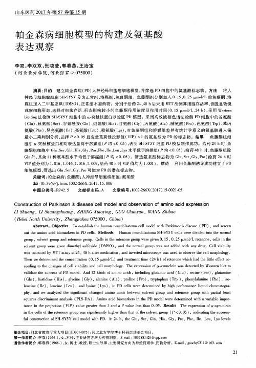

电针对鱼藤酮诱导的帕金森病模型大鼠黑质内ERK1/2及TNF-α的影响王述菊;马骏;王彦春;曾晓玲;龚元勋;梁艳;孙国杰【期刊名称】《中国老年学杂志》【年(卷),期】2015(000)020【摘要】目的:探讨电针对帕金森病(PD)模型大鼠黑质内ERK1/2信号通路及炎症因子肿瘤坏死因子(TNF)-α的作用及机制。
方法雄性健康SD大鼠32只,随机分为正常组、假手术组、模型组、电针组,每组8只。
模型组和电针组经颈背部注射鱼藤酮造模14 d,造模后进行行为学评价,电针组给予电针“风府”和“太冲”两穴治疗,连续治疗14 d;其余各组不做治疗,假手术组给予相同剂量的 DMSO和生理盐水混合液。
采用免疫组化法检测大鼠黑质区磷酸化的ERK1/2、酪氨酸羟化酶( TH)、TNF-α阳性细胞表达情况。
结果各组大鼠神经行为学表现存在差异,与正常组、假手术组相比,模型组大鼠黑质区TH阳性细胞数减少(P<0.05),磷酸化的ERK12/、TNF-α阳性细胞数增加(P<0.05);与模型组相比,电针组大鼠黑质区TH阳性细胞数增加( P<0.05),磷酸化的ERK1/2、TNF-α阳性细胞数减少( P<0.05)。
结论电针可以通过调节 PD模型大鼠体内 MAPK/ERK1/2通路,降低p-ERK1/2在PD模型大鼠黑质区的表达,进而减少TNF-α的表达,对PD病的发生发展起到一定的调节作用。
%Objective To observe the effect of electroacupuncture ( EA ) onERK1/2 signaling pathway and inflammatory cytokines TNF-αin substantia nigra(SN)cells of rotenone-induced rats model with Parkinson'sdisease(PD),and explore the mechanism of EA on PD . Methods A total of32 male SD rats were randomly and averagely divided into normal ,sham-operation,model and EA groups.Model and EA groups were injected intradermally with rotenone ( dissolved in DMSO and saline in certain percentage ) on the nape of the neck for 14 d to establish PD modelrats .Behavioral assessment was conducted after the establishment of PD rats model .EA group was applied to "Fengfu"and"Taichong"points for 20 min once daily for 14 d.Other groups were not given treatments .After the treatments ,the rats were killed for sampling substantia nigra tissue to detect the number of TH ,p-ERK1/2 and TNF-αpositive cells by inmmuno-histochemistry .Results Rats in each group performed differently in neurobehavior .Compared with normal and sham-operation groups ,the expressions of TH positive cells were significantly reduced in model group (P<0.05),the expressions of p-ERK1/2 and TNF-αposi tive cells were significantly increased in model group(P<0.05).Compared with that of model group,the expression of TH positive cells was significantly increased in EA group (P<0.05),the expressions of p-ERK1/2 and TNF-αpositive cells were significantly reduced in EA group (P<0.05).Conclusions EA therapy might reduce the expression of TNF-αin SN of PD rats by regulating the expression of p-ERK1/2(MAPK pathway),which might delay the process of PD.【总页数】4页(P5694-5696,5697)【作者】王述菊;马骏;王彦春;曾晓玲;龚元勋;梁艳;孙国杰【作者单位】湖北中医药大学针灸骨伤学院,湖北武汉 430065;湖北中医药大学针灸骨伤学院,湖北武汉 430065;湖北中医药大学针灸骨伤学院,湖北武汉430065;湖北中医药大学针灸骨伤学院,湖北武汉 430065;湖北中医药大学针灸骨伤学院,湖北武汉 430065;湖北中医药大学针灸骨伤学院,湖北武汉 430065;湖北中医药大学针灸骨伤学院,湖北武汉 430065【正文语种】中文【中图分类】R749【相关文献】1.电针对帕金森病模型大鼠黑质内Bip、CHOP蛋白表达的影响 [J], 马骏;王中明;王述菊;余沛豪;王彬;王琪2.电针对鱼藤酮诱导的帕金森病模型大鼠黑质GDNF mRNA表达的影响 [J], 马骏;梁少荣;王述菊;王彦春;甘水咏;王培俊;许永海3.电针对帕金森病模型大鼠黑质超氧阴离子、超氧化物岐化酶的影响 [J], 程宇核;张少武;朱小虎;张晨;邹旭丹;王俊华;万超4.电针对帕金森病模型大鼠黑质细胞线粒体形态及复合物Ⅰ的影响 [J], 程宇核;万超;王俊华;朱小虎;彭俊良;余新华;邹旭丹5.松果菊苷对鱼藤酮诱导的大鼠帕金森病模型黑质多巴胺能神经元的选择性保护作用(英文) [J], 封新影;朱敏;张琦琪;陈依萍;李文伟因版权原因,仅展示原文概要,查看原文内容请购买。

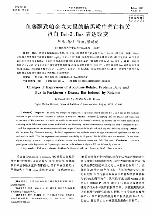
鱼藤酮致帕金森病大鼠黑质中Smac和Bcl-2的表达及意义
鱼藤酮是一种天然产物,被广泛应用于中药领域,具有镇痛、抗炎、抗癌和免疫调节等生物活性。
最近的研究表明,鱼藤酮还可能对帕金森病有一定的保护作用。
其中涉及的机制尚不明确。
研究人员使用鱼藤酮注射大鼠,并研究它们的Smac和
Bcl-2蛋白表达,探索这两种蛋白在鱼藤酮防治帕金森病中的
作用。
帕金森病是一种中枢神经系统退行性疾病,其特征为黑质多巴胺能神经元的死亡和大脑内酪氨酸代谢障碍。
研究发现,鱼藤酮可能促进黑质多巴胺能神经元的生存,因此对于提高帕金森病患者的生活质量具有一定的意义。
Smac是一种细胞凋亡信号转导途径的重要因子,与钙离子平
衡以及线粒体通过西南线形聚集体C的释放有关。
Bcl-2则属
于Bcl-2家族,具有抗凋亡的作用。
研究人员通过Western
blot分析发现,鱼藤酮可以显著提高大鼠黑质中Smac的表达,同时抑制Bcl-2的表达。
这种处理方案可能是通过在线粒体膜
上调节Smac和Bcl-2之间的平衡,从而改善帕金森病的症状。
总的来说,这项研究表明鱼藤酮对帕金森病有一定的保护作用。
Smac和Bcl-2蛋白对于线粒体膜的稳定非常重要,鱼藤酮可
能通过调节它们的表达来由此来调整线粒体膜的功能。
这一发现为进一步研究帕金森病的治疗提供了启示,也为我们深入了解这种疾病的病理机制提供了一个新的途径。

鱼藤酮对慢性帕金森病小鼠中脑黑质致密部α-突触核蛋白表达的影响万金城;张玉平;龙汉春;赵臣勇;周长青;彭国光【摘要】目的观察长期小剂量鱼藤酮暴露对C57BL小鼠行为学和中脑黑质区病理学的改变,并观察鱼藤酮对中脑黑质区α-突触核蛋白(α-syn)表达的影响.方法将雄性C57BL小鼠随机分为鱼藤酮组(n=14)和对照组(n=10).鱼藤酮组给予背部皮下注射鱼藤酮1mg/kg,1次/d,连续注射40d;对照组经相同方式给予相同体积的DMSO和生理盐水混合液.采用自由活动实验和游泳实验评价小鼠的行为学改变.免疫组化法检测小鼠中脑黑质致密部酪氨酸羟化酶(TH)和α-syn的表达,RT-PCR检测α-syn mRNA的表达情况.结果行为学观察发现,鱼藤酮组自由活动实验和游泳实验显示,末次给药后3d鱼藤酮组和对照组小鼠穿梭距离分别为165.4±5.5、257.6±4.6格,下肢站立次数分别为20.3±3.3、34.9±3.5次;游泳实验活动能力评分分别为1.8±0.4、2.8±0.2;两组间差异均有统计学意义(P<0.05).免疫组化检测发现,鱼藤酮组TH阳性细胞计数(18.5±4.0个)比对照组(24.2±2.4个)明显减少(P<0.05).鱼藤酮组中脑黑质部存在α-syn阳性包涵体,且α-syn阳性细胞的积分光密度值(2160.00α±86.20)较对照组(1698.00±78.22)明显增加(P<0.01).结论慢性鱼藤酮中毒能诱导C57BL小鼠发生PD样的行为学和病理学改变,导致中脑黑质TH阳性多巴胺能神经元减少,促使α-syn表达增高并聚集.【期刊名称】《解放军医学杂志》【年(卷),期】2010(035)006【总页数】4页(P667-670)【关键词】帕金森病;黑质;鱼藤酮;突触核蛋白【作者】万金城;张玉平;龙汉春;赵臣勇;周长青;彭国光【作者单位】400016,重庆,重庆医科大学附属第一医院神经内科;400016,重庆,重庆医科大学附属第一医院神经内科;400016,重庆,重庆医科大学附属第一医院神经内科;400016,重庆,重庆医科大学附属第一医院神经内科;400016,重庆,重庆医科大学附属第一医院神经内科;400016,重庆,重庆医科大学附属第一医院神经内科【正文语种】中文【中图分类】R74帕金森病(Parkinson′s disease,PD)是中老年人最常见的神经系统变性疾病之一,临床上以静止性震颤、运动迟缓、肌强直和姿势步态异常为主要表现,其主要病理特征是黑质多巴胺(DA)能神经元变性缺失及残存的DA能神经元内嗜酸性包涵体-路易小体(Lewy's body)形成。

非受体型酪氨酸蛋白激酶介导内皮细胞通透性改变的研究进展张伟金(综述);郭晓华(审校)
【期刊名称】《微循环学杂志》
【年(卷),期】2014(24)1
【摘要】作为一层选择性半透膜,内皮细胞通过细胞间以及细胞与基质的相互作用,构成物质交换的屏障,不仅为血流提供光滑的表面,还参与许多生理调节,包括免疫反应、血管生成及组织体液的稳态。
内皮细胞受损导致屏障功能下降将导致疾病的发生,如蛋白渗出肺间质引起肺水肿和肺部急性损伤等[1]。
而非受体型酪氨酸蛋白激酶(Src)是最早发现的蛋白质酪氨酸磷酸激酶之一,在细胞内信号转导中起着重要的作用。
目前对其介导的信号转导通路的研究是一大热点,特别是其对内皮细胞通透性的影响引起了广泛关注。
本文就 Src 介导内皮细胞通透性改变的研究进展综述如下。
【总页数】4页(P61-64)
【作者】张伟金(综述);郭晓华(审校)
【作者单位】南方医科大学病理生理学教研室,广州 510515;南方医科大学病理生理学教研室,广州 510515
【正文语种】中文
【中图分类】R331.3+5
【相关文献】
1.RhoA介导凝血酶或脂多糖诱导内皮细胞株单层通透性增高
2.血管内皮细胞特异性敲除cdc42基因的杂合子小鼠与非基因敲除小鼠在急性肺损伤肺组织病理改变以及肺微血管通透性变化的比较
3.人脐静脉内皮细胞单层通透性改变的效应因子
4.LOX-1介导oxLDL诱导的人脐静脉内皮细胞高通透性反应
5.核因子-κB及Toll 样受体4介导脂多糖诱导内皮细胞单层通透性增高
因版权原因,仅展示原文概要,查看原文内容请购买。
鱼藤酮PLGA纳米粒的制备及诱导大鼠帕金森病模型的实验研究毛全高【摘要】目的:制备鱼藤酮聚合物纳米分散液,建立与人类帕金森病发病特征相类似的动物模型.方法:采用溶剂扩散法制备鱼藤酮聚乳酸-羟基乙酸共聚物[poly(lactic-co-glycolic acid),PLGA]纳米粒,检测冷冻干燥保护剂(海藻糖、甘露醇、葡萄糖)及其浓度对冻干样品粒径的影响.将PLGA纳米粒制成冻干粉,28只SD大鼠分为4组,分别为空白对照组(6只),鱼藤酮组(6只),高剂量鱼藤酮PLGA纳米粒组(R-PLGA,8只)以及低剂量R-PLGA组(8只).各组均为皮下注射给药,空白对照组给予生理盐水,鱼藤酮组给予普通鱼藤酮溶液,R-PLGA组给予鱼藤酮纳米粒冻干粉.除高剂量组每4天给药1次(剂量为3.5 mg/kg)外,其他组均为每3天给药1次,每次剂量均为2.5 mg/kg.各组整个处理过程均为27 d.采用纳米粒度仪/Zeta电位仪测定纳米粒的粒径与Zeta电位;差示扫描量热仪(DSC)分析纳米粒中鱼藤酮的熔点,HPLC法测定鱼藤酮含量.观察大鼠外观行为表现(评分)、体质量、实格试验中移动潜伏期,用免疫组化法测定大鼠黑质酪氨酸羟化酶(TH)免疫阳性细胞数.结果:制得的R-PLGA 纳米粒平均粒径为306.5 nm,Zeta电位为-2.66 mV,包封率为89.14%,载药量为18.22%.2%海藻糖作为冷冻干燥保护剂保护作用最好,冻干粉平均粒径312.6 nm.与对照组比较,普通鱼藤酮组与高、低剂量R-PLGA组的大鼠行为学评分升高(P<0.01),移动潜伏期从(0.93±0.21)s延长至(7.35±3.27)s,差异有统计学意义(P<0.05).对照组TH免疫阳性细胞数为82.06±10.20,低剂量模型组(17.54±4.20)明显减少(P<0.01).结论:制得可用于制作帕金森病大鼠模型的R-PLGA纳米粒冻干粉.【期刊名称】《江苏大学学报(医学版)》【年(卷),期】2016(026)001【总页数】6页(P82-87)【关键词】帕金森病;鱼藤酮;聚乳酸-羟基乙酸共聚物;免疫组织化学染色法;大鼠【作者】毛全高【作者单位】如皋市人民医院药剂科,江苏如皋226500【正文语种】中文【中图分类】R944;R994.39[Abstract] Objective:To establish a method for preparing animal models with onset characteristics simi-lar to that of human Parkinson′s disease(PD).Methods:The rotenone poly(lactic-co-glycolic acid)(PL-GA)nanoparticles was prepared by using solvent diffusion method.Inspecting the types of freeze-drying pro-tectants and concentration on the influence of sample diameter of freeze-drying powder.The Parkinson′s dis-ease model was induced in adult SD rats by subcutaneous injection of rotenone PLGA,the model of low dose 2.5 mg/kg every 3 days,and high dose 3.5 mg/kg every 4 days.Altogether the drug treatment lasted for 27 days.Diameter and Zeta potential of nanoparticles was determined by nanometer particle size analyzer.With differential scanning calorimetry(DSC)the physical state of rotenone in nanoparticles was analyzed.Rote-none of the contents was determined by HPLC.Grid experiment test were applied to investigateanimal be-havior change and prolonged start latency before and after rotenone was exposed.The injury of dopaminergic neuron was examined by the method of immunohistochemisty.Results:The mean diameter of the particle was 306.5 nm and Zeta potention was-2.66 mV.Its entrapement ratio reached 89.14%with the drugloading of 18.22%.The effectiveness of protection is best when using 2%concentration trehalose.The mean diameter of the particle was 312.6 nm.Compared with the control group,the rats of general rotenone group and model group,behavior score was rised(P<0.01);the start latency was prolonged to(7.35±3.27)s (P<0.05),respectively,compared with in control group(0.93±0.21)s.The number of nigral tyrosine hydroxylase(TH)positive cells decreased significantly from82.06±10.2 in control group to 17.54±4.2 (P<0.01).Conclusion:Good quality freeze-dried preparations can be obtained and the adult rat PD models were established successfully under the condition.[Key words]Parkinson′s disease;rotenone;poly(lactic-co-glycolic acid);immunohistochemical stai-ning method;rat帕金森病是常见的神经退行性疾病,临床表现为静止性震颤、肌僵直、运动迟缓和姿势不稳。
Pael-R基因未参与鱼藤酮诱导帕金森病细胞模型的变化
近来研究发现,环境毒素和遗传缺陷是引发帕金森病的分子机制。
中国中南大学湘雅二医院邹婷所在团队前期研究发现,Parkin的作用底物之一Pael-R参与了Parkin基因突变引发多巴胺神经元凋亡的过程,但其是否也同时参与了环境毒素鱼藤酮引发多巴胺神经元凋亡的过程尚未知。
在本次实验中探讨了在鱼藤酮诱导的帕金森模型中降低Pael-R的表达对模型细胞凋亡的影响,结果表明下调Pael-R表达后对帕金森病模型细胞的活率和凋亡没有影响,证实下调Pael-R基因的表达对鱼藤酮制作的帕金森病细胞模型没有保护作用。
相关结果发表于《中国神经再生研究(英文版)》杂志2014年2月第4期。
Hoechst33258染色显示部分RNA干扰的帕金森病模型细胞细胞出现核固缩和核分裂
Article: " The Pael-R gene does not mediate the changes in rotenone-induced Parkinson’s disease model cells," by Ting Zou, Xiangqi Tang, Zhiling Huang, Niangui Xu, Zhiping Hu (Department of Neurology, Second Xiangya Hospital, Central South University, Changsha, Hunan Province, China)
Zou T, Tang XQ, Huang ZL, Xu NG, Hu ZP. The Pael-R gene does not mediate the changes in rotenone-induced Parkinson’s dise ase model cells. Neural Regen Res. 2014;9(4):402-406.
欲获更多资讯:
Neural Regen Res
The Pael-R gene does not mediate the changes in rotenone-induced PD model cells
Currently, the pathogen esis of Parkinson’s disease is not entirely clear, but it has been generally considered to be the result of interactions among various genetic and environmental factors. Ting Zou and coworkers from Second Xiangya Hospital, Central South University in China previously found that Parkin mutation causes Parkin-associated endothelin receptor-like receptor (Pael-R) protein deposition, and the associated cytotoxicity leads to dopaminergic neuronal apoptosis. These researchers speculated that the Pael-R gene is possibly involved in the action of rotenone on cells. Therefore, they investigated the role of the Pael-R gene in rotenone-induced Parkinson’s disease model cells using RNA interference. IN their recent study, Pael-R expression was decreased after RNA interference compared with the control group (no treatment) and the model group (rotenone treatment), while the rate of apoptosis and survival of dopaminergic cells did not differ
significantly between groups. Experimental findings, published in the Neural Regeneration Research (Vol. 9, No. 4, 2014), indicate that the Pael-R gene has no role in the changes in rotenone-induced Parkinson’s disease model cells.
In the RNA interference group, some cells showed nuclear condensation and division detected by Hoechst 33258 staining.
Article: " The Pael-R gene does not mediate the changes in rotenone-induced Parkinson’s disease model cells," by Ting Zou, Xiangqi Tang, Zhiling Huang, Niangui Xu, Zhiping Hu (Department of Neurology, Second Xiangya Hospital, Central South University, Changsha, Hunan Province, China)
Zou T, Tang XQ, Huang ZL, Xu NG, Hu ZP. The Pael-R gene does not mediate the changes in rotenone-induced Parkinson’s disease model cells. Neural Regen Res. 2014;9(4):402-406.。