抗菌肽6
- 格式:pdf
- 大小:493.96 KB
- 文档页数:12


抗菌肽概况1.1 抗菌肽的基本概况抗菌肽又称抗微生物肽(antimicrobialpeptide)或肽抗生素(peptide antibiotics),在动植物体内分布广泛,是天然免疫防御系统的一部分。
抗菌肽是近年来发现的广泛存在于自然界的一类阳离子抗菌活性肽。
越来越多的证据表明它们在宿主先天性免疫和适应性免疫中有着重要的作用。
目前国内外对抗菌肽的研究开发正不断深入。
抗菌肽(antibacterialpeptides)广义上是指存在于生物体内具有抵抗外界微生物侵害、消除体内突变细胞的一类小分子多肽。
抗菌肽是由生物细胞特定基因编码,经特定外界条件诱导产生的一类多肽。
1972年,瑞典科学家Boman对惜古比天蚕(Hyalophoracecropia)蛹注射蜡状芽孢杆菌(Bacilluscereus),首次发现了抗菌肽cecropin。
此后,对抗菌肽的研究取得了很大的进展,目前在昆虫、植物、哺乳动物、病毒、两栖类以及人类中已发现类似的抗菌活性物质达2000多种。
抗菌肽广泛存在于动物的免疫细胞(如吞噬细胞)、各种脏器的粘膜、皮肤以及植物的花、果、叶中。
有专家推测,抗菌肽在进化意义上最早可能参与了早期真核细胞的噬菌作用,这种作用既是细胞自身防御的需要,而且有可能通过降解微生物为自身生长提供需要的营养,并且最终在生物进化过程中作为防御分子被保留下来。
由于抗菌肽具有小分子的特点,可以快速合成并易于大量存储,与特异性免疫反应相比能更加迅速地对病原菌作出反应,使其成为生物机体先天性非特异性防御系统的重要组分,此外,抗菌肽还具有稳定、水溶性好、抗菌机制独特、对高等动物正常细胞无害等特点,显示了在医学和农业上潜在的研究价值和应用价值。
近年来,有关抗菌肽及其应用逐渐成为动物学、植物学、药理学及生理学等领域的研究热点。
1.2 抗菌肽的理化性质包括细菌、真菌、昆虫、被囊动物(tunicate)、两栖类动物、甲壳类动物、鸟类、鱼类、哺乳动物(包括人类)以及植物在内的所有生物体都可产生抗菌肽。

抗菌肽抗菌肽(antibacterial peptides,ABPs)是生物体产生的一类具有广谱抗菌、抗病毒、杀寄生虫、抗肿瘤等活性的小分子多肽的总称,广泛分布于自然界的各种生物体内。
国际文献中趋向于称为抗微生物肽(antimicrobial peptides,AMPs)或者“肽抗生素”(peptide antibiotics)。
抗菌肽是宿主天然防御的重要参与者,在先天性免疫和获得性免疫中发挥重要作用,因其产生比IgM快100多倍,因此也被称为机体的第一道防线[1]。
单细胞生物体产生的抗菌肽能够增强自身的竞争优势[2,3],而多细胞生物体产生的抗菌肽是先天性免疫系统极其重要的组成成分, 在抵抗外来微生物的入侵方面发挥重要作用[4,5]。
抗菌肽因其具有抗菌谱广、不受传统抗生素耐药突变株影响、不易产生耐药菌株、与传统抗生素有协同作用、中和内毒素等特性,90年代以来已成为国内外免疫学和分子生物学的研究热点,研究内容主要包括抗菌肽的分离与纯化、蛋白质构型与功能的关系、作用机理、应用基因工程克隆与表达抗菌肽基因、改造合成抗菌肽基因以及动植物的转抗菌肽基因工程等[6]。
由于抗菌肽具有天然抗菌性,作用迅速,选择性强,而且很少有耐药性的发生,因此很有可能成为新一代绿色抗菌剂和免疫调节剂。
1抗菌肽的分布、种类和理化特性抗菌肽最早是在1972年由瑞典的科学家Boman等[7]在果蝇中发现,而第一个真正意义上的抗菌肽是由Steiner等[8]从惜古比天蚕蛹中通过注射阴沟肠杆菌及大肠埃希氏菌诱导分离出来,定名为天蚕素(Cecropins)。
目前已知的抗菌肽有1200多种,已从生物界包括细菌、真菌、昆虫、鱼类、两栖类动物、甲壳类动物、鸟类、哺乳动物和植物体中都发现了抗菌肽或类似抗菌肽的小分子肽类,主要由消化道、呼吸道、泌尿生殖道和皮肤的上皮细胞或淋巴细胞等产生[9]。
而每个有机体内都含有多种不同种类的抗菌肽,如在牛体内就发现了包括defensin、BMAP、indolicidin、bactenecin以及其它具有抗菌活性的蛋白片段在内的38种抗菌肽[10]。

抗菌肽与抗生素的区别一、来源不同抗生素(antibiotics)是由微生物(包括细菌、真菌、放线菌属)在生活过程中所产生的具有抗病原体或其它活性的一类次级代谢产物。
如青霉素来源青霉菌产生的次级代谢产物,硫酸粘杆菌素来源于粘细菌产生的次级代谢产物。
而抗菌肽(antimicrobial peptide)又称宿主防御肽,指生物体内经诱导而产生的一类具与先天免疫相关的有抗菌活性小分子多肽物质,最早是在昆虫中发现,随后在各种生物体都有发现,包括细菌、植物、动物甚至人体中都有。
而一些微生物产生的抗菌肽都是初级代谢产物,是生物体直接产生的。
二、产生方式不同抗菌肽是通过基因编码合成,由DNA到RNA到蛋白(多肽),故所有的抗菌肽中的氨基酸为20种常见氨基酸。
即信使RNA分子中每相邻的三个核苷酸编成一组,在蛋白质合成时所代表一种氨基酸。
如AAA为赖氨酸,AUG为蛋氨酸等。
而抗生素大部分为酶促反应生成。
涉及到许多合成酶、水解酶、转移酶等的作用,将一些化学基团组合,生成复杂的结构。
即使一些肽类抗生素,也经过了大量酶促反应的作用,故许多肽类抗生素很难有明确的氨基酸序列,因为存在大量的氨基酸衍生物或者稀有氨基酸。
如杆菌肽由9-12个氨基酸及其衍生物组成,同时存在噻唑环的修饰,很难确定其氨基酸序列。
如硫酸粘杆菌素由10个氨基酸组成,其中6个氨基酸为二氨基丁酸这种稀有氨基酸,也就是非编码氨基酸,还存在一个甲基辛酰这种非氨基酸结构,故也很难确定其氨基酸序列。
这些结构大部分是通过酶促反应生成。
三、对机体的免疫作用不同抗菌肽为宿主防御肽,在宿主先天免疫中有重要的调节作用,能提高机体的非特异免疫,如天蚕素抗菌肽能通过提高CD4+和CD8+的比值,从而提高机体的非特异免疫。
而许多抗生素不能提高免疫,还对免疫有抑制作用或者损伤作用,如氯霉素对骨髓造血功能有很强的破坏作用,从而降低免疫细胞的生成。
因为抗菌肽是机体先天免疫产生的对抗外源病原物,除了对细菌有抑制杀灭作用,对真菌、病毒、原虫都有一定的抑制作用,不同的抗菌肽抗菌谱不同。
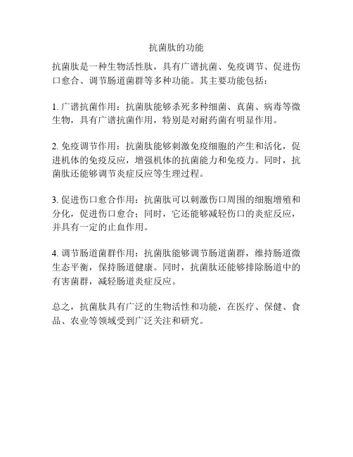
抗菌肽的功能
抗菌肽是一种生物活性肽,具有广谱抗菌、免疫调节、促进伤口愈合、调节肠道菌群等多种功能。
其主要功能包括:
1. 广谱抗菌作用:抗菌肽能够杀死多种细菌、真菌、病毒等微生物,具有广谱抗菌作用,特别是对耐药菌有明显作用。
2. 免疫调节作用:抗菌肽能够刺激免疫细胞的产生和活化,促进机体的免疫反应,增强机体的抗菌能力和免疫力。
同时,抗菌肽还能够调节炎症反应等生理过程。
3. 促进伤口愈合作用:抗菌肽可以刺激伤口周围的细胞增殖和分化,促进伤口愈合;同时,它还能够减轻伤口的炎症反应,并具有一定的止血作用。
4. 调节肠道菌群作用:抗菌肽能够调节肠道菌群,维持肠道微生态平衡,保持肠道健康。
同时,抗菌肽还能够排除肠道中的有害菌群,减轻肠道炎症反应。
总之,抗菌肽具有广泛的生物活性和功能,在医疗、保健、食品、农业等领域受到广泛关注和研究。

抗菌肽—一种治疗幽门螺旋杆菌的新型药物随着抗菌素的广泛使用,针对幽门螺旋杆菌(Hp)治疗出现的耐药率也相应增加,标准三联疗法的Hp根除率已降至80%或更低,以至于更迫切需要一种新型的有效抗菌药物。
抗菌肽作为一种不易产生耐药性的抗微生物药物,在抗幽门螺旋杆菌方面具有較好的前期。
本文主要综述目前抗菌肽针对幽门螺旋杆菌的治疗情况。
标签:抗菌肽;细菌素;幽门螺旋杆菌;治疗方案幽门螺杆菌(Hp)是一种常见的微需氧的革兰阴性菌,是上消化性溃疡和慢性胃炎等疾病的重要致病因素,也与胃癌的发生密切相关。
Hp感染在世界范围普遍存在,约有半数以上的人口胃内存在Hp定居,其中发展中国家Hp感染率相对较高。
1.目前治疗Hp的常用方法杀灭Hp往往需要几种抗生素联合使用,但由于抗生素在感染疾病中的广泛应用,令Hp产生耐药的情况日趋严重,杀灭Hp所需的抗生素治疗也从二联到三联或四联不断增加。
目前关于Hp的治疗方法相对较多,其中一线治疗就包括标准三联疗法(PPI+克拉霉素+阿莫西林/甲硝唑7~14d)、含铋四联疗法(PPI+铋剂+四环素+甲硝唑10~14d)、序贯疗法(二联治疗”PPI+阿莫西林”5d+三联治疗”PPI+克拉霉素+甲硝唑”5d)、伴同疗法(PPI+克拉霉素+阿莫西林+甲硝唑7~10d)、混合疗法(双联疗法7d+伴同疗法7d),其余的还有二线治疗如喹诺酮三联疗法、三线治疗如药敏试验及经验治疗等[1]。
但是对于日趋严重的耐药性问题还是没有得到有效的解决,因此我们需要针对幽门螺杆菌产生耐药机制进行更深入的研究及研发新型的抗菌药物。
2.抗菌肽的定义抗菌肽(antimicrobial peptides,AMPs)是由多种生物细胞特定基因编码经外界条件诱导产生的一类具有广谱抗细菌、真菌、病毒、原虫、抑杀肿瘤细胞等活性作用的多肽。
AMPs种类繁多,可由生物体内直接产生,如细菌、植物、昆虫、两栖动物、哺乳动物等。
它具有强碱性、热稳定性等理化性质,并且最重要的是因其灭菌机制主要是作用于细胞膜而不易产生耐药性。
一、摘要抗菌肽是一类具有广谱抗菌活性的小分子肽,具有高效、低毒、不易产生耐药性等优点。
本实验旨在通过高效液相色谱(HPLC)技术分离纯化抗菌肽,并通过抑菌圈法测定其抗菌活性。
实验结果表明,成功分离纯化了抗菌肽,并测定了其最小抑菌浓度(MIC)。
二、引言抗菌肽是一类具有广谱抗菌活性的小分子肽,广泛存在于各种生物体内,如昆虫、鱼类、两栖动物和哺乳动物等。
抗菌肽具有高效、低毒、不易产生耐药性等优点,因此在抗菌药物的研究和开发中具有广阔的应用前景。
本实验通过HPLC技术分离纯化抗菌肽,并对其抗菌活性进行测定。
三、实验材料与方法1. 实验材料抗菌肽粗提物:购自某生物科技公司;金黄色葡萄球菌、大肠杆菌、白色念珠菌:购自某微生物研究所;甲醇、乙腈:色谱纯;其他试剂:分析纯。
2. 实验仪器高效液相色谱仪(HPLC):某品牌;细菌培养箱:某品牌;无菌操作台:某品牌;电子天平:某品牌;抑菌圈测定仪:某品牌。
3. 实验方法(1)抗菌肽的分离纯化将抗菌肽粗提物溶解于适量甲醇中,采用反相高效液相色谱法进行分离纯化。
色谱柱:C18柱(4.6×250mm,5μm);流动相:乙腈-水(梯度洗脱);流速:1.0mL/min;检测波长:215nm。
(2)抗菌活性的测定采用抑菌圈法测定抗菌肽的抗菌活性。
将金黄色葡萄球菌、大肠杆菌、白色念珠菌分别接种于琼脂平板上,待菌落长成后,将抗菌肽溶液滴加于平板上,37℃恒温培养24小时,观察抑菌圈的大小。
四、实验结果与分析1. 抗菌肽的分离纯化通过HPLC技术分离纯化抗菌肽,得到单一峰,证明抗菌肽已成功分离纯化。
2. 抗菌活性的测定(1)金黄色葡萄球菌抗菌肽对金黄色葡萄球菌的MIC为10-7mol/L,抑菌圈直径为16mm。
(2)大肠杆菌抗菌肽对大肠杆菌的MIC为10-6mol/L,抑菌圈直径为15mm。
(3)白色念珠菌抗菌肽对白色念珠菌的MIC为10-6mol/L,抑菌圈直径为14mm。
Dissociation of Antimicrobial and Hemolytic Activities in Cyclic Peptide Diastereomers by Systematic Alterations in Amphipathicity*(Received for publication,December 1,1998,and in revised form,February 20,1999)Leslie H.Kondejewski‡,Masood Jelokhani-Niaraki‡,Susan W.Farmer§,Bruce Lix‡,Cyril M.Kay‡¶,Brian D.Sykes‡¶,Robert E.W.Hancock§,and Robert S.Hodges‡¶ʈFrom the ‡Protein Engineering Network of Centres of Excellence and ¶Department of Biochemistry,University of Alberta,Edmonton,Alberta T6G 2S2,Canada and the §Department of Microbiology and the Canadian Bacterial Diseases Network,University of British Columbia,Vancouver,British Columbia V6T 1Z3,CanadaWe have investigated the role of amphipathicity in a homologous series of head-to-tail cyclic antimicrobial peptides in efforts to delineate features resulting in high antimicrobial activity coupled with low hemolytic activ-ity (i.e.a high therapeutic index).The peptide GS14,cyclo(VKLKV d -YPLKVKL d -YP),designed on the basis of gramicidin S (GS),exists in a preformed highly am-phipathic -sheet conformation and was used as the base compound for this study.Fourteen diastereomers of GS14were synthesized;each contained a different single enantiomeric substitution within the framework of GS14.The -sheet structure of all GS14diastereomers was disrupted as determined by CD and NMR spectros-copy under aqueous conditions;however,all diaste-reomers exhibited differential structure inducibility in hydrophobic environments.Because the diastereomers all have the same composition,sequence,and intrinsic hydrophobicity,the amphipathicity of the diaste-reomers could be ranked based upon retention time from reversed-phase high performance liquid chroma-tography.There was a clear correlation showing that high amphipathicity resulted in high hemolytic activity and low antimicrobial activity in the diastereomers.The latter may be the result of increased affinity of highly amphipathic peptides to outer membrane components of Gram-negative microorganisms.The diastereomers possessing the most favorable therapeutic indices pos-sessed some of the lowest amphipathicities,although there was a threshold value below which antimicrobial activity decreased.The best diastereomer exhibited 130-fold less hemolytic activity compared with GS14,as well as greatly increased antimicrobial activities,resulting in improvement in therapeutic indices of between 1,000-and 10,000-fold for a number of microorganisms.The therapeutic indices of this peptide were between 16-and 32-fold greater than GS for Gram-negative microorgan-isms and represents a significant improvement in spec-ificity over GS.Our findings show that a highly am-phipathic nature is not desirable in the design of constrained cyclic antimicrobial peptides and that an optimum amphipathicity can be defined by systematic enantiomeric substitutions.The ever-increasing development of bacterial resistance to traditional antibiotics has reached alarming levels,making it essential that new antibiotics be developed (1).Ideally,these new antibiotics should possess both novel modes of action as well as different cellular targets compared with existing anti-biotics to decrease the likelihood of development of cross-resis-tance.Antimicrobial peptides may represent such a new class of antibiotics,and their design and structure-activity relation-ships have become an area of active research in recent years (see Refs.2and 3and references therein).Although their exact mode of action has not been established,it has been proposed that the cytoplasmic membrane is the main target of these peptides,where their accumulation results in increased perme-ability and loss of barrier function.The development of resist-ance to these membrane active peptides is not expected because this would require substantial changes in the lipid composition of cell membranes.Indeed,the induction of resistance to such peptides has not been seen for a number of the antimicrobial peptides (2).Because both their mode of action and cellular targets are different from those of the traditional antibiotics,antimicrobial peptides represent a truly new class of antibiotics and are therefore attractive candidates for development as such.Two major classes of the cationic antimicrobial peptides are the ␣-helical and the -sheet peptides.The ␣-helical class (for example cecropins,magainins,and melittin)are linear pep-tides that exist as disordered structures in aqueous media and become helical upon interaction with hydrophobic solvents or phospholipid vesicles.Unlike the ␣-helical peptides,-sheet peptides are cyclic peptides constrained in this conformation either by disulfide bonds (tachyplesins,protegrins,and polyphemeusins)or by cyclization of the backbone (gramicidin S and tyrocidines).Although the -sheet conformations of these peptides may be further stabilized in the presence of a hydro-phobic or lipid environment,they exist largely in a “preformed”-sheet conformation in aqueous environments due to their structural constraints.From numerous structure-activity stud-ies on both natural and synthetic antimicrobial peptides,a number of factors believed to be important for antimicrobial activitiy have been identified.These include the presence of both hydrophobic and basic residues,as well as a defined sec-ondary structure (␣-helical or -sheet),either preformed or inducible,and an amphipathic nature that segregates basic and hydrophobic residues to opposite sides of the molecule in lipid or lipid-mimicking environments (2–5).Many of the antimicrobial peptides show poor selectivity for bacteria in that they are also toxic to higher eukaryotic cells.To make the antimicrobial peptides useful as therapeutics there-fore requires delineation of the features responsible for antimi-*This study was supported by the Canadian Government through grants to the Canadian Bacterial Diseases Network (to R.E.W.H.)and the Protein Engineering Network of Centers of Excellence (to R.S.H.).The costs of publication of this article were defrayed in part by the payment of page charges.This article must therefore be hereby marked “advertisement ”in accordance with 18U.S.C.Section 1734solely to indicate this fact.ʈTo whom correspondence should be addressed.Tel.:780-492-2758;Fax:780-492-1473;E-mail:robert.hodges@ualberta.ca.T HE J OURNAL OF B IOLOGICAL C HEMISTRYVol.274,No.19,Issue of May 7,pp.13181–13192,1999©1999by The American Society for Biochemistry and Molecular Biology,Inc.Printed in U.S.A.This paper is available on line at 13181by guest, on June 29, 2011 Downloaded fromcrobial activity from those responsible for toxicity to higher eukaryotic cells (typically measured as hemolytic activity).The obvious goal is to design peptides that have high antimicrobial activity coupled with low toxicity,i.e.a high specificity or high therapeutic index.Recent studies on a number of linear pep-tides have attempted to delineate features responsible for these activities and found that high amphipathicity (6,7),high hy-drophobicity (6,8),as well as high helicity (9,10)were corre-lated with increased hemolytic activity.Antimicrobial activity on the other hand was found to be less dependent on peptide helicity (9,10).Furthermore,decreases in either hydrophobic-ity or amphipathicity were either found to increase (7,8)or to decrease antimicrobial activity (6,11),depending on the pep-tides studied.In both cases,however,specificity for bacteria over erythrocytes could be increased either by increasing activ-ity coupled with decreased hemolysis (7,8)or because the hemolytic activity was decreased more readily than antimicro-bial activity (6,11).Relatively few studies have investigated structural features responsible for the hemolytic and antimicrobial properties of the cyclic -sheet peptides.The fact that these peptides are constrained and therefore have less conformational freedom compared with the linear ␣-helical peptides suggests that the properties of these peptides may be different.We have utilized the 10residue head-to-tail cyclic peptide gramicidin S (GS)1(12)as the basis of our design for novel antimicrobial agents.GS has the sequence cyclo(Val-Orn-Leu-d -Phe-Pro)2and exists in an antiparallel -sheet conformation with the strands fixed in place by two type II Ј-turns (5,13,14).The -sheet struc-ture gives the molecule a preformed amphipathic nature with four hydrophobic residues (Val and Leu)making up one face of the molecule and two basic Orn residues making up the other face.This amphipathicity,along with high hydrophobicity,has long been thought to be important for the antimicrobial prop-erties of GS-like peptides (5,15–18).A previous study with cyclic -sheet antimicrobial peptides based on GS indicated that it is possible to dissociate antimicrobial and hemolytic activities through gross manipulation of -sheet structure and amphipathicity (19).In this manuscript,we report on the effect of small incremental changes in amphipathicity (directed hy-drophobicity and positive charge)on the antimicrobial and hemolytic properties of cyclic 14residue peptides.We have utilized the cyclic tetradecapeptide,GS14,cyclo(VKLKV d -YPLKVKL d -YP),as the model peptide in this study.This pep-tide has been shown to exist in a highly amphipathic -sheet structure,with six hydrophobic residues on one face of the molecule and four basic residues on the opposite face (19,20).Unlike GS,which exhibits broad spectrum antimicrobial activ-ity as well as hemolytic activity,GS14was found to possess limited antimicrobial activity and very high hemolytic activity (19).Because amphipathicity is intimately linked to -sheet structure in GS14,any change in -sheet structure was pre-dicted to directly affect the amphipathicity of the molecule.We have created a series of GS14diastereomers in which each contains a different single residue enantiomeric substitution within the framework of GS14resulting in a series of cyclic peptides possessing gradated disruption of -sheet structure and amphipathicity.The present method of enantiomeric sub-stitutions within a constrained backbone system has the ad-vantage that all peptides retain the same sequence,intrinsic hydrophobicity,and basicity but differ only in structure.Weshow that the amphipathicity of these peptides has a large effect on their biological properties and that by defining the optimum amphipathicity in the framework of these cyclic pep-tides,the balance between hemolytic and antimicrobial activi-ties can be optimized.MATERIALS AND METHODSPeptide Synthesis,Purification,and Cyclization—All peptides were synthesized by solid phase peptide synthesis using standard t -butyloxy-carbonyl chemistry,cleaved from the resin,and purified by preparative RP-HPLC as reported previously (19).For all peptides proline was the C terminus because racemization can occur during the cyclization re-action with other residues.2Purity of linear peptides was verified by analytical RP-HPLC,and correct peptide masses were verified by elec-trospray mass spectrometry on a Fisons VG Quattro triple quadrupole mass spectrometer (Manchester,UK).Pure linear side chain protected peptides were cyclized,deprotected,and purified by preparative RP-HPLC as described (19).Purified cyclic peptides were homogeneous by analytical RP-HPLC and gave correct primary ion molecular weights by mass spectrometry as well as appropriate amino acid analysis ratios.Peptide concentrations of stock solutions were determined by amino acid analysis for subsequent use in biological assays.Analytical Reversed-phase Analysis of Diastereomers—Peptides were analyzed by RP-HPLC on a Zorbax SB-C8column (150ϫ2.1-mm inner diameter,5m particle size,300Åpore size;Rockland Technologies,Wilmington,DE)using a Hewlett Packard 1100chromatograph at 70°C with a linear AB gradient of 1%B/min (where solvent A was 0.5%aqueous trifluoroacetic acid and solvent B was 0.5%trifluoroacetic acid in acetonitrile)at a flow rate of 0.25ml/min.Circular Dichroism Measurements—CD spectra were recorded on a Jasco J-500C spectropolarimeter (Jasco,Easton,MD)as described (19).Spectra were recorded in either 5m M sodium acetate buffer,pH 5.5,or 5m M sodium acetate buffer,pH 5.5,containing 50%trifluoroethanol (TFE).NMR Spectroscopy—NMR spectroscopy was carried out under aque-ous conditions on a Varian Unity 300MHz spectrometer equipped with a 5-mm inverse detection probe.Each peptide was dissolved in 500l of 90%H 2O/10%D 2O (or 100%D 2O)giving a sample conentration of 1–2m M ,and the pH was adjusted to 5.5.1H double quantum filtered two-dimensional correlated spectroscopy,rotating frame Overhauser effect spectroscopy,and total correlation spectroscopy spectra were collected at 25°C and processed as described (21).The chemical shift index was calculated for selected peptides as described by Wishart et al.(22).Molecular Modelling—A model of GS14was constructed using In-sight II (Biosym Technologies Inc.,San Diego,CA)on a Silicon Graphics workstation starting with the linear peptide LKV d-YPLKVKL d-YPVK.The model was constructed by specifying standard antiparallel -sheet ,values of Ϫ139°and ϩ135°,respectively (23).Two type II Ј-turns were incorporated into the model designating D -Tyr and Pro residues as residues i ϩ1and i ϩ2of the turns.Dihedral angles (,)used for the turns were 60°,Ϫ120°and Ϫ80°,0°for i ϩ1and i ϩ2residues,respectively (23).The formation of the turns brought the N and C termini into close proximity,and an amide bond was formed between the termini.The model was subjected to energy minimization using the consistent valence force field (24)with a distance-dependent dielectric constant of 4at pH 7with no cross-terms.The potential energy of the model was minimized in two steps,first using the steepest descent algorithm for 100iterations followed by 100iterations using the VA09A algorithm.Calculation of Peptide Hydrophobic Moment and Hydrophobicity—The mean residue hydrophobic moment ()of GS14and a comparable linear ␣-helical peptide (7)was calculated using the consensus hydro-phobicity scale of Eisenberg et al.(25)to facilitate comparison with other antimicrobial peptides.Due to the cyclic nature of GS14,calcula-tion of was carried out for each half of the molecule,and the value reported represented the average of these values.This procedure was necessary to account for the two -turns in the molecule and appears to be a reasonable assumption based on the molecular model.The two segments used for the calculations were:VKLKVYP and LKVKLYP;a value of ␦ϭ180°was used for the angle at which successive side chains emerge from the backbone of the -sheet.Measurement of Antibacterial,Antifungal,and Hemolytic Activity—Minimal inhibitory concentrations (MICs)were measured using a1The abbreviations used are:GS,gramicidin S;LPS,lipopolysaccha-ride;MIC,minimal inhibitory concentration;NPN,1-N -phenylnaphth-ylamine;RP-HPLC,reversed-phase high performance liquid chro-matography;TFE,trifluoroethanol;dansyl,5-dimethylamino-naphthalene-1-sulfonyl.2L.H.Kondejewski and R.S.Hodges,unpublished results.Peptide Diastereomer Amphipathicity-Activity Relationships13182 by guest, on June 29, 2011 Downloaded fromstandard microtitre dilution method in Luria Broth no salt medium utilizing the same bacterial strains as reported(19).MICs were deter-mined as the lowest peptide concentration that inhibited growth after 24h at37°C.The hemolytic activity of peptides was measured in saline utilizing human erythrocytes as described(16,19).The concentration of peptide required for complete hemolysis was determined visually after 24h at37°C.Measurement of Peptide Outer Membrane Interactions—Dansyl-poly-myxin B displacement from Pseudomonas aeruginosa lipopolysaccha-ride(LPS)was measured to determine the binding affinity of the peptides to LPS(19).Permeabilization of bacterial outer membranes was measured by monitoring peptide mediated1-N-phenylnaph-thalamine(NPN)fluorescence increases utilizing Escherichia coli UB1005cells(16,19).RESULTSDesign of Cyclic PeptidesIn this study we systematically replaced each residue in the sequence of the highly amphipathic-sheet peptide,GS14, with its enantiomer(Table I).The rationale for these substitu-tions was based on the following observations:(a)In a previous study(19)we found that with certain cyclic peptides the lack of -sheet structure and amphipathicity under aqueous condi-tions was the key to achieving a high specificity for microbes over human erythrocytes(i.e.a high therapeutic index).In that study,the disruption of-sheet structure and amphipathicity was accomplished by utilizing peptides that could not form -sheet structures due to the number of residues in the ring (19,20).(b)During the initial synthesis and cyclization of GS14 we found that racemization of the C terminus could occur during the cyclization reaction when the C terminus was a non-Pro residue and that this racemization led to loss of -sheet structure.2Together,these observations led us to hy-pothesize that GS14could be transformed into a peptide pos-sessing a high therapeutic index by disrupting the-sheet character,and hence the amphipathicity,of the molecule.To accomplish this,we synthesized all14possible diastereomers of GS14;each contains a different single amino acid enantio-meric substitution.We also synthesized two diastereomers with two and four enantiomeric substitutions,respectively(Ta-ble I).The peptides were characterized with respect to their structural and biological properties.Structure of GS14DiastereomersCircular Dichroism Spectroscopy—The CD spectra of GS14 and representative single residue substitution diastereomers under aqueous conditions are shown in Fig.1A.It has been shown by NMR spectroscopy that both GS and GS14possess a similar-sheet conformation and that they also exhibit similar CD spectra with large negative ellipticities at206and223nm (19,20),reminiscent of a combination of-sheet structure and type II-turns(26).All of the single replacement diaste-reomers as well as those containing two or four enantiomeric substitutions exhibited CD spectra more typical of disordered structures,indicating that enantiomeric substitutions within the framework of GS14resulted in the disruption of-sheet structure.The CD spectra of GS14and representative diaste-reomers recorded in the presence of the lipid-mimicking sol-vent,TFE,are shown in Fig.1B.TFE is generally thought of as a helix-inducing solvent(see Ref.27and references therein); however,recent studies have shown that aqueous solutions containing TFE can also stabilize-hairpin and-turn struc-tures(28,29).In50%TFE the molar ellipticities around206 and223nm were significantly enhanced for GS14as well as all the diastereomers,suggesting an enhancement or stabilization of-sheet structure in the peptides.However,because the shape of the observed CD spectra of these cyclic peptides is a combination of contributions by-turns,aromatic residues, and-sheet structure,it is difficult to assign particular second-ary structural elements to these spectra(26).One striking difference in the CD spectra of all diastereomers in the pres-ence of TFE was displayed by GS14K4(and GS14K11;not shown),with a large blue-shifted negative ellipticity in the vicinity of200nm when compared with all other diastereomers (Fig.1B).This large increase in molar ellipticity at205nm for GS14K4and GS14K11is also evident from Fig.2,where molar ellipticity changes in the different environments at both205 and220nm are summarized graphically.The majority of pep-tides exhibited greater negative molar ellipticities at both these wavelengths in the presence of TFE.It is noteworthy,however, that although all diastereomers exhibited essentially identical spectra under aqueous conditions,all displayed considerablyT ABLE ISequences and biological and physical properties of GS14diastereomersPeptide name Linear sequence a Retentiontime b LPS bindingaffinity cHemolyticactivity dminMg/mlGS VOLFPVOLFP54.929512.5GS14VKLKVYPLKVKLYP50.63 1.5GS14V10VKLKVYPLKVKLYP45.418 6.2GS14L3VKLKVYPLKVKLYP44.23512.5GS14P14VKLKVYPLKVKLYP42.920 6.2GS14P7VKLKVYPLKVKLYP42.61812.5GS14V1VKLKVYPLKVKLYP41.34140GS14L12VKLKVYPLKVKLYP41.33225GS14V5VKLKVYPLKVKLYP41.133150GS14Y13VKLKVYPLKVKLYP40.62412.5GS14L8VKLKVYPLKVKLYP40.36250GS14Y6VKLKVYPLKVKLYP40.22025GS14K2VKLKVYPLKVKLYP39.85050GS14K9VKLKVYPLKVKLYP38.860100GS14K4VKLKVYPLKVKLYP37.893200GS14K11VKLKVYPLKVKLYP37.150150GS14K2K4VKLKVYPLKVKLYP33.1120Ͼ200GS14K2K4K9K11VKLKVYPLKVKLYP28.5200Ͼ200a Linear sequences of cyclic peptides.Underlined residues represent D-amino acids.O is ornithine.b Retention time on RP-HPLC as determined from Fig.6.c Peptide concentration to displace50%of dansyl-polymyxin B from LPS as described under“Materials and Methods.”d Peptide concentration required for100%lysis of human erythrocytes.Peptide Diastereomer Amphipathicity-Activity Relationships13183by guest, on June 29, Downloaded fromdifferent CD spectra in TFE,indicating the induction of differ-ent backbone conformations in a hydrophobic environment,depending on the position of the enantiomeric substitution.The diastereomers also displayed similar structural changes in the presence of either SDS micelles or phospholipid vesicles (data not shown).NMR Spectroscopy—The extent of disruption of the -sheet structure in the GS14diastereomers was confirmed/demon-strated by NMR spectroscopy for selected analogs.3The regions of the 1H NMR spectrum containing the amide and aromatic proton resonances are shown in Fig.3for GS14(top spectrum )and one diastereomer,GS14K2(bottom spectrum ).The GS14spectrum showed 12well resolved amide resonances (each is a doublet corresponding to the amide resonances of Val 1to Tyr 6and Leu 8to Tyr 13),and a single pair of doublets corresponding to the meta (2,6)and ortho (3,5)protons of both Tyr 6and Tyr 13.The diverse chemical shifts for the amide resonances indicate a well ordered structure,and the downfield chemical shifts indi-cate significant -sheet structure.The single pair of doublets for Tyr 6and Tyr 13indicates that both are in equivalent envi-ronments.By comparison,the amide resonances observed for GS14K2were much less diverse and centered more around the random coil value,indicating disruption of -sheet structure.In addition,two pairs of doublets were observed for Tyr 6and Tyr 13,indicating that they are not equivalent in the structure.The H ␣chemical shift deviations from random coil values for GS14and three diastereomers are shown in Fig.4.Positive chemical shift deviations greater than 0.1ppm indicate down-field shifted resonances relative to random coil values,and a cluster of three or more continuous positive chemical shifts is indicative of -sheet structure (22).It is apparent that the H ␣chemical shifts for GS14(Fig.4A )were all -sheet-like within the -strands of the molecule (see molecular model below)and non--sheet chemical shifts in the turns defined by the D -Tyr-Pro sequence.It is clear from the chemical shift analysis of representative diastereomers (Fig.4,B ,C ,and D )that the strands contained less -sheet structure and the turns became more disrupted compared with GS14.It is also noteworthy that the three diastereomers differed in both the extent and location of disruption,indicating that they were structurally different under aqueous conditions.Molecular Model of GS14—Both CD and NMR spectroscopy indicated that GS14possesses a -sheet structure similar to GS.A model of GS14was constructed to contain the essential features as present in GS,namely,an antiparallel -sheet structure with two type II Ј-turns defined by the D -Tyr-Pro sequence.As shown in Fig.5,the incorporation of both the cyclic constraint (due to backbone cyclization of the peptide)as well as secondary structural constraints (-sheet and turns)results in a highly amphipathic molecule where Val and Leu residues make up the hydrophobic face and Lys residues make up the basic face of the molecule (Fig.5,lower panel ).There are3The complete 1H NMR assignments for these analogs are available athttp://www.pence.ualberta.ca/ftp/kondejewski.html.F IG .1.CD spectra of GS14and representative GS14diaste-reomers.A ,spectra were recorded in 5m M sodium acetate buffer,pH 5.5,at 20°C at a peptide concentration of approximately 0.3m M .Sam-ples were GS14(q ),GS14V1(E ),GS14K2(Ⅺ),GS14L3(f ),GS14K4(‚),and GS14V5(Œ).B ,spectra were recorded in 5m M sodium acetate buffer,pH 5.5,containing 50%TFE,at 20°C.Samples were as in A.F IG .2.Environment-dependent CD spectral changes in GS14diastereomers.Molar ellipticities at 205(A )and 220nm (B )are shown for the diastereomers under aqueous conditions (black bars )and in the presence of 50%TFE (gray bars ).Peptide Diastereomer Amphipathicity-Activity Relationships13184 by guest, on June 29, 2011 Downloaded frompotentially six hydrogen bonds that could be formed to stabilize the -sheet structure of GS14located between all Val and Leu residues.The non-H-bonded sites are all occupied by Lys resi-dues (Fig.5,upper panel ).Reversed-phase HPLC Analysis—Retention time on re-versed-phase HPLC can be used as a measure of peptide hy-drophobicity (30,31).It is well known,however,that the for-mation of a hydrophobic binding domain due to peptide secondary structure can affect peptide interactions with re-versed-phase matrices,this effect having been observed both with ␣-helical peptides (32,33)and -sheet peptides (21,34).GS14and the GS14diastereomers have exactly the same com-position and sequences,and therefore all have the same intrin-sic hydrophobicity.Any differences in retention times wouldbeF IG .3.One-dimensional 1H NMR spectra of the amide regions for GS14and GS14K2.Spectra were recorded in H 2O,pH 5.5,at 20°C.F IG .4.Chemical shift analysis of GS14and representative GS14diastereomers.The chemical shift deviations of ␣-proton resonances relative to random coil values are shown for GS14(A ),GS14V1(B ),GS14K2(C ),and GS14V10(D ).Three or more continuous positive chemical shift deviations greater than 0.1ppm (dashed line )are indicative of -sheet structure.Peptide Diastereomer Amphipathicity-Activity Relationships 13185by guest, on June 29, 2011 Downloaded fromdue to differences in their effective hydrophobicity,which is related to the ability of the peptide to form a hydrophobic preferred binding domain for interaction with the hydrophobic surface of the HPLC matrix.Conversely,the positioning of positive charges in the vicinity of the hydrophobic binding domain would also serve to destabilize the hydrophobic inter-actions and substantially decrease the overall hydrophobicity of the preferred hydrophobic binding domain (7,35,36).Be-cause retention time of the GS14diastereomers on RP-HPLC is a measure of the ability to form a large hydrophobic bindingdomain and at the same time segregate hydrophobic and hy-drophilic side chains to opposite sides of the molecule,it is therefore also a measure of amphipathicity in these peptides.The RP-HPLC separation of a mixture of GS14and the 14diastereomers is shown in Fig.6.There was a wide range of retention times observed for the analogs,and all had a lower retention time than GS14.As seen from the model of GS14(Fig.5)the parent molecule exists in a highly amphipathic -sheet conformation with a large hydrophobic preferred binding do-main formed by six hydrophobic residues on one face of the molecule,as well as the basic residues sequestered on the opposite face of the molecule.All diastereomers had lower retention times compared with GS14due to decreased am-phipathicity caused by either a decreased size of the hydropho-bic preferred binding domain or the relative positioning of hydrophobic and basic residues.Measurement of retention times of the diastereomers on RP-HPLC is therefore a conven-ient means for ranking and comparing peptide amphipathicity in this homologous series of peptides.The extent of disruption of amphipathicity was dependent on the position of the enan-tiomeric substitution,with diasteromers containing substitu-tions in the non-H-bonded sites (Lys in this case)being much more disruptive than those with substitutions in the H-bonded sites (Val and Leu).Enantiomeric substitutions in the center of the -sheet (Leu 3and Val 10)as well as Pro residues in the turns were least disruptive.Membrane Interactions of GS14DiastereomersInteraction with Bacterial Lipopolysaccharide—Cationic peptides interact with Gram-negative bacteria by initially binding to LPS prior to self-promoted uptake across the outer membrane (37,38).We investigated the interaction between GS14diastereomers and the bacterial outer membranes by monitoring the displacement of LPS-bound dansyl-polymyxin B by the peptides (19,39).The LPS binding affinity of GS14was extremely strong,approaching that of polymyxin B itself (19),whereas all the diastereomers had lower affinity (Table I).There is a good correlation between binding affinity and reten-tion time on RP-HPLC (Fig.7),with those peptides having a longer retention time exhibiting higher affinity for LPS.This indicates that the peptides that are more amphipathic (a greater hydrophobicity of the preferred binding domain and therefore also a larger oriented basic face)also bind tighter to LPS.The binding affinity of GS was lower than all the diaste-reomers,although GS has the greatest retention time onRP-F IG .5.Molecular model of GS14.The model was constructed to contain -sheet dihedral angles for residues in the strands and two type II Ј-turns defined by the Xaa-D -Tyr-Pro-Xaa sequence.An amide bond was formed between the N and C termini,and energy minimization of the structure carried out as described under “Materials and Methods.”Upper panel ,a top view of the backbone of GS14indicating the positions of potential interstrand hydrogen bonds.Lower panel ,a side view of GS14indicating the relative positioning of hydrophobic and basicresidues.F IG .6.RP-HPLC separation of GS14and single residue substitution diastereomers.A mixture of GS14and the 14diastereomers was separated by RP-HPLC as described under “Materials and Methods.”The position of the enantiomeric substitution is shown for each peak on the chromatogram.Peptide Diastereomer Amphipathicity-Activity Relationships13186 by guest, on June 29, 2011 Downloaded from。