2016超声年会造影
- 格式:pptx
- 大小:16.70 MB
- 文档页数:60
超声造影简介
超声造影在临床上应用非常普遍,最早的心脏声学造影技术自上世纪六十年代末,应用于临床以来发展很快,在诊断先天性心脏病方面的价值已得到了充分的肯定,其方法是将含有微泡的造影剂直接经外周静脉注入,抵达冠脉循环,来评价心肌微循环的。
完整性的心肌灌注声学造影也逐渐的进入临床,由单纯定性诊断进入定量诊断阶段,声学造影在其它脏器,肝肾、胰腺、甲状腺、乳腺等的临床应用中已经证实在肿瘤的检出和定性诊断中有着重要的意义。
该技术对血管狭窄的诊断也发挥了较好的作用,对腹部外伤的损伤程度范围以及评价,评定的准确率非常高。
除此之外,超声造影技术还在、前列腺、输卵管、卵巢、胆囊、脾脏等组织器官病变的诊断与鉴别诊断中具有非常重要的临床意义。
超声造影科普
超声造影是将一种对人体无害,在进入人体后变成微小气泡的造影剂,就像超声医师派到人体内部的一名侦查员,随时将人体的变化报告给超声医师。
可以早期确诊轻微的先天性心脏病,可以早期鉴别肝,肾、胰腺、甲状腺、乳腺肿瘤的良恶性程度。
是一种无损伤,对人体无害的检查方法。
缺点是造影剂都是国外进口,比较昂贵,所以检查费用比较贵。
四维彩色超声诊断仪是目前世界上最先进的彩色超声设备。
:能够显示您未出生的宝宝在妈妈肚子了的实活动图像。
同其它超声诊断相比,四维彩超能够多方位、多角度地
观察宫内胎儿的生长发育情况,且可以为早期诊断胎儿先天性体表畸形和先天性心脏疾病提供准确的科学依据。
过去的
发育不良等,以便尽早的进行治疗。
如果宝宝的位置合适,还可以留一张宝宝在妈妈肚子里的肖像或者活动录像。
谈一谈什么是超声造影?郭健伟发布时间:2023-06-14T09:48:23.582Z 来源:《健康世界》2023年7期作者:郭健伟[导读] 说起超声,大家应该都不陌生,B超、彩超是最常规的超声检查,也是现阶段临床最常见的检查方式,超声就像是透视眼一般能够直接看透人体内部,并借助先进的超声设备以及前沿超声成像技术,再加上超声医师丰富的临床经验为临床诊断工作提供有力支持。
都江堰市人民医院四川成都 611830说起超声,大家应该都不陌生,B超、彩超是最常规的超声检查,也是现阶段临床最常见的检查方式,超声就像是透视眼一般能够直接看透人体内部,并借助先进的超声设备以及前沿超声成像技术,再加上超声医师丰富的临床经验为临床诊断工作提供有力支持。
近年来,随着医学技术不断发展,诸多新型技术被临床广泛应用,为疾病诊疗开辟了新的领域,超声造影就是近几年飞速发展且获得显著成效的超声新技术,当你被脏器肿块确诊困难而焦虑时,当你为观察输卵管及血管是否通畅而犯愁时,当你害怕放射线有伤害时,超声造影都能够提供帮助,但作为新型技术,人们对它并不了解,因此,本篇文章就来谈一谈什么是超声造影。
一、超声造影的基本定义超声造影是继B超、彩超之后的超声医学第三次革命,是目前最为先进的超声成像技术,现已被誉为无创性微循环血管造影,医学又称其为声学造影,主要是在常规超声检查基础上,经由外周静脉注射超声造影剂,实时观察组织微血管灌注信息,以提高病变检查率,鉴别病变良恶性质,该检查方式应用范围广泛,一般常规超声可以显示的部位均可进行超声检查检查,包括消化系统、泌尿系统、浅表器官、子宫附件、输卵管等,且能够对介入、脏器肿块穿刺活检以及肿瘤治疗后的疗效进行观察,还能够评价心脏、大血管以及动脉粥样斑块的稳定性。
超声造影包括散射、背向散射、机械指数、基频与基波五种基本概念,其中散射指的是超声波遇到长度小于其波长的小界面时产生的向西面八方分散转移的能量,人体内实质性器官和软组织细胞是最为经典的散射体;背向散射即朝探头方向散射,属于散射的范畴,但散射方向特定,对超声诊断更为有用,现代超声诊断仪主要利用组织内无数小界面背向散射原理显示人体内复杂且细微的组织结构;机械指数主要指超声脉冲传播过程中张弛期负压峰值与探头中心频率平方根的比值,主要可评价超声作用在组织时瞬间声场中的最大声波负压;基频指的是声源最低固有频率;基波指的是基频的振动波。
科普一下你不知道的超声造影知识随着医学技术的快速发展,影像学检查的方式也在逐渐增多,增强CT、MRI等检测已经逐渐被人们所熟知,但很多人对超声造影的了解却很有限。
超声造影实际上属于增强超声的一种,被视为超声发展史上的第三次革命,当前超声造影在临床上的应用也越来越普遍,成为超声诊断工作当中不可或缺的组成部分。
那么超声造影的优势究竟在哪些方面体现?它和CT、MRI等检查又存在哪些区别呢?今天就让我来给大家科普关于超声造影相关的小知识吧!超声造影是什么?常规超声检测想必大家都见过或者经历过,就是通过使用探头在检测位置按压,就能够得到检测位置内部的图像。
超声检测是利用振动来传递信息,通过体内器官组织对振动的反射得到图像。
而超声造影则是在常规超声检查的基础之上,通过静脉注射超声造影剂的方式,让检查区域的血流信号能够得到显示,进而更好地对检测者血流灌注的情况进行反应,有助于医生对患者的疾病做出诊断。
既然提到超声造影剂,我们就来聊聊这是一种什么物质。
超声造影剂实际上是一种微型气泡,当前常用的造影剂为六氟化硫微气泡,其直径大约在2—5微米左右,通过静脉注射的方式能够进入到人体的血液当中,跟随人体进行循环,进而达到靶器官当中,但是由于气泡就有一定的体积,它也不会穿透血管的内皮进入到组织间隙当中。
有人好奇,向血液当中注射其他的物质,不会对我们的身体造成不良的影响吗?事实上注射超声造影剂是非常安全的,超声造影剂在使用之前,不需要进行过敏实验以及肝肾功能的监测,因为其基本上不会造成不良反应,经过临床测试,注射超声造影剂,发生过敏或者类过敏反应的人只有万分之一,因此相比于CT和MRI造影剂,超声造影剂是非常安全的。
同时前文也提到了,超声造影剂本质上是一种微型泡沫,因此在注射到身体中之后,这些物质能够通过呼吸系统排出体外,是非常安全的。
超声造影在临床上应用越来越广泛,主要得益于超声造影独特的优势,超声造影本质上还是超声检查,其不会产生任何的辐射,只是通过振动的传导和反射获取信息,不会受到角度和呼吸作用的影响,可以在短时间内进行多次检测。
超声造影临床应用什么是超声造影?超声造影是指向心血管腔内、空腔脏器内以及组织内注入某种能产生声学对比效应的物质,以便清晰显示组织结构、血流状态及病变。
超声造影是继二维超声、多普勒和彩色血流成像之后超声医学领域的第三次革命。
超声造影是目前最先进的超声成像技术,被誉为无创伤性微循环血管显影。
它提高了人体组织低速血流以及微小血管显示的敏感性,提供比彩色多普勒超声更丰富、更明确的诊断信息。
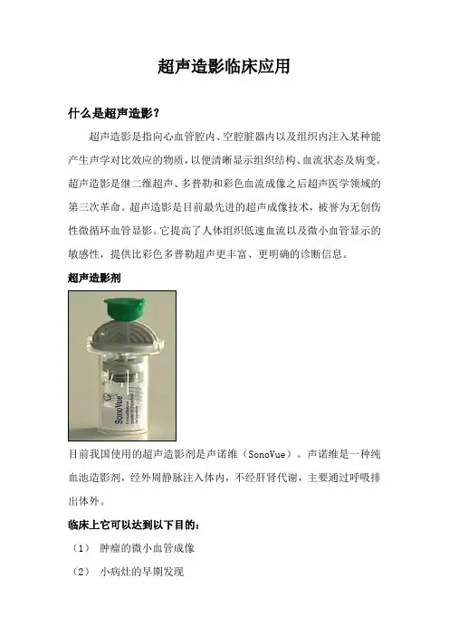
超声造影剂目前我国使用的超声造影剂是声诺维(SonoVue)。
声诺维是一种纯血池造影剂,经外周静脉注入体内,不经肝肾代谢,主要通过呼吸排出体外。
临床上它可以达到以下目的:(1)肿瘤的微小血管成像(2)小病灶的早期发现(3)介入治疗后的疗效评估(4)血管病变的诊断(5)实质脏器外伤快速诊断超声医学科超声造影技术临床应用成效超声医学科自2006年开展超声造影技术以来,在肝脏,肾脏,乳腺,前列腺等脏器的检查方面积累了丰富的经验。
目前超声造影在我科的临床应用体现在以下几个方面:(1)肿瘤的检出和定性诊断。
在肝肿瘤数量的诊断方面,尤其在检测1cm以下的亚厘米病灶方面,声学造影优于常规超声。
在肝、胆及肾占位性病变的良恶性鉴别诊断方面体现出了临床实用价值。
超声造影:符合肝细胞癌“快进快出”的血流动力学特征。
病理:肝细胞癌II级(6*6*5mm)(2)靶向引导。
我科开展了超声造影靶向引导肝脏肿瘤射频治疗和超声造影靶向引导前列腺穿刺技术。
在肝肿瘤射频治疗中,通过超声造影发现常规超声难以显示的病灶,并引导射频针准确进入肿瘤内。
通过超声造影靶向引导前列腺穿刺,提高了穿刺的单针阳性率,检出更高Gleason评分的前列腺癌及更多的阳性病例。
超声造影靶向引导穿刺(造影剂高灌注区域)病理:前列腺癌,Gleason4+4=8(3)疗效评估。
通过超声造影评估肝、肾肿瘤射频治疗后区域有无残存肿瘤组织。
评估肿瘤的化疗疗效。
小肝癌射频治疗前:造影剂高灌注治疗后:无造影剂灌注超声造影与增强CT、核磁共振(MRI)相比有何特点?超声造影最大的优势就在于实时动态扫查(即可不间断地观察到整个连续过程),而CT/MRI是间歇断层扫描成像,这一特点有助于医生对病灶进行鉴别诊断以及发现更多更小的病灶。
●卫生与管理●健康女性2021年第2期·174· 什么是超声造影,超声造影可以检查哪些疾病?杨桂枝攀钢集团成都医院 四川成都 610000在临床中很常用到的影像学检查方法之一就是超声检查,想必大家都非常的熟悉,它就是超声医生们的放大镜,今天在超声检查中需要给大家介绍另外一种比放大镜更有精准度的显微镜,它就是超声造影,它被誉为是超声医学界发展史上的第三次革命。
超声造影是超声医生们的神秘利器,如果患者的肝上有结节,想要了解结节的具体情况,那就可以通过超声造影检查,将造影剂打到血管里,让病变部位显示得更清楚。
传统的造影一定要借助CT,但是CT对人体有辐射,很多患者会对此检查存在一定的忧虑。
超声也能够进行造影检查,不仅没有辐射,而且可以全程录像显示。
大大提高超声诊断的准确性,能够全面鉴别病变的良性恶性,让疾病得到及时治疗,更重要的是没有明显的不良反应,那么今天就让我们跟着文章了解一下超声造影究竟是什么?哪些疾病能够利用超声造影来进行检查吧!你知道什么是超声造影吗?1.超声造影的基本情况超声造影它还有一个别称,叫做声学造影,就是利用造影剂使后散射回声增强,显著提高超声诊断的分辨和灵敏度,在临床上的作用和增强CT 和MRI类似,主要是解决普通超声难以确诊的一些疑难病症。
借助常规的超声造影,看到的是一个模糊的病变部位,而利用超声造影可以将该部位的病变情况清晰地展现在医生眼前,便于医生进行相关的诊断和治疗。
2.超声造影的显著优势超声造影的优势自然是不必多说了,首先超声造影是没有辐射的,放射检查过量的辐射会导致人体细胞出现损伤,甚至导致染色体畸形病变,影响到患者身体甚至是后代的情况,所以面对辐射,很多患者内心是存在抵触的。
但是超声造影是安全无辐射的;其次它的优势是可以进行实时动态的检查,不需要像CT和MRI那样事先设定固定扫描时间,超声造影可以把整个造影过程录制下来,这样可以帮助医生在后期对信息进行全面筛查,避免遗漏的情况,一般患者在当天进行超声造影后就能够明确诊断;再次在使用超声造影的过程中,超声造影剂使用的量很小,无需要皮试,经过肺循环,没有肝肾毒性,安全可靠,对于那些肝肾功能不全,或者要进行肝肾器官移植的患者非常实用;最后它作为超声界中的“显微镜”,对于一些特别微小病灶,尤其是小于一厘米的肿瘤显示率高,超声造影的费用低,在使用中可以发挥突出作用。
为什么要做超声造影?在现代社会发展中医学技术不断进步和快速发展,超声造影在各种疾病诊疗中的应用范围不断扩大。
超声造影属于一种新型的影像技术,在疾病的性质的判断以及后续治疗中都具有实质性意义。
什么是超声造影?超声造影是继B超、彩色多普勒超声之后,在现有超声医学的基础上进行的第三次革命,也是目前医学领域最为先进的超声成像技术手段之一。
该技术在应用时主要是在常规超声检查的基础上,将造影剂通过静脉注射到人体内,一般每次的使用量在1ml左右即可,可以根据需求反复注射,以此来保证病灶显示的对比度得到提升,为诊疗的准确性提供基本保证。
如果将彩超比喻是宏观的方式,那么其可以显示出0.5mm以上的血管,而造影剂就是以一种微观的方式呈现出来,其整个直径只有5μm左右,比红细胞还要小一些,所以可以将微循环更好的体现出来。
究其原因主要是由于即便如此细微,也可以利用肉眼观察到,超声机器可以直接将其信号放大到数千倍以上,直接将其显示在显示屏当中。
由此可以看出,超声造影在使用时根本目的是为了保证对比度得到提升,为诊疗结果的准确性提供基本保证。
超声造影的优势有哪些?与常规彩超相比:在医学领域上良性病变与恶性病变的微循环模式之间具有非常明显的差异性,尤其是对于常规彩超来说,只单纯能够显示出0.5mm以上的血管,但是对于超声造影来说,在使用时的造影剂只有5μm。
造影剂在经过人体静脉注射之后,会直接分布在全身组织当中,在各组织以及各器官中实现微循环,促使组织以及脏器的超声回声越来越强,原本常规下的彩超手段无法发现明确的病灶,或者病灶的表现并不是很明确。
由此可以看出,通过超声造影在其中的合理利用,可以对普通彩超鉴别不了的病灶进行鉴别及诊断。
除此之外,通过超声造影技术的合理利用,还可以更好的弥补彩超技术中存在的不足之处,对器官组织的具体血液循环灌注情况等展开深入了解,以一种持续动态化的方式呈现出来,为疾病的诊断提供便利条件。
由此可以看出,超声造影本身的使用与常规彩超相比更具有明显的优势特点。
超声造影描述超声造影是一种常用的医学检查方法,通过利用超声波在人体内部的传播和反射特性,获取有关人体器官和组织结构的影像信息。
它不仅可以提供高分辨率的图像,还可以实时观察器官的功能和血流情况,因此在临床上应用广泛。
超声造影的原理是利用超声波在不同组织界面的反射来形成图像。
当超声波通过人体组织时,会遇到不同组织的声阻抗差异,从而产生反射信号。
超声仪器会将这些信号转化为图像,显示出组织的形态和结构。
超声造影可以用于检查人体各个部位的器官和组织,如心脏、肝脏、肾脏、乳腺等。
在心脏超声造影中,医生可以观察心脏的结构和功能,诊断心脏病变。
在肝脏超声造影中,可以检测肝脏的大小、形态和血流情况,帮助诊断肝病。
在乳腺超声造影中,可以发现乳腺肿块和乳腺癌等病变。
超声造影具有无创、无辐射、操作简便等优点,适用于各个年龄段的患者,尤其适用于孕妇和儿童。
同时,超声造影还可以与其他检查方法相结合,如CT、MRI等,提高诊断的准确性。
超声造影的应用范围广泛,不仅可以用于检查病变,还可以用于指导诊疗过程。
在肿瘤治疗中,超声造影可以用于引导穿刺活检或者介入治疗,提高手术的准确性和安全性。
在妇科领域,超声造影可以用于监测卵泡发育和排卵情况,指导人工受孕或者试管婴儿等治疗。
除了临床应用,超声造影还在科研领域发挥重要作用。
科研人员可以通过超声造影技术研究人体器官和组织的生理和病理变化,探索疾病的发生机制。
同时,超声造影还可以用于药物输送和基因治疗等领域的研究,提高治疗效果。
尽管超声造影有很多优点,但也存在一些限制。
首先,超声波在穿过骨骼或气体时会产生反射,影响图像的质量。
其次,超声波在穿透深部组织时会有衰减,限制了其应用范围。
此外,超声造影的分辨率相对较低,对于微小病变的检测有一定的局限性。
总的来说,超声造影是一种安全、无创的医学检查方法,具有广泛的应用前景。
随着技术的不断进步,超声造影的图像质量和应用范围将进一步提高,为临床诊断和治疗提供更加精确和可靠的支持。

超声检查是现代医疗中必不可少的检查方法,临床上常作为首选检查手段。
但是常规彩色超声检查在观察病灶血管动态灌注状态、病灶良恶性诊断、介入引导方面具有局限性。
随着医学发展,诊断方法不断更新,现在很多临床医师建议患者做超声造影检查。
今天,给大家详细讲一下超声造影的相关知识。
超声造影和普通超声的区别普通超声,俗称B 超,或灰阶超声,20世纪80年代开始应用于我国,其利用超声波探测出来的图像是黑白的,只能观测到基本的组织结构。
彩色超声现已基本普及我国各个级别的医院,其在B 超基础上增加了清晰度,而且能显示血流分布状态。
超声造影(CEUS ,又称造影显像)是一项无创、无电离辐射的新型影像学技术,它在常规二维超声检查基础上,通过静脉注射超声造影剂,增强人体的血流散射信号,为疾病的超声诊断提供新的信息。
超声造影是目前最先进的超声成像技术,它能实时、动态地观察组织微血管灌注信息,比二维超声更能清晰显示病灶的形态、大小及边界,以提高病变的检出率,并鉴别病变的良恶性,具有高敏感性、诊断准确性。
如果说B 超是黑白照相机,彩超是彩色照相机,那超声造影就是摄像机,它能记录病灶的动态视频情况。
近年来,超声造影广泛应用于二级甲等以上医院,临床上反响良好,深受医生和患者认可。
超声造影的安全性目前我国使用的是二代造影剂为声诺维(Sono Vue ),通用名为注射用六氟化硫微泡,使用的是卵磷脂蛋白包裹惰性气体(六氟化硫SF 6)微泡,是一种非常安全的微气泡悬浮液制剂。
微气泡的平均直径小于红细胞,通过呼吸排出体外,对人体无害。
静脉注射15分钟后,几乎所有六氟化硫气体都可排出,该造影剂过敏反应发生率低于0.002%,因此不必进行常规皮试,无心脏毒性和肝、肾毒性。
什么情况需要做超声造影1.对病灶的定性(良/恶)诊断。
2.早期发现微小病灶。
3.超声造影引导下对肿瘤的穿刺活检,可观察肿瘤病变的活性区、坏死区,定位更精准,提高检出率。
4.肝脏肿瘤介入治疗前后的评估。
关于超声造影你应该了解这些超声造影属于超声诊断重要方法之一,你对超声造影了解多少?超声造影如何诊断疾病?使用超声造影有哪些注意事项?一、关于超声造影第一,概述。
超声造影是一种使用造影剂、增强后散射回声,分辨率、敏感度较高的技术,该技术相较于常规造影来说优点众多,其通过注射造影剂辅助造影,增强造影效果,能够察觉微小病灶,及时发觉患者身体不适之处。
与CT相比,该技术血流信号信噪比更强,成像更清晰,能清晰反映病灶低速血流向,诊断水平较高。
超声造影术使用范围较广,肝脏、肾脏等器官病变都可通过该技术诊断。
第二,超声造影剂。
造影剂根据应用对象选用所使类型,微气泡造影剂使用次数最多,微泡散射性较好。
造影剂又根据微泡内包裹的气体划分为第一代造影剂和第二代造影剂。
超声造影剂作为超声造影与其他超声诊断方法相区别的方面之一,具有较多特点,包括:(1)安全性能高,副作用较少;(2)稳定性较好;(3)产生谐波较为丰富;(4)微气泡大小均匀,能自由通过毛细血管。
第三,超声造影术。
超声造影术较多如造影剂爆破成像法、低机械指数成像法等,前者使用第一代造影剂,为获取丰富谐波常用爆破微泡方式,例如利用超声造影诊断腹部脏器,手动触发获取成像;后者使用第二代造影剂,能够获取血流连续谐波成像。
第四,相应禁忌证。
患有以下疾病的人群不适用于超声造影诊断技术,包括:(1)心绞痛、心肌梗塞;(2)严重贫血;(3)有注射声学造影剂过敏史等。
二、超声造影如何诊断第一,原发性肝癌。
该病较常见,前期症状不明显,病情发展较迅速,确诊时大多已至中晚期,尽早诊断才能及时治疗,影像学检查作为重要诊断方法之一发挥极其重要的作用,常规超声效果不大理想,超声造影术出现后尽可能的弥补了常规超声存有的缺陷,准确度较高,能够有效反映肝脏病变具体情况,快速捕捉有效信息,准确评估肿瘤细胞情况,而且该技术价格低,创伤小,副作用少,因此该技术在临床被广泛应用。
使用超声造影术诊断该病的流程如下:(1)准备超声诊断仪,设置探头频率;(2)患者取平卧位;(3)进行常规超声检查;(4)稀释造影剂并注射,用5毫升生理盐水冲管;(5)开启造影设备,设置低机械指数,观察回声强度;(6)注射造影剂后30秒为动脉期,31至120秒之间为门脉期,121至360秒之间为延迟期。
超声造影(Contrast-enhanced ultrasound)PrefaceReview the history of the development of medical ultrasound, we see that the ultrasound rise in 70s (real-time grey-scale ultrasound) or B type ultrasound or gray-scale ultrasound tomography technology, laid the foundation of modern ultrasonic diagnosis, paving the way for the clinical application of ultrasound is very wide; color Doppler imaging technology developed in the 80s, the modern ultrasonic imaging diagnosis is very characteristic of nondestructive examination and cardiovascular hemodynamics and blood flow of the body organs and tissues to create a new field; since 90s, the emergence of many new ultrasound technology is one of the medical ultrasound emerge in an endless stream, the most influential and can further improve its status in modern imaging technology, than ultrasound contrast imaging. The contrast-enhanced ultrasonography (contrast enhanced ultrasound). With the help of intravenous contrast ultrasound and contrast harmonic imaging technology, can clearly show the micro blood vessels and tissue perfusion, increase the image contrast resolution, greatly improve the sensitivity and specificity of ultrasound detected lesions. This is very similar to the enhanced CT scan. Nowadays, radiography not only opens up the scope of clinical application, but also improves the diagnostic level of conventional grey scale / color Doppler ultrasonography, and has a good prospect in targeted therapy. In conclusion, CEUS is a major technological innovation and research direction, and is a new milestone in the development of medical ultrasound.The concept of contrast-enhanced ultrasoundBarry B. Goldberg is a pioneer in the research and development of new ultrasound contrast agents in the world. He has shown great interest in the research and application of various ultrasound contrast agents. Goldberg et al compared microbubbles to ultrasound contrast agents (vascularcontrast, agents) or vascular enhanced ultrasound contrast agents, which are different from oral contrast agents (oralagents) commonly used in gastrointestinal imaging. Therefore, ultrasound contrast has 2 kinds of angiography agents and oral or enema contrast agents, and the former is also called microbubble contrast medium. For the past more than 10 years, contrast enhanced ultrasound or vascular contrast enhanced ultrasound has been the most rapidly developed. Microbubble ultrasound contrast agent in the initial research stage, the first gas is mainly used for imaging of air and oxygen, then, is to CO2 as the representative of the micro bubble shell membrane contrast agent intravenous injection and transcatheter arterial injection of contrast-enhanced ultrasound. In 90s, a new type of ultrasound contrast agent came into being. The membrane contrast agent containing microbubbles was represented by Levovist (Li Fa), Albunex and Echvist, which is called the first generation of new contrast agents. Since then, more inert gas containing SonoVue (SonoVue) and Options angiography as the representative of the shell membrane type agent, also known as the second generation of new contrast agent. The average diameter of the new contrast microbubbles is about 3~5 m, which can successfully pass through the pulmonary circulation, and realize the contrast enhancement of the left ventricle, the myocardium, and the organs, tissues and lesions of the wholebody. The safety of microbubble ultrasound contrast agents: a large number of experimental studies and more than 10000 cases of clinical experience have proved that microbubble contrast agents are safe. According to estimates, each time the intravenous injection of ultrasound contrast microbubbles containing air / gas amount is less than 200 L (0.2 ml), any no danger of air embolism; the contrast agent listed only in shell membrane was composed of acoustic ctose, the contrast agent with albumin, phospholipids or polymer. That is easy for human metabolism, toxic effects on the human body does not. Therefore, it is an ideal ultrasound contrast agent. The study pointed out that the second generation of new ultrasound contrast agent with high molecular weight and low solubility and low dispersion of fluorine containing inert gas such as SF6, C3F8 etc., can significantly extend the microbubbles in the human body life, increase the stability of microbubbles.Principle of contrast-enhanced ultrasoundStudy of ultrasound contrast agent has experienced three stages, namely the first generation of CO2 free micro shell membrane type bubble contrast agent as the representative, with Albunex and Levovist (Levovist) as the representative of the second generation of air containing micro bubble shell membrane model and contrast agent, containing an inert gas microbubble contrast agents such as SonoVue, Optison, Echogen etc..The basic principle of these contrast agents are by changing the sound attenuation, velocity and enhanced scattering, change the basic role of sonic and between organizations, namely the absorption, reflection and refraction, so that thelocation of the echo signal enhancement. Ultrasound contrast agent microbubbles to ideal through small lung, heart and capillary circulation, so that the peripheral intravenous contrast can be simple, and can stably maintain its effect in acoustic imaging. It is found that low solubility and low dispersion of high molecular gas, such as fluorine gas, can increase the lifetime of microbubbles in blood and increase stability. With the development of polymer chemistry, some foreign scholars using biodegradable polymeric materials to replace the Human Albumin and other natural phospholipid substances, changing the composition of micro bubble shell, thereby avoiding the problem of acoustic effect caused by the limitation of the nature of the material itself is not stable. At present the domestic and foreign research shows that polymer microbubble ultrasound contrast agent development is the most promising, it can change the polymerization conditions the acoustic properties can be designed for a "tailored" for the imaging condition of contrast agent, make the particle size distribution is more concentrated, after the sound attenuation is weak and prolonged in vivo the remaining time to be used in different physiological and pathological conditions and targeted drug delivery. With microbubbles especially halothane gas ultrasound contrast agent was developed and used in clinical, ultrasound contrast imaging technology is in rapid development, from the color Doppler imaging, intermittent harmonic gray-scale ultrasonography, contrast-enhanced ultrasound imaging quality has been greatly improved, the resolution is obviously improved, and greatly reduce the the artifacts, and can display real-time perfusion.Clinical application of contrast enhanced ultrasoundThe development of ultrasound contrast agents andcontrast-enhanced ultrasound has promoted the application of contrast-enhanced ultrasound in clinical diagnosis and treatment.1. contrast echocardiography (contrast, echocar-diography): it is reported that routine echocardiography results in technical difficulties in some 10% to 20% of patients. Microbubble contrast echocardiography is a useful method for improving endocardial findings and making cardiac function more accurate. In addition, it can visually display myocardial perfusion and improve the quantitative determination of myocardial viability, which can be successfully used to determine myocardial ischemia and necrosis.The room of ultrasonography in the diagnosis of septal defect sensitivity was 97%, higher than the same group of patients with cardiac catheterization sensitivity. It can also be used for the diagnosis of primary pulmonary hypertension,2. liver harmonic contrast enhanced ultrasonography:The effect of liver ultrasound contrast: 1., improve the detection rate of 2., location and qualitative changes of the lesions, 3. curative effect judgment, 4. portal vein blood flow study. Contrast observation contents: 1. enhanced mode: the overall enhanced peripheral enhanced central enhanced (radial) enhanced 2.. Distribution after the peak: homogeneous, cyclic, homogeneous enhancement of the 3. phase: according to the CT staging can be divided into arterial portal venous phase delay0-40S 41-120S 121-200S 4. enhanced continuously the time.A large number of studies have confirmed that harmonic ultrasound is the most successful clinical application in the liver, and has made a breakthrough in the application of liver tumors, and can be compared with CT angiography. First of all, the sensitivity of small tumors was improved significantly, especially for tumors less than 1cm. Secondly, the specificity was enhanced significantly. (1) liver tumors or lesions of benign and malignant differentiation, including hepatocellular carcinoma (HCC and metastasis), hemangioma, adenoma and focal nodular hyperplasia (FNH) with non-uniform distribution of fatty liver and other identification; (2) liver cancer preoperative routine liver ultrasound, and further determine the size and scope of invasion tumor metastasis, there are no small hidden intrahepatic or multifocal tumors;(3) application in the diagnosis of liver tumors: interventional ultrasound contrast helps small tumor of liver in suspicious especially nodule biopsy puncture echo; (4) the application of interventional therapy liver neoplasms: in hepatic artery embolization chemotherapy aftercontrast-enhanced ultrasound, but also in the RF, microwave ablation, HIFU immediately after treatment of bedside ultrasound angiography, determine the ablation effect and there is no residual tumor tissue, to determine further treatment, improve the curative effect.For without further treatment in patients with long-term follow-up; (5) the application of contrast-enhanced ultrasound in liver cancer operation: it is reported that intraoperative angiography can detect / except other smaller tumors, surgicaltreatment or change range of ways in time; the primary lesion resection into intrahepatic metastasis occult examination and immediately decided to use resection or ablation; evaluate the nature and extent of liver injury. In addition, in other liver diseases such as liver transplantation without vascular stenosis or occlusion of portal hypertension in patients with TIPS stent patency and Buchashi comprehensive ultrasound color Doppler syndrome detection is difficult, the application value of contrast-enhanced ultrasound have certain liver.3. renal contrast radiography: renal artery stenosis is frequently encountered by ultrasonography. Renal contrast angiography can routinely display renal artery, improve the detection rate of renal artery stenosis, and make up for the deficiency of color Doppler ultrasonography. It is also helpful for the color Doppler examination of renal vessels. Animal studies have shown that radiography helps to detect renal tumors more sensitively, and the clinical significance remains to be further studied.4. splenic contrast ultrasonography: CEUS is helpful for the diagnosis of splenic tumor, splenic trauma and splenic infarction and the evaluation of its range. Chinese scholars have begun to evaluate the effect of microwave ablation in the treatment of hypersplenism.5. contrast-enhanced ultrasonography of pancreatic masses: reports of Levovist ultrasound contrast agents in pancreatic tumors have been reported by Dai Lu Shu, which increased blood flow in the tumor by up to 100%. Malignant tumors were mainly vascularized, whereas benign tumors were less vascular. Itimproves the judgment of benign and malignant tumors.6. breast tumor ultrasound contrast: by means ofcontrast-enhanced ultrasound, it can display the characteristics of tumor microvascular distribution, and can be used to identify benign and malignant tumors by using the time echo intensity curve matched with microbubbles.7. lymphoangiography enhanced (contrast): experimental studies have confirmed that microbubble ultrasound angiography is helpful in showing drainage of lymph vessels and lymph nodes. CEUS is helpful in identifying lymph node metastasis of cancer. Therefore, it is possible to have sentinel lymph node metastasis in patients with breast cancer. It is of potential clinical value.8. contrast enhanced ultrasonography is helpful to enhance two-dimensional transcranial Doppler (TCD) and color Doppler angiography of intracranial vessels and its real-timethree-dimensional display.9. microbubble ultrasound contrast can be used to evaluate the function of liver, kidney and heart transplantation patients.Application of contrast-enhanced ultrasound in liver transplantation: 1. Preoperative evaluation of portal status, direction of blood flow, presence of portal vein thrombosis, portal cavernous degeneration, and hepatic artery occlusion;2, whether the postoperative hepatic artery stenosis and occlusion (for patients after liver transplantation,two-dimensional and color Doppler can not see this point, it is very important, compared to the radiology department, DSA and a lot of convenience)Is there a false aneurysm?. The ultrasound contrast system of transplanted liver consists of selective arterial cannulation (invasive) or by peripheral vein (minimally invasive) injection of ultrasound contrast agent, which forms the ultrasonic interface of liver and liver in liver tissue, and the image is dense and strong echo. CEUS can observe the changes of the 3 dynamic phases of the liver blood flow, that is, the arterial phase, the portal vein phase, and the liver parenchyma phase. Therefore, CEUS is helpful to detect the abnormal blood flow and intravascular lesions and the dilatation of the small bile duct. To observe the blood supply of the hepatic artery after orthotopic liver transplantation with contrast-enhanced ultrasound, it is important to evaluate the prognosis after orthotopic liver transplantation.10. other: the microbubble contrast agent injected into the bladder will help to diagnose bladder ureter reflux; contrast agents injected into the uterine cavity will help to confirm the patency of the fallopian tubes in infertile women. It is reported that CEUS has good effect and is expected to replace traditional radiographic radiography with radioactivity. Gu Yurong reported that the application of ultrasound contrast agent Levovist can help CDFI to evaluate tumor blood vessels more accurately, and is of great value in the diagnosis, differential diagnosis and treatment of ovarian benign and malignant tumors..Application of ultrasound contrast agents in targeted therapyThe broad prospects of microbubble ultrasound contrast agents have been reported. In fact,The therapeutic effect of this new microbubble contrast agent is also promising and has attracted much attention. It has become one of the most important research and development directions. Because the micro bubble mean diameter of less than red blood cells, can run on the microcirculation, and because of the successful development of a new type of fluorine and sulfur or fluorocarbon gas contrast agent, the contrast agent in the microcirculation lived significantly longer (according to some reports of tissue-specific contrast agent can be kept for several hours in liver tissue), scholars are actively to explore the use of ultrasonic irradiation for treatment of non-invasive. For example, intravascular thrombolysis, or / and the micro bubble as drug or gene carrier in combination with micro bubble or shell membrane surface, with the help of ultrasonic drug or gene transfer into specific tissues, the antitumor activity and promote angiogenesis. The mechanism of ultrasound enhanced gene transfection and how to further improve the gene transfection rate have also been actively explored, and a lot of positive results have been achieved.Modern medical imaging techniques, such as CT and MR, have been widely used and relied on contrast agents for many years. In contrast, ultrasound still rarely exerts its great potential with contrast media. In recent years, microbubble enhanced ultrasound imaging has been widely applied in clinical diagnosis, and has shown unique advantages. Microbubbleultrasound has an attractive prospect in targeted therapy.(SonoVue) is bolaike company (Bracco) pharmaceutical company produces at present a new medical imaging agent. SonoVue is on the market all over the world, and has been widely used in the United States and europe. In the United States, it is mostly used in the study of the heart, while in Europe it is mainly used in the study of the abdomen. Approved by the State Drug Administration review, China entered the market, SonoVue entered China in 2002 March 8, 2004 after phase III clinical study; 21 hospital first began to use Yum products to match the use of SonoVue. Develop very rapidly. It is an effective tool for clinicians. Compared with MRI and CT, the cost of ultrasonic examination is low and has very good market value.Bracco in Shanghai Pudong pharmaceutical joint venture Bracco-Sine Joint Venture, Chinese named Bracco Xinyi Pharmaceutical Co. Ltd., began operating this year, part of the joint venture by contrast agent will plant to produce. Therefore, the product can be purchased at home, the price of about 700 yuan. Most patients can afford it.。
超声造影的不足之处
超声造影是在常规超声检查的基础上,通过静脉注射超声造影剂,增强人体组织的回声信号,实时动态地观察组织的微血管灌注信息,以提高病变的检出率并对病变的良恶性进行鉴别,它具有安全性好、无过敏反应,实时性、可重复性强等优势,但仍存在一些不足之处:
1. 空间分辨率相对较低:相对于其他影像学检查方法(如CT、MRI),超声造影的空间分辨率较低。
这可能导致对小病灶或细微结构的显示不够清晰。
2. 操作者依赖性:超声造影的结果在一定程度上依赖于操作者的经验和技术水平。
不同操作者之间可能存在差异,这可能影响诊断的准确性。
3. 深度限制:由于超声的传播特性,超声造影在检测深层组织或器官时可能受到限制。
对于一些位于深层或胃肠道等部位的病变,超声造影的应用可能受到一定限制。
4. 肺气、肠气等干扰:肺部或肠道内的气体可能对超声造影的传播和成像造成干扰,影响对这些区域病变的检测和诊断。
5. 假阳性和假阴性结果:尽管超声造影可以提供血流灌注信息,但它并不能完全取代组织病理学诊断。
超声造影可能出现假阳性和假阴性结果,需要结合其他影像学和临床信息进行综合判断。
需要指出的是,超声造影作为一种辅助诊断工具,在临床实践中应结合其他影像学检查和临床资料进行综合分析,以提高诊断的准确性。
随着技术的不断发展和研究的深入,超声造影的不足之处也在不断得到改进和克服。
超声造影技术优势、基本原理、调节机器及注意事项增强超声就是超声造影技术,它被称为超声发展史上的“第三次革命”,已经越来越被临床认可和依赖,成为超声诊断工作重要部分。
超声造影超声造影,是在常规超声基础上,通过静脉注射超声造影剂,有效增强组织器官的血流信号,更好地反映血流灌注情况,有助于对疾病进行诊断(图1)。
图 1 右侧为常规超声,箭头所示肝脏结节,隐约可见且性质不明确;左侧为超声造影,箭头所示为常规超声上不确定的结节,造影后清楚的显示,且其增强特征符合肝细胞肝癌目前常用超声造影剂的是六氟化硫微气泡(图2),直径通常为2-5微米,经外周静脉注入后,能通过肺循环、体循环到达靶器官,而不能穿透血管内皮进入组织间隙,因此超声造影是纯血池造影,这是和CT、MRI增强的最大区别。
同时作为造影剂的微泡,其半衰期短,不通过肝肾循环,通过呼吸道排出,这是和CT、MRI增强的不同之处。
基本原理是超声波反射,而超声造影基本物理原理不同,它是利用血液中的超声造影剂在声场中的非线性效应和产生的背向散射所获得的对比增强图像。
微泡造影剂在不同机械指数(MI)的声场中会产生不同的变化,只有在低MI声场中,才能产生非线性谐波信号。
超声造影要在低MI条件下进行。
如何调节机器超声造影检查时想要达到最佳效果,需要充分调节机器:MI调节。
根据目前的超声造影剂特性,MI不能超过0.2,需要我们根据病灶的位置,深度等,适当调节,当病灶位置较深时可以适度提高MI,助于观察病灶增强情况,也会增强对微泡破坏,缩短观察时间。
深度调节。
需要以显示完整的病灶以及部分邻近组织可以作为对比观察为佳。
聚焦点调节。
检查时,焦点常规放置于病灶的底部水平,焦点个数不宜太多,最好不超过2个。
增益调节。
在注射造影剂之前需要调节二维图像的增益,比如甲状腺造影,需要将甲状腺组织调成无回声,仅显示气管、筋膜等为线状回声。
图像显示方式。
造影模式下,可以采用单幅显示,也可以采用二维模式和造影模式的双幅对比显示,双幅对比显示,尤其适用于不易显示,易脱靶的病灶。
美国放射学院超声造影LI-RADS指南(2016版)
杨丹
【期刊名称】《临床超声医学杂志》
【年(卷),期】2017(19)10
【摘要】指南形成的时间节点 2014年4月美国放射学院对比增强超声肝脏影像学报告及数据系统(ACR CEUS LI-RADS)工作组成立,2015年11月至2016年3月先后经北美放射学会2015年教学展示、美国超声医学会2016年口头报告及2016年壁报,工作组于2016年5月将超声造影(CEUS)LI-RADS最终版(2016版)工作流程提交至指导委员会,2016年6月24日指导委员会正式通过该工作流程,并公开发布。
【总页数】7页(P712-718)
【作者】杨丹
【作者单位】401120 重庆市,重庆医科大学附属第三医院超声科
【正文语种】中文
【相关文献】
1.美国放射学院(ACR)关于定量CT(QCT)骨密度测量操作指南 [J], 程晓光;李娜
2.美国心脏病学学院/美国心脏学会/欧洲心脏病学会2006年心房纤颤控制指南——执行摘要——来自美国心脏病学学院/美国心脏学会行医指南与政策会议工作组和欧洲心脏病学会行医指南委员会的联合报告(2001年房颤控制指南修订版) [J], 宋田
3.慢性稳定型心绞痛诊疗指南(1999年)--美国心脏病学院(ACC)/美国心脏学会
(AHA)/美国医师学院及美国内科学会ACP-ASIM联合议定 [J], 马虹;廖晓星
4.2020年美国放射肿瘤学会宫颈癌放射治疗指南解读 [J], 王焜煜;王元景;孔为民
5.2014 ACC/AHA非ST段抬高型急性冠脉综合征诊治指南概述——美国心脏病学院/美国心脏协会研究小组实践指南报告 [J], 陈研;吴小盈
因版权原因,仅展示原文概要,查看原文内容请购买。