骨嗜酸性肉芽肿影像诊断
- 格式:ppt
- 大小:1.97 MB
- 文档页数:15
骨嗜酸性肉芽肿影像表现及病理分析发布时间:2022-10-14T08:01:42.893Z 来源:《医师在线》2022年14期作者:张树云[导读] 目的探讨骨嗜酸性肉芽肿影像特点及病理改变。
方法收集2018年6月-2022年6月间5例,张树云云南省红河州泸西县人民医院放射科云南省红河州 652499摘要:目的探讨骨嗜酸性肉芽肿影像特点及病理改变。
方法收集2018年6月-2022年6月间5例,分析临床影像表现及病理改变。
结果5例中男3例,女2例,都有局部间歇性疼痛,软组织肿胀。
结论骨嗜酸性肉芽肿结合临床病史及影像表现和病理分析不难做出正确诊断。
关键词:骨嗜酸性肉芽肿;影像表现;病理分析骨嗜酸性肉芽肿是以骨破坏及有大单核细胞、嗜酸细胞集聚为特征。
最多见于小儿及青年,约2/3患者发病于20岁前,男性较女性发病率高。
骨嗜酸性肉芽肿易见于扁平骨,尤以头颅及肋骨为著,还可发生于脊椎、股骨、下颌骨和肱骨,而手及足骨则不受累。
1材料与方法1.1一般资料 2018-2022年我院经手术病理证实骨嗜酸性肉芽肿共6例有,其中男性4例,女2例,年龄5月-12岁,平均7岁。
部位:颅骨4例,长骨1例,骨盆1例,多发者2例。
经手术后病理确诊5例,经穿刺病理确诊1例。
有局部疼痛、软组织肿胀及骨质破坏性改变4例,仅有骨质破坏1例,无症状者1例。
1.2头颅X线片使用岛津DR机,上缘包括顶骨,下缘包下颌骨,患儿贴近平板探测器,球管与患儿的距离1.2m。
1.3 CT片使用128排螺旋CT,层厚1.5mm,螺距1.3,重建间隔0.7mm,5例增强的扫描。
1.4病理标本HE染色,电子光镜观察,免疫组化采用Envision两步法标记。
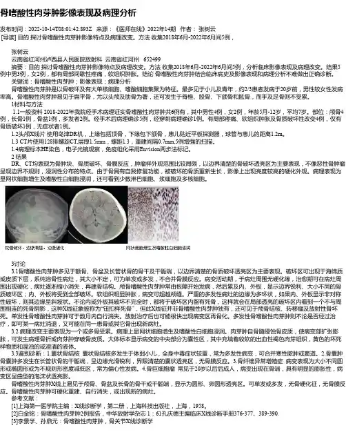
2 结果DR、CT均表现为骨肿块、骨质破坏、骨膜反应,肿瘤样外观范围比较局限,以边界清楚的骨破坏透亮区为主要表现,不像恶性骨肿瘤呈现边界不规则,浸润性分布的特点。
由于骨具有自我修复功能,被破坏的骨质重新生长,影像上出现亮度较高的硬化外观。

儿童颅骨嗜酸性肉芽肿的影像学诊断叶圣利;林晓荷;罗莉莉;许思恩;何家维【期刊名称】《温州医学院学报》【年(卷),期】2014(000)010【摘要】Objective: To investigate the image manifestation and improve the ability of diagnosis of skull eosinophilic granuloma in chidlren.Methods: Imaging data of skull eosinoplilic granuloma in children-retrospec-tive analysis of 20 cases by X-ray, CT and MRI scan, included: 2 cases of X-ray, 20 cases of CT scan, 4 cases of MRI scan (including 2 cases of enhancement scanning).Results: Out of 20 cases, single lesion was found in 16 cases, and multiple lesions were found in 4 cases. There were totally 26 lesions in all 20 patients, temporal bone (n=13), frontal bone (n=7), occipital bone (n=4), parietal bone (n=2) were respectively being involved. The X-ray ifndings of 2 cases were clear, sharp edge of the circular hole shaped bone destruction. The CT scanning found 26 lesions, including 7 cases of osteolytic destruction, 19 cases of punched-out like bone defect, 21 cases of soft tissue mass. The MRI scanning revealed 4 cases of soft tissue mass adjacent to the osteodestructions. Compared with gray matter of the brain, MR showed isosignal intensity on T1WI and T2WI after intravenous injection of Gd-DTPA, the lesions showed enhanced remarkably.Conclusion: The CT and MR manifestations of children skull EG have certain characteristics. Combination of medical record may improvethe diagnostic accuracy in the majority of cases. The MRI can accurately show the location, shape and extent of skull eosinophilic granuloma in children which is valuable in differential diagnosis of various causes.%目的:探讨儿童颅骨嗜酸性肉芽肿(EG)的影像学表现,以提高临床对此病的诊断水平。

骨的嗜酸性肉芽肿骨的嗜酸性肉芽肿即局灶性郎罕氏细胞组织细胞增生症,是一种良性的局灶性的病变。
本病多见于儿童和青少年,男性多见。
病变一般局限于骨骼,多为单发性,好发于颅骨、肋骨和股骨。
1 X线表现1.1 发病部位颅骨、长骨、骨盆等部位。
1.2 X线所见主要是骨质溶骨性破坏,可呈圆形、卵圆形或不规则形。
早期病灶境界较清,周围骨质无异常改变,至后期,破坏区周围骨质常硬化而致密。
不同部位的病灶各有一定特点:1)颅骨:多累及顶骨和额骨,单发病灶多见。
病变始于板障,逐渐累及内外板和邻近软组织,大小约1~5 cm。
病灶呈类圆形穿凿样破坏或多房性破坏,边缘硬化带少见,可跨越颅缝,也可破坏外板向外突出形成软组织肿块;2)长骨:病变多累及骨干和干骺端,骨破坏区呈卵圆形或双房状轮廓,长轴与骨干一致,边缘清楚,少有硬化。
病变部位可见层状骨膜增生。
病变大小约1.1 cm×1.3 cm~3 cm×5 cm 大小;3)骨盆:病变部位不定,均呈局限性单房或多发骨质破坏,周围伴有环形骨质增生带,骨皮质连续性中断,破坏区内均可见斑点状“钮扣”样死骨。
2 讨论2.1 骨嗜酸性肉芽肿是以骨骼损害为主,或局限于骨的一种网状内皮细胞增生症。
病因不明,多认为与原发性免疫缺陷有关。
它能使组织细胞增生,发生感染,使免疫缺陷加重,但随年龄增大,这种免疫缺陷有所减轻。
镜下见网状内皮细胞增生,有大量嗜酸性粒细胞浸润,可有组织坏死。
晚期,组织细胞内形成泡沫细胞,嗜酸性粒细胞消失,最后结缔组织增生而纤维化,甚至可骨化。
2.2 临床上本病好发于20 岁以下青少年,男多于女,临床上常有较明显之局部症状和体征,主要为疼痛、压痛、肿胀和肿块。
颅骨肿块可有波动感。
少数病人可出现全身症状,如低热、乏力、食欲不振。
2.3 实验室检查可有嗜酸性细胞稍多和血沉中度加速。
2.4 本病预后良好,病灶经治疗后可修复,也可自愈。
2.5 本病需与以下疾病相鉴别:2.5.1 急性骨髓炎:临床表现为发病急,高热和明显中毒症状,患肢活动障碍,局部红肿和压痛,X线表现为发病2 周后干骺端骨松质中出现局限性骨质疏松,继而形成多数分散不规则的骨质破坏区,骨小梁模糊,消失,破坏区边缘模糊,以后骨质破坏向骨干延伸,可达骨干2/3 或全骨干。