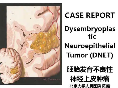
胚胎发育不良性神经上皮肿瘤ppt课件
- 格式:ppt
- 大小:13.90 MB
- 文档页数:25
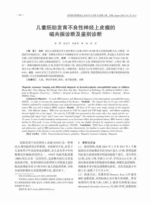
•298实用医学影像杂志2018年8月第19卷第4期 JPMI,August2018,V〇1.19.N〇.4儿童胚胎发育不良性神经上皮瘤的磁共振诊断及鉴别诊断郑彬陈志平时胜利陈琬焦丹【摘要】目的探讨儿童胚胎发育不良性神经上皮瘤(DNET)的M(特点及鉴别诊断方法,以期进一步 提高对病的。
方法回顾性分析经手病理的15 D N E T儿的,儿均行M(,4行磁共振波谱(M(S)分。
结果15 病均,,-M( TiWI ( T2WI ,DWI 或*T2-FLAI(序列6 心低、的“环征”,5例呈“倒三角征”。
增强扫描病无强化12例,轻度不均匀强化3 。
病边界较为清晰,均无占位效应及瘤水肿。
MRS表"A A峰均下降,3例Ch。
峰大致,1 升。
病变区与对照区对比,差无计学义(!>0.05)。
结论DNET好于儿童及青少年,MR 具有一定特征性,熟悉掌握这些特点并做好相似疾病的鉴别诊断,可诊断供可靠的影像依据。
【关键词】儿童;神经外胚瘤,原始;磁共振成像;诊断Magnetic resonance imaging and differential diagnosis of dysembryoplastic neuropithelial tumor in childrenZheng Bin*, Chen Zhiping, Shi ShengH, Chen Wan, Jiao Dan. ^Department of R adiology, the Affiliated Children's Hospital of Zhengzhou University! Children's Hospital of Henan Province; Zhengzhou Children's Hospital, Henan450018, China【Abstract】Objective To s/udy feD/ures D<d differentia1 diagnosis of dysembryop1astic neuropithe1ia1 /umor (DNET),in order to increase the understanding of the disease. Methods The clinical data of 15 cases with DNETchildren confirmed by surgical pathology were analyzed retrospectively, and the children were selected for the preoperative MR scan and 4 routines (MRS) analysis. Results All focus of 15 cases were single,mainly in the temporallobe,with different shapes; MRI scan was based on T1WI low signal and T!WI high signal,and diffuse weightingshowed slightly low or low signal. Six cases by T2-FLAIR were presented in the center with a low signal and the surrounding high signal nringn,and 5 cases were ninverted trianglen. The enhanced scanning lesion was not enhanced in12 cases,3 cases of mild nonuniform enhancement,as no local mass effect and peripheral edema. MRS showed a slightdecline in NAA peak,3 cases of Cho peak were normal,1 case was slightly elevated. In cmparison to normal controlarea,the difference was not statistically significant (P>0.05). Conclusion DNET have a high incidence in childrenand adolescents,and its MRI performance has a certain characteristic,be familiar to master these features and differential diagnosis of the disease, it can provide reliable imaging evidence for preoperative diagnosis of the disease.【Key words】Child; Neuroectodermal tumors,primitive; Magnetic resonance imaging; Diagnosis胚胎发育不良性神经上皮瘤(DNET)是一种神 经元-神经胶质混合性肿瘤。

pat hogenesis of cerebral m icrovascular d is eas e .Exp Mol Patho l ,2003,74:1482159.[4] Ono K,Hirohat a M ,YamadaM.α2Li po i c acid exh i b its anti 2a m yl oi 2dogenicity for β2amyl o i d fibrils in vitro .B i oche m B i ophys Res C om 2mun,2006,341:104621052.[5] Lee J M ,Yi n K,Hsin I,et al .M atrix metall op rotei nas e 29in cerebral 2amyl oid 2angiopathy 2relat ed he mo rrhage .J Neu r o l Sci,2005,15:2292230,2492254.[6] 刘江红,黎健,许贤豪,等.OX 2LDL 促进Aβ产生的实验研究及其临床意义.中国神经免疫学和神经病学杂志,2003,10,35238.[7] Coma M ,Gu i x FX,Ill 2R aga G,et al .Oxi dative stress triggers t heamyl oidogen i c pathway in human vas cu lar s mooth m us cle cells .Neu 2r obi o l Aging,2008,29:9692980.[8] 于明,邬巍,邬英全,等.依达拉奉对淀粉样β蛋白25~35致PC12细胞氧化损伤的神经保护作用.中国临床康复,2005,9:57259.[9] Ono K,Hamaguchi T,Naiki H,et al .Anti 2a m yloidogenic effects ofan ti oxidants:I m p licati on s f o r t he preventi on and t herap eut i cs ofA l zhei mer ’s disease .B i och i m B i ophys Acta,2006,1762:5752586.[10] R ensi nk AA ,Verbeek MM ,Otte 2H �ll er I,et al .I nhibi ti on of amy 2l oid 2β2induced cell deat h i n hum an brain p ericytes i n vitro.B rain Res,2002,952:1112121.(2008-02-10 收稿)论 著胚胎发育不良性神经上皮瘤的临床及影像学诊断吴 晶 吴 杰 贾秀川 【摘要】目的 提高对胚胎发育不良性神经上皮瘤(DNT )的临床及影像特征的认识。
胚胎发育不良性神经上皮肿瘤临床及病理分析胚胎发育不良性神经上皮肿瘤(Dysembryoplastic neuroepithelial tumor, DNT)是最近才被公认的独立性病理实体,并于1993年被收入WHO神经系统肿瘤新分类,属于较少见的神经上皮起源肿瘤。
由于DNT被认识较晚,且发病率较低,故在国内尚未引起重视,但因其外科治疗预后良好,无需放疗和化疗,故熟悉其临床、影像学、电生理及病理学特征对DNT的正确诊断与治疗尤为重要。
为此,将我院1999年9月~2004年4月收治,经手术及病理证实的5例DNT结合文献进行分析。
资料与方法1. 一般资料: 5例均于全麻下行开颅肿瘤切除术, 经病理检查确定诊断, 男3例, 女2例, 年龄6~32岁, 平均17岁, 病程1.5月~8年。
病变部位为左颞2例,右颞1例,右额颞1例,左顶1例。
2.临床表现:临床特点均表现为难治性癫痫,其中4例为复杂部分发作,1例为单纯部分发作演变为复杂部分发作;3例表现有记忆力、计算力下降,1例合并头痛、眼底水肿及中枢性面瘫,余无神经功能缺损。
3.影像学及电生理检查:术前5例患者均行CT及MRI检查,2例做了X-Ray检查,2例做了PET检查,以便了解病变部位及影像学表现。
5例均于术前行常规及长程脑电图检查,观察电生理改变。
4. 免疫组化染色:组织标本经3.7%中性甲醛固定,常规石蜡包埋,5mm厚连续切片,常规脱蜡入水后,用单克隆I抗ABC法标记突触素(SYN,1:150)、神经元特异性烯醇化酶(NSE,1:200)、二聚性钙结合蛋白(S-100,1:200)、神经微丝蛋白(NF,1:200)和神经胶质原纤维酸性蛋白(GFAP,1:200),所有单抗均购自美国Santa Cruz公司,ABC药盒购自美国Vector公司。
每种免疫组化染色均设阳性和阴性对照。
结果1.影像学表现:CT检查显示,病灶多呈低密度改变,1例含钙化斑。
MRI检查的共性特征为T1低信号,T2高信号,边界清楚,除1例有周围水肿外,余无占位效应;增强扫描示1例病灶呈明显囊壁及结节样强化,1例呈轻度增强,3例无增强效应。

