DOI 10.1111j.1469-8986.2006.00490.x Comparison of time and frequency domain measures of RSA
- 格式:pdf
- 大小:221.36 KB
- 文档页数:13
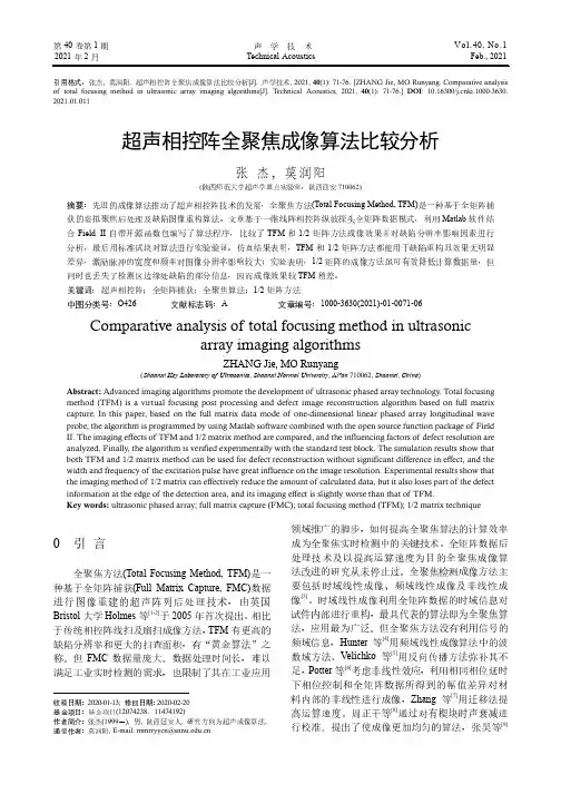
Journal of Alloys and Compounds 492 (2010) 496–499Contents lists available at ScienceDirectJournal of Alloys andCompoundsj o u r n a l h o m e p a g e :w w w.e l s e v i e r.c o m /l o c a t e /j a l l c omMixing of iron and molybdenum and photo-doping effect on Sr 2FeMoO 6Y.C.Hu a ,Q.Ji a ,J.J.Ge a ,R.B.Xie a ,Z.S.Jiang a ,X.S.Wu a ,∗,G.F.Cheng b ,Hairui Liu c ,Qingfeng Lu caNanjing National Laboratory of Microstructures,Key Lab of Solid State Microstructures,Department of Physics,Nanjing University,Hankou Road,Nanjing 210093,China bShanghai Institute of Ceramics,CAS,1295Dingxi Road,Shanghai 200050,China cDepartment of Physics,Henan Normal University,Xinxiang 453007,Henan,Chinaa r t i c l e i n f o Article history:Received 17August 2009Received in revised form 19November 2009Accepted 20November 2009Available online 26 November 2009Keywords:Magnetically ordered materials X-ray diffraction Crystal structurePositron spectroscopya b s t r a c tThe structure and photo-doping effect on Sr 2FeMoO 6are investigated using the X-ray powder diffraction and positrons annihilation technique,respectively.Anti-site defect is about 8.8%,can be estimated directly from the XRD pattern,which is well consistent with that of obtained from the XRD refinements.The integral intensity ratio of the reflection (101)and the reflections (200)and (112)may used to estimate the concentration of the anti-site defect.The positron annihilation lifetime in Sr 2FeMoO 6is sensitive to photo-doping.The average lifetime and the electron density n e vary with photo-doping.© 2009 Elsevier B.V. All rights reserved.1.IntroductionThe double perovskite compound Sr 2FeMoO 6(SFMO),with a high magnetic transition temperature (T c ≈410K)[1]and a remarkable magneto-resistance around room temperature,has become one of the most promising candidates in magnetic storage materials [2].SFMO is a typical ordered double perovskite structure A 2BB O 6,where A is the alkali-earth ion,B and B are the transition metals.As reported in [3,4],the crystal structure of SFMO is cubic or tetragonal,where the alternating FeO 6and MoO 6octahedral are arranged regularly in a rock salt superlattice with the voluminous Sr cation occupying the voids among the octahedral.The existence of anti-site defects (Mo on Fe site and vice versa)has great influence on the magnetic and transport properties of Sr 2FeMoO 6[5,6].Defects are important structural information in understanding the properties of material [7].Positron annihilation technique (PAT)can provide unique information about defects and has many advan-tages in structural characteristic.It is a nondestructive and effective method to detect the information of defects [8],whereas there are few studies about SFMO using this technique recently.Photo-doping,which remains the component and structure of material unchanged,is an effective way to improve the property [9,10].It has been extensively used in semiconductor material [11,12],but∗Corresponding author.Tel.:+862583594402;fax:+862583595535.E-mail address:xswu@ (X.S.Wu).seldom studies are performed on magnetic materials.In order to understand the photo-doping effect on defects in SFMO,we synthe-size the compounds using standard solid-state reaction.The crystal structure and photo-doping effect on defects have been studied by means of X-ray powder diffraction and the positrons annihilation technique,respectively.2.ExperimentalSample of Sr 2FeMoO 6is prepared by standard solid-state reaction.Stoichiomet-ric powders of SrCO 3,Fe 2O 3and MoO 3are mixed,ground and heated at 900◦C for 10h in air.The pre-reacted mixture is then finely ground,pressed into pellets and sintered at 1280◦C in a stream of 5%H 2/Ar gas for 15h with several intermediate grindings.The samples are heated and cooled at a rate of 5◦C/min under the same atmosphere.Structure of the sample is examined by X-ray powder diffraction (XRD)using a Rigaku D/max 2500diffractometer with CuK ␣radiation (50kV 250mA)and a graphite monochromator.The XRD pattern shows that the sample crystallizes in sin-gle phase.The XRD data are analyzed by means of the Rietveld refinement program GSAS [13].Sample is illuminated by halogen lamp (60mW/cm 2)with varying illumina-tion time from 0min to 40min in a space of 10min.Power of the lamp is 100W and distance between sample and lamp is 7.9cm.The sample temperature values reached at 28±1◦C after 40min irradiation.The heating rate of the samples is about 2±1◦C/10min through the illumination.We use pieces of the samples at the same time.After each illumination,the positron lifetime measurements are carried out using a conventional fast–fast coincidence ORTEC-100U system with a prompt time resolution of 228ps (FWHM,full width at half maximum)and 10Ci 22Na source in sandwich geometry with the pellets.The total count for spectrum is 1million with the counting rate of 1000cps.Lifetime spectrum is analyzed by the PATFIT computer program with necessary source corrections.The measurements of positron lifetime are all performed at room temperature (20±1◦C).0925-8388/$–see front matter © 2009 Elsevier B.V. All rights reserved.doi:10.1016/j.jallcom.2009.11.148Y.C.Hu et al./Journal of Alloys and Compounds492 (2010) 496–499497Fig. 1.Observed(circles)and calculated(continuous line)XRD pattern for Sr2FeMoO6.The lowest curve is the difference between the observed and the calcu-lated XRD patterns.The vertical bars at the bottom indicate the Bragg reflection positions.(101)is the Bragg reflection at2Â≈19◦and(200)+(112)is that at 2Â≈32◦.3.Results and discussionFig.1presents the XRD pattern for Sr2FeMoO6.The excellent crystalline quality of sample used in this work can be appreci-ated.The XRD data can be used to refine the structure of the sample and abundance of information could be revealed due to precise counting statistics.Rietveld refinements of XRD diffraction pattern are carried out using the I4/m and the corresponding Wyck-off positions:Sr,4d(1/2,0,1/4);Fe1,2a(0,0,0);Fe2,2b(0,0,1/2); Mo1,2b(0,0,1/2);Mo2,2a(0,0,0);O1,8h(x,y,0);O2,4e(0,0,z). The refinement is performed according to the following group order[14,15]:(1)scale factor;background;zero point shift/sample displacement,transparency coefficient;(2)cell parameters;(3) peak shape;half width;asymmetry parameter and preferred ori-entation;(4)atom position parameter;(5)site occupancies;(6) overall thermal parameters;(7)isotropic thermal parameters.The Gaussian function is considered to refine the profile.No absorp-tion correction is taken into account.The wavelengths of CuK␣1, CuK␣2and the intensity ratio arefixed as1.5406A,1.5444A,and 0.497A,respectively.Full occupancy at every site in the unit cell is assumed during the refinements,i.e.the occupancies of Fe and Mo at2a and2b in total are1,respectively.The preferred orien-tations are very small and the temperature factors B Sr,B Fe1,B Fe2,B Mo1,B Mo2,B O1,and B O2are0.01304Å2,0.00611Å2,0.00611Å2,0.00392Å2,0.00392Å2,0.02301Å2,and0.01271Å2,respectively. The refinement process is smooth and leads to good quality fac-tors Rwp=10.00%and Rp=7.26%.All reflections can be indexed and a=b=5.58235(1)Å,c=7.87965(4)Å.The occupancy of Fe on Mo site (which is the occupancy of Fe2,equivalent in value to that of Mo on Fe site)is8.8%,which means the order concentrationÁis82.4% due toÁ=1−2x,where x is fraction of Fe on Mo site.Superstructure reflection(101)at2Â≈19◦can be seen clearly,which may indicate the existence of cation order-ing between Fe/Mo.Fig.2is the sketch of the unit cell of Sr2FeMoO6,in which Fe and Mo are completely ordered.Each unit cell is composed of10ions and the coordinates ofthe Fig.2.The unit cell of Sr2FeMoO6,in which Fe and Mo is completely ordered. ions are as following:Sr(1/2,1/2,1/4),(1/2,1/2,3/4),Fe(0,0,1/2), Mo(0,0,0),and O(1/2,0,0),(0,1/2,0),(0,0,1/4),(0,0,3/4),(1/2,0,1/2), (0,1/2,1/2).Based on the structure factor formula F(h k l)= nj=1f j e2 i(hx j+ky j+lz j),we can calculate the F(h k l)of Sr2FeMoO6as:F(h k l)=f Sre2 i(h/2+k/2+l/4)+e2 i(h/2+k/2+(3/4)l)+f Oe2 i(h/2)+e2 i(k/2)+e2 i(l/2)+e2 i(3/4)l+e2 i(h/2+l/2)+e2 i(k/2+l/2)+f Fe e2 i(l/2)+f Mo e2 i0when(h k l)=(101),F(101)=f Sr(e(3/2) i+e(5/2) i)+f O(e i+e0i+e(1/2) i+e(3/2) i+e2 i+e i)+f Mo−f Fe =f Mo−f FeSo we can obtain:I101∝P101|F(101)|2=P101|f Mo−f Fe|2,whereP101=(1+cos22Â)/(4sin2ÂcosÂ)is the Lorentz polarization fac-tor.The reflection of(101)is indeed observed at2Â≈19◦.Infact,as reported in Ref.[16],there is a fraction of Mo atoms onthe B site,equivalent in value to that of Fe atoms on the B site,which is defined as the anti-site defect.Suppose there are x Moions on Fe sites in the unit cell.The F(101)can be modified as:F(101)=(1−x)f Mo+xf Fe−(1−x)f Fe−xf Mo=(1−2x)f Mo−(1−2x)f Fe=(1−2x)(f Mo−f Fe)Four interesting results can be seen from above consequences.Firstly,the intensity of(101)reflection decreases with the increaseof the anti-site defect.Secondly,the integral intensity ratio betweenthe reflection peak of about2Â≈19◦and the reflection peak ofabout2Â≈32◦,I19◦/I32◦,which is used to indicate the order of Fe/Mo[17],can be interpreted as following:I32◦=I200+I112I200∝P200F2002,I112∝P112F1122,Thus,I32◦∝P200F2002+P112F1122Due to F(200)=(f Fe+f Mo)+2(f Sr+3f O),F(112)=(f Fe+f Mo)−2(f Sr+3f O).So I32◦∝P200|(f Fe+f Mo)+2(f Sr+3f O)|2+P112|(f Fe+f Mo)−2(f Sr+3f O)|2,which has no relation to Fe/Mo order.I19◦/I32◦can498Y.C.Hu et al./Journal of Alloys and Compounds 492 (2010) 496–499indicate the Fe/Mo order concentration as I 19◦is induced by Fe/Mo order.The larger I 19◦/I 32◦,the higher order of Fe/Mo.Thirdly,we define R is the integral intensity ratio of I 19◦/I 32◦for ideal sample without anti-site defect and R e as that for our sample which is prepared by standard solid-state reaction.R =I 19◦I 32◦∝P 101 F (101)2P 200F 2002+P112F1122∝P 101 f Mo −f Fe2P 200 (f Fe +f Mo )+2(f Sr +3f O ) 2+P 112 (f Fe +f Mo )−2(f Sr +f O )2When consider the fraction of anti-site defect x ,R e ∝(1−2x )2P 101 f Mo −f Fe2P 200(f Fe +f Mo )+2(f Sr +3f O ) 2+P 112(f Fe +f Mo )−2(f Sr +f O )2,soR e /R =1−2x .Due to Á=1−2x ,thus Á=R e /R .R e =I 19◦/I 32◦=2.56%from our experiment and we cancalculate R =3.76%[18,19]as sin Â(101)/ =0.11and sin Â(200)/ ≈sin Â(112)/ =0.18.ThusR e /R =82.5%,which is also similar with the order concentration Áfrom Rietveldrefinement.So we think Á=R e /R is a convenient method to calculate the order concentration approximately.Fourthly,based on the third result,we can define the disorder concentration as:=⎧⎨⎩2x,0<x ≤12(1−x )+(1−x ),12≤x <1.When x =1/2,F (101)=0, =100%.This means that the peak at 19◦will disappear as Fe/Mo complete disorder.When x =0or 1,the Fe/Mo is perfect ordered.Analysis on the positron lifetime spectra usually tells us three meaningful components of 1, 2and 3in the ratio I 1+I 2+I 3=100%.The second lifetime component 2with the typ-ical value of 278–280ps and a relative intensity of 1.5–2%,is due to the partial trapping of positrons at residual extrinsic vacancy-type defects. 2suggests that photo-doping has little effect on vacancy-type defect,according to our experiments [20],we here do not consider for further discussion.Fig.3(a)left shows the short lifetime component 1against different illumination time in the Sr 2FeMoO 6.The short lifetime component 1,having a value of 180–200ps,is due to the free annihilation of positrons.Anti-site defect is not open volume defect and contributes to 1.Electrons in the sample are activated by photo-doping and the concentration of electrons at the site of free annihilation increases,which improves the annihilation rate 1. 1decreases because of 1=1/ 1.The increase of 1is due to the decrease of the electron concentration at the site of free annihilation when t ≥30min.The longest lifetime component 3against different illumina-tion time in SFMO is shown in Fig.3(a)right.The longest component 3,with the typical value >0.5ns,results from positronium formed and annihilated through the ‘pick-off’process.The three main mod-els that describe the positronium formation in condensed matter are the Ore model (OM)[21],the spur model (SM)[22]and the free volume model (FVM)[23].Our work can be better explained according to FVM.Illumination may make electrons transit into the free volume and improve the annihilation rate 3. 3decreases because of 3=1/ 3. 3continually decreases because more elec-trons might locate the free volume.This is similar with Ref.[24],which suggests that two or more electrons can locate one vacancy.The concentration of electron at the free volume is saturated when t =30min due to Coulomb compel potential betweenelectrons.Fig.3.Positron lifetime parameters (a)left 1,right 3;(b)left I 1,right I 3;and (c)left AV ,right n e against different illumination time in the Sr 2FeMoO 6.The solid lines are guides for the eyes.Fig.3(b)shows I 1(left)and I 3(right)against different illu-mination time in the Sr 2FeMoO 6.I 1is found first to increase (0min ≤t ≤30min)and then decrease (t >30min)with a relative intensity of 85–88%,which is the main part of the positron lifetime spectra.This can be explained in light of the following scenario:electrons at the site of free annihilation is easier to active by illu-mination than at the free volume.When t =30min,electrons at the site of free annihilation is completely activated.When t >30min,electrons at the free volume is continually activated but at the site of free annihilation has already saturated,thus the intensity of 1decreases.Fig. 4.Observed (circles)and calculated (continuous line)XRD pattern for Sr 2FeMoO 6after 30min illumination,which is the representative XRD pattern for illuminated samples.The lowest curve is the difference between the observed and the calculated XRD patterns.The vertical bars at the bottom indicate the Bragg reflection positions.Y.C.Hu et al./Journal of Alloys and Compounds 492 (2010) 496–499499Table 1Unit cell parameters for Sr 2FeMoO 6after irradiation obtained from Rietveld refinement of XRD data.0min10min 20min 30min 40mina 5.58235(1) 5.58246(9) 5.58236(5) 5.58250(5) 5.58261(9)c7.87965(4)7.87968(8)7.87982(4)7.87985(6)7.87990(6)v245.55084(2)245.56228(3)245.55737(1)245.57068(5)245.58227(3)Rwp (%)10.0010.4110.289.9210.12Rp (%)7.267.596.866.767.41We have calculated the average positron lifetime AV ,defined as AV =I i i ,where I i is the relative intensity of the i th lifetime com-ponent. AV is better to describe the mean size of the defects in the samples.As shown in Fig.3(c)left, AV of the photo-doped is smaller than that of photo-free.According to Navarro et al.[25],Sr 2FeMoO 6may be unstable when heated,and that may have important oxida-tion effects if exposure it to air for several days.We measured the structure of the samples after PALS measurements.Fig.4shows the XRD pattern for Sr 2FeMoO 6after 30min illumination,which is the representative XRD pattern for illuminated samples.There is no trace of SrMoO 4is detected,due to that 40min might be a very short time for oxidation procedure.Structure parameters after irra-diation obtained from Rietveld refinement are shown in Table 1.The increase of unit cell makes the size of defects small.This might be important reason for decrease of average lifetime.Ions may absorb photons and occupy part of defects,thus also reduce the mean size of defects.We calculated the electron density n e ,defined asn e =1/(¯ r 20c ),where c is the speed of light and r 0is classical elec-tron radius.The variation of n e against illumination time is shown in Fig.3(c)right.The minimum value of AV suggests the maximum of electrons density when t =30min.4.ConclusionTo investigate the structure and photo-doping effect on defect in SFMO,we performed X-ray powder diffraction and the positrons annihilation technique.The result of X-ray powder diffraction sug-gests that the superstructure reflection (101)appearing due to the Fe/Mo order on the B/B site can index the concentration of anti-defect.Positron lifetime measurements after illumination suggest that the positron annihilation lifetime is sensitive to photo-doping.The minimum of defects size and the maximum of electrons den-sity when t =30min indicate that photo-doping may be an effective way to improve the property of the material.AcknowledgementsThis work is supported by NNSFC (10774065,10523001)and NKPBRC (2006CB921802,2010CB923404).Professor QFL thanks for the financial support from Henan Education foundation (No.2007140008).References[1]K.-I.Kobayashi,T.Kimura,H.Sawada,K.Terakura,Y.Tokura,Nature 395(1998)677.[2]A.Gary,Prinz,J.Magn.Magn.Mater.200(1999)57.[3]F.K.Patterson,C.W.Moeller,R.Ward,Inorg.Chem.2(1963)196.[4]F.S.Galasso,F.C.Douglas,R.J.Kasper,J.Chem.Phys.44(1966)1672.[5]D.Stoeffler,S.Colis,Mater.Sci.Eng.B 126(2006)133.[6]M.F.LÜ,J.P.Wang,J.F.Liu,W.Song,X.F.Hao,D.F.Zhou,X.J.Liu,Z.J.Wu,J.Meng,J.Alloy Compd.428(2007)214.[7]Z.Bajnok,Zs.Simon,Nucl.Phys.B 802(2008)307.[8]X.J.Hu,J.S.Ye,H.J.Liu,S.Mariazzi,R.S.Brusa,Thin Solid Films 516(2008)1699.[9]J.H.Hao,W.D.Si,X.X.Xi,Appl.Phys.Lett.76(2000)3100.[10]J.H.Hao,X.T.Zeng,H.K.Wong,J.Appl.Phys.79(1996)1810.[11]M.Idrish Miah,Mater.Chem.Phys.111(2008)249.[12]O.V.Prezhdo,Chem.Phys.Lett.460(2008)1.[13]B.H.Toby,J.Appl.Crystallogr.34(2001)210.[14]X.S.Wu,W.M.Chen,X.Jin,S.S.Jiang,Physica C 273(1996)99–106.[15]X.S.Wu,S.S.Jiang,N.Xu,F.M.Pan,X.R.Huang,W.Ji,Z.Q.Mao,G.J.Xu,Y.H.Zhang,Physica C 266(1996)296–302.[16]M.T.Anderson,K.B.Greenwood,G.A.Taylor,K.R.Poppelmeier,Prog.Solid StateChem.22(1993)197.[17]Ll.Balcells,J.Navarro,M.Bibes,A.Roig,B.Martínez,J.Fontcuberta,Appl.Phys.Lett.78(2001)781.[18]/xray/comp/scatfac.htm .[19]A.Guinier,X-ray Diffraction,W.H.Freeman and Company,San Francisco,1963.[20]Y.C.Hu,P.F.Wang,B.Lv,Q.Ji,X.S.Wu,Q.F.Lu,J.Appl.Phys.105(2009)07D726.[21]A.Ore,Univ.Bergen Arbok,Naturvet Rekke,1949,p.9.[22]O.E.Mogensen,J.Chem.Phys.60(1974)998.[23]W.Brandt,S.Berko,W.W.Walker,Phys.Rev.120(1960)1289.[24]P.Hautojarvt,Positrons in Solid,Springer-Verlag,Berlin,Heidelberg/New York,1979.[25]J.Navarro,C.Frontera,D.Rubi,N.Mestres,J.Fontcuberta,Mater.Res.Bull.38(2003)1477–1486.。
第40卷第1期声学技术Vol.40, No.1引用格式:张杰, 莫润阳. 超声相控阵全聚焦成像算法比较分析[J]. 声学技术, 2021, 40(1): 71-76. [ZHANG Jie, MO Runyang. Comparative analysis of total focusing method in ultrasonic array imaging algorithms[J]. Technical Acoustics, 2021, 40(1): 71-76.] DOI: 10.16300/ki.1000-3630. 2021.01.011超声相控阵全聚焦成像算法比较分析张杰,莫润阳(陕西师范大学超声学重点实验室,陕西西安710062)摘要:先进的成像算法推动了超声相控阵技术的发展,全聚焦方法(Total Focusing Method, TFM)是一种基于全矩阵捕获的虚拟聚焦后处理及缺陷图像重构算法。
文章基于一维线阵相控阵纵波探头全矩阵数据模式,利用Matlab软件结合Field II自带开源函数包编写了算法程序,比较了TFM和1/2矩阵方法成像效果并对缺陷分辨率影响因素进行分析,最后用标准试块对算法进行实验验证。
仿真结果表明,TFM和1/2矩阵方法都能用于缺陷重构且效果无明显差异,激励脉冲的宽度和频率对图像分辨率影响较大;实验表明,1/2矩阵的成像方法虽可有效降低计算数据量,但同时也丢失了检测区边缘处缺陷的部分信息,因而成像效果较TFM稍差。
关键词:超声相控阵;全矩阵捕获;全聚焦算法;1/2矩阵方法中图分类号:O426 文献标志码:A 文章编号:1000-3630(2021)-01-0071-06Comparative analysis of total focusing method in ultrasonicarray imaging algorithmsZHANG Jie, MO Runyang(Shaanxi Key Laboratory of Ultrasonics, Shaanxi Normal University, Xi’an 710062, Shaanxi, China) Abstract: Advanced imaging algorithms promote the development of ultrasonic phased array technology. Total focusing method (TFM) is a virtual focusing post processing and defect image reconstruction algorithm based on full matrix capture. In this paper, based on the full matrix data mode of one-dimensional linear phased array longitudinal wave probe, the algorithm is programmed by using Matlab software combined with the open source function package of Field II. The imaging effects of TFM and 1/2 matrix method are compared, and the influencing factors of defect resolution are analyzed. Finally, the algorithm is verified experimentally with the standard test block. The simulation results show that both TFM and 1/2 matrix method can be used for defect reconstruction without significant difference in effect, and the width and frequency of the excitation pulse have great influence on the image resolution. Experimental results show that the imaging method of 1/2 matrix can effectively reduce the amount of calculated data, but it also loses part of the defect information at the edge of the detection area, and its imaging effect is slightly worse than that of TFM.Key words: ultrasonic phased array; full matrix capture (FMC); total focusing method (TFM); 1/2 matrix technique0 引言全聚焦方法(Total Focusing Method, TFM)是一种基于全矩阵捕获(Full Matrix Capture, FMC)数据进行图像重建的超声阵列后处理技术,由英国Bristol大学Holmes等[1-2]于2005年首次提出。
Advances in Clinical Medicine 临床医学进展, 2021, 11(4), 1889-1894Published Online April 2021 in Hans. /journal/acmhttps:///10.12677/acm.2021.114272体外膈肌起搏对呼吸机相关膈肌功能障碍的有效性研究王晓红1*,单亮1,徐祥美2,张超凡1,刘鑫1,李连弟11青岛大学附属医院重症医学科,山东青岛2青岛大学附属医院肝病中心,山东青岛收稿日期:2021年3月22日;录用日期:2021年4月20日;发布日期:2021年4月27日摘要目的:呼吸机相关膈肌功能障碍在临床应用呼吸机患者中普遍存在,与ICU患者的脱机拔管及病死率密切相关,本研究通过对全麻术后转入ICU继续应用呼吸机患者应用体外膈肌起搏器刺激膈神经来间断锻炼膈肌功能,通过超声、呼吸肌力学指标来评估对膈肌功能的影响。
方法:研究对象为全麻术后转入ICU 继续呼吸机治疗的患者,试验组间断应用体外膈肌起搏器,对照组不予膈神经刺激,在0 h、5 h、10 h、15 h、20 h分别在PSV模式下测量膈肌增厚率、最大吸气负压来评估膈肌功能。
结果:t0时对照组和试验组的膈肌增厚率:(15.47 ± 0.67)%、(15.48 ± 0.44)%和最大吸气负压:(−4.29 ± 0.61) cm H2O、(−4.30 ± 0.52) cm H2O,t检验,差异无统计学意义;在5 h、10 h、15 h、20 h时试验组TFdi依次为:(17.59 ±0.50)%、(22.98 ± 0.83)%、(24.95 ± 0.63)%、(28.28 ± 0.58)%;对照组为:(17.36 ± 0.64)%、(22.55± 0.92)%、(24.69 ± 0.94)%、(28.04 ± 0.61)%;两组间进行独立样本t检验,差异有明显的统计学意义(P < 0.05)。
普罗雌烯阴道胶丸联合电刺激生物反馈疗法在盆底功能障碍患者中的应用张霞① 方岚① 【摘要】 目的:探讨普罗雌烯阴道胶丸联合电刺激生物反馈疗法在盆底功能障碍(PFD)患者中的疗效。
方法:选择无锡市第二中医医院2022年2月—2023年1月收治的100例PFD患者为研究对象,采取随机数字表法分为观察组(50例)与对照组(50例)。
对照组给予电刺激生物反馈疗法,观察组给予普罗雌烯阴道胶丸联合电刺激生物反馈疗法,两组均治疗3个月。
治疗前及治疗3个月后,通过生物刺激反馈仪测定两组Ⅰ、Ⅱ类肌纤维肌力、肌纤维疲劳度及盆底肌电位,通过盆底功能障碍问卷(PFDI-20)评估患者盆底功能障碍程度;记录两组不良反应。
结果:治疗后,两组Ⅰ、Ⅱ类肌纤维肌力明显均优于治疗前,且观察组Ⅰ、Ⅱ类肌纤维肌力均明显优于对照组,差异均有统计学意义(P<0.05)。
治疗后,两组肌纤维疲劳度、PFDI-20评分均优于治疗前,且观察组均优于对照组,差异均有统计学意义(P<0.05);两组盆底肌电位均高于治疗前,且观察组高于对照组,差异均有统计学意义(P<0.05)。
两组不良反应总发生率比较,差异无统计学意义(P>0.05)。
结论:普罗雌烯阴道胶丸联合电刺激生物反馈疗法可有效提高Ⅰ、Ⅱ类肌纤维肌力、盆底肌电位,改善肌纤维疲劳度及盆底功能障碍,且不良反应发生较少。
【关键词】 盆底功能障碍 电刺激生物反馈疗法 普罗雌烯阴道胶丸 肌纤维肌力 Application of Promestriene Vaginal Capsules Combined with Electrical Stimulation Biofeedback Therapy in Patients with Pelvic Floor Dysfunction/ZHANG Xia, FANG Lan. //Medical Innovation of China, 2024, 21(05): 148-152 [Abstract] Objective: To investigate the efficacy of Promestriene Vaginal Capsules combined with electrical stimulation biofeedback therapy in patients with pelvic floor dysfunction (PFD). Method: A total of 100 patients with PFD admitted to Wuxi No.2 Chinese Medicine Hospital from February 2022 to January 2023 were selected as research objects, and were divided into observation group (50 cases) and control group (50 cases) by random number table method. The control group was given electrical stimulation biofeedback therapy, and the observation group was given Promestriene Vaginal Capsules combined with electrical stimulation biofeedback therapy, both groups were treated for 3 months. Before and 3 months after treatment, the muscle fibers muscle strength, muscle fiber fatigue and pelvic floor muscle potential of class Ⅰ and Ⅱ of the two groups were measured by the pelvic floor rehabilitation treatment instrument; the pelvic floor dysfunction degree of the patients was assessed by the pelvic floor distress inventory-short form 20 (PFDI-20); the adverse reactions of the two groups were recorded. Result: After treatment, the muscle fibers muscle strength of class Ⅰ and Ⅱ in the two groups were significantly better than those before treatment, and the muscle fibers muscle strength of class Ⅰ and Ⅱ in the observation group were significantly better than those in the control group, the differences were statistically significant (P<0.05). After treatment, muscle fiber fatigue and PFDI-20 score in two groups were better than those before treatment, and those in the observation group were better than those in control group, the differences were statistically significant (P<0.05); pelvic floor muscle potential in two groups were higher than those before treatment, and that in the observation group was higher than that in the control group, the differences were statistically significant (P<0.05). There was no significant difference in the total incidence of adverse reactions between the two groups (P>0.05). Conclusion: Promestriene Vaginal Capsules combined with electrical stimulation biofeedback therapy can effectively improve the muscle fibers muscle strength of class Ⅰ and Ⅱ, pelvic floor muscle potential, improve muscle fiber fatigue and pelvic floor dysfunction, and less adverse reactions.①无锡市第二中医医院妇产科 江苏 无锡 214000通信作者:张霞- 148 - 盆底功能障碍(PFD)是由盆底支持结构缺陷、损伤及功能障碍导致的一组疾病,在妇女尤其是有分娩经历的妇女中发生率较高[1]。

2016年2月份统计已经被踢出EI的中文期刊:1009-6264 材料热处理学报1006-3021 地球学报1005-0388 电波科学学报1001-3555 分子催化1673-0224 粉末冶金材料科学与工程1672-3813 复杂系统与复杂性科学1001-9731 功能材料1006-2793 固体火箭技术1005-0086 光电子-激光1001-6929 环境科学研究0253-2417 林业化学与工业1000-0925 内燃机工程1001-4322 强激光与粒子束1000-985X 人工晶体学报1000-1441 石油物探1672-1926 天然气地球科学1004-0277 稀土1673-9078 现代食品科技1009-6582 现代隧道技术1002-0861 烟草科技1671-8879 长安大学学报(自然科学版)1672-7126 真空科学与技术学报1003-0174 中国粮油学报1004-5708 中国烟草学报1004-5341 China Welding (English Edition)1022-4653 Chinese Journal of Electronics1934-6344 International Journal of Agricultural and Biological Engineering 1672-5220 Journal of Donghua University (English Edition)1005-9113 Journal of Harbin Institute of Technology (New Series)1869-1951 Journal of Zhejiang University: Science C1674-4926 Journal of Semiconductors。
·临床研究·传统的右室起搏曾是治疗缓慢型心律失常的标准方法,但其使心室正常的电激动顺序发生改变,可导致心室的电-机械活动不同步,引起心房纤颤,加重心室重构,心功能恶化,增加心力衰竭死亡和住院风险[1]。
右室流出道起搏虽然起搏位点接近于希氏束,更接近心脏正常的电激动顺序,但右室电活动主要通过心肌传导激活[2],传导速度远慢于希氏-浦肯野系统,因此不能维持左室良好的同步性。
2017年,Huang等[3]实时三维超声心动图对比评价左束支区域起搏与右室流出道起搏术后左室整体收缩功能及同步性张丽娟邓晓奇王淑珍严霜霜徐敏刘春霞谭焜月熊峰摘要目的应用实时三维超声心动图(RT-3DE)技术对比分析左束支区域起搏(LBBP)与右室流出道起搏(RVOP)术后左室整体收缩功能及同步性。
方法收集我院成功植入永久起搏器的患者47例,根据起搏部位不同分为LBBP患者25例(LBBP组)和RVOP患者22例(RVOP组)。
应用RT-3DE获取两组患者术后1个月左室整体收缩功能参数即左室射血分数(LVEF)、左室每搏量(LVSV)、左室舒张末期容积(LVEDV)及收缩末期容积(LVESV),以及左室收缩同步性参数即左室达最小收缩容积时间的标准差(Tmsv-SD)、最大时间差(Tmsv-Dif)及经心率校正的标准差及时间差(Tmsv-SD%、Tmsv-Dif%),比较两组上述各参数差异。
结果两组LVEF、LVSV、LVEDV、LVESV比较差异均无统计学意义;LBBP组左室16节段、12节段及6节段Tmsv-SD、Tmsv-Dif、Tmsv-SD%、Tmsv-Dif%均较RVOP组小,差异均有统计学意义(均P<0.05)。
结论RT-3DE能定量评价LBBP患者和RVOP患者左室收缩同步性,且LBBP患者术后左室收缩同步性优于RVOP。
关键词超声心动描记术,三维;左束支区域起搏;右室流出道起搏;同步性[中图法分类号]R540.45;R541[文献标识码]AAssessment of left ventricular global systolic function and synchrony in patients with left bundle branch area pacing and right ventricular outflow tract pacing byreal-time three-dimensional echocardiographyZHANG Lijuan,DENG Xiaoqi,WANG Shuzhen,YAN Shuangshuang,XU Min,LIU Chunxia,TAN Kunyue,XIONG Feng Department of Cardiology,the Third People’s Hospital of Chengdu,Chengdu610031,ChinaABSTRACT Objective To compare and analyze the left ventricular global systolic function and synchrony in patients with left bundle branch area pacing(LBBP)and right ventricular outflow tract pacing(RVOP)by real-time three-dimensional echocardiography(RT-3DE).Methods Twenty-five patients with LBBP(LBBP group)and22patients with RVOP(RVOP group)were enrolled in the study.The global systolic function parameters of the left ventricle,including left ventricular ejection fraction(LVEF),left ventricular stroke volume(LVSV),left ventricular end-diastolic volume(LVEDV),left ventricular end-systolic volume(LVESV)and systolic synchronic parameters(Tmsv-SD,Tmsv-Dif,Tmsv-SD%,Tmsv-Dif%)were obtained by RT-3DE.The differences of above parameters were compared between two groups.Results There were no significant differences in LVEF,LVSV,LVEDV and LVESV between the LBBP group and RVOP group.Tmsv-SD,Tmsv-Dif,Tmsv-SD%,Tmsv-Dif% of16,12,6segments of left ventricle in LBBP group were lower than those in RVOP group,there were statistically significant differences(all P<0.05).Conclusion RT-3DE can quantitatively assess left ventricular systolic synchrony,and LBBP is superior to RVOP in left ventricular systolic synchrony.KEY WORDS Echocardiography,three-dimensional;Left bundle branch area pacing;Right ventricular outflow tract pacing;Synchrony作者单位:610031成都市,西南交通大学附属医院成都市第三人民医院心内科成都市心血管病研究所通讯作者:熊峰,Email:********************首次报道了一种新的、安全、可行的生理性起搏方法,即“左束支区域起搏(LBBP)”,其电学参数更优。
超声与数字断层摄影检查致密乳腺进展安琪;李晶【摘要】The dense breast notification legislation,which was passed by Connecticut in 2009,has attracted more attention to the dense breast.Now there is still no unified criterion nor global guide for reference to indicate additional supplemental screening tool suitable for women with dense breast.Ultrasonography and digital breast tomosynthesis became research focus.The progresses of these two technologies in dense breast were reviewed in this article.%2009年美国康涅狄格州通过的致密乳腺告知法案,引发对于致密乳腺的关注.目前全球仍无统一的标准或指南来指导如何选择适合致密乳腺的辅助检查.超声与数字乳腺断层摄影技术是目前的研究热点.本文对二者在致密乳腺检查中的进展进行综述.【期刊名称】《中国介入影像与治疗学》【年(卷),期】2018(015)004【总页数】4页(P251-254)【关键词】致密乳腺;超声检查;数字乳腺断层摄影检查【作者】安琪;李晶【作者单位】中国医科大学附属盛京医院超声科,辽宁沈阳 110004;中国医科大学附属盛京医院超声科,辽宁沈阳 110004【正文语种】中文【中图分类】R737.9;R4452012年WHO的数据统计显示,乳腺癌居全球女性肿瘤的第2位。
我国乳腺癌的发病率约22.1/10万,居我国女性肿瘤首位[1],尤其55~59岁的城市居民是重点防控人群[2]。
第30卷 第4期系统工程与电子技术Vol.30 No.42008年4月Systems Engineering and Electronics Apr.2008文章编号:10012506X (2008)0420621204收稿日期:2007203229;修回日期:2007207208。
作者简介:王旭东(19782),男,博士研究生,主要研究方向为电子侦察,FP GA 应用。
E 2mail :xudong_nuaa @基于修正Rife 算法的正弦波频率估计及FPGA 实现王旭东,刘 渝,邓振淼(南京航空航天大学信息科学与技术学院,江苏南京210016) 摘 要:Rife 算法的基础上,通过对输入信号进行频谱搬移,给出了一种修正Rife (MRife )算法。
该算法易于并行实现。
Monte Caro 仿真表明,MRife 算法具有频率估计精度高、整个量化频率范围内性能平稳等优点。
当SNR (信噪比)大于0dB 时,MRife 算法频率估计均方根误差接近克拉美-罗限(CRB ,Cramer 2Rao bound )。
为了提高算法FP GA 实现时的系统运行速度,提出使用FF T 运算后的实/虚部代替FF T 模进行插值,仿真表明对MRife 算法性能影响不大。
最后,将MRife 算法在单片FP GA 芯片内进行了硬件设计。
布局布线后的时序仿真结果表明,该设计能够对输入数据速率为200M Hz 的信号进行实时频率估计,数据不堆积。
关键词:修正Rife 算法;频率估计;FP GA ;FF T 中图分类号:TN 911 文献标志码:AModif ied Rife algorithm for frequency estimation of sinusoid andimplementation in FPGAWAN G Xu 2dong ,L IU Yu ,DEN G Zhen 2miao(Coll.of I nf ormation Science &Technology ,N anj ing Univ.of A eronautics &A st ronautics ,N anj ing 210016,China ) Abstract :Based on Rife ’s met hod ,a modified 2Rife (MRife )algorit hm is proposed which is derived from moving t he frequency of inp ut signal.This algorit hm is easy to be realized wit h a parallel structure.Monte Caro simulation result s indicate t hat MRife algorit hm has good and steady frequency estimation capacity in t he wholefrequency range.When t he input SNR (signal 2to 2noise ratio )exceeds 0dB ,t he RMS error of MRife algorit hm can approach CRB (Cramer 2Rao bound ).In order to improve system speed ,only real or image part of t he FF T coefficient s is used when implementing MRife algorit hm in FP GA.Simulation result s show t hat t his simplifica 2tion doesn ’t modify t he efficiency of MRife algorithm too much.Finally ,t he MRife algorit hm is implemented in a single FP GA IC.According to the after 2place 2and 2route simulation result s ,t his design can achieve real 2time frequency estimation even t hough the input data rate is as fast as 200M Hz.There is no data deposit.K eyw ords :modified Rife algorit hm ;frequency estimation ;field programmable gate array ;FF T0 引 言 正弦波频率估计是电子侦察、雷达、通信等领域研究的一个热门问题,也是工程实践中一个重要问题[2]。
当代医学2010年4月第16卷第10期总第201期ContemporaryMedicine,Apt.2010,V01.16No.10IssueNo.201doi:10.3969/j.issn.1009—4393.2010.10.019组织因子及组织因子途径抑制物在PCI中无复流的研究进展何凌宇夏勇[摘要】研究发现组织因子及组织因子途径抑制物与PCI中无复流的发生密切相关,现就组织因子及组织因子途径抑制物与无复流做一综述。
【关键词1组织因子,组织因子途径抑制物;无复流[Abstract]Manystudieshavefoundthattissuefactorandtissuefactorpathwayinhibitoriscloselyrelatedtono—rcflowofPCI.Thisarticlesummarizesresearchintotheprogressinno-reflowofPCIintissuefactorandtissuefactorpathwayinhibitor.【Keywords]tissuefactor;tissuefactorpathwayinhibitor;no-rcflow近年来,组织因子及组织因子途径抑制物在PCI中冠状动脉无复流(noreflow,NR)中的作用备受关注,研究表明,组织因子及组织因子途径抑制物可能不但参与无复流的发生发展,而且与无复流的预后密切相关。
组织因子(tissuefactor,TF)作为外源性凝血途径起始启动子,是血栓形成的关键成分。
组织因子途径抑制物(tissuefactorpathwayinhibitor,TFPI)是调节TF介导的凝血过程的主要生理抑制物,是新确定的一种抗凝物质。
在PCI中TF及TFPI与微血栓形成相互关联,共同影响着无复流的发生发展。
现就TF及TFPI在PCI中无复流的研究进展作一综述。
1组织因子与无复流1.1组织因子在无复流发病机制中的作用目前研究表明PCI术中冠状动脉无复流核心发病机制是微血栓形成和心肌再灌注损伤所造成的冠脉微循环障碍…,TF在微血栓形成方面起着重要作用。
图7APN 各组大鼠子宫内膜胞饮突发育的影响Fig.7Effects of APN on the development of endometrial pinopodes (PP)in each group.Red arrows indicate the pinopodes.P C O S +A P N +V E H P C O S +A P N +G WP C O S +A P NP C O S C O N ×5000×20000P COS +A P N +V E H P C O S +AP N +G W P CO S+A P NP CO S C ON 120%100%80%60%40%20%0%P r e g n a n c y r a tePC O S +A P N+VE HP CO S +A PN +G W PC O S +APN PC O S C ONP CO S+A P N+VE HP CO S+A P N+GW P CO S +A P NP CO S C ON*******N u m b e r o f e m b r y o s20181614121086420图8APN 对各组大鼠妊娠的影响Fig.8Effects of APN on pregnancy of the rats ineach group.A :Statistics of pregnancy rate in each group.B :Pregnant uterine specimens of the rats in each group.C :Statistics of the number of embryos.*P <0.05,**P <0.01,***P <0.001.BCAJ South Med Univ,2024,44(2):298-307··306参考文献:[1]Kostroun KE,Goldrick K,Mondshine JN,et al.Impact of updated international diagnostic criteria for the diagnosis of polycystic ovary syndrome[J].F&S Rep,2022,4(2):173-8.[2]李予,苏圣梅,杨冬梓.多囊卵巢综合征患者子宫内膜容受性的特点[J].生殖与避孕,2014,34(7):571-4.[3]Huang L,Liang A,Li T,et al.Mogroside V improves follicular development and ovulation in young-adult pcos rats induced by letrozole and high-fat diet through promoting glycolysis[J].Front Endocrinol(Lausanne),2022,Mar28Online ahead of print.[4]Jiang NX,Li XL.The disorders of endometrial receptivity in PCOS and Its mechanisms[J].Reprod Sci,2022,29(9):2465-76.[5]Pathare ADS,Hinduja I,Mahadik RC.Basic aspects of endometrial receptivity in PCOS patients[J].Mol Biol Rep,2022,49(2):1519-28.[6]龚斐,郭慧,彭炬,等.改良超长方案改善多囊卵巢综合征(PCOS)不孕患者子宫内膜容受性的研究[J].生殖与避孕,2015,35(5): 300-9.[7]Shan H,Luo R,Guo X,et al.Abnormal endometrial receptivity and oxidative stress in polycystic ovary syndrome[J].Front Pharmacol, 2022,Jul25Online ahead of print.[8]Achari AE,Jain SK.Adiponectin,a Therapeutic target for obesity, diabetes,and endothelial dysfunction[J].Int J Mol Sci,2017,18(6): 1321.[9]Dos Santos E,Pecquery R,de Mazancourt P,et al.Adiponectin and reproduction[J].Vitam Horm,2012,90:187-209.[10]Michalakis KG,Segars JH.The role of adiponectin in reproduction: from polycystic ovary syndrome to assisted reproduction[J].Fertil Steril,2010,94(6):1949-57.[11]Zhang N,Hao C,Liu X,et al.A potential determinant role of adiponectin and receptors for the early embryo development in PCOS patients with obesity hinted by quantitative profiling[J].Gynecol Endocrinol,2017,33(2):113-8.[12]Bannigida DM,Nayak SB,R V.Serum visfatin and adiponectin-markers in women with polycystic ovarian syndrome[J].Archives of Physiology&Biochemistry,2020,126(4):283-6.[13]Benrick A,Chanclón B,Micallef P,et al.Adiponectin protects against development of metabolic disturbances in a PCOS mouse model[J].Proceedings of the National Academy of Sciences,2017, 114(34):E7187-96.[14]Wagner N,Wagner KD.The role of PPARs in disease[J].Cells, 2020,9(11):2367.[15]Yang J,Chen L,Zhang X,et al.PPARs and female reproduction: evidence from genetically manipulated mice[J].PPAR Res,2011,31(23):4692-705.[16]尹天晓,温菁,魏巍等.小檗碱对PCOS大鼠子宫内膜PPARs的表达及内分泌代谢的影响[J].中国医学创新,2019,16(7):1-6.[17]Froment P,Gizard F,Defever D,et al.Peroxisome proliferator-activated receptors in reproductive tissues:from gametogenesis toparturition[J].The Journal of endocrinology,2006,189(2):199-209.[18]李维宏,牟晓玲,漆洪波.脂联素通过p38MAPK信号通路影响URSA患者外周血AdipoR1和免疫细胞平衡[J].免疫学杂志,2018,34(5):436-42.[19]Silber M,Miller I,Bar-Joseph H,et al.Elucidating the role of pigment epithelium-derived factor(PEDF)in metabolic PCOSmodels[J].J Endocrinol,2020,244(2):297-308.[20]Fauser BC,Tarlatzis BC,Rebar RW,et al.Consensus on women's health aspects of polycystic ovary syndrome(PCOS):theamsterdam ESHRE/ASRM-Sponsored3rd PCOS consensusworkshop group[J].Fertil Steril,2012,97(1):28-38.[21]Oróstica L,Rosas C,Plaza-Parrochia F,et al.Altered steroid metabolism and insulin signaling in PCOS endometria:Impactin tissue function[J].Bentham Science,2016,22(36):5614-24.[22]Wang J,Wu D,Guo H,et al.Hyperandrogenemia and insulin resistance:The chief culprit of polycystic ovary syndrome[J].LifeSci,2019,11(1):236:116940.[23]Lessey BA,Young SL.What exactly is endometrial receptivity[J].Fertil Steril,2019,111(4):611-7.[24]Bui AH,Timmons DB,Young SL.Evaluation of endometrial receptivity and implantation failure[J].Curr Opin Obstet Gynecol,2022,34(3):107-13.[25]韩华,薛改,李洁,等.宫腔粘连模型大鼠子宫内膜胞饮突发育和整合素β3表达[J].现代妇产科进展,2017,26(5):345-8.[26]Kara M,Ozcan SS,Aran T,et al.Evaluation of endometrial receptivity by measuring HOXA-10,HOXA-11,and leukemiainhibitory factor expression in patients with polycystic ovarysyndrome[J].Gynecol Minim Invasive Ther,2019,8(3):118-22.[27]Michalakis KG,Segars JH.The role of adiponectin in reproduction: from polycystic ovary syndrome to assisted reproduction[J].Fertility and Sterility,2010,94(6):1949-57.[28]Cermik D,Selam B,Taylor HS.Regulation of HOXA-10expression by testosteronein vitroand in the endometrium of patients withpolycystic ovary syndrome[J].J Clinical Endocrinol Metab,2003,88(1):238-43.[29]Cabrera-Cruz H,Orostica L,Plaza-Parrochia F,et al.The insulin-sensitizing mechanism of myo-inositol is associated with AMPKactivation and GLUT-4expression in human endometrial cellsexposed to a PCOS environment[J].Am J Physiol EndocrinolMetab,2020,318(2):E237-48.[30]Zhang W,Zuo M,Lu J,et al.Adiponectin reduces embryonic loss rate and ameliorates trophoblast apoptosis in early pregnancy ofmice with polycystic ovary syndrome by affecting the AMPK/PI3K/Akt/FoxO3a signaling pathway[J].Reprod Sci,2020,27(12):2232-41.(编辑:吴锦雅) J South Med Univ,2024,44(2):298-307··307动脉粥样硬化(AS )是泛指动脉壁增厚、失去弹性、硬化性的疾病,以脂质沉积和内膜纤维化为特征,所导致的冠状动脉粥样硬化性心脏病(CHD )、心肌梗死、脑梗死等心脑血管疾病严重威胁人类生命健康[1,2]。
第49卷 第12期 2016年12月天津大学学报(自然科学与工程技术版)Journal of Tianjin University (Science and Technology )V ol.49 No.12Dec. 2016收稿日期:2015-05-06;修回日期:2015-11-10.基金项目:国家自然科学基金资助项目(51405340,61503283);精密测试技术及仪器国家重点实验室开放基金资助项目(PIL1302); 天津市应用基础与前沿技术研究计划资助项目(15JCQNJC02400). 作者简介:李醒飞(1966— ),男,博士,教授. 通讯作者:李醒飞,lxftju@.全相位FFT 时移相位差频谱校正分析及改进李醒飞1,李 立1,寇 科1,吴腾飞1,杨 颖2(1. 天津大学精密测试技术及仪器国家重点实验室,天津 300072; 2. 天津职业技术师范大学自动化与电气工程学院,天津 300222)摘 要:全相位快速傅里叶变换(apFFT )拥有良好的频谱泄漏抑制能力.然而,现有全相位时移相位差校正方法只能在特殊时移位发挥效果,未能完全消除相位测量中的相位模糊,在部分情形下校正结果会出现较大偏差.针对该缺陷,对全相位相位差校正进行理论分析,提出了利用相位压缩运算在时移位数L 不大于有效数据点数N 时消除相位模糊的一般性校正方法,并对能省略相位压缩运算的时移1位和N 位两种特殊情形进行分析.数值仿真结果表明,改进后方法的稳定性明显优于改进前,无噪时时移N 位和时移1位的频率校正误差可到10-7数量级.关键词:全相位;频谱泄漏;频谱校正;时移相位差中图分类号:TN911.6 文献标志码:A 文章编号:0493-2137(2016)12-1290-06Analysis and Improvement of Time -Shift Phase Difference SpectralCorrection Based on All -Phase FFTLi Xingfei 1,Li Li 1,Kou Ke 1,Wu Tengfei 1,Yang Ying 2(1.State Key Laboratory of Precision Measuring Technology and Instruments ,Tianjin University , Tianjin 300072,China ;2.School of Electrical Engineering and Automation ,Tianjin Universityof Technology and Education ,Tianjin 300222,China )Abstract :All phase fast Fourier transform (apFFT )has eminent ability to restrain spectral leakage .However ,theexisting time-shift phase difference correction method cannot eliminate phase ambiguity in all cases but only at par-ticular time-shift positions ,which degrades the accuracy .In light of this ,the analysis and the improvement of phase difference correction method was discussed in this paper .A phase difference correction method based on w rapping was proposed to eliminate phase ambiguity when the time shifting digit L is not more than the effective point number N .In addition ,one-point and N -point time shifting methods were discussed to omit phase wrapping .The results of numerical simulation show that the improvement reaches greater stability and the tw o methods can achieve a fre-quency precision of 10-7 order of magnitudes.Keywords :all-phase ;spectral leakage ;spectral correction ;time-shift phase difference许多科研活动及工业生产都需要对信号频率进行准确估计,例如电能系统中克服电压失步、电能质量检测[1],激光测距系统利用频率对距离进行估计[2]等.现阶段,过零检测[3]、卡尔曼滤波[4]、人工神经网络[5]、泰勒序列展开[6]、进化算法[7]等方法被应用于信号频率估计.上述算法能够实现较高精度,但其测量精度依赖于算法模型阶数,为得到准确的频率估计需要大量的计算,难以满足测量实时性的要求.而以离散傅里叶变换[8](discrete Fourier transf orm ,DFT )为基础的算法,因快速傅里叶变换(ast Fourier trans-form ,FFT )的引入,在保证测量精度的前提下能够极大提高计算速度,可以满足实时准确测量的需要,因此得到广泛应用.然而,FFT 因其固有缺陷限制了频率测量精度.一是因为实际数字计算是对有限数据进行的傅里叶变换,需要对信号进行截取,在此过程中会产生DOI:10.11784/tdxbz2015050382016年12月 李醒飞等:全相位FFT 时移相位差频谱校正分析及改进 ·1291·截断误差,造成频谱泄漏,进而影响测量精度.通常对截断序列进行加窗处理,能够对频谱泄漏进行抑 制[9-11].另一缺陷是离散傅里叶变换存在栅栏效应,造成测量结果存在分辨误差,因而需要对测量结果进行校正.目前常用频率校正方法有多次FFT 迭代 法[12]、能量重心法[13]、比值法[14]、时移相位差法[15]. 上述方法中,时移相位差法的频谱校正精度最高.虽然FFT 经过了加窗函数处理以及频谱校正,测量精度有所改善,但其改善程度有限.针对FFT 的上述问题,文献[16]提出了一种具有更优频谱泄漏抑制能力的全相位快速傅里叶变换(all phase f ast Fou-rier transf orm ,apFFT ),能提高测量精度并保持FFT 快速运算的特点.为对apFFT 进行校正,文献[17-18]提出了全相位时移相位差频谱校正及其改进方法,都对相位模糊[19]进行了补偿.然而,这两种方法提出的相位补偿只能在特殊情形下有效,在测量中可能会出现较大偏差.本文对上述提出的全相位时移相位差频谱校正进行理论分析和改进,提出了基于相位压缩运算的全相位时移相位差校正方法,能有效消除相位模糊,并对可省略相位压缩运算的特殊情形进行分析和改进,对全相位时移相位差校正作了有效补充.1 全相位频谱分析原理apFFT 为改善截断误差而提出,实质是在普通FFT 变换之前添加数据预处理环节,即将2N -1个数据点分割成N 段各包含N 点的数据段,并对这N 段数据循环移位相加成一段N 点的数据后进行傅里叶变换.文献[16]证实加窗函数对信号处理有改善作用.下面根据FFT 和apFFT 信号处理过程,对二者的幅值和相位特性进行分析.设输入初相为θ0(θ0∈[-π,π])、数字角频率为ω*、幅值为A 的单频复指数序列 *0j()[0,1]()e n N x n A n ωθ+−=∈ (1)添加窗函数f 处理后,进行FFT 变换得到*012πj[()]*2g ()()e 2π/N kN N X k AF k N θωω−−=− (2)式中:真实谱线位置k ∈[0,N -1];F g (ω)为窗函数f 的傅里叶幅度谱.截取2N -1点复指数序列*j()[1,1]()e n N N x n A n ωθ+−+−=∈ (3)对信号添加双窗函数,处理后结果为2j ()()e N X k Y k Aθ==j *g (2π/)e 2AF k N θω− (4)式中:| X N (k )|表示对X N (k )取模.对比式(2)和式(4),可看出FFT 谱线上对应的相位随谱线变化而变化,而apFFT 谱线上对应的相位值都是初相位值,具有相位不变性,能不经过校正就得到准确相位值.同时,apFFT 的幅值大小与普通FFT 幅值大小的关系中包含平方项,该特点能加大主谱线幅值与旁谱线幅值的差异,有益于准确区分主谱线.文献[16]通过数值仿真证实,apFFT 具有比普通FFT 更强的频谱泄漏抑制能力.2 全相位时移相位差法校正分析由上述分析可知,apFFT 主谱线上对应的相位值就是初相位,不需要校正.故而可以利用apFFT 这一良好的相位特性对频率进行校正.文献[17]提出全相位时移相位差校正,对该方法的原理进行了论述.先利用apFFT 求出原序列初相位,再求出延时L 个数据点后序列的相位,最后利用二者的差Δφ对主谱线的值进行校正.由于apFFT 求取的相位在[-π,π]之间,会造成相位模糊.文献[17]直接添加2k *L π/N 作为相位补偿项,其中k *是apFFT 求得的最大谱线值.当L =N 时,相位补偿项2k *π,文献[17]认为可以忽略相位模糊问题,即傅里叶变换因为栅栏效应不能识别的谱线位置Δk 为 =/2πk ϕΔΔ (5)文献[18]提出了改进方法,将数据循环1位,采用相同的分析方法,可看作是上述方法的特例,即 L =1.文献[18]认为1位相移可以忽略相位模糊,有 *=/2πk k N ϕ+ΔΔ (6)文献[17]和文献[18]都引入相位补偿以消除相位模糊,但未考虑测量中的全部情况,这会导致测量结果在某些时候出现较大偏差.下面通过理论推导对偏差产生的原因进行分析.设原始序列apFFT 处理后主谱线上理论相位φ1*和实际测得相位φ1为*1100= [π,π]ϕϕθθ=∈− (7)延时L 个数据点后进行apFFT 变换.设不能分辨出的谱线部分为Δk (Δk ∈[-0.5,0.5]).根据apFFT 特性,主谱线上的相位理论上为原始初相位延迟L 点后的值**212()k k L N ϕϕπ=−+Δ (8)即为·1292· 天津大学学报(自然科学与工程技术版) 第49卷 第12期**21 = 2π/2π/k L N kL N ϕϕ−−Δ (9)式(9)中第2项即为文献[17]引入的相位补偿项.仅添加2k *L π/N 即可消除相位模糊的前提条件是*12π/=2ππ2π/πk L N n n kL N ϕ⎧⎪⎨−−Δ⎪⎩为整数≤≤(10)显然,对于任意选取的L ,该条件不恒成立.即使对于特殊移位L =N ,也不能保证式(10)中的第2个条件始终成立,从而导致最终的校正结果可能出现错误.而对于时移L =1,由式(8)可知,可以忽略补偿相位的条件是*1π2()/πk k N ϕ−−π+Δ≤≤ (11)对于任意φ1∈[-π,π],该条件不能恒成立,不能有效消除相位模糊,在某些情况可能出现偏差.3 全相位时移相位差法校正改进3.1 基于相位压缩运算的全相位时移相位差校正针对上述未考虑所有消除相位模糊的情况,本文提出能有效消除相位模糊的校正方法.式(9)中第1项φ1为时移前的初相位,可由式(7)求得.第2项2πk *L /N 中N 、L 已知,k *为apFFT 求出的主谱线位置,故而可推算出该项值的大小.设 *1wrapToPi(2π/k L N ϕϕ=−) (12)式中wrapToPi (φ1-2πk *L /N )表示将φ1-2πk *L /N 压缩至[-π,π]范围内.当L ≤N 时,2πΔkL /N ∈[-π,π],实际测得φ2为 22/ 2/[,]2/2 2/2/2 2/kL N kL N kL N kL N kL N kL N ϕϕϕϕϕϕϕ−πΔ−πΔ∈−ππ⎧⎪=−πΔ+π−πΔ−π⎨⎪−πΔ−π−πΔπ⎩<>(13)设22Nk L ϕϕ−′Δ=π (14)可以由式(13)和式(14)求出Δk .实际上由于Δk ∈[-0.5,0.5],对不属于该范围的Δk 需要校正,即//k N Lk k k N L ′Δ+⎧⎪′Δ=Δ⎨⎪′Δ−⎩ 0.50.50.50.5k k k ′Δ−′−Δ′Δ<≤≤>(15)则最终的数字角频率值为**2ˆk k N ωπ=+Δ() (16)式(16)对于任意L ≤N 时移位都能消除相位模糊.apFFT 具有相位不变性,相位不需要校正.即 *01ˆθϕ= (17)结合式(2)和式(4),并考虑消除不同窗函数的影响,采用文献[20]的幅值校正公式,即幅度值校正为 2***ˆ()/()A X k Y k = (18)综合式(12)~(18),L ≤N 时改进的幅值、相位、频率校正流程如图1所示.图1 L ≤N 时改进的全相位时移相位差校正流程Fig.1Flow of improved all -phase time -shift phase dif -ference correction when L ≤N3.2 省略相位压缩运算的全相位时移相位差校正在改进的频率校正方法中,需要进行相位压缩运算.实际上,由于移位L 可以设定,实际运用可通过调节时移位数L 省略相位压缩运算,即为文献[17]和文献[18]中提出的校正方法,但文献中提出的校正方法不完善.现在讨论这2种能省略相位压缩运算的情形.3.2.1 可省略相位压缩运算的第1种情形当L =N 时,计算式(9)可得 *11wrapToPi(2)=k ϕϕϕ=−π (19)可以直接用φ1替代式(14)中的φ得到k ′Δ并利用式(15)求得Δk ,即11k k k k ′Δ+′Δ′Δ−⎧⎪Δ=⎨⎪⎩ 0.50.50.50.5k k k ′Δ<−′−Δ′Δ>≤≤ (20)对比式(20)和式(5),可见改进的算法对求出的k ′Δ进行了进一步校正,即对不满足式(10)中第2个条件的相位进行了进一步相位补偿,从而可有效消除相位模糊.L =N 时校正流程如图2所示.图2 L =N 时改进的全相位时移相位差校正流程Fig.2Flow of improved all -phase time -shift phase dif -ference correction when L =N2016年12月 李醒飞等:全相位FFT 时移相位差频谱校正分析及改进 ·1293·3.2.2 可省略相位压缩运算的第2种情形由模拟信号与数字信号关系,有 *s()k k L LfN f +Δ= (21)式中:f 为模拟频率;f s 为信号采样率.如果 *s ()12k k L Lf N f +Δ=≤ (22)成立,则有*2π0()πL k k N +Δ≤≤ (23)此时可直接得到真实谱线位置k *+Δk ,即1212*12122π2π2πNL k k N L ϕϕϕϕϕϕϕϕ−⎧⎪⎪+Δ=⎨−+⎪⎪⎩≥< (24)特别地,L =1时,如果满足采样定理,式(22)一定成立.则此时式(24)可简化为1212*12122π2π2πN k k N ϕϕϕϕϕϕϕϕ−⎧⎪⎪+Δ=⎨−+⎪⎪⎩≥<(25)对比式(25)和式(6),可看出改进的方法对k *+Δk 进行了进一步校正,即对不满足式(11)的相位进行相位补偿,以消除相位模糊.则L =1时,校正流程如图3所示.图3 L =1时改进的全相位时移相位差校正流程Fig.3 Flow of improved all -phase time -shift phase dif -ference correction when L =1对比式(20)和式(25),无相位压缩运算的第1种情形L =N 求出的是Δk ,其总共需要3N -1个数据点;第2种情形中求出的是k *+Δk ,对于L =1只需要2N 个数据点.4 数值仿真第3节推导了全相位时移相位差法校正原理,基于相位压缩运算提出了L ≤N 时能消除相位模糊的校正方法,并对省略相位压缩运算的2种特殊情形进行了分析.本节通过数值仿真,对原理推导进行验证.4.1 L ≤N 时改进前后的校正方法性能对比下面运用数值仿真,对比在不同时移位下,式(15)改进后校正方法和文献[17]中改进前直接添加补偿项方法的频率校正效果.设定采样率为100Hz ,信号幅值为1,初相为0.2π,信号频率为20.9Hz 、25.6Hz 、32.1Hz ,选取有效点数N 为20,即频率分辨率为5Hz ,通过频率换算,可得到设定值k *分别为4、5、6.L 从1逐次增加到N ,每次截取2N +L -1个点进行计算,并换算成模拟频率求出偏差.改进前结果如图4所示,改进后结果如图5所示.图4 L ≤N 时改进前不同时移的频率偏差 Fig.4Frequency deviation of unimproved correctingmethod versus time shifting when L ≤N图5 L ≤N 时改进后不同时移的频率偏差 Fig.5Frequency deviation of improved correcting me -thod versus time shifting when L ≤N从图4可以看出,改进前的校正公式对于任意选取的时移位L ,在20个点中,对于频率为20.9Hz 的信号,只有4个点偏差较小,这4个点处L 分别为5、10、15、20;对于频率为25.6,Hz 的信号,只有5个点偏差较小,这5个点处L 分别为4、8、12、16、20;对于频率为32.1,Hz 的信号,只有2个点偏差较小,这2个点处L 分别为10、20.在这些点处恰好满足式(10)的条件,其他点不满足该式条件,校正出现较大偏差.图5为改进后的偏差结果,从图中可以看出改进后的校正方法效果良好,时移点数L 从1变化到·1294·天津大学学报(自然科学与工程技术版)第49卷 第12期20过程中,解算出的频率误差在10-7数量级.从而验证了提出的改进校正方法的有效性和稳定性,能够在时移位L≤N时克服相位模糊,避免某些时移点处出现较大偏差.4.2 L=N和L=1时改进前后的性能对比实际测量中,可用3.2节中讨论的特殊情形省略相位压缩运算.为此,对这2种情形改进前后效果进行数值仿真,对比频率校正性能.第1种情形中L=N,对比了式(20)改进后方法和式(5)改进前方法;第2种情形中取L=1,对比了式(25)改进后方法和式(6)改进前方法.设定采样率f s为1000Hz,信号幅值为1,初相为0.45π,信号起始频率为20Hz,变化步长为2Hz,共计算16次.设定N为100,对L=N方法,取3N-1=299个点进行加双汉宁窗apFFT处理.对L=1方法,共取2N=200个点进行加双汉宁窗apFFT 处理.L=N时改进前后频率校正效果对比如图6所示.L=1时改进前后频率校正效果对比如图7所示.从图6和图7中可以看出,2种情形中,在改进之前,在某些点由于未对相位值进行处理和未对求出的Δk进行校正,造成较大偏差.校正后能克服该缺陷,证实了改进后全相位时移相位差校正的有效性和稳定性.图6L=N时改进前后不同频率的频率偏差Fig.6Frequency deviation of improved method and un-improved method versus frequency when L=N图7L=1时改进前后不同频率的频率偏差Fig.7Frequency deviation of improved method and un-improved method versus frequency when L=12种情形校正原理不同,所以出现偏差的大小有所差异.由图6可以看出,L=N方法得出Δφ后有1/N运算,所以其偏差大小为f s/N,此仿真中为10Hz.而由图7可以看出,L=1方法求出相位差Δφ后求得k*+Δk,因此其偏差大小为采样率f s的大小,即为1000Hz.5 结 论本文对全相位时移相位差频谱校正方法进行了一般性讨论,提出了能在时移位数不大于数据点数情况下消除相位模糊的校正方法,并对时移1位和时移N位2种特殊情况进行了分析,对原有校正方法进行了改进.采用数值仿真对提出的改进方法进行验证.通过理论推导和数值仿真,可以得出以下结论.(1) 原有的相位补偿方法不能在所有情形下消除相位模糊.对于任意L≤N,需要根据计算值对相位进行相位压缩运算并根据相位值关系实施校正,方可消除相位模糊.(2) 当L=N时,可以省略相位压缩运算.根据时移前后的测量相位可以得到谱线校正值Δk,为避免校正偏差,需要根据Δk的值对其再次校正.(3) 当L=1时,也可以省略相位压缩运算.根据时移前后的测量相位差,可以直接得到准确的数字角频率,但需要根据时移前后2个主谱线上的相位大小关系对相位差进行补偿校正.(4) L=N和L=1 两种方法都能获取较高频率精度,无噪环境频率精度可达10-7数量级.表明改进后的校正方法具有良好的校正性能.参考文献:[1]Jones D. Estimation of power system parameters[J].IEEE Transactio ns o n Po wer Systems,2004,19(4):1980-1989.[2]Kou Ke,Li Xingfei,Li Li,et al. Absolute distance estimation with improved genetic algorithm in laser self-mixing scheme[J]. Optics & Laser Technology,2015,68:113-119.[3]Rodriguez J C C,Lopez J V,Olay C C,et al. Dual-tap chopping stabilizer with subcyclic ac soft switching[J].IEEE Transactio ns o n Industrial Electro nics,2010,57(9):3060-3074.[4]张鑫明,叶 锋,杨 波,等. S igma-Point卡尔曼滤波用于 OFDM 载波频偏估计[J]. 天津大学学报:自然科学与工程技术版,2013,46(5):458-462.2016年12月李醒飞等:全相位FFT时移相位差频谱校正分析及改进·1295·Zhang Xinming,Ye Feng,Yang Bo,et al. Applicationof S igma-Point Kalman filter to carrier frequency offsetestimation of OFDM systems[J]. Journal of Tianjin Uni-versity:Science and Techno lo gy,2013,46(5):458-462(in Chinese).[5]Álvarez-Vigil A E,González-Nicieza C,Gayarre F L,et al. Predicting blasting propagation velocity and vibra-tion frequency using artificial neural networks[J]. Inter-nati o nal J o urnal o f R o ck Mechanics and MiningSciences,2012,55:108-116.[6]Ren J,Kezunovic M. A hybrid method for power system frequency estimation[J]. IEEE Transactio ns o n Po werDelivery,2012,27(3):1252-1259.[7]Li Li,Li Xingfei,Kou Ke,et al. Approach of self-mixing interferometry based on particle swarm optimiza-tion for absolute distance estimation[J]. Jo urnal o f theOptical Society of Korea,2015,19(1):95-101. [8]Fornaro T,Biczysko M,Monti S,et al. Dispersion corrected DFT approaches for anharmonic vibrationalfrequency calculations:Nucleobases and their dimers[J]. Physical Chemistry Chemical Physics,2014,16(21):10112-10128.[9]Chen Kuifu,Cao Xu,Li Yangfeng. Sine wave fitting to short records initialized with the frequency retrieved fromHanning windowed FFT spectrum[J]. Measurement,2009,42(1):127-135.[10]Hidalgo R M,Fernandez J G,Rivera R R,et al. A simple adjustable window algorithm to improve FFTmeasurements[J]. IEEE Transactions on Instrumentationand Measurement,2002,51(1):31-36.[11]Wen He,Teng Zhaosheng,Guo S iyu,et al. Hanning self-convolution window and its application to harmonicanalysis[J]. Science in China Series E:TechnologicalSciences,2009,52(2):467-476.[12]Aboutanios E,Mulgrew B. Iterative frequency estima-tion by interpolation on Fourier coefficients[J]. IEEETransactio ns o n Signal Pro cessing,2005,53(4):1237-1242.[13]丁 康,郑春松,杨志坚. 离散频谱能量重心法频率校正精度分析及改进[J]. 机械工程学报,2010,46(5):43-48.Ding Kang,Zheng Chunsong,Yang Zhijian. Fre-quency estimation accuracy analysis and improvement ofenergy barycenter correction method for discrete spectrum[J]. Jo urnal o f Mechanical Engineering,2010,46(5):43-48(in Chinese).[14]Schoukens J,Pintelon R,van Hamme H. The interpo-lated fast Fourier transform:A comparative study[J].IEEE Transactio ns o n Instrumentatio n and Measure-ment,1992,41(2):226-232.[15]丁 康,钟舜聪. 通用的离散频谱相位差校正方法[J]. 电子学报,2003,31(1):142-145.Ding Kang,Zhong Shuncong. A universal phase differ-ence correcting methods on discrete spectrum[J]. ActaElectro nica Sinica,2003,31(1):142-145(in Chi-nese).[16]王兆华,侯正信,苏 飞. 全相位 FFT 频谱分析[J].通信学报,2003,24(B11):16-19.Wang Zhaohua,Hou Zhengxin,Su Fei. All phase FFTspecterm analysis[J]. Jo urnal o n Co mmunicatio ns,2003,24(B11):16-19(in Chinese).[17]黄翔东,王兆华. 全相位时移相位差频谱校正法[J].天津大学学报,2008,41(7):815-820.Huang Xiangdong,Wang Zhaohua. All-phase time-shiftphase difference correcting spectrum method[J]. Journalo f Tianjin University,2008,41(7):815-820(in Chi-nese).[18]张 涛,任志良,陈 光,等. 改进的全相位时移相位差频谱分析算法[J]. 系统工程与电子技术,2011,33(7):1468-1472.Zhang Tao,Ren Zhiliang,Chen Guang,et al. Im-proved spectrum analysis algorithm for all-phase time-shift phase difference[J]. Systems Engineering and Elec-tronic,2011,33(7):1468-1472(in Chinese). [19]齐国清,贾欣乐. 基于 DFT 相位的正弦波频率和初相的高精度估计方法[J]. 电子学报,2001,29(9):1164-1167.Qi Guoqing,Jia Xinle. High-accuracy frequency andphase estim ation of single-tone based on phase of DFT[J]. Acta Electro nica Sinica,2001,29(9):1164-1167(in Chinese).[20]黄翔东,王兆华. 基于全相位频谱分析的相位差频谱校正法[J]. 电子与信息学报,2008,30(2):293-297.Huang Xiangdong,Wang Zhaohua. Phase differencecorrecting spectrum method based on all-phase spectrumanalysis[J]. Journal of Electronics & Information Tech-nology,2008,30(2):293-297(in Chinese).(责任编辑:王晓燕)。
ComparisonoftimeandfrequencydomainmeasuresofRSAinambulatoryrecordings
ANNEBETD.GOEDHART,aSOPHIEVANDERSLUIS,aJANH.HOUTVEEN,bGONNEKEWILLEMSEN,aandECOJ.C.DEGEUSa
aDepartmentofBiologicalPsychology,VrijeUniversiteitAmsterdam,TheNetherlands
bDepartementofHealthPsychology,UniversityofUtrecht,Utrecht,TheNetherlands
AbstractTheextenttowhichvariousmeasuresofambulatoryrespiratorysinusarrhythmia(RSA)capturethesameinformationacrossconditionsindifferentsubjectsremainsunclear.Inthisstudytherootmeansquareofsuccessivedifferences(RMSSD),peakvalleyRSA(pvRSA),andhighfrequencypower(HFpower)wereassessedduringambulatoryrecordingin84subjects,ofwhich64wereretestedafterabout3years.WeusedcovariancestructuremodelingtotesttheequalityofthecorrelationsamongthreeRSAmeasuresovertwotestdaysandthreeconditions(daytimesittingorwalkingandnighttimesleep)andingroupswithlow,medium,andhighmeanheartrate(HR),orlow,medium,andhighmeanrespirationrate(RR).ResultsshowedthatambulatoryRMSSD,pvRSA,andHFpowerarehighlycorrelatedandthattheircorrelationisstableacrosstime,ambulatoryconditions,andawiderangeofrestingHRandRRvalues.RMSSDappearstobethemostcost-efficientmeasureofRSA.
Descriptors:Heartratevariability,Ambulatory,Parasympathetic,Interbeatinterval,Respiration
Measuresofheartratevariability(HRV)provideawindowonthemodulationofheartratebytheautonomicnervoussystemandhavebroadapplicationsinbothhumanandanimalphysi-ology(Berntsonetal.,1997;TaskForceoftheEuropeanSocietyofCardiologyandtheNorthAmericanSocietyofPacingandElectrophysiology,1996).Within-subjectstudiesshowthatHRVisresponsivetochangesinpsychologicalstate,particular-lymentalloadandemotionalstress(Allen&Crowell,1989;Kamphuis&Frowein,1985;Langewitz&Ruddel,1989;Mulder,1992;Sakakibara,Takeuchi,&Hayano,1994),andtochangesinpostureandphysicalactivity(Hatfieldetal.,1998;Houtveen,Groot,&deGeus,2005;Houtveen,Rietveld,&deGeus,2002;Tulppo,Makikallio,Takala,Seppanen,&Huikuri,1996).Between-subjectsstudiesfurthershowthatlowerlevelsofHRVindependentlypredictcardiacdiseaseandcardiacmortal-ity(Bigger,Fleiss,Rolnitzky,&Steinman,1993;Dekkeretal.,1997,2000;Hayanoetal.,1991;Lombardietal.,1987;Nolanetal.,1998;Sauletal.,1988;Singeretal.,1988;Singhetal.,1998;Tsujietal.,1996.)MostresearchhasfocusedonHRVintherespiratoryfre-quencyrange,alsoknownasrespiratorysinusarrhythmia(RSA).RSAisthedifferenceinheartperiodduringtheinspi-ratoryandexpiratoryphasesoftherespiratorycycle.RSAshows
virtuallynosensitivitytosympatheticnervoussystemactivitybutisaffectedinadose–responsewaybymuscarinergicblockersinhumans(Martinmaki,Rusko,Kooistra,Kettunen,&Saalasti,2006)orvagalcoolinginanimals(Katona&Jih,1975).ThishasledtotheuseoftonicRSAlevelsasaproxyforindividualdif-ferencesinvagalcardiaccontrol(Berntsonetal.,1997;TaskForceoftheEuropeanSocietyofCardiologyandtheNorthAmericanSocietyofPacingandElectrophysiology,1996),al-thoughnotwithoutcontroversybecauseofpotentialconfound-ingbyindividualdifferencesinsensitivityofchemoreceptorandbaroreceptorreflexes(Berntsonetal.,1997;Houtveenetal.,2002)andbyindividualdifferencesinrespiratorybehavior(Grossman&Kollai,1993;Grossman,Wilhelm,&Spoerle,2004;Ritz&Dahme,2006).RSAcanbederivedfromtheinterbeatinterval(IBI)timeseriesinthetimedomainbytakingtherootmeansquareofdifferencesbetweensuccessiveinterbeatintervals(RMSSD;Penttilaetal.,2001)or,inthefrequencydomain,byFourieranalysis(Akselrodetal.,1981,1985)orWaveletanalysis(Pichotetal.,1999;Wiklund,Akay,&Niklasson,1997).RSAcanalsobederivedbypeak–valleyestimation(pvRSA;Katona&Jih,1975)usingthetimeseriesofIBIsincombinationwiththeres-pirationsignal.Animportantfeatureofthesetime-andfrequency-domainRSAmeasuresisthattheycanbereliablymeasuredundernat-uralisticconditionswiththeuseofambulatorymonitoring(deGeus,Willemsen,Klaver,&vanDoornen,1995;Houtveenetal.,2005;Wilhelm,Roth,&Sackner,2003).Instressresearch,ambulatoryrecordingisahugeadvantage.Differentbetween-
Preparationofthisarticlewas,inpart,financiallysupportedbyNWO/MaGWVIDI-016-065-318.Addressreprintrequeststo:AnnebetD.Goedhart,VrijeUniversiteit,DepartmentofBiologicalPsychology,vanderBoechorststraat1,1081BT,Amsterdam,TheNetherlands.E-mail:ad.goedhart@psy.vu.nl.
Psychophysiology,44(2007),203–215.BlackwellPublishingInc.PrintedintheUSA.Copyrightr2007SocietyforPsychophysiologicalResearchDOI:10.1111/j.1469-8986.2006.00490.x
203