Oleanane triterpenoid CDDO-Me inhibits growth and induces apoptosis
- 格式:pdf
- 大小:840.23 KB
- 文档页数:11
SteroidBiochemꎬ2020ꎬ199:105548.[2] TAMAUCHISꎬKAJIYAMAHꎬUTSUMIFꎬetal.Efficacyofmedroxyprogesteroneacetatetreatmentandretreatmentforatypicalendometrialhyperplasiaandendometrialcancer[J].JObstetGynaecolResꎬ2018ꎬ44(1):151-156.[3] BAIKSꎬMEHTAFFꎬCHUNGSH.Medroxyprogesteroneacetatepreventionofcervicalcancerthroughprogesteronereceptorinahumanpapillomavirustransgenicmousemodel[J].AmJPatholꎬ2019ꎬ189(12):2459-2468.[4] SUNRꎬGUOYꎬYINNꎬetal.Preparationofsterilelong ̄actinginjectablemedroxyprogesteroneacetatemicrocrystalsbasedonanti ̄solventprecipitationandcrystalhabitcontrol[J].ExpertOpinDrugDelꎬ2019ꎬ16(10):1133-1144.[5] 杨梦瑞ꎬ李鹏ꎬ简凌波ꎬ等.甲砜霉素纯度标准物质定值研究及其不确定度评估[J].农产品质量与安全ꎬ2019ꎬ(5):63-68.[6] YANGDZꎬSUBꎬBIYCꎬetal.Preparationandcertifca ̄tionofanewsalvianolicacidareferencematerialforfoodanddrugresearch[J].NaturProdBioprospꎬ2020ꎬ10(2):67-75.[7] TANGPAISARNKULNꎬTUCHINDAPꎬWILAIRATPꎬetal.Developmentofpurecertifiedreferencematerialofstevioside[J].FoodChemꎬ2018ꎬ255:75-80.[8] 刘金涛ꎬ李玲ꎬ董家吏ꎬ等.柚皮素标准物质的研制及不
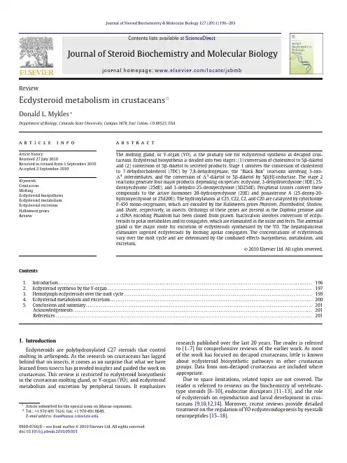
Journal of Steroid Biochemistry &Molecular Biology 127 (2011) 196–203Contents lists available at ScienceDirectJournal of Steroid Biochemistry and MolecularBiologyj o u r n a l h o m e p a g e :w w w.e l s e v i e r.c o m /l o c a t e /j s b mbReviewEcdysteroid metabolism in crustaceans ଝDonald L.Mykles ∗Department of Biology,Colorado State University,Campus 1878,Fort Collins,CO 80523,USAa r t i c l ei n f oArticle history:Received 27July 2010Received in revised form 1September 2010Accepted 2September 2010Keywords:Crustacean MoltingEcdysteroid biosynthesis Ecdysteroid metabolism Ecdysteroid excretion Halloween genes Reviewa b s t r a c tThe molting gland,or Y-organ (YO),is the primary site for ecdysteroid synthesis in decapod crus-taceans.Ecdysteroid biosynthesis is divided into two stages:(1)conversion of cholesterol to 5-diketol and (2)conversion of 5-diketol to secreted products.Stage 1involves the conversion of cholesterol to 7-dehydrocholesterol (7DC)by 7,8-dehydrogenase,the “Black Box”reactions involving 3-oxo- 4intermediates,and the conversion of 4-diketol to 5-diketol by 5[H]-reductase.The stage 2reactions generate four major products,depending on species:ecdysone,3-dehydroecdysone (3DE),25-deoxyecdysone (25dE),and 3-dehydro-25-deoxyecdysone (3D25dE).Peripheral tissues convert these compounds to the active hormones 20-hydroxyecdysone (20E)and ponasterone A (25-deoxy-20-hydroxyecdysone or 25d20E).The hydroxylations at C25,C22,C2,and C20are catalyzed by cytochrome P-450mono-oxygenases,which are encoded by the Halloween genes Phantom ,Disembodied ,Shadow ,and Shade ,respectively,in insects.Orthologs of these genes are present in the Daphnia genome and a cDNA encoding Phantom has been cloned from prawn.Inactivation involves conversion of ecdys-teroids to polar metabolites and/or conjugates,which are eliminated in the urine and feces.The antennal gland is the major route for excretion of ecdysteroids synthesized by the YO.The hepatopancreas eliminates ingested ecdysteroids by forming apolar conjugates.The concentrations of ecdysteroids vary over the molt cycle and are determined by the combined effects biosynthesis,metabolism,and excretion.© 2010 Elsevier Ltd. All rights reserved.Contents 1.Introduction ..........................................................................................................................................1962.Ecdysteroid synthesis by the Y-organ ...............................................................................................................1973.Hemolymph ecdysteroids over the molt cycle ......................................................................................................1994.Ecdysteroid metabolism and excretion ..............................................................................................................2005.Conclusions and summary ...........................................................................................................................201Acknowledgements ..................................................................................................................................201References ...........................................................................................................................................2011.IntroductionEcdysteroids are polyhydroxylated C27steroids that control molting in arthropods.As the research on crustaceans has lagged behind that on insects,it comes as no surprise that what we have learned from insects has provided insights and guided the work on crustaceans.This review is restricted to ecdysteroid biosynthesis in the crustacean molting gland,or Y-organ (YO),and ecdysteroid metabolism and excretion by peripheral tissues.It emphasizesଝArticle submitted for the special issue on Marine organisms.∗Tel.:+19704917616;fax:+19704910649.E-mail address:don@ .research published over the last 20years.The reader is referred to [1–7]for comprehensive reviews of the earlier work.As most of the work has focused on decapod crustaceans,little is known about ecdysteroid biosynthetic pathways in other crustacean groups.Data from non-decapod crustaceans are included where appropriate.Due to space limitations,related topics are not covered.The reader is referred to reviews on the biochemistry of vertebrate-type steroids [8–10],endocrine disruptors [11–13],and the role of ecdysteroids on reproduction and larval development in crus-taceans [9,10,12,14].Moreover,recent reviews provide detailed treatment on the regulation of YO ecdysteroidogenesis by eyestalk neuropeptides [15–18].0960-0760/$–see front matter © 2010 Elsevier Ltd. All rights reserved.doi:10.1016/j.jsbmb.2010.09.001D.L.Mykles /Journal of Steroid Biochemistry &Molecular Biology 127 (2011) 196–2031972.Ecdysteroid synthesis by the Y-organCholesterol is the precursor for ecdysteroid synthesis in the YO.The YOs are located in the cephalothorax,anterior to the branchial chamber.They are epithelioid tissues with cells contain-ing numerous mitochondria and extensive endoplasmic reticulum,which is characteristic of steroidogenic tissues of vertebrates [1,2,19].Arthropods cannot synthesize cholesterol de novo ,and therefore acquire it from their diet [3,20,21].Cholesterol binds to high-density lipoproteins in the hemolymph and is taken up by the YO [20–26].The uptake of cholesterol is an important rate-limiting step in the synthesis of ecdysteroids and is regulated by eyestalk neuropeptides,such as molt-inhibiting hormone (MIH)[21–23,27,28].Ecdysteroid biosynthesis in the molting glands of insects and crustaceans is similar,but the crustacean YO synthesizes a greater diversity of ecdysteroids [1,3,4,29,30].The biosynthetic pathway in the crustacean YO is complex.For convenience,ecdysteroid biosyn-thesis can be divided into two stages:(1)conversion of cholesterol to 5-diketol (Fig.1)and (2)conversion of 5-diketol to secreted products (Fig.2).The first stage of ecdysteroid synthesis is common to all decapod species.The second stage has four pathways with branching points at 5-diketol and 5-ketodiol [1,31,32].These pathways are named for the final ecdysteroid product secreted by the YO:ecdysone,3-dehydroecdysone (3DE),25-deoxyecdysone (25dE),and 3-dehydro-25-deoxyecdysone (3D25dE).YOs usually secrete two products,indicating that only two of the potential four pathways are operational at any given time in one species.This appears to be due to the relative activities of enzymes at the branch points,which directs metabolites along particular pathways.Cholesterol7-Dehydrocholesterol (7DC)5β-Diketol (3D2,22,25,dE)Non-molting glossy (Nm-g)/Shroud (Sro)Spook (Spo)/Spookier (Spok)Spookiest (Spot)Neverland (Nvd)(7,8-Dehydrogenase)5β[H]-Reductase“B l a c k B o x ”Δ -Diketol4Fig.1.Stage 1of the ecdysteroid biosynthetic pathway in crustacean molting gland (Y-organ).In the first reaction,cholesterol is converted to 7-dehydrocholesterol (7DC)by 7,8-dehydrogenase,encoded by the Neverland (nvd )gene in insects.In a series of reactions involving enzymes encoded by non-molting glossy /shroud ,Spook /Spookier ,and Spookiest ,termed the “Black Box”,7DC is converted to 4-Diketol,which is converted to 5-diketol by 5[H]-reductase.Modified from [1,29,31].Much of what we know about the biosynthetic pathway in the YO comes from metabolic studies using radiolabelled precursors.The first step is the conversion of cholesterol to 7-dehydrocholesterol (7DC)by 7,8-dehydrogenase;the two H()at carbons #7and #8are removed,resulting in the formation of a double bond between those carbons (Fig.1).Studies on a vari-ety of decapod crustacean species have shown that radiolabelled cholesterol injected into animals is converted to ecdysone,3DE,20-hydroxyecdysone (20E),25dE,3-dehydro-20-hydroxyecdysone (3D20E),and/or ponasterone A (PA;25-deoxy-20E or 25d20E)[21,26,33–35].A [3H]3-hydroxy- 4compound is poorly metab-olized by YO cells,indicating that the 3-oxo group is required for efficient 5-reduction [36].[3H]25-hydroxycholesterol (25C)is converted to 7-dehydro-25C by the YOs of green crab (Carci-nus maenas ),shrimp (Penaeus vannamei ),and crayfish (Orconectes limosus );the 7-dehydro-25C is converted to 3DE in C.maenas or to ecdysone and 3DE in P.vannamei and O.limosus [37].The YOs from Cancer antennarius can also synthesize 3DE from 25C [38].Although it is not a normal intermediate in the pathway,these results show that 25C can be a substrate for 7,8-dehydrogenase.The uptake of 25C is much faster than cholesterol,which makes it a useful tool for the study of the regulation of ecdysteroidogenesis [37,38].The 7DC undergoes a series of reactions involving 3-oxo- 4intermediates [36],the so-called “Black Box”,the details of which are becoming clearer [39].YO cells from C.maenas convert a 25-deoxy-3-oxo- 4intermediate to 3DE and 25dE;no ecdysone was detected [36].However,YO cells convert a 3-oxo- 4intermediate hydroxylated at C25to 3DE and ecdysone [36].This is consistent with studies showing that these ecdysteroids are synthesized from 25C as the precursor [32,37,38].The part of the pathway after the “Black Box”reactions is bet-ter understood,particularly the hydroxylations at C2,C22,and C25,which increase the polarity of the secreted products. 4-Diketol is first reduced at C5by 5[H]-reductase to form 5-diketol (Fig.2;3-dehydro-2,22,25-deoxyecdysone or 3D2,22,25dE).5-Diketol is converted to 5-ketodiol (2,22,25-deoxyecdysone or 2,22,25dE)by 3-dehydroecdysteroid-3-reductase (Fig.2)[31].In C.maenas ,[3H]5-diketol is ultimately converted to 3DE and 25dE by C.mae-nas YO cells [36].[3H]5-ketodiol is hydroxylated at C2,C22,or C25in C.maenas YOs to produce 22,25-deoxyecdysone (22,25dE),2,25-deoxyecdysone (2,25dE),or 2,22-dideoxyecdysone (2,22dE),respectively [40–42].2,22dE and 2,25dE are converted to ecdysone and 25dE,respectively,and secreted by the YO [40,41].22,25dE is not processed further and is not secreted [40,41].In the YOs of crayfish,[3H]5-diketol is converted to ecdysone and 3DE,whereas [3H]5-ketodiol is converted to ecdysone [31].The major secreted products of the YO are ecdysone,3DE,3D25dE,and 25dE,but the relative amounts vary among species.Four patterns are observed in decapod crustaceans (Table 1).TheTable 1Major ecdysteroid products secreted by the Y-organs of decapod crustaceans.The production of 25dE in U.pugilator is suggested by high hemolymph titers of ponas-terone A in premolt animals [30,49].SpeciesE a3DE a 25dE a 3D25dE aPachygrapsus crassipes •Cancer antennarius ••Orconectes limosus ••Procambarus clarkii ••Penaeus vannamei••Macrobrachium rosenbergii ••Carcinus maenas ••Callinectes sapidus ••Uca pugilator••Menippe mercenaria••aAbbreviations :E,ecdysone;3DE,3-dehydroecdysone;25dE,25-deoxyecdysone;3D25dE,3-dehydro-25-deoxyecdysone.198 D.L.Mykles /Journal of Steroid Biochemistry &Molecular Biology 127 (2011) 196–2035b- Diketol (3D2,22,25dE)5b -Ketodiol (2,22,25dE)5b- Ketotriol (2,22dE)20-Hydroxyecdysone (20E)b -reductase3-Dehydro-2,22-deoxyecdysone (3D2,22dE)3-Dehydro-2-deoxyecdysone (3D2dE)Disembodie 2,25-deoxyecdysone (2,25dE)Y-OrganPeripheral tissues3-Dehydroecdysone25-deoxyecdysone (25dE)Ponasterone A (PA)3-Dehydro-2,25-deoxyecdysone (3D2,25dE)3-Dehydro-25-deoxyecdysone (3D25dE)3-Dehydro-25-deoxyecdysone b -R(3D20E)Ponasterone A (PA)Fig.2.Stage 2of the ecdysteroid biosynthetic pathway in crustacean molting gland (Y-organ).Data indicate that there are two branching points,resulting in four pathways and four products secreted from the Y-organ.The products vary with species and molt stage,but no more than two major products are usually secreted at one time.3-Reductase and 20-hydroxylase activities in peripheral tissues convert the secreted products to the compounds circulating in the hemolymph.Therefore,the ecdysteroids in the hemolymph are determined by the compounds secreted by the YO and the activities of 3-reductase and 20-hydroxylase.All the compounds metabolized by peripheral tissues are detected in hemolymph samples;the major ecdysteroids in hemolymph are 20E,PA,3D20E,and 25d20E.In M.mercenaria ,a potential metabolite,3-dehydro-25-deoxy-20-hydroxyecdysone (3D25d20E),resulting from the 20-hydroxylation of 3D25dE has not been identified [32].The terminal hydroxylations at carbons #25,#22,#2,and #20are catalyzed by cytochrome P450mono-oxygenases encoded by Phantom (phm ),Disembodied (dib ),Shadow (sad ),and Shade (shd ),respectively.The specificities of these enzymes are such that the hydroxylations occur in the same order:C25→C22→C2→C20.Hence,hydroxylation at a specific carbon prevents hydroxylation by enzymes “upstream”of that step.For example,hydroxylation at C2prevents hydroxylations at C25and C22.Modified from [1,29,31,32].YOs in crayfish (O.limosus and Procambarus clarkii ),shrimp (P.vannamei and Macrobrachium rosenbergii ),and rock crab (C.anten-narius )secrete ecdysone and 3DE [25,31,43–46],the YOs in green crab (C.maenas )and blue crab (Callinectes sapidus )secrete ecdysone and 25dE [34,41](unpublished data on C.sapidus cited in [32]),theYOs in stone crab (Menippe mercenaria )secrete 3DE and 3D25dE [27,32],and the YOs in lined shore crab (Pachygrapsus crassipes )and fiddler crab (Uca pugilator )secrete ecdysone [47,48].However,the high levels of PA in the hemoymph of premolt U.pugilator indicate that the YO of this species also secretes 25dE in addition to ecdysoneD.L.Mykles/Journal of Steroid Biochemistry&Molecular Biology127 (2011) 196–203199[49].This suggests that the biosynthetic pathway has branching points at5-diketol and5-ketodiol,leading to fourfinal ecdys-teroid products(Fig.2).At thefirst branching point5-diketol can be converted to5-ketodiol,3-dehydro-2,22-deoxyecdysone (3D2,22dE),or3-dehydro-2,25-deoxyecdysone(3D2,25dE)(Fig.2). At the second branching point5-ketodiol can be converted to either5-ketotriol or2,25-deoxyecdysone(2,25dE),which leads to the production of ecdysone or25dE,respectively(Fig.2).In the pathway leading to ecdysone,5-ketodiol is hydroxylated sequen-tially at C25,C22,and C2[1].At thefirst branching point,5-diketol is hydroxylated at C25to3D2,22dE;subsequent serial hydroxyla-tions of3D2,22dE at C22and C2produce3DE[31].[3H]-3D2,22dE is converted to3DE and ecdysone by C.maenas YO cells[36].In M.mercenaria,5-diketol is apparently hydroxylated at C22to 3D2,25dE;subsequent hydroxylation at C2produces3D25dE[32]. At the second branching point,5-ketodiol is hydroxylated at C22 to2,25-deoxyecdysone(2,25dE);subsequent hydroxylation at C2 produces25dE.The branching points in the YO ecdsteroid synthetic pathway suggest that the P450mono-oxygenases,encoded by the Hal-loween genes Phantom(phm),Disembodied(dib),Shadow(sad), and Shade(shd)can bind multiple substrates.Phm apparently can hydroxylate5-diketol or5-ketodiol at C25;Dib apparently can hydroxylate5-diketol,3D2,22dE,5-ketodiol,or5-ketotriol at C22;Sad apparently can hydroxylate3D2dE,2,25dE,or2dE at C2; and Shd apparently can hydroxylate25dE,3DE,or ecdysone at C20 (Fig.2).However,the specificities of the Phm,Dib,Sad,and Shd enzymes are such that the C25→C22→C2→C20order of hydrox-ylations is maintained(Fig.2)[3,29].For example,compounds hydroxylated at C22(2,25dE and25dE)cannot be hydroxylated at C25in the YO from C.maenas[40].Hydroxylation at C2(22,25dE) prevents hydroxylations at C22and C25[1,40].Ecdysteroid biosynthetic reactions are catalyzed by reductases and P450mono-oxygenases(CYPs).The reduction of cholesterol to 7DC(Fig.1)is catalyzed by a7,8-dehydrogenase,which is encoded by the Neverland(nvd)gene in insects and nematode[50].The Nvd protein has two conserved domains involved in catalysis:a Rieske-like domain containing a2Fe-2S binding motif and a C-terminal domain containing a non-heme Fe(II)-binding motif[50]. Halloween genes constitute a family of P450mono-oxygenases, which have been well characterized in insects[29].They are char-acterized by havingfive conserved structural motifs(Helix-C or WxxxR sequence,Helix-I or GxE/DDT/S sequence,Helix-K or ExxR sequence,PERF motif or PxxFxPE/DRF sequence,and heme-binding or PFxxGxRxCxG/A sequence),as well as six substrate recogni-tion sites(SRS1,2/3,4,5,and6)[51].The“Black Box”reactions are catalyzed by non-molting glossy(nm-g)/shroud(sro)and sev-eral Halloween genes:spook(spo)and its paralog spookier(spok); and spookiest(spot)[51,52].Spo and Phm belong to the CYP2clade of P450enzymes and are associated with the endoplasmic reticu-lum(ER),whereas Dib,Sad,and Shd are associated with the inner membrane of the mitochondrion[29,53–55].Nm-g/sro encodes a short-chain NAD(P)H dehydrogenase/reductase,which appears to act upstream of Spo[52].Nm-g/sro has an N-terminal NADP/NAD binding domain and a central catalytic domain and is expressed primarily in prothoracic gland and ovary[52].Crustaceans appear to have the same enzymes for ecdysteroid biosynthesis as insects.An inhibitor of steroid5␣-reductase,L-645390,blocks the conversion of cholesterol to7DC in the YO of M.mercenaria[22].The5-reductase that converts 4-diketol to 5-diketol is a cytosolic enzyme in YO cells that requires NADPH for activity[36].Orthologs of nvd,nm-g/sro,spo,phm,dib,sad,and shd have been identified in the Daphnia pulex genome[52,54,56], and a cDNA encoding Phm has been cloned from Kuruma prawn, Marsupenaeus japonicus[57].Nvd and spo are located adjacent to each other in the D.pulex genome[54].The M.japonicus Phm and D.pulex Phm havefive conserved motifs present in insect Phm(WxxxR,GxE/DTT/S,ExxR,PxxFxPE/DRF,and PFxxGxRxCxG/A heme-binding motif),five of six substrate recognition sites(SRS 1,2/3,4,and5),and N-terminal ER-targeting and Pro/Gly-rich sequences[57].Mj-Phm is a target of eyestalk neuropeptides,as its expression in the YO is increased as much as7-fold during premolt and is decreased about2.5-fold by sinus gland extract and recom-binant MIH[57].None of the crustacean Halloween enzymes have been characterized biochemically.A member of the clade4of cytochrome P450enzymes has been cloned from O.limosus(CYP4C15)and C.maenas(CYP4C39) [58,59].Both genes encode proteins with hydrophobic N-terminal ER-targeting and Pro/Gly-rich sequences characteristic of microso-mal cytochrome P450enzymes[58,59].CYP4C15is expressed in the YO and the protein is associated with the ER[58,60].More-over,CYP4C15expression in the YO is increased during premolt and is decreased by MIH[58,60].These data suggest that they are involved in ecdysteroid biosynthesis,but their precise role remains to be determined.It has been hypothesized that the gene cat-alyzes7,8-dehydrogenation or25-hydroxylation[58,60].However, CYP4C15and CYP4C39have low sequence identity/similarity to nvd and phm,making it unlikely that these genes are involved in7,8-dehydrogenation and25-hydroxylation,respectively,in the YO. CYP4C39is expressed in the hepatopancreas,which suggests it may have a role in ecdysteroid metabolism by peripheral tissues[59].Ecdysteroids secreted by the YO are converted to20-hydroxyecdysone(20E)and related compounds by peripheral tissues,such as gonad,hindgut,abdominal ganglia,eyestalk ganglia,hepatopancreas,antennal gland,and epidermis(Fig.2) [38,61–66].YOs cannot convert3DE to ecdysone or ecdysone to20E[31,43,61].In peripheral tissues,ecdysone is hydroxy-lated at C20,probably by Shd,to produce20E[38,40,41,62–64]. A20-hydroxylase activity in the hepatopancreas of the blackback land crab(Gecarcinus lateralis)requires NADPH,is inhibited by metyrapone(an inhibitor of cytochrome P450enzymes),and is associated with the mitochondrial fraction[62].A3-reductase converts3DE to ecdysone,which is hydroxylated at C20,to produce 20E[32,38,45,61,63].In M.mercenaria,3-reductase in peripheral tissues converts3D25dE to25dE,which is then converted to PA [32].In C.maenas,25dE is converted to PA by a20-hydroxylase in the testis[34,40,41].PA can be hydroxylated at C25or C26 to produce20E or inokosterone,respectively[67].In crayfish(O. limosus),abdominal ganglia convert3DE to ecdysone,20E,and 3D20E[63].Antennal glands of crayfish express an aldoketore-ductase,but its precise function is unknown;it may catalyze 3-reduction or may have an osmoregulatory role by converting D-glucose to sorbitol[68].These data indicate that peripheral tissues have3-reductase,25-hydroxylase,and20-hydroxylase activities, which can convert products secreted by the YO to20E and related compounds.3.Hemolymph ecdysteroids over the molt cycleTotal ecdysteroid titers in the hemolymph vary over the molt cycle.In general,ecdysteroids are low during intermolt and postmolt;during premolt,concentrations rise and reach a peak shortly before molting(ecdysis)in a variety of crustacean species [2,5–7,46,62,69–89].A precipitous drop in hemolymph ecdys-teroids triggers the shedding of the exoskeleton[90].Eyestalk ablation,which removes the primary source of inhibitory neu-ropeptides(e.g.,MIH),activates the YO and increases hemolymph ecdysteroid levels[1,2,21,23,70,79,86,91,92].In addition to the peak in hemolymph titers at the end of the premolt stage,there are smaller peaks during postmolt and early premolt stages,which may have physiological functions[30,48,93–95].For species with terminal molts,the YOs degenerate and ecdysteroids remain at200 D.L.Mykles/Journal of Steroid Biochemistry&Molecular Biology127 (2011) 196–203low concentrations in the hemolymph,even after eyestalk ablation [2,96].The major ecdysteroids identified in the hemolymph of decapod crustaceans are ecdysone,20E,3D20E,and PA, but the specific compounds and their levels vary between species,developmental stage,molt stage,and season [32,44,48,49,61,64,66,67,70,78,81,82,87,93,97].In stone crabs (M.mercenaria)that have been eyestalk ablated to activate the YO, 20E,3D20E,and PA are present in about equal amounts;a fourth large peak eluting from a reverse-phase column was not identified [32].Infiddler crab(U.pugilator)induced to molt by eyestalk ablation,there are two transitory peaks of hemolymph ecdysteroid that differ in the relative titers of ecdysteroids.Peak I,which occurs at early premolt,has a higher ratio of ecdysone to20E than peak II,which occurs at late premolt[48].Eyestalk ablation alters the composition of ecdysteroids circulating in the hemolymph, but the effect differs between species.In blue crabs(C.sapidus) at late premolt,PA is the major ecdysteroid in intact animals, whereas20E is the major ecdysteroid in eyestalk-ablated animals [70].In U.pugilator,the PA:20E ratio in the hemolymph is higher in animals induced to molt by eyestalk ablation than in animals induced to molt by multiple leg autotomy[49].In Kuruma prawn (Penaeus japonicus),20E and PA are the major ecdysteroids during the postmolt and intermolt stages,while20E alone is the major ecdysteroid during early and late premolt stages[87].In C.maenas, ecdysone and20E are detected at low levels in the hemolymph of intermolt animals,whereas the PA level is higher than the20E level in the hemolymph of premolt animals[34,67,78].At late premolt(stage D3),the titers of PA in the hemolymph decline more rapidly than those of ecdysone and20E[78].This corresponds to a change in the relative amounts of ecdysone and25dE,a precursor to PA,secreted by the YO between intermolt and premolt animals; YOs from premolt animals secrete greater amounts of25dE than ecdysone than YOs from intermolt animals[40,41].This appears to depend on the activities of enzymes at the branching points (Fig.2).For example,in rock crab and crayfish YOs,an increase in3-reductase activity would shift production and secretion from3DE to ecdysone[31].In green andfiddler crabs,an increase in25-hydroxylase activity would shift secretion from25dE to ecdysone[31].Ecdysteroid composition can be affected by developmental stage.During the last three molts,blue crabs(C.sapidus)transi-tion through juvenile,prepubertal,and adult stages.PA is the major hemolymph ecdysteroid at all three stages.However,the titers of PA in juveniles are significantly higher than and the titers of20E significantly lower than the titers in prepubertal and adult stages [70].In juvenile C.sapidus,eyestalk ablation increases hemolymph ecdysteroid titers,but there is no peak in ecdysteroids prior to ecd-ysis and titers during postmolt remain elevated compared to intact animals[70].These data indicate that there are stage-specific dif-ferences in ecdysteroid metabolism,but the functional significance of these changes remains to be investigated.4.Ecdysteroid metabolism and excretion20E and PA are the major active ecdysteroids circulating in the hemolymph of decapod crustaceans.Inactivation involves conver-sion to more polar metabolites and/or formation of conjugates, which are excreted in the urine or eliminated in the feces.Thus,the two major organs responsible for removing ecdysteroids from the hemolymph are the antennal gland and hepatopancreas(midgut gland)in decapods.The diversity of inactivation pathways in crustaceans is reminiscent to those used by insects to detoxify ecdysteroids ingested from plants(see[98]for review).The pri-mary site for excretion of ecdysteroids is the antennal gland.In lobster(Homarus americanus),between81%and96%of excreted ecdysteroids is in the urine,depending on the molt stage;the remainder is in the feces[66,97].In crayfish(O.limosus),there is a preferential excretion of ecdysone over20E.About two-thirds of the[3H]ecdysone injected into the hemolymph appears in the water after1h[99].In contrast,most of the[3H]20E injected into the hemolymph remains in the hemolymph,with less than5%of the[3H]20E appearing in the water after1h[99].This suggests that selective excretion of unmetabolized ecdysone by the antennal gland keeps hemolymph ecdysone titers low.Conversion of ecdysteroids to more polar compounds involves two modifications of the side chain at the#26carbon:hydrox-ylation and oxidation.In insects,20E is inactivated by a cytochrome P45026-hydroxylase[100,101].In decapod crus-taceans,a26-hydroxylase activity in various tissues converts20E to20,26-dihydroxyecdysone(20,26E)and PA to inokosterone(25-deoxy-20,26-dihydroxyecdysone or25d20,26E)[63,65–67,97]. Oxidation at the C26forms ecdysonoic acids(EAs).For example, 20E and PA are metabolized to20-hydroxyecdysonoic acid(20EA) and25-deoxy-20-hydroxyecdysonoic acid(25d20EA),respec-tively,and are excreted in the urine[65–67,97,102].Ecdysteroids can also be converted to their3␣-hydroxy epimers by periph-eral tissues.In crayfish(P.clarkii),a3␣-reductase activity in the epidermis converts ecdysone and20E to3␣-hydroxyecdysone and3␣,20-hydroxyecdysone,respectively,which are selectively excreted[61].Various tissues can convert ecdysteroids and their metabolites to highly polar(HP)conjugates,which can be excreted in the urine[61,63,65,66,97].The levels of HP conjugates vary over the molt cycle and can at times be the predominant ecdysteroid in the hemolymph[61,77,82,87,88,93].In H.americanus,the rapid drop in ecdysteroids at the end of premolt is associated with an increase in HP metabolites in the hemolymph and excretion in the urine[93,97].Using[3H]ecdysone as a tracer,a greater proportion (about75%after72h)is converted into HP conjugates in postmolt (stage A)and intermolt(stage C)lobsters than is converted in early premolt(stages D0and D1)lobsters(about25%after72h)[66]. The conversion by late premolt(stage D2)lobsters increases after injection and reaches early premolt levels by96h as animals tran-sition through the late premolt ecdysteroid peak[66].In C.maenas, intermolt and premolt animals differ in the metabolism of[3H]PA injected into the hemolymph.Forty-eight hours after injection into intermolt animals,the major metabolites were conjugates and 20EA,whereas24h after injection into premolt animals,the major metabolites were conjugates only[67].Enzymatic hydrolysis with snail sulfatase or porcine liver esterase is used to analyze the com-position of conjugates[66,93].HP conjugates in the hemolymph contain various ecdysteroids and metabolites usually in proportion to their hemolymph concentrations[93,97].This indicates that the modifications are generally nonspecific.However,HP conjugates in the urine contain relatively high proportions of polar metabo-lites,such as20,26E,3␣-hydroxy compounds,and ecdysonoic acids [61,66,97].In addition,excretion rates of H.americanus are higher in intermolt and late premolt(D2)stages than early premolt(D0 and D1)stages[66].These data suggest that the conversion of ecdysteroids to inactive polar metabolites and their preferential excretion by the antennal gland can determine the ecdysteroid titers in the hemolymph,especially at the end of premolt when titers drop precipitously.The hepatopancreas is the primary site for the production of apolar conjugates(perhaps fatty acyl esters),which are then elim-inated in the feces[65,66,99].These conjugates can be formed from ecdysteroids in the hemolymph[63,65,66,84,97]or from ecdysteroids ingested with food[97].When lobsters were fed [3H]ecdysone,less than0.4%of the radioactivity appeared in the hemolymph and urine;essentially all of the[3H]ecdysone was in the form of apolar conjugates in the feces[97].The amounts of apo-。

Remnant PbI2, an unforeseen necessity in high-efficiency hybrid perovskite-based solar cells?a)Duyen H. Cao, Constantinos C. Stoumpos, Christos D. Malliakas, Michael J. Katz, Omar K. Farha, Joseph T. Hupp, and Mercouri G. KanatzidisCitation: APL Materials 2, 091101 (2014); doi: 10.1063/1.4895038View online: /10.1063/1.4895038View Table of Contents: /content/aip/journal/aplmater/2/9?ver=pdfcovPublished by the AIP PublishingArticles you may be interested inParameters influencing the deposition of methylammonium lead halide iodide in hole conductor free perovskite-based solar cellsAPL Mat. 2, 081502 (2014); 10.1063/1.4885548Air stability of TiO2/PbS colloidal nanoparticle solar cells and its impact on power efficiencyAppl. Phys. Lett. 99, 063512 (2011); 10.1063/1.3617469High efficiency mesoporous titanium oxide PbS quantum dot solar cells at low temperatureAppl. Phys. Lett. 97, 043106 (2010); 10.1063/1.3459146Near-IR activity of hybrid solar cells: Enhancement of efficiency by dissociating excitons generated in PbS nanoparticlesAppl. Phys. Lett. 96, 073505 (2010); 10.1063/1.3292183Effects of molecular interface modification in hybrid organic-inorganic photovoltaic cellsJ. Appl. Phys. 101, 114503 (2007); 10.1063/1.2737977APL MATERIALS2,091101(2014)Remnant PbI2,an unforeseen necessity in high-efficiency hybrid perovskite-based solar cells?aDuyen H.Cao,1Constantinos C.Stoumpos,1Christos D.Malliakas,1Michael J.Katz,1Omar K.Farha,1,2Joseph T.Hupp,1,band Mercouri G.Kanatzidis1,b1Department of Chemistry,and Argonne-Northwestern Solar Energy Research(ANSER)Center,Northwestern University,2145Sheridan Road,Evanston,Illinois60208,USA2Department of Chemistry,Faculty of Science,King Abdulaziz University,Jeddah,Saudi Arabia(Received2May2014;accepted26August2014;published online18September2014)Perovskite-containing solar cells were fabricated in a two-step procedure in whichPbI2is deposited via spin-coating and subsequently converted to the CH3NH3PbI3perovskite by dipping in a solution of CH3NH3I.By varying the dipping time from5s to2h,we observe that the device performance shows an unexpectedly remark-able trend.At dipping times below15min the current density and voltage of thedevice are enhanced from10.1mA/cm2and933mV(5s)to15.1mA/cm2and1036mV(15min).However,upon further conversion,the current density decreases to9.7mA/cm2and846mV after2h.Based on X-ray diffraction data,we determinedthat remnant PbI2is always present in these devices.Work function and dark currentmeasurements showed that the remnant PbI2has a beneficial effect and acts as ablocking layer between the TiO2semiconductor and the perovskite itself reducingthe probability of back electron transfer(charge recombination).Furthermore,wefind that increased dipping time leads to an increase in the size of perovskite crys-tals at the perovskite-hole-transporting material interface.Overall,approximately15min dipping time(∼2%unconverted PbI2)is necessary for achieving optimaldevice efficiency.©2014Author(s).All article content,except where otherwisenoted,is licensed under a Creative Commons Attribution3.0Unported License.[/10.1063/1.4895038]With the global growth in energy demand and with compelling climate-related environmental concerns,alternatives to the use of non-renewable and noxious fossil fuels are needed.1One such alternative energy resource,and arguably the only legitimate long-term solution,is solar energy. Photovoltaic devices which are capable of converting the photonflux to electricity are one such device.2Over the last2years,halide hybrid perovskite-based solar cells with high efficiency have engendered enormous interest in the photovoltaic community.3,4Among the perovskite choices, methylammonium lead iodide(MAPbI3)has become the archetypal light absorber.Recently,how-ever,Sn-based perovskites have been successfully implemented in functional solar cells.5,6MAPbI3 is an attractive light absorber due to its extraordinary absorption coefficient of1.5×104cm−1 at550nm;7it would take roughly1μm of material to absorb99%of theflux at550nm.Further-more,with a band gap of1.55eV(800nm),assuming an external quantum efficiency of90%,a maximum current density of ca.23mA/cm2is attainable with MAPbI3.Recent reports have commented on the variability in device performance as a function of perovskite layer fabrication.8In our laboratory,we too have observed that seemingly identicalfilmsa Invited for the Perovskite Solar Cells special topic.b Authors to whom correspondence should be addressed.Electronic addresses:j-hupp@ and m-kanatzidis@2,091101-12166-532X/2014/2(9)/091101/7©Author(s)2014FIG.1.X-ray diffraction patterns of CH3NH3PbI3films with increasing dipping time(%composition of PbI2was determined by Rietveld analysis(see Sec.S3of the supplementary material for the Rietveld analysis details).have markedly different device performance.For example,when ourfilms of PbI2are exposed to MAI for several seconds(ca.60s),then a light brown coloredfilm is obtained rather than the black color commonly observed for bulk MAPbI3(see Sec.S2of the supplementary material for the optical band gap of bulk MAPbI3).23This brown color suggests only partial conversion to MAPbI3and yields solar cells exhibiting a J sc of13.4mA/cm2and a V oc of960mV;these values are significantly below the21.3mA/cm2and1000mV obtained by others.4Under the hypothesis that fully converted films will achieve optimal light harvesting efficiency,we increased the conversion time from seconds to2h.Unexpectedly,the2-h dipping device did not show an improved photovoltaic response(J sc =9.7mA/cm2,V oc=846mV)even though conversion to MAPbI3appeared to be complete.With the only obvious difference between these two devices being the dipping time,we hypothesized that the degree of conversion of PbI2to the MAPbI3perovskite is an important parameter in obtaining optimal device performance.We thus set out to understand the correlation between the method of fabrication of the MAPbI3layer,the precise chemical compositions,and both the physical and photo-physical properties of thefilm.We report here that remnant PbI2is crucial in forming a barrier layer to electron interception/recombination leading to optimized J sc and V oc in these hybrid perovskite-based solar cells.We constructed perovskite-containing devices using a two-step deposition method according to a reported procedure with some modifications.4(see Sec.S1of the supplementary material for the experimental details).23MAPbI3-containing photo-anodes were made by varying the dipping time of the PbI2-coated photo-anode in MAI solution.In order to minimize the effects from unforeseen variables,care was taken to ensure that allfilms were prepared in an identical manner.The composi-tions offinal MAPbI3-containingfilms were monitored by X-ray diffraction(XRD).Independently of the dipping times,only theβ-phase of the MAPbI3is formed(Figure1).9However,in addition to theβ-phase,allfilms also showed the presence of unconverted PbI2(Figure1,marked with*) which can be most easily observed via the(001)and(003)reflections at2θ=12.56◦and38.54◦respectively.As the dipping time is increased,the intensities of PbI2reflections decrease with a concomitant increase in the MAPbI3intensities.In addition to the decrease in peak intensities of PbI2,the peak width increases as the dipping time increases indicating that the size of the PbI2 crystallites is decreasing,as expected,and the converse is observed for the MAPbI3reflections.This observation suggests that the conversion process begins from the surface of the PbI2crystallites and proceeds toward the center where the crystallite domain size of the MAPbI3phase increases and that of PbI2diminishes.Interestingly,the remnant PbI2phase can be seen in the data of other reports, but has not been identified as a primary source of variability in cell performance.8,10 Considering that the perovskite is the primary light absorber within the device,we wantedto further investigate how the optical absorption of thefilm changes with increasing dipping timeFIG.2.Absorption spectra of CH3NH3PbI3films as a function of unconverted PbI2phase fraction.FIG.3.(a)J-V curves and(b)EQE of CH3NH3PbI3-based devices as a function of unconverted PbI2phase fraction.(Figure2).11,12The pure PbI2film shows a band gap of2.40eV,consistent with the yellow color of PbI2.As the PbI2film is gradually converted to the perovskite,the band gap is progressively shifted toward1.60eV.The deviation of MAPbI3’s band gap(1.60eV)from that of the bulk MAPbI3 material(1.55eV)could be explained by quantum confinement effects related with the sizes of TiO2and MAPbI3crystallites and their interfacial interaction.13,14Interestingly,we also noticed the presence of a second absorption in the light absorber layer,in which the gap gradually red shifts from1.90eV to1.50eV as the PbI2concentration is decreased from9.5%to0.3%(Figure2—blue arrow).Having established the chemical compositions and optical properties of the light absorberfilms, we proceeded to examine the photo-physical responses of the corresponding functional devices in order to determine how the remnant PbI2affects device performance.The pure PbI2based device remarkably achieved a0.4%efficiency with a J sc of2.1mA/cm2and a V oc of564mV (Figure3(a)).Upon progressive conversion of the PbI2layer to MAPbI3,we observe two different regions(Figure4,Table I).In thefirst region,the expected behavior is observed;as more PbI2is converted to MAPbI3,the trend is toward higher photovoltaic efficiency,due both to J sc and V oc, until1.7%PbI2is reached.The increase in J sc is attributable,at least in part,to increasing absorption of light by the perovskite.We speculate that progressive elimination of PbI2,present as a layer between TiO2and the perovskite,also leads to higher net yields for electron injection into TiO2and therefore,higher J values.For a sufficiently thick PbI2spacer layer,electron injection would occur instepwise fashion,i.e.,perovskite→PbI2→TiO2.Finally,the photovoltage increase is attributable toFIG.4.Summary of J-V data vs.PbI2concentration of CH3NH3PbI3-based devices(Region1:0to15min dipping time, Region2:15min to2h dipping time).TABLE I.Photovoltaic performance of CH3NH3PbI3-based devices as a function of unconverted PbI2fraction.Dipping time PbI2concentration a J sc(mA/cm2)V oc(V)Fill factor(%)Efficiency(%) 0s100% 2.10.564320.45s9.5%10.10.93352 4.960s7.2%13.40.96052 6.72min 5.3%14.00.964557.45min 3.7%14.70.995578.315min 1.7%15.1 1.036629.730min0.8%13.60.968648.51h0.4%12.40.938657.62h0.3%9.70.84668 5.5a Determined from the Rietveld analysis of X-ray diffraction data.the positive shift in TiO2’s quasi-Fermi level as the population of photo-injected electrons is higher with increased concentration of MAPbI3.The second region yields a notably different trend;surprisingly,below a concentration of2% PbI2,J sc,V oc,and ultimatelyηdecrease.Considering that the light-harvesting efficiency would increase when the remaining2%PbI2is converted to MAPbI3(albeit to only a small degree),then the remnant PbI2must have some other role.We posit that remnant PbI2serves to inhibit detrimental electron-transfer processes(Figure5).Two such processes are back electron transfer from TiO2to holes in the valence band of the perovskite(charge-recombination)or to the holes in the HOMO of the HTM(charge-interception).This retardation of electron interception/recombination observation is reminiscent of the behavior of atomic layer deposited Al2O3/ZrO2layers that have been employed in dye-sensitized solar cells.15–18It is conceivable that the conversion of PbI2to MAPbI3occurs from the solution interface toward the TiO2/PbI2interface and thus would leave sandwiched between TiO2and MAPbI3a blocking layer of PbI2that inhibits charge-interception/recombination.For this hypothesis to be correct,it is crucial that the conduction-band-edge energy(E cb)of the PbI2be higher than the E cb of the TiO2.19–21 The work function of PbI2was measured by ultraviolet photoelectron spectroscopy(UPS)and was observed to be at6.35eV vs.vacuum level,which is0.9eV lower than the valence-band-edge energy(E vb)of MAPbI3(see Sec.S7of the supplementary material23for the work function of PbI2);the E cb(4.05eV)was calculated by subtracting the work function from the band gap(2.30eV).solar cell.FIG.6.Dark current of CH3NH3PbI3-based devices as a function of unconverted PbI2phase fraction.The E cb of PbI2is0.26eV higher than the E cb of TiO2and thus PbI2satisfies the conditions of a charge-recombination/interception barrier layer.In order to probe the hypothesis that PbI2acts as a charge-interception barrier,dark current measurements,in which electronsflow from TiO2to the HOMO of the HTM,were made.Consistent with our hypothesis,Figure6illustrates that the onset of the dark current occurs at lower potentials as the PbI2concentration decreases.In the absence of other effects,the increasing dark current with increasing fraction of perovskite(and decreasing fraction of PbI2)should result in progressively lower open-circuit photovoltages.Instead,the photocurrent density and the open-circuit photovoltage bothincrease,at least until to PbI2fraction reaches1.7%.As discussed above,thinning of a PbI2-basedFIG.7.Cross-sectional SEM images of CH3NH3PbI3film with different dipping time.sandwich layer should lead to higher net injection yields,but excessive thinning would diminish the effectiveness of PbI2as a barrier layer for back electron transfer reactions.Given the surprising role of remnant PbI2in these devices,we further probed the two-step conversion process by using scanning-electron microscopy(SEM)(Figure7).Two domains of lead-containing materials(PbI2and MAPbI3)are present.Thefirst domain is sited within the mesoporous TiO2network(area1)while the second grows on top of the network(area2).Area2initially contains 200nm crystals.As the dipping time is increased,the crystals show marked changes in size and morphology.The formation of bigger perovskite crystals is likely the result of the thermodynamically driven Ostwald ripening process,i.e.,smaller perovskite crystals dissolves and re-deposits onto larger perovskite crystals.22The rate of charge-interception,as measured via dark current,is proportional to the contact area between the perovskite and the HTM.Thus,the eventual formation of large, high-aspect-ratio crystals,as shown in Figure7,may well lead to increases in contact area and thereby contributes to the dark-current in Figure6.Regardless,we found that the formation of large perovskite crystals greatly decreased our success rate in constructing high-functioning,non-shorting solar cells.In summary,residual PbI2appears to play an important role in boosting overall efficiencies for CH3NH3PbI3-containing photovoltaics.PbI2’s role appears to be that of a TiO2-supported blocking layer,thereby slowing rates of electron(TiO2)/hole(perovskite)recombination,as well as decreasing rates of electron interception by the hole-transporting material.Optimal performance for energy conversion is observed when ca.98%of the initially present PbI2has been converted to the perovskite. Conversion to this extent requires about15min.Pushing beyond98%(and beyond15min of reaction time)diminishes cell performance and diminishes the success rate in constructing non-shorting cells.The latter problem is evidently a consequence of conversion of small and more-or-less uniformly packed perovskite crystallites to larger,poorly packed crystallites of varying shape and size.Finally,the essential,but previously unrecognized,role played by remnant PbI2 provides an additional explanation for why cells prepared dissolving and then depositing pre-formed CH3NH3PbI3generally under-perform those prepared via the intermediacy of PbI2.We thank Prof.Tobin Marks for use of the solar simulator and EQE measurement system. Electron microscopy was done at the Electron Probe Instrumentation Center(EPIC)at Northwestern University.Ultraviolet Photoemission Spectroscopy was done at the Keck Interdisciplinary SurfaceScience facility(Keck-II)at Northwestern University.This research was supported as part of theANSER Center,an Energy Frontier Research Center funded by the U.S Department of Energy, Office of Science,Office of Basic Energy Sciences,under Award No.DE-SC0001059.1R.Monastersky,Nature(London)497(7447),13(2013).2H.J.Snaith,J.Phys.Chem.Lett.4(21),3623(2013).3M.M.Lee,J.Teuscher,T.Miyasaka,T.N.Murakami,and H.J.Snaith,Science338(6107),643(2012).4J.Burschka,N.Pellet,S.J.Moon,R.Humphry-Baker,P.Gao,M.K.Nazeeruddin,and M.Gratzel,Nature(London) 499(7458),316(2013).5F.Hao,C.C.Stoumpos,D.H.Cao,R.P.H.Chang,and M.G.Kanatzidis,Nat.Photonics8(6),489(2014);F.Hao,C.C. Stoumpos,R.P.H.Chang,and M.G.Kanatzidis,J.Am.Chem.Soc.136,8094–8099(2014).6N.K.Noel,S.D.Stranks,A.Abate,C.Wehrenfennig,S.Guarnera,A.Haghighirad,A.Sadhanala,G.E.Eperon,M.B. Johnston,A.M.Petrozza,L.M.Herz,and H.J.Snaith,Energy Environ.Sci.7,3061(2014).7H.S.Kim,C.R.Lee,J.H.Im,K.B.Lee,T.Moehl,A.Marchioro,S.J.Moon,R.Humphry-Baker,J.H.Yum,J.E.Moser, M.Gratzel,and N.G.Park,Sci.Rep.2,591(2012).8D.Y.Liu and T.L.Kelly,Nat.Photonics8(2),133(2014).9C.C.Stoumpos,C.D.Malliakas,and M.G.Kanatzidis,Inorg.Chem.52(15),9019(2013).10J.H.Noh,S.H.Im,J.H.Heo,T.N.Mandal,and S.I.Seok,Nano Lett.13(4),1764(2013).11Diffuse reflectance measurements of MAPbI3films were converted to absorption spectra using the Kubelka-Munk equation,α/S=(1-R)2/2R,where R is the percentage of reflected light,andαand S are the absorption and scattering coefficients, respectively.The band gap values are the energy value at the intersection point of the absorption spectrum’s tangent line and the energy axis.12L.F.Gate,Appl.Opt.13(2),236(1974).13O.V oskoboynikov,C.P.Lee,and I.Tretyak,Phys.Rev.B63(16),165306(2001).14X.X.Xue,W.Ji,Z.Mao,H.J.Mao,Y.Wang,X.Wang,W.D.Ruan,B.Zhao,and J.R.Lombardi,J.Phys.Chem.C 116(15),8792(2012).15E.Palomares,J.N.Clifford,S.A.Haque,T.Lutz,and J.R.Durrant,J.Am.Chem.Soc.125(2),475(2003).16C.Prasittichai,J.R.Avila,O.K.Farha,and J.T.Hupp,J.Am.Chem.Soc.135(44),16328(2013).17A.K.Chandiran,M.K.Nazeeruddin,and M.Gratzel,Adv.Funct.Mater.24(11),1615(2014).18M.J.Katz,M.J.D.Vermeer,O.K.Farha,M.J.Pellin,and J.T.Hupp,Langmuir29(2),806(2013).19M.J.DeVries,M.J.Pellin,and J.T.Hupp,Langmuir26(11),9082(2010).20C.Prasittichai and J.T.Hupp,J.Phys.Chem.Lett.1(10),1611(2010).21F.Fabregat-Santiago,J.Garcia-Canadas,E.Palomares,J.N.Clifford,S.A.Haque,J.R.Durrant,G.Garcia-Belmonte, and J.Bisquert,J.Appl.Phys.96(11),6903(2004).22Alan D.McNaught and Andrew Wilkinson,IUPAC Compendium of Chemical Terminology(Blackwell Scientific Publica-tions,Oxford,1997).23See supplementary material at /10.1063/1.4895038for experimental details,absorption spectrum of bulk CH3NH3PbI3,fraction,size,absorption spectrum,work function of unconverted PbI2,and average photovoltaic perfor-mance.。
齐墩果酸抗肿瘤作用机制的研究进展闫月乔(综述);杨宇(审校)【摘要】恶性肿瘤已经成为严重威胁人类生命与健康的疾病之一,随着对恶性肿瘤治疗方面研究的不断进展,越来越多的抑制肿瘤的有效物质被发现。
齐墩果酸( OA)是近年来发现的具有抑癌作用的重要植物提取物,多年来的研究表明其具有明显的抑制肿瘤生长的作用,随着对齐墩果酸研究的不断深入,其抗肿瘤作用的机制也逐渐被发现。
研究结果表明齐墩果酸的抗肿瘤机制存在多条途径。
%Malignant tumor has become one of the serious problems threatening human life and health . With the development of the research on malignant tumor treatment , more and more antitumor active sub-stances have been found.Oleanolic acid( OA) is an important plant extract that can significantly inhibit the growth of cancer,which is found in recent years .With the deepening of research of OA ,its anti-tumor mecha-nisms have also been identified ,The results show that the anti-tumor mechanism of oleanolicacid in the pres-ence of multiple way.【期刊名称】《医学综述》【年(卷),期】2016(022)005【总页数】4页(P914-917)【关键词】齐墩果酸;抗肿瘤作用;凋亡【作者】闫月乔(综述);杨宇(审校)【作者单位】哈尔滨医科大学附属第二医院肿瘤内科,哈尔滨150001;哈尔滨医科大学附属第二医院肿瘤内科,哈尔滨150001【正文语种】中文【中图分类】R691齐墩果酸(oleanolic acid,OA) 是一种天然植物提取物,也是自然界中分别最广泛的五环三萜类化合物,近年来,研究表明,在肝癌[1]、白血病[2]、肺癌[3]等恶性肿瘤中具有抑制肿瘤生长的作用,但其具体的分子生物学作用机制还不清楚。
熊猫豆提取物抑制肿瘤活性及诱导结肠癌细胞Caco-2凋亡机制研究胡冬梅;彭吕杨;薛照辉【摘要】为了探究熊猫豆的抑制肿瘤活性及其诱导Caco-2凋亡的机制,采用80%丙酮浸提法制备熊猫豆粗提物,通过MTT法测定其对人结肠癌细胞Caco-2增殖的抑制效果,琼脂糖凝胶电泳、流式细胞术、酶联免疫分析等技术与方法检测细胞周期、DNA及凋亡相关蛋白相对表达量的变化.结果发现,与空白对照组相比,0.1~1.0 mg/mL熊猫豆丙酮提取物对Caco-2细胞增殖有明显的抑制作用,且呈现剂量效应关系,IC50为0.72 mg/mL.0.4、0.6、0.8 mg/mL熊猫豆丙酮提取物处理72 h后的Caco-2细胞,可以观察到典型的凋亡细胞的梯状DNA条带,细胞周期G1期延长,CytC、caspase-9、caspase-3蛋白的相对表达量均有显著上升.熊猫豆丙酮能够抑制结肠癌细胞Caco-2增殖,这种抑制作用是通过阻滞细胞周期和诱导线粒体通路介导的细胞凋亡实现的.【期刊名称】《中国粮油学报》【年(卷),期】2015(030)004【总页数】5页(P6-10)【关键词】熊猫豆;人结肠癌细胞Caco-2;细胞凋亡;线粒体通路【作者】胡冬梅;彭吕杨;薛照辉【作者单位】天津大学化工学院食品科学系,天津300072;天津大学化工学院食品科学系,天津300072;天津大学化工学院食品科学系,天津300072【正文语种】中文【中图分类】R735.3结肠癌是全球最常见的恶性肿瘤之一,全球每年新发病例约800万人,占所有恶性肿瘤的10%~15%,死亡率仅次于肺癌和肝癌[1]。
目前,结肠癌的治疗方法仍以手术治疗、放射治疗和化学治疗为主,但手术创伤性大、恢复时间长,在很大程度上限制其应用;放射治疗和化学治疗具有非特异性的细胞毒素作用,在杀伤肿瘤细胞的同时对机体正常组织器官也会造成一定程度的损伤,在应用方面仍存在争议。
食物治疗因其成本低廉、方便无毒害等优点,日益成为防癌抗癌的研究热点。
OleananetriterpenoidCDDO-MeinhibitsgrowthandinducesapoptosisinprostatecancercellsthroughaROS-dependentmechanism
DorrahDeeba,XiaohuaGaoa,HaoJiangb,BranislavaJanicc,AliS.Arbabc,YonRojanasakuld,ScottA.Dulchavskya,SubhashC.Gautama,*
aDepartmentofSurgery,HenryFordHealthSystem,Detroit,MI,UnitedStates
bDepartmentofNeurology,HenryFordHealthSystem,Detroit,MI,UnitedStates
cDepartmentofRadiology,HenryFordHealthSystem,Detroit,MI,UnitedStates
dDepartmentofPharmaceuticalSciences,WestVirginiaUniversity,Morgantown,WV,UnitedStates
1.IntroductionCarcinomaoftheprostate(CaP)isthemostcommonlydiagnosedcancerandthesecondleadingcauseofcancerrelatedmortalityinmenintheUnitedStates.Currenttherapies,suchasradicalprostatectomy,localradiotherapyorbrachytherapyhaveonlylimitedefficacyagainstthemetastaticdisease[1,2]andeffectivechemotherapeuticagentsareunavailableatpresent.High
fatdiethasbeenlinkedtotheincreasedriskofmetastaticprostatecancer[3].Incontrast,consumptionoflowfatdietalongwithhighintakeofdarkgreenleafyvegetables,fruits,andsoyproductsisconsideredtosignificantlyreducetheincidenceofprostatecancer.Thecancer-preventingeffectsofplant-derivedfoodshavebeenattributedtothepresenceofpolyphenolicphytochemicalswithstrongantioxidantactivity[4].Indeed,therehasbeenanincreasinguseofdietarysupplementstopreventand/ortreatprostatecancer.Thus,clinicaldevelopmentofplant-derivedflavonoidsandphenolic/polyphenolicantioxidantsaschemopre-ventiveagentscouldsignificantlyreducedisease-relatedmorbid-ityandmortalityandimproveprognosis.Triterpenesortriterpenoidsaremembersofalargerfamilyofstructurallyrelatedcompoundsknownascyclosqualenoidsthatarewidelydistributedintheplantkingdom[5].Oleanolicacidandursolicacidarenaturallyoccurringtriterpenoidsthathavebeenusedintraditionalmedicineforcenturiesasantibacterial,antifungal,anticancer,andanti-inflammatoryagents[6–8].
BiochemicalPharmacology79(2010)350–360ARTICLEINFOArticlehistory:Received15July2009Accepted2September2009Keywords:CCDO-MeProstatecancerROSApoptosisMitochondrialdepolarizationCellsurvivalsignalingABSTRACTCDDO-Me,asynthetictriterpenoidderivedfromoleanolicacid,isapromisinganticanceragentthathasshownstrongactivityagainstawidevarietyofcancertypesinvitroandinvivo.WehavepreviouslyshownthatCDDO-Meinducesapoptosisinprostatecancercellsirrespectiveoftheirhormonalstatus.TofurtherunderstandtheproapoptoticmechanismofCDDO-Me,weinvestigatedtheroleofreactiveoxygenspecies(ROS)inmediatingtheapoptosisinducingactivityofCDDO-MeinLNCaPandPC-3prostatecancercelllines.Here,weshowthatCDDO-MeinducesROSgenerationfrombothnonmitochondrialandmitochondrialsources,whichisassociatedwiththeinductionofapoptosisascharacterizedbyincreasedannexinV-binding,cleavageofPARP-1andprocaspases-3,-8,-9,lossofmitochondrialmembranepotentialandreleaseofcytochromec.Inaddition,CDDO-MeinhibitedcellsurvivalAkt,NF-kBandmTORsignalingproteins.TheinhibitionofROSgenerationbyN-acetylcysteine(NAC)orbyoverexpressionofantioxidantenzymesglutathioneperoxidase(GPx)andsuperoxidedismutase-1(SOD-1)preventedCDDO-Me-inducedapoptosis.PretreatmentwithNACblockedannexinV-binding,cleavageofPARP-1andprocaspases-3,-8,-9,lossofmitochondrialmembranepotentialandreleaseofcytochromecbyCDDO-Me.NACalsopreventedtheinhibitionofconstitutivelyactiveAkt,NF-kBandmTORbyCDDO-Me.Together,thesedataindicatethatROSplaysanessentialroleinthe
inductionofapoptosisbyCDDO-Meinprostatecancercells.ß2009ElsevierInc.Allrightsreserved.
Abbreviations:CDDO,2-cyano-3,12-dioxooleana-1,9(11)-dien-28-oicacid;CDDO-Me,C-28methylesterderivativeofCDDO;H2DCFDA,6-carboxy-2,7-dichlorodihy-droflurosceindiacetate;DHE,dihydroethidiumbromide;ROS,reactiveoxygenspecies;PARP-1,poly-ADP-ribosepolymerase-1;NF-kB,nuclearfactorkappaB;mTOR,mammaliantargetofrapamycin;NAC,N-acetylcystein;SOD-1,superoxidedismutase-1;GPx,glutathioneperoxidase.*Correspondingauthorat:SurgicalResearch4D,OneFordPlace,Detroit,MI48202,UnitedStates.Tel.:+13138746998;fax:+13138743770.E-mailaddress:sgautam1@hfhs.org(S.C.Gautam).
ContentslistsavailableatScienceDirectBiochemicalPharmacology
journalhomepage:www.elsevier.com/locate/biochempharm
0006-2952/$–seefrontmatterß2009ElsevierInc.Allrightsreserved.doi:10.1016/j.bcp.2009.09.006