第二部分 药学生化实验
实验一. 疏水作用层析
【实验目的】
通过实验了解疏水作用层析的原理与方法。
【实验原理】
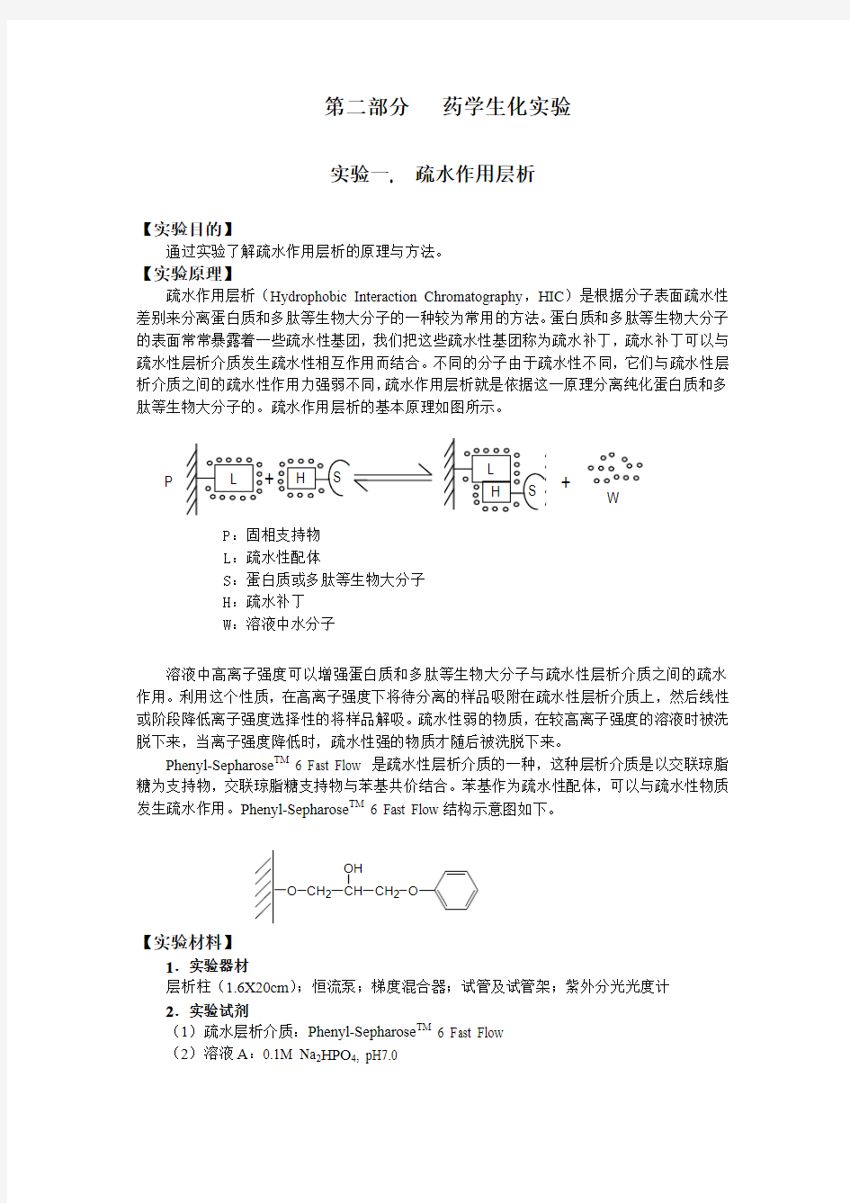
疏水作用层析(Hydrophobic Interaction Chromatography ,HIC )是根据分子表面疏水性差别来分离蛋白质和多肽等生物大分子的一种较为常用的方法。蛋白质和多肽等生物大分子的表面常常暴露着一些疏水性基团,我们把这些疏水性基团称为疏水补丁,疏水补丁可以与疏水性层析介质发生疏水性相互作用而结合。不同的分子由于疏水性不同,它们与疏水性层析介质之间的疏水性作用力强弱不同,疏水作用层析就是依据这一原理分离纯化蛋白质和多肽等生物大分子的。疏水作用层析的基本原理如图所示。
溶液中高离子强度可以增强蛋白质和多肽等生物大分子与疏水性层析介质之间的疏水作用。利用这个性质,在高离子强度下将待分离的样品吸附在疏水性层析介质上,然后线性或阶段降低离子强度选择性的将样品解吸。疏水性弱的物质,在较高离子强度的溶液时被洗脱下来,当离子强度降低时,疏水性强的物质才随后被洗脱下来。
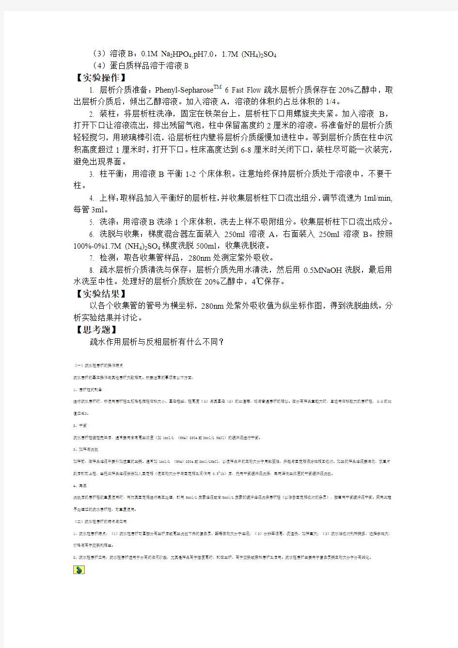
Phenyl-Sepharose TM 6 Fast Flow 是疏水性层析介质的一种,这种层析介质是以交联琼脂糖为支持物,交联琼脂糖支持物与苯基共价结合。苯基作为疏水性配体,可以与疏水性物质发生疏水作用。Phenyl-Sepharose TM 6 Fast Flow 结构示意图如下。
O CH 2CH CH 2O
OH
【实验材料】
1.实验器材
层析柱(1.6X20cm );恒流泵;梯度混合器;试管及试管架;紫外分光光度计
2.实验试剂
(1)疏水层析介质:Phenyl-Sepharose TM 6 Fast Flow
(2)溶液A :0.1M Na 2HPO 4, pH7.0
+ W
P :固相支持物
L :疏水性配体
S :蛋白质或多肽等生物大分子
H :疏水补丁
W :溶液中水分子
P
(3)溶液B:0.1M Na2HPO4,pH7.0,1.7M (NH4)2SO4
(4)蛋白质样品溶于溶液B
【实验操作】
1. 层析介质准备:Phenyl-Sepharose TM 6 Fast Flow疏水层析介质保存在20%乙醇中,取出层析介质后,倾出乙醇溶液。加入溶液A,溶液的体积约占总体积的1/4。
2. 装柱:将层析柱洗净,固定在铁架台上,层析柱下口用螺旋夹夹紧。加入溶液B,打开下口让溶液流出,排出残留气泡,柱中保留高度约2厘米的溶液。将准备好的层析介质轻轻搅匀,用玻璃棒引流,沿层析柱内壁将层析介质缓慢加进柱中。等到层析介质在柱中沉积高度超过1厘米时,打开下口。柱床高度达到6-8厘米时关闭下口,装柱尽可能一次装完,避免出现界面。
3. 柱平衡:用溶液B平衡1-2个床体积。注意始终保持层析介质处于溶液中,不要干柱。
4. 上样:取样品加入平衡好的层析柱,并收集层析柱下口流出组分,调节流速为1ml/min,每管3ml。
5. 洗涤:用溶液B洗涤1个床体积,洗去上样不吸附组分。收集层析柱下口流出成分。
6. 洗脱与收集:梯度混合器左面装入250ml溶液A,右面装入250ml溶液B。按照100%-0%1.7M (NH4)2SO4梯度洗脱500ml,收集洗脱液。
7. 检测:取各收集管样品,280nm处测定紫外吸收。
8. 疏水层析介质清洗与保存:层析介质先用水清洗,然后用0.5MNaOH洗脱,最后用水洗至中性。处理好的层析介质放在20%乙醇中,4℃保存。
【实验结果】
以各个收集管的管号为横坐标,280nm处紫外吸收值为纵坐标作图,得到洗脱曲线。分析实验结果并讨论。
【思考题】
疏水作用层析与反相层析有什么不同?
(一)疏水柱层析的操作特点
疏水层析的基本操作与其他层析大致相同。所要注意的事项有以下方面:
1、层析柱的制备
进行疏水层析时,所使用层析柱之规格包括柱体积大小、直径粗细、柱高度(h)与其直径(d)的比值等,均与普通层析的相似。在分离样品量较大时,宜选用体积较大的层析柱,h:d的比
值应≤3。
2、平衡
疏水层析柱装柱完毕后,通常要用含有高盐浓度(如1mol/L (NH4)2SO4或2mol/L NaCl)的缓冲液进行平衡。
3、加样与洗脱
加样前,在样品溶液中要补加适量的盐类。通常加1mol/L (NH4)2SO4或2mol/LNaCl,以使样品中的生物大分子局部变性,并能与固定相很好地相互吸附。加盐的样品溶液要混匀,放置片
刻后即可上柱,当把此样品溶液徐徐加入固定相(使生物大分子与固定相之间作用0.5~1h)后,先用平衡缓冲液洗涤,再用降低盐浓度的平衡缓冲液洗脱。
4、再生
洗脱后的层析柱欲重复使用时,需对其固定相进行再生处理,即用8mol/L尿素溶液或含8mol/L尿素的缓冲溶液洗涤层析柱(以除去固定相吸附的杂质),接着用平衡缓冲液平衡。采用此程
序处理过的疏水层析柱,可重复使用。
(二)疏水柱层析的特点与应用
1、疏水柱层析特点:(1)疏水柱层析可直接分离盐析后或高盐洗脱下来的蛋白质、酶等生物大分子溶液;(2)分辨率很高、流速快、加样量大;(3)疏水性吸附剂种类多,选择余地大,价格与离子交换剂相当。
2、疏水柱层析应用:疏水柱层析适用于分离的任何阶段,尤其是样品离子强度高时,即在盐析、离子交换或亲和层析之后用。疏水柱层析主要用于蛋白质类生物大分子分离纯化。
Experiment 13 Hydrophobic Interaction Chromatography
【Purpose 】
To understand the principle and method of hydrophobic interaction chromatography.
【Principle 】
Hydrophobic Interaction Chromatography (HIC) is a versatile method for the purification and separation of biomolecules based on differences in their surface hydrophobicity.
Proteins and peptides usually sequester hydrophobic amino acids in domains away from the surface of the molecule. However, many biomolecules considered hydrophilic have sufficient hydrophobic groups exposed to allow interaction with hydrophobic ligands attached to the chromatographic matrix.
As shown in the following figure, substances are separated on the basis of their varying strength of their hydrophobic interaction with hydrophobic groups attached to the uncharged matrix.
Hydrophobic interaction between a biomolecule and the matrix is enhanced by high ionic strength buffers. This technique is usually performed in the presence of moderately high concentrations of salts in the adsorption buffer. Elution is achieved by a linear or step wise decrease in concentration of the salt.
Phenyl-Sepharose TM 6 Fast Flow is one of the HIC medium. In Phenyl-Sepharose TM 6 Fast Flow, phenyl groups as HIC ligand which have interaction with hydrophobic biomolecules are coupled to the cross-linked agarose matrices via glycidyl ether bonds (shown in the following figure)。
O CH 2CH CH 2O
OH
【Materials 】
1. Apparatus:
(1)Column (16/20)
(2)Pump
(3)Gradient maker
+ W
P :Polymer matrix
L :Ligand attached to polymer matrix
S :Solute molecule
H :Hydrophobic patch on surface of solute molecule
W :water molecules in the bulk solution
P
(4)Ultraviolet meters
2.Reagents:
(1)Phenyl-Sepharose TM 6 Fast Flow
(2)Buffer A: 0.1M Na2HPO4 pH7.0
(3)Buffer B: 0.1M Na2HPO4 pH7.0 1.7M (NH4)2SO4
(4)Protein sample solved in buffer B
【Procedure】
1. Preparing the gel
(1)Equilibrate all material to room temperature.
(2)Phenyl-Sepharose TM6 Fast Flow medium is supplied pre-swollen in 20% ethanol. Decant the ethanol solution and replace it with buffer A to a total volume of 32.5ml (75% settled gel 25% buffer).
(3)Degas the slurry under vacuum.
2. Assembling the column
(1)Details of the column parts can be found in the instructions supplied with the column. Before package, ensure that all parts, particularly the nets, net fasteners and glass tubes, are clean and intact.
(2)Mount the end piece on the column and close it with a stopper, add buffer B into the column. Open the bottom outlet of the column to let out some buffers to ensure that there is no air bubbles trapped under the net. Close the column with a stopper.
(3)Flush the column with buffer B. leaving a few ml at the bottom. Mount the column vertically on a laboratory stand.
3. Packing the column
(1)Pour the gel slurry into the column in one continuous motion. Pouring down a glass rod held against the wall of the column helps prevent the introduction of air bubbles. Fill the remainder of the column with buffer.
(2)Mount the top piece on the column and connect it with the pump.
4. Equilibration
(1)Open the bottom outlet of the column and start the pump.
(2)To equilibrate, pump approximately 100ml of buffer B through the column at a flow rate of 1ml/min. The column is fully equilibrated when the pH and /or conductivity of the effluent is the same as buffer B.
5. Banding
(1)Applied the sample prepared onto the equilibrated column.
(2)Collect the solutions which has flown out at a fraction of 3 ml per test tube
6. Wash
Wash the column with buffer B to get rid of samples do not bind and collect the solutions which has flown out.
7. Elution
Elute the column with a gradient form 100% B to 100% A, total volume is 500ml and collect the elution.
8. Assay
Assay the optical density of the sample in the test tubes by ultraviolet meter at 280nm.
9. Clean of the medium
(1)After every run, wash the column with 50ml of distilled H2O.
(2)To remove precipitated proteins by washing the column with 80ml 1M NaOH solution, followed immediately with 50ml of distilled H2O.
(3)Equilibrate the column with 20% ethanol. The medium should be stored in 20% ethanol at 4℃.
【Results】
Take the test tube numbered as X axis and the optical density at 280nm as Y axis, draw out the elute curve, and analysis the result and discuss.
【Questions】
What is the difference between HIC and reverse phase chromatography?