Caspase-independent_cell_deaths.1
- 格式:pdf
- 大小:136.75 KB
- 文档页数:7

hippocampus after hyp〇xia-ischemia(J]. Glia, 2015,63(12): 2220-2230. [18] Wang S, Xue H, Xu Y, et al. Sevoflurane postconditioning inhibitsautophagy through activation of the extracellular signal-regulated kinase cascade, alleviating hypoxic-ischem ic brain injury in neonatal rats[J]. Neurochem Res, 2019, 44(2):347-356.[19] Li X, Wang MH, Qin C, et al. Fingolimod suppresses neuronalauophagy through the mTOR/p70S6K pathway and alleviates ischemic brain damage in mire[J]. PLoS One, 2017, 12(1 l):e0188748.[20] Yan YQ, Zhang B, Wang L, et al. Induction of apoptosis andautophagic cell death by the vanillin derivative 6-brom ine-5-hydroxy-4-methoxybenzaldehyde is accompanied by the cleavage ofDNA-PKcs and rapid destruction of c-Myc oncoprotein in HepG2cells[J]. Cancer Lett, 2007, 252(2):280-289.[21 ]Requejo-Aguilar R, Lopez-Fabuel I, Fernandez E, et al. PINK1deficiency sustains cell proliferation by reprogramming glucosemetabolism through HIF1[J]. Nat Commun, 2014,5: 4514.[22] Devireddy S, Liu A, I^ampe T, et al. The organization of milochondrialquality control and life cycle in the nervous system in vivo in theabsence of PINKlfJ]. J Neurosci, 2015, 35(25): 9391-9401.(收稿日期:2020-10-10)•论著•芍药苷对急性脑梗死大鼠小胶质细胞激活调节的神经元焦亡的作用祖力菲亚•艾克木麦合莆热提•乌莆尔邢辉辉樊明新张俊江【摘要】目的研究芍药苷对急性脑梗死(AC1)大鼠神经元焦亡的影响。
细胞焦亡及相关疾病的研究进展何明月;袁芳;刘映红【摘要】细胞焦亡是由Gasdermin蛋白介导的一种程序性炎性细胞死亡方式,分为依赖caspase-1的经典途径和依赖caspase-4/5/11的非经典途径.炎性小体的激活在此过程中扮演重要角色.细胞焦亡参与肾脏疾病、动脉粥样硬化、神经系统疾病、感染性疾病等的发生发展,并发挥重要作用.通过对细胞焦亡作用机制及相关疾病的研究,为临床疾病的防治提供新思路.【期刊名称】《基础医学与临床》【年(卷),期】2019(039)004【总页数】4页(P560-563)【关键词】细胞焦亡;炎性小体;caspase-1/4/5/11;GSDMD【作者】何明月;袁芳;刘映红【作者单位】中南大学湘雅医学院湘雅二医院肾内科,湖南长沙410011;中南大学湘雅医学院湘雅二医院肾内科,湖南长沙410011;中南大学湘雅医学院湘雅二医院肾内科,湖南长沙410011【正文语种】中文【中图分类】R34细胞焦亡(pyroptosis)是一种由GSDMD介导的细胞程序性坏死。
炎性小体(inflammasome)激活半胱天冬酶(caspase-1/4/5/11),进而切割GSDMD,使其N端与C端分离,打破自抑制,通过正负电荷之间的相互作用,在细胞膜上通过寡聚体化形成蜂窝状的孔道(内径约10~14 nm),产生水分内流、细胞膜内外离子梯度消失,细胞发生肿胀、渗透性溶解最终导致破裂死亡[1-2]。
细胞焦亡参与肾脏疾病、动脉粥样硬化、神经系统疾病、感染性疾病等的发病机制[3-6],并发挥重要作用。
对细胞焦亡的深入研究已经成为近年来国际炎性反应领域的研究热点。
文章就细胞焦亡及相关疾病的研究进展进行综述。
1 Caspase-1或caspase-4/5/11切割GSDMD引起细胞焦亡1.1 细胞焦亡的“执行者”-GSDMDGSDMD属于功能未知的gasdermin蛋白家族,其他成员包括gasdermin A、gasdermin B、gasdermin C、DFNA5和DFNB59。
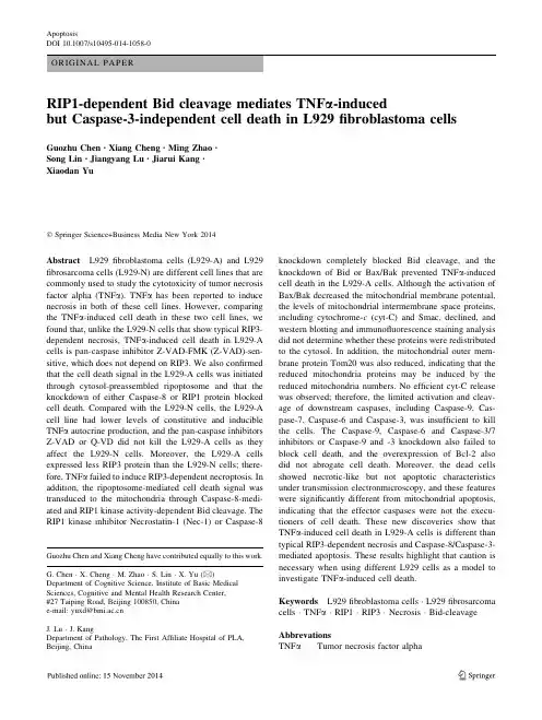
ORIGINAL PAPERRIP1-dependent Bid cleavage mediates TNF a -inducedbut Caspase-3-independent cell death in L929fibroblastoma cellsGuozhu Chen •Xiang Cheng •Ming Zhao •Song Lin •Jiangyang Lu •Jiarui Kang •Xiaodan YuÓSpringer Science+Business Media New York 2014Abstract L929fibroblastoma cells (L929-A)and L929fibrosarcoma cells (L929-N)are different cell lines that are commonly used to study the cytotoxicity of tumor necrosis factor alpha (TNF a ).TNF a has been reported to induce necrosis in both of these cell lines.However,comparing the TNF a -induced cell death in these two cell lines,we found that,unlike the L929-N cells that show typical RIP3-dependent necrosis,TNF a -induced cell death in L929-A cells is pan-caspase inhibitor Z-VAD-FMK (Z-VAD)-sen-sitive,which does not depend on RIP3.We also confirmed that the cell death signal in the L929-A cells was initiated through cytosol-preassembled ripoptosome and that the knockdown of either Caspase-8or RIP1protein blocked cell pared with the L929-N cells,the L929-A cell line had lower levels of constitutive and inducible TNF a autocrine production,and the pan-caspase inhibitors Z-VAD or Q-VD did not kill the L929-A cells as they affect the L929-N cells.Moreover,the L929-A cells expressed less RIP3protein than the L929-N cells;there-fore,TNF a failed to induce RIP3-dependent necroptosis.In addition,the ripoptosome-mediated cell death signal was transduced to the mitochondria through Caspase-8-medi-ated and RIP1kinase activity-dependent Bid cleavage.The RIP1kinase inhibitor Necrostatin-1(Nec-1)or Caspase-8knockdown completely blocked Bid cleavage,and the knockdown of Bid or Bax/Bak prevented TNF a -induced cell death in the L929-A cells.Although the activation of Bax/Bak decreased the mitochondrial membrane potential,the levels of mitochondrial intermembrane space proteins,including cytochrome-c (cyt-C)and Smac,declined,and western blotting and immunofluorescence staining analysis did not determine whether these proteins were redistributed to the cytosol.In addition,the mitochondrial outer mem-brane protein Tom20was also reduced,indicating that the reduced mitochondria proteins may be induced by the reduced mitochondria numbers.No efficient cyt-C release was observed;therefore,the limited activation and cleav-age of downstream caspases,including Caspase-9,Cas-pase-7,Caspase-6and Caspase-3,was insufficient to kill the cells.The Caspase-9,Caspase-6and Caspase-3/7inhibitors or Caspase-9and -3knockdown also failed to block cell death,and the overexpression of Bcl-2also did not abrogate cell death.Moreover,the dead cells showed necrotic-like but not apoptotic characteristics under transmission electronmicroscopy,and these features were significantly different from mitochondrial apoptosis,indicating that the effector caspases were not the execu-tioners of cell death.These new discoveries show that TNF a -induced cell death in L929-A cells is different than typical RIP3-dependent necrosis and Caspase-8/Caspase-3-mediated apoptosis.These results highlight that caution is necessary when using different L929cells as a model to investigate TNF a -induced cell death.Keywords L929fibroblastoma cells ÁL929fibrosarcoma cells ÁTNF a ÁRIP1ÁRIP3ÁNecrosis ÁBid-cleavage Abbrevations TNF a Tumor necrosis factor alphaGuozhu Chen and Xiang Cheng have contributed equally to this work.G.Chen ÁX.Cheng ÁM.Zhao ÁS.Lin ÁX.Yu (&)Department of Cognitive Science,Institute of Basic Medical Sciences,Cognitive and Mental Health Research Center,#27Taiping Road,Beijing 100850,China e-mail:yuxd@J.Lu ÁJ.KangDepartment of Pathology,The First Affiliate Hospital of PLA,Beijing,ChinaApoptosisDOI 10.1007/s10495-014-1058-0Z-VAD Z-VAD-FMKNec-1Necrostatin-1MMP Mitochondrial membrane potentialROS Reactive oxygen speciescyt-C Cytochrome-cIntroductionProgrammed cell death is essential to the normal functions of multi-cellular organisms and plays a critical role in development,immunity,inflammation,and cancer pro-gression.Based on morphological and biochemical fea-tures,programmed cell death is classified into apoptosis, necrosis(also termed necroptosis),and autophagic cell death[1,2].Although many of the initiators,regulators, and effectors in these three types of cell death are different, increasing evidence indicates that apoptosis,necrosis,and autophagic cell death are not completely independent cell death pathways and that the various death pathways are closely intertwined with crosstalk between the regulating components.TNF a is a pleiotropic cytokine that induces a variety of cellular responses,including the promotion of cell survival and induction of programmed cell death.In addition to inducing Caspase-dependent apoptosis,TNF a also induces RIP1and RIP3kinase-dependent necroptosis as an alter-native during apoptosis-deficient conditions[3,4].The mechanisms by which TNF a induces either apoptosis or necroptosis have been widely studied in recent years,and the cell death type is mediated by the formation of apop-totic or necrotic signaling complexes[5–7].Apoptosis is mediated by complex IIa,which contains RIP1,FADD, Caspase-8,and TRADD.In complex IIa,Caspase-8 becomes activated and then activates downstream Caspase-3to induce apoptosis[8–10].In certain cell lines,activated Caspase-8also induces apoptosis by cleaving Bid,which then activates the Bax/Bak pore and subsequent events, causing the activation of Caspases-9and-3[11,12].In cells with high RIP3expression and treatment with the broad-spectrum Caspase-inhibitor Z-VAD to inhibit the Caspase-8-induced cleavage of RIP1,RIP3,and CYLD, RIP3interacts with RIP1to form complex IIb(also known as the necrosome),which then initiates necroptosis[13–16]. Notably,in addition to death receptor activation-induced necrosis,many other inducers such as ischemia–reperfu-sion,oxidative stress,calcium overload,and others also induce necrosis[17].However,the mechanisms by which these intrinsic stimuli induce necroptosis remain primarily undefined.Similar to TNF a inducing complex IIa forma-tion,genotoxic stress and the loss of IAPs are known to induce ripoptosome formation.Although the ripoptosome comprises the same components as complex IIa,the rip-optosome pre-assembles or spontaneously assembles and does not require pre-existing TNFR signaling platforms.In addition,the ripoptosome,in the absence or presence of RIP3,can either lead to RIP1kinase-dependent apoptosis or RIP1kinase-dependent necrosis,respectively[18,19]. In addition to influencing inducible ripoptosome assembly, whether RIP1kinase also involves the regulation of mito-chondria dysfunction and downstream effector caspase activation during ripoptosome-initiated apoptosis is cur-rently unknown.L929cells are commonly used to test the cytotoxic effects of TNF a;however,this short name actually corre-sponds to two different cell lines(L-929fibroblastoma cells ATCCÒCCL-1TM and WEHI-13VARfibrosarcoma cells ATCCÒCRL-2148TM)is prone to cause some con-fusion.The common feature of these two L929cell lines is that both are sensitive to TNF a,and TNF a alone induces cell death.Although several early studies have reported Fig.1Z-VAD differentially regulates TNF a-induced cell death in L929-A and L929-N cells.a Nec-1inhibited TNF a-induced cell death in both L929-A and L929-N cells.The cells were treated with TNF a (10ng/mL for L929-A cells and50ng/mL for L929-N cells in all experiments)in the absence or presence of Nec-1(50l M in all experiments)for24h,and cell death was examined by microscopy (9200)as based on morphological changes.More than threefields in each group were observed,and representative images are shown.The cells were also stained with Annexin V-FITC and PI and analyzed by flow cytometry.Representative measurements of at least three independent experiments are shown.The cell death values reported represent the mean±SD of three separate experiments.*P\0.01.b TNF a-induced cytotoxicity was inhibited in L929-A cells but promoted in L929-N cells in response to Z-VAD treatment.The cells were treated with TNF a in the absence or presence of Z-VAD(20l M in all experiments)for24h,and cell death was examined by morphological changes via microscopy(9200)andflow cytome-try.*P\0.01,#P\0.05.c Z-VAD and Nec-1suppressed the TNF a-induced cleavage of PARP and Caspase-3.The cells were treated with TNF a in the absence or presence of Z-VAD and Nec-1for12h. Western blotting was used to detect the cleavage of PARP and Caspase-3induced by TNF a.d Pan-caspase inhibitors induced cell death in L929-N cells but not L929-A cells.The cells were treated with Z-VAD or Q-VD(50l M)for48h.Cell death was examined by microscopy(9200)as based on morphological changes.The cells were also stained with propidium iodide and analyzed byflow cytometry.*P\0.01.e Z-VAD significantly promoted TNF a tran-scription in L929-N cells but not L929-A cells.The cells were treated with Z-VAD for12h and then collected for RNA extraction.The TNF a mRNA level was determined by RT-PCR.The representative images of three independent experiments are shown.f The effect of Z-VAD on the activation of Erk in L929-A and L929-N cells.The cells were treated with20l M Z-VAD for the indicated time points and then lysed.The phosphorylated and total Erk levels were determined by western Blotting.g RIP3was differentially expressed in the L929-A and L929-N cells.The L929-A and L929-N cells were treated with TNF a for12h,and western blotting was used to determine the RIP1and RIP3levels.GAPDH was used as a loading controlcApoptosisthat TNF a induces apoptosis or atypical apoptosis in L929fibroblastoma cells(for clarification we refer to this cell line here as L929-A)[20–22],a recent study demonstrated that TNF a induces necrotic cell death[23].In contrast, TNF a induces typical necroptosis in the L929fibrosarcoma cell line(L929-N)[24–26].Although L929cells have been widely used as a model to study TNF a-induced necropto-sis,the results obtained from different studies involving the regulation of TNF a-induced necrosis are surprisingly var-iable or even contradictory.For example,RIP1knockdown has been reported to either enhance or inhibit the resistance of L929cells to TNF a[1,27],and autophagy initiation may either block necrosis or enhance necrosis[28,29].No explanations currently exist clarifying these differences; however,the respective L929cells used in these studies may have actually been different variants of this cell line. In this study,we compared the TNF a-induced cells death in these two L929cell lines,and unlike L929-N cells, TNF a-induced cell death in the L929-A cells was not RIP3-dependent necrosis but was RIP1-and Caspase-8-dependent and Caspase-3-independent cell death with necrotic morphology.These results indicate that we needto Apoptosisbe cautious when using different L929cells as a model to investigate TNF a-induced cell death.ResultsZ-VAD inhibits TNF a-induced cell death in L929-A cells but enhances death in L929-N cells via autocrine TNF a productionFirst,we treated L929-A and L929-N cells with the TNF a and RIP1kinase inhibitor,Nec-1,or the pan-caspase-inhibitor,Z-VAD,to observe their effects on cell death as evaluated either using microscopy orflow cytometry ana-lysis.Both cell lines were sensitive to TNF a,with the L929-A cells being more sensitive than the L929-N cells. Nec-1blocked TNF a-induced cell death in both cell lines, indicating that the cell death was RIP1-dependent(Fig.1a).However,the effects of Z-VAD on TNF a-induced cell death were contrasting.Specifically,Z-VAD exacerbated TNF a-induced cell death in the L929-N cells as previously reported[30],whereas Z-VAD inhibited cell death in the L929-A cells(Fig.1b).Western blot analysis also showed the cleaved Caspase-3and PARP levels in the L929-A cells treated with TNF a but not in the L929-N cells.More importantly,like Z-VAD,Nec-1also blocked the cleavage of Caspase-3and PARP(Fig.1c).A previous study reported that Z-VAD alone induces necroptosis in L929cells through the activation of MAPKs and AP-1and subsequent autocrine TNF a production[31]. We treated the cells with pan-caspase-inhibitors and found that both Z-VAD and Q-VD induce cell death in the L929-N cells but not in the L929-A cells(Fig.1d).We then assessed the effect of Z-VAD on TNF a transcription by RT-PCR.The results showed that L929-N cells constitu-tively expressed a high level of TNF a and that TNF a mRNA levels remarkably enhanced following Z-VAD stimulation,whereas the L929-A cells showed very low levels of constitutive TNF a expression,which were only faintly enhanced by Z-VAD(Fig.1e).In addition,we found that the L929-N cells have high levels of constitutive ERK activation(Fig.1f).Thesefindings support the hypothesis that the absence of autocrine TNF a in L929-A cells may allow the cells to escape Z-VAD-induced necrosis.A recent study showed that protein kinase RIPK3 activity determines whether cells die by necroptosis or apoptosis[32];therefore,we evaluated the expression of RIP1and RIP3proteins in these two L929cell lines fol-lowing TNF a exposure,and the expression of RIP3in the L929-N cells was much higher than that in L929-A cells. Moreover,TNF a reduced RIP1protein levels in both cell lines;however,in the L929-A cells,the RIP3protein levels were reduced at greater amounts compared with in the levels in the L929-N cells(Fig.1g).TNF a-induced L929-A cell death is RIP1-and Caspase-8-dependent but RIP3-independentThe key step for TNF a-induced apoptosis is the formation of complex IIa;therefore,wefirst investigated the associ-ation between RIP1,Caspase-8,and FADD in L929-A cells via immunoprecipitation analysis.Unexpectedly,a pre-existing interaction between the RIP1,Caspase-8,and FADD proteins was observed prior to TNF a stimulation, which is consistent with an assembled ripoptosome[19]; moreover,TNF a significantly decreased the protein level of RIP1and Caspase-8in the whole cell lysate,causing less RIP1to be immunoprecipitated compared with the control. However,a higher level of Caspase-8was co-immuno-precipitated with RIP1,indicating that the association between RIP1and Caspase-8was remarkably enhanced in TNF a-treated cells(Fig.2a).Although TNF a induced time-dependent cell death,cleaved PARP did not exhibit the same increase,and the Caspase-8,RIP1,and cFLIP L proteins decreased(Fig.2b).The Caspase-8decrease was reversed by Nec-1and partially recovered by Z-VAD treatment but not by treatment with the Caspase-8inhibitor Z-IETD(Fig.2c).The effects of the components of com-plex II on TNF a-induced cell death were further investi-gated via the separate knockdown of Caspase-8,RIP1,and RIP3.The results confirmed that TNF a-induced cell death in the L929-N cells occurred through RIP3-dependent necrosis(Fig.2d).In the L929-A cells,only the knock-down of RIP1or Caspase-8,not RIP3,markedly decreased the cytotoxicity of TNF a(Fig.2e).Fig.2Effect of TNF a on the composition of complex II proteins inL929-A cells.a TNF a promoted the binding between Caspase-8and RIP1.The cells were treated with or without TNF a for12h.An anti-RIP1antibody was used for immunoprecipitation,and western blotting was then used to analyze the binding between RIP1and Caspase-8.b The effects of TNF a on inducing time-dependent cell death and protein expression.The cells were treated with TNF a for the indicated times,cell death was examined by microscopy(9200), and the protein levels of FLIP L,RIP1,Caspase-8,and PARP were detect by western blotting.Actin was used as a loading control.c Nec-1and Z-VAD reversed the Caspase-8downregulation induced by TNF a.The cells were treated with TNF a in the absence and presence of Nec-1or Z-VAD for24h,and the Caspase-8protein level was detected by western blotting.d,e RIP1,RIP3,and Caspase-8 differentially regulated cell death induced by TNF a in the L929-N and L929-A cells.The cells were transfected with RIP1,RIP3,or Caspase-8siRNA for48h and then treated with TNF a for24h.Cell death was examined by microscopy(9200)and analyzed byflow cytometry.The effect of siRNA knockdown was determined by western blotting.*P\0.01cApoptosisApoptosisBid cleavage is important for TNF a-induced cell deathin L929-A cellsAs a BH3-only family member,Bid has been shown to play a crucial role in death receptor-induced apoptosis in certain type II cells[11,12].In addition to transferring the extrinsic apoptotic signal from the cytosol to the mitochondria,Bid cleavage also amplifies the caspase cascade via the active intrinsic apoptosis pathway[11,12,33].We isolated mito-chondria and detected the Bid protein by western blotting.As shown in Fig.3a,full-length Bid was found in the mito-chondria and cytosol of the L929-A cells,and TNF a resulted in significant Bid cleavage,which was only detected in iso-lated mitochondria(Fig.3a).Furthermore,the levels of several mitochondrial proteins,including cyt-C,AIF,and Smac,were also decreased concomitant with Bid cleavage (Fig.3a).TNF a-induced Bid cleavage and cyt-C loss were partially inhibited by Z-VAD(Fig.3b),whereas the Cas-pase-8inhibitor Z-IETD did not inhibit Bid cleavage or reverse cyt-C loss(Fig.3b).However,siRNA targeting Caspase-8completely blocked Bid cleavage and reversed AIF and cyt-C decline(Fig.3c),indicating that Caspase-8is involved in mediating Bid pared withZ-VAD,Nec-1completely blocked Bid cleavage and reversed cyt-C decrease,indicating that TNF a-induced Bid cleavage is RIP1kinase activity-dependent(Fig.3d).The importance of Bid in mediating TNF a-induced cell death was further confirmed by the knockdown of Bid expression, which promoted cell death in the L929-N cells but not in the L929-A cells(Fig.3e).Effect of other proteases on Bid cleavageIn addition to Caspase-8,TNF a-induced JNK activation has been reported to induce the cleavage of Bid and pro-duce jBid,which functions similarly to tBid to promote apoptosis[34].TNF a stimulation leads to the accumulation of reactive oxygen species(ROS),an essential step for prolonged JNK activation and the induction of cell apop-tosis or necrosis[35,36].We also detected strong TNF a-induced sustained JNK activation in the L929-A cells,and the JNK inhibitor SP600125partially blocked TNF a-induced cell death;however,inhibiting JNK activity did not remarkably decrease Bid cleavage(Fig.4a).Further-more,the ROS scavengers affected cell viability and Bid cleavage in TNF-treated L929-A cells.Interestingly,these two ROS scavengers,butylated hydroxyanisole(BHA)and N-acetylcysteine(NAC),showed different effects.BHA effectively blocked TNF a-induced Bid cleavage and pro-tected the cell from TNF a-induced cell death,whereas NAC was not able to block Bid cleavage or prevent cell death(Fig.4b,c).Recently,Cabon et al.reported that alkylating DNA-damage agents mediate necroptosis,a process that involves calpain-mediated Bid cleavage[37].Thus,we treated the L929-A cells with the calpain inhibitors E-64-c and ALLN and found that these agents did not block TNF a-induced cell death(Fig.4d,e),and E-64-c did not inhibit Bid cleavage (Fig.4d).Similar to calpain,the possibility that lysosomal cathepsins and chymotrypin B mediate Bid cleavage[38,39] was also excluded because E-64-c also inhibited cathepsin B, cathepsin H,and cathepsin L.In addition,the cathepsin D inhibitor pepstatin A and chymotrypsin B inhibitor N-p-tosyl-L-phenylalaninechloromethylketone(TPCK)also failed to rescue the L929-A cells from TNF a-induced cell death (Fig.4e).Bid cleavage induces mitochondrial dysfunctionUsing a mitochondrial membrane potential assay kit with JC-1,we detected changes in the mitochondrial membrane potential(MMP).TNF a caused a significant loss of MMP, whereas Z-VAD,Nec-1,and BHA significantly rescued the loss of MMP(Fig.5a),which are consistent with the blockage of Bid cleavage by these molecules,indirectly confirming that Bid cleavage mediates MMP loss.Cleaved Bid was previously demonstrated to induce a downstream cell death signal by activating Bax and Bak[40,41],and the knockdown of both Bax and Bak(but not Bax or Bik alone)also effectively inhibited TNF a-induced cell death (Fig.5b).We also detected the effects of other Bcl-2 family proteins on TNF a-induced cell death.The results Fig.3Bid cleavage is involved in mediating TNF a-induced necrop-tosis in L929-A cells.a TNF a induced Bid cleavage in L929-A cells. The cells were treated with or without TNF a for12h,the mitochondria and cytoplasm were separated,and the proteins were extracted.Western blotting was used to detect AIF,cyt-C,Smac and Bid cleavage.b Z-VAD but not Z-IETD partially reversed the Bid cleavage induced by TNF a.The cells were treated with TNF a in the absence or presence of Z-VAD or Z-IETD(50l M)for12h,then the mitochondria and cytoplasm were separated,and the proteins were extracted.Western blotting was used to detect Tubulin,Actin,cyt-C, and Bid cleavage.c Caspase-8knockdown reversed Bid cleavage induced by TNF a.The cells were infected with Caspase-8or control shRNA lentivirus for72h and then treated with or without TNF a for 12h.The mitochondria and cytoplasm were separated,and the proteins were extracted.Western blotting was used to detect Tubulin, COX IV,cyt-C,AIF,Caspase-8and the cleavage of Bid.d Nec-1 significantly inhibited TNF a-induced Bid cleavage in L929cells.The L929-A cells were treated with TNF a in the presence or absence of Nec-1for12h.The mitochondria and cytoplasm were separated,and the proteins were extracted.Western blotting was used to detect Tubulin,Actin,cyt-C,and Bid cleavage.e Bid knockdown decreased L929-A cell death induced by TNF a.The cells were transfected with Bid siRNA or control siRNA for48h and then treated with or without TNF a for24h.Cell death was examined by microscopy(9200)and analyzed byflow cytometry.The effect of siRNA knockdown was determined by western blotting.*P\0.01cApoptosisApoptosisshowed that the transient overexpression of Bcl-xL par-tially decreased TNF a-induced cell death,but the overex-pression of Bcl-2had no protective effect(Fig.5c).The western blotting results showed that following Bid cleav-age,the mitochondrial intramembrane space proteins cyt-C and Smac were reduced,and these proteins did not appear in the ing MitoTracker to co-stain the cells,the distribution of cyt-C and AIF proteins in the cells was studied.The cyt-C protein did not show a remarkable release from mitochondria;however,the AIF protein was re-distributed in the nucleus following TNF a exposure (Fig.5d).Western blot analysis also showed increased AIF protein in the nucleoprotein fraction(Fig.5d),proving that the AIF protein entered the nucleus following TNF a exposure.Then,we detected the level of the mitochondria outer membrane protein Tom20,and TNF a reduced the levels of both cyt-C and Tom20proteins,indicating that the decline of mitochondrial intramembrane space proteins may be related to the decreasing number of mitochondria in the cells(Fig.5e).The effect of inhibiting caspase activation on TNF a-induced L929-A cell deathBid cleavage induces Bax/Bak-dependent cell death,but no obvious mitochondrial cyt-C release was observed;there-fore,we next investigated whether Bax/Bak activation was accompanied with apical Caspase-8and downstream Cas-pase-9and Caspase-3/7or Caspsase-6activation similar to typical apoptosis.First,western blot analysis of Caspase-activation in L929-A cells treated with TNF a showed that TNF a only induced faint cleavage of Caspase-8,Caspase-9,Caspase-7and Caspase-3,and Caspase-6did not show any cleavage.The cleaved Caspase-s and PARP did not show a time-dependent increase,which was inconsistent with time-dependent cell death(Figs.6a,2b).We then used several Caspase-inhibitors,including the pan-Cas-pase-inhibitor Q-VD,Caspase-8inhibitor Z-IETD,Cas-pase-9inhibitor Z-LEHD,Caspase-6inhibitor Z-VEID and Caspase-3/7inhibitor Z-DEVD,to separately pretreat L929-A cells and then stimulated with TNF a for24h. Only Q-VD prevented TNF a-induced cell death,whereas Z-IETD,Z-LEHD,Z-VEID and Z-DEVD had no signifi-cant effects on TNF a-induced cell death(Fig.6b),and the knockdown Caspase-9did not affect TNF a-induced cell death(Fig.6b).The efficiency of these caspase inhibitors was demonstrated by using etopside-induced Jurkat cell apoptosis as a positive control(Fig.6c),and western blot analysis showed that Z-IETD and Z-DEVD block Caspase-8and Caspase-3/7cleavage separately in Jurkat cells but failed to block this cleavage in L929-A cells(Fig.6d).To further evaluate the effect of Caspase-3,we used siRNA to knockdown Caspase-3and further confirmed that Caspase-3knockdown did not inhibit TNF a-induced cell death (Fig.6e)or affect TNF a-induced PARP cleavage(Fig.6f). In addition,cell morphological changes were observed by transmission electron microscopy.The results showed that TNF a exposure did not induce the characteristic apoptotic nuclear morphological features such as chromatin con-densation and the formation of apoptotic bodies;however, massive cytosolic vacuolation was observed,characterizing necrotic cell death(Fig.6g).DiscussionAs a cell model,both L929-A and L929-N cell lines have been previously used to study TNF a-induced necrosis[9, 23,28].In this study,we report that TNF a-induced cell death in these two L929cell lines is different,with several important different characteristics summarized in Table1. L929-A cells display features suggesting that TNF a alone induces cell death;however,the dead cells did not show sole AnnexinV positivity but were AnnexinV/propidium iodine(PI)-double positive,and their cell death was blocked by the RIP1kinase inhibitor Nec-1,a cell death that is easily miscategorized as necrosis[23].Compared with the L929-N cells,the L929-A cells differed in their response to the pan-caspase inhibitor,Z-VAD,and Q-VD did not induce but blocked TNF-induced cell death.Several studies have reported that the mechanism by which Z-VAD induces either autophagic cell death or necrosis in L929-N cells is via initiating autocrine TNF a[31,42],and the effect of autocrine TNF a has also been confirmed in cad-mium exposure-triggered necrosis[43].These data indicate that the autocrine production of TNF a plays an important Fig.4Effect of TNF a-induced JNK activation,ROS accumulation, and calpain and cathepsin activity on Bid cleavage.a The effect of SP600125on TNF a-induced L929-A cell death and Bid cleavage. The cells were treated with TNF a in the absence or presence of 40l M SP600125for24h.The cells were stained with Annexin V-FITC and PI and analyzed byflow cytometry.The mitochondria and cytoplasm were separated,and the proteins were extracted. Western blotting was used to detect Bid cleavage.*P\0.01.b, c BHA and NAC differentially regulated TNF a-induced Bid cleavage and cell death in L929-A cells.The cells were treated with TNF a in the absence or presence of100l M BHA or5mM NAC for24h.Cell death was determined by staining the cells with PI and analyzed by flow cytometry.Western blotting was used to detect Bid cleavage. *P\0.01.d The effect of E-64-c on TNF a-induced L929-A cell death and Bid cleavage.The cells were treated with TNF a in the absence or presence of E-64-c(16l g/mL)for24h.Cell death and Bid cleavage were detected as described above.e ALLN,pepstatin A and TPCK have no effect on TNF a-induced cell death in L929-A cells.The L929-A cells were treated TNF a in the absence or presence of ALLN(20l M),pepstatin A(10l M)and TPCK(20l M)for24h. Cell death was examined by microscopy(9200)and analyzed byflow cytometrycApoptosisrole in mediating necrosis,at least in L929-N cells.Unlike L929-N cells,which have high levels of constitutive and Z-VAD-inducible TNF a transcription,the L929-A cells only had a low level of TNF a expression that was barely enhanced by Z-VAD stimulation,the absence of autocrine TNF a may explain why Z-VAD failed to kill the L929-A cells.The key role of RIP3in mediating necroptosis has been shown in many studies;however,recent studies have also found that RIP3expression is important for caspase inhibitors to switch the apoptotic response to necrosis[14] and that RIP3protein kinase activity is essential for nec-roptosis but also governs whether a cell activates Caspase-8and dies by apoptosis[32].The L929-A cells express comparatively low levels of RIP3;therefore,thisprotein Apoptosis。
摘要内皮细胞通过分泌的细胞间通讯的重要介质血管壁细胞阵列和张力的调节中起着重要的作用。
人血管内皮细胞(EC)营养剥夺引起的非常规分泌导致nanovesicles不同于凋亡小体的多泡体标记和承载释放形式(MVB)。
营养的缺乏也是一个有效的诱导的自噬小泡运输途径可以帮助细胞自噬。
营养缺乏引起的一个显着和自噬功能的迅速增加,通过电子显微镜和免疫印迹分析LC3-II / LC3-I比成像。
增加自噬通量在血清饥饿细胞巴弗洛霉素A确认1。
诱导的自噬后的凋亡反应的指标,如通过显微镜和聚(ADP-核糖)评估在细胞膜通透性的指示坏死缺失聚合酶裂解。
与zvad-fmk泛caspase抑制并没有阻止细胞自噬的发展而受到负面影响自噬泡(AV)成熟。
采用免疫印迹法验证多维蛋白质组学的方法,我们确定营养剥夺EC释放AV组件(lc3i,LC3-II,ATG16L1和LAMP2)而zvad-fmk 泛caspase抑制剂阻断AV释放。
同样,在主动脉的小鼠EC营养剥夺的分离研究/半胱天冬酶3-缺失的小鼠没有凋亡自噬LC3和未能迅速释放。
总的来说,本研究结果表明,自噬成分释放的营养剥夺细胞凋亡的人类细胞的细胞膜通透性的缺失。
这些结果也确定caspase-3作为一种新型的AV释放器。
关键词自噬,自噬,caspase的激活,caspase-3,营养剥夺,非常规分泌说明内皮细胞通过分泌的细胞间通讯的重要介质血管壁细胞阵列和张力的调节中起着重要的作用。
响应于各种细胞应力是免疫或非免疫内皮细胞凋亡是血管疾病最关键。
凋亡细胞的凋亡小体膜结合主动释放来自皱缩,细胞膜。
1,2我们最近证明,除了膜起泡,非常规形式的分泌在人类凋亡激活的内皮细胞(EC)导致纳米囊泡的生化和功能不同的凋亡小体的多泡体标记和承载释放(MVB)。
3泛半胱天冬酶失活或小干扰靶向研究显著降低凋亡细胞释放的MVB成分RNA。
然而,MVB成分释放被描述在一个实验中,在人类EC敏锐地剥夺生长因子,一个经典的促凋亡刺激,但也是一种有效的诱导细胞自噬。
(一)caspase家族Caspases是近年来发现的一组存在于胞质溶胶中的结构上相关的半胱氨酸蛋白酶,它们的一个重要共同点是特异地断开天冬氨酸残基后的肽键。
Caspase一词是从Cysteine aspartic acid specific protease 的字头缩写衍生而来,就反映了这个特征,而这种高度的特异性,在蛋白酶中是很少见的。
由于这种特异性,使caspase能够高度选择性地切割某些蛋白质,这种切割只发生在少数(通常只有1个)位点上,主要是在结构域间的位点上,切割的结果或是活化某种蛋白,或使某种蛋白失活,但从不完全降解一种蛋白质。
Caspase的研究源于线虫(C. elegans)细胞程序化死亡的研究。
线虫在发育过程中,有131个细胞将进入程序化死亡;研究发现有11个基因与PCD 有关,其中ced3和ced4基因是决定细胞凋亡所必需的,ced9基因抑制PCD。
线虫细胞程序化死亡的研究促进了其他动物特别是哺乳类动物中细胞凋亡的研究。
人们发现哺乳类细胞中存在着Ced3的同源物ICE(interleukin-1b converting enzyme),它催化白介素-1b的活化,即从其前体上将IL-1b 切割下来。
在大鼠成纤维细胞中过量表达ICE和Ced3都会引起细胞凋亡,表明了ICE和Ced3在结构和功能上的相似性;然而敲除ICE基因的小鼠其表现型正常,并未发现细胞凋亡发生明显改变。
进一步的研究发现,另一个ICE成员,后来被称为apopain,CPP32或Yama的半胱氨酸蛋白酶,催化poly(ADP-ribose)Polymerase(PARP),即聚(ADP-核糖)聚合酶的裂解,结果导致细胞的凋亡,因而认为apopain执行着与线虫中的ced3相同的功能。
Apopain被称为是“死亡酶”,而PARP被认为是“死亡底物”。
Apopain/CPP32/Yama是在1995年由两个实验室分别同时报导,时间上只有两周之差。
中国法医学杂志CH I N J FO R EN S I C M ED 2006年第21卷第2期【基金项目】国家自然科学基金资助项目(30271347);教育部留学归国基金资助项目(教外留司[2000]367号)【作者简介】韩阳(1980-),男,山东省德州人,硕士研究生,主要从事皮肤损伤愈合机制的研究。
【通讯作者】官大威(1963-),男,教授,博士生导师,主要从事皮肤损伤愈合机制及钝力性心脏外伤研究。
综 述cas pase-8及其研究进展韩 阳,官大威,侯震寰,赵 锐,路 斌(中国医科大学法医学院法医病理学教研室,辽宁沈阳110001)【摘要】细胞凋亡是细胞生理性自主死亡的过程,一组被称为cas pase的蛋白酶蛋白水解系统是该过程的核心。
Fas(AP O-1/C D95)是一种跨细胞膜受体,其活化后会引发细胞凋亡。
在Fas引发的凋亡过程中一系列cas pase级联式地活化,其中cas pase-8的活化是其第一步反应。
活化后的cas pase-8会引发包括cas pase-9在内的下游cas pase活化,进而诱导细胞凋亡。
cas pase-8与人类的诸多疾病及损伤的病理变化有关,如癌症、神经退化性疾病、寄生虫病及创伤等。
随着对cas pase-8的结构、功能和调控等方面研究的不断深入,将会发现更多有关cas pase-8的生物学作用。
【关键词】法医病理学;cas pase-8;凋亡;死亡受体;损伤【文献标识码】A 【文章编号】100125728(2006)02-0094-03Ca spa se-8and its advances i n rel a ted stud i es/(△HAN Yang,G UAN Da2wei,HOU Zhen2huan,et al./△D epart m ent of Forensic Pathology,China M edical U niversity School of Forensic M edicine,Shenyang 110001,China)【Abstract】Apop t osis is a physi ol ogical p r ocess of cellular aut odestructi on.The central component of this machinery is a p r oteolytic syste m involving a fa m ily of p r oteases called cas pases.Fas(AP O-1/CD95) is a trans me recep t or p r otein which induces apop t osis upon activati on.I n apop t osis triggered by Fas, a subset of cas pases is activated.Among the m,cas pases-8(F L I CE/MACH)is the first step in the cas2 cade of apop t otic events induced by Fas.Cleavaged cas pase-8then activates other downstrea m cas pases including cas pase-9,thereby comm iting the cell t o undergo apop t osis.Many studies show that cas pase-8 is related t o the pathogenesis of certain diseases and injuries such as cancers,neur odegenerative diseases, parasit osis,trau ma and s o on in hu man beings.W ith the continuous researches in the structure,functi on and regulati on of cas pase-8,more as pects on cas pase-8and its bi ol ogical functi ons will be underst ood.【Key words】Forensic pathol ogy;Cas pase-8;Apop t osis;Death recep t or;I njury 细胞凋亡(apop t osis)是基因控制的细胞自主性死亡。
化疗药物激活caspase-3切割GSDME 引发细胞焦亡共3篇化疗药物激活caspase-3切割GSDME引发细胞焦亡1化疗药物激活caspase-3切割GSDME引发细胞焦亡癌症是当前世界范围内疾病的主要死亡原因之一,对于大多数癌症的治疗,放化疗是其中的重要方法。
放化疗是通用的癌症治疗方法,通过抑制癌细胞增殖达到治疗的目的。
目前化疗后引发的不良反应很多,如厌食、腹泻、脱发、恶心等副作用是治疗患者过程中的常见问题。
一方面,为了改善化疗的疗效,许多科学家和医生历经多年研究提出了许多新型的化疗药物。
但是,新型的化疗药物副作用依旧严重。
因此,另一方面,科学家越来越聚焦于对化疗药物作用机理的深入研究,以便发现和创造更有效和安全的抗肿瘤药物。
近年来,越来越多的研究发现,许多化疗药物能够通过激活细胞原有的凋亡机制进而诱导肿瘤细胞死亡。
凋亡是一种不可逆的,有限细胞死亡方式,它可以被分为内源性和外源性依据死亡信号是来源于细胞内还是细胞外。
目前,已知的凋亡存在几条不同的通路,包括线粒体途径、膜死亡受体途径、端粒脱氧核糖核酸( DNA)损伤途径和内质网途径。
其中,线粒体通路是非常重要和复杂的一个通路,它能够在化疗药物和放疗等外源性或内源性压力下释放细胞色素c并激活casapase酶,从而引发凋亡的发生。
然而,近几年来,一种新型谷胱甘肽富含配体亲和性低的细胞凋亡相关蛋白(GSDME)被广泛发现,能与下游效应蛋白3(caspase-3)相互作用,GSDME全称为“gasdermin E”。
GSDME一方面能够催化精细的细胞凋亡,是通过激活细胞凋亡与促进卵裂的多功能垫底蛋白;另一方面,GSDME也被证明是一种细胞磷脂酰肌醇依赖性的开孔蛋白,从而控制细胞运动、分化和凋亡周期。
GSDME的结构包含两个区域:一个N-端调节器和一个C-端汉堡状域。
N端调节器通过将C端的域“保护”在超长柄中来抑制C端的生物学活性,而C-终端则通过向细胞膜插入域来触发炸裂孔插入膜破裂,并释放pro-inflammatory因子,如Ⅱ型胶原酶和interleukin-1β。

细胞凋亡相关的基因和蛋白2009-04-01 21:11细胞凋亡的调控涉及许多基因,包括一些与细胞增殖有关的原癌基因和抑癌基因。
其中研究较多的有ICE、Apaf-1、Bcl-2、Fas/APO-1、c-myc、p53、ATM等。
1.Caspase家族Caspase属于半胱氨酸蛋白酶,相当于线虫中的ced-3,这些蛋白酶是引起细胞凋亡的关键酶,一旦被信号途径激活,能将细胞内的蛋白质降解,使细胞不可逆的走向死亡。
它们均有以下特点:①酶活性依赖于半胱氨酸残基的亲核性;②总是在天冬氨酸之后切断底物,所以命名为caspase(cysteine aspartate-specific protease),方便起见本文称之为凋亡酶;③都是由两大、两小亚基组成的异四聚体,大、小亚基由同一基因编码,前体被切割后产生两个活性亚基。
最早发现人类中与线虫ced-3同源的基因是ICE,即:白介素-1 β转换酶(Interleukin-1 β-converting enzyme)基因,因该酶能将白介素前体切割为活性分子,故名。
通过cDNA杂交和查找基因组数据库,在人类细胞中已发现11个ICE同源物,分为2个亚族(subgroup):ICE亚族和CED-3家族(图1),前者参与炎症反应,后者参与细胞凋亡,又分为两类:一类为执行者(executioner或effector),如caspase-3、6、7,它们可直接降解胞内的结构蛋白和功能蛋白,引起凋亡,但不能通过自催化(autocatalytic)或自剪接的方式激活;另一类为启动者(initiator),如caspase-8、9,受到信号后,能通过自剪接而激活,然后引起caspase级联反应,如caspase-8可依次激活caspase-3、6、7。
细胞中还具有caspase的抑制因子,称为IAPs(inhibitors of apoptosis proteins),属于一个庞大的蛋白家族。
中国细胞生物学学报 Chinese Journal of Cell Biology2021,43⑴:39~46DOI: 10.11844/cjcb.2021.01.0006一种新的细胞死亡形式---parthanatos研究进展杨希瑶张娜杨洋徐天瑞*安输*(昆明理工大学生命科学与技术学院,云南省高校靶点药物筛选与利用重点实验室,昆明650000)摘要 作为一种不同于凋亡的新型调控性细胞死亡形式,parthanatos与神经退行性疾病、中风、谷氨酸兴奋性中毒、活性氧(R O S)诱导的损伤和肿瘤等诸多疾病的发生发展密切相关。
由于 多聚A D P核糖聚合酶-l(ADP-ribose p〇lymerase-l,P A R P-1)的异常活化是诱发parthanatos发生的先 决条件,所以parthanatos亦被称为P A R P-1介导的细胞调亡。
此夕卜,调亡诱导因子(apoptosis inducing factor,A I F)和巨嗤细胞迁移抑制因子(migration inhibitory factor,M I F)亦是parthanatos发生过程的关 键因子。
该文欲对parthanatos发生的分子机制及parthanatos对相关疾病发生发展影响的最新研究 进展作一综述。
关键词 parthanatos;渦亡诱导因子;多聚A D P核糖聚合酶-1;巨嗤细胞迁移抑制因子;细胞凋亡Recent Progress in Parthanatos—a New Form of Cell DeathY A N G Xiy a o,Z H A N G N a,Y A N G Y a n g,X U Tianrui*,A n S h u*(Key Laboratory o f Target Drug Screening and Utilization o f Yunnan Province, College o f L ife Sciences and Technology,Kunming University o f S cience and Technology, Kunming 650000, China)Abstract A s a n e w form of regulated cell death,parthanatos occurs by a m e c h a n i s m different from apoptosis,and plays a key role in the pathological process of neurodegenerative diseases,stroke,glutamate-induced excitotoxicity,R O S(reactive o x y g e n species)-induced injury and tumors.Because the abnormal activation of P A R P-1i s required for the induction of parthanatos,parthanatos i s also k n o w n as P A R P-1-mediated apoptosis. Additionally,A I F(apoptosis inducing factor)and M I F(macrophage migration inhibitory factor)are also the two key factors in the process of parthanatos.This review will describe the molecular m e c h a n i s m of parthanatos and discuss i t s influence on the occurrence and development of s o m e diseases.Keywords parthanatos;A I F;P A R P-1;M I F;apoptosis多聚A D P核糖聚合酶-1(ADP-ribose polymerase-1, P A R P-1)依赖性细胞死亡(parthanatos)是一种新型调 控性细胞死亡,于2008年被H A R R A Z等⑴命名为 parthanatos,“par”代表 P A R(p o l y A D P-ribose),后缀 “thanatos”来源于古希腊神话,意思是“死亡”。
凋亡小体的蛋白标志物
凋亡小体(apoptosome)是一个复合物,由多个蛋白质组成,
其中包括以下蛋白标志物:
1. 细胞凋亡蛋白1(Apaf-1):是凋亡小体的核心蛋白,其在
细胞凋亡过程中发挥重要作用。
Apaf-1通过与胞质中释放的
细胞色素c结合形成凋亡小体,进而激活半胱天冬酶活化的蛋白酶caspase-9,最终引发细胞凋亡。
2. 细胞色素c(cytochrome c):细胞色素c是线粒体内的一
种蛋白质,通常位于线粒体的内膜空间中。
在细胞凋亡过程中,由于细胞发生损伤或受到信号刺激,线粒体膜破裂,使细胞色素c从线粒体内逸出到细胞质中,与Apaf-1结合形成凋亡小体。
3. caspase-9:是一种半胱天冬酶活化的蛋白酶,属于凋亡执行者caspase家族的成员之一。
在凋亡小体形成后,caspase-9被Apaf-1活化,进而激活其他凋亡执行者caspase,引发细胞内
的级联反应,最终导致细胞凋亡。
综上所述,凋亡小体的蛋白标志物主要包括Apaf-1、细胞色
素c和caspase-9。
新细胞死亡方式——Parthanatos的死亡因子转自生物通对王莹飞博士的专访细胞死亡有很多种方式,今年荣膺诺贝尔生理或医学奖的细胞自噬研究也属于其中之一(即II型程序性细胞死亡)。
2007年,约翰霍普金斯大学医学院T ed Dawson与Valina Dawson夫妻发现了程序性脑细胞死亡的一种新形式,他们将其命名为parthanatos(thanatos: 希腊神话中死亡的象征)。
生物通报道:细胞死亡有很多种方式,今年荣膺诺贝尔生理或医学奖的细胞自噬研究也属于其中之一(即II型程序性细胞死亡)。
2007年,约翰霍普金斯大学医学院Ted Dawson与Valina Dawson 夫妻发现了程序性脑细胞死亡的一种新形式,他们将其命名为parthanatos(thanatos: 希腊神话中死亡的象征),这种新的细胞死亡方法出现在中风、阿尔茨海默症、帕金森病和罕见的致命遗传疾病亨廷顿病中,因此对其展开研究具有重要的意义。
今年10月,约翰霍普金斯大学医学院和德州大学西南医学中心的一组研究人员通过蛋白芯片筛选发现了parthanatos通路中的细胞死亡因子,并发现了一种特殊蛋白会导致中风过程中脑细胞的最终死亡,这种被称为巨噬细胞移动抑制因子(MIF)。
这个蛋白在线粒体凋亡诱导因子(AIF)帮助下进入细胞核后,就发动了细胞核中基因组的切割,导致细胞死亡。
AIF本身并不能切割DNA,因此当时Dawson实验室的博士后研究员王莹飞(Yingfei Wang)博士(她目前任职德州大学西南医学中心助理教授)用蛋白芯片来筛选成千上万个人类蛋白质,来寻找与AIF相互作用最密切的蛋白质,最终她找到了巨噬细胞移动抑制因子(MIF)。
而且更重要的是,他们也发现了一些化合物,可在实验室培养的细胞中阻断MIF的作用,从而保护它们免于parthanatos,因此这一发现将有助于研发针对大脑损伤的新治疗方法。
这一研究成果公布在Science杂志上,为了进一步了解这项重要的成果,生物通特联系了王博士,就读者感兴趣的问题请教了她。
727A very common and the best understood of the mechanisms of physiological cell death is apoptosis, resulting from theactivation, through either of two primary pathways, ofsite-specific proteases called caspases. There are, however,many other routes to cell death, prominently including autophagyand proteasomal degradation of critical constituents of cells.These routes are frequently seen in experimental situations inwhich initiator or effector caspases are inhibited or blockedthrough genetic means, but they are also encountered duringnormal physiological and pathological processes. Mostfrequently, autophagic or proteasomal degradation is used toeliminate massive cytoplasm of very large cells, especiallypost-mitotic cells, and these pathways are prominent eventhough caspase genes, messages, and pro-enzymes are foundin the cells. These forms of cell death are fully physiologicaland not simply a default pathway for a defective cell; and theyare distinct from necrosis. We do not yet understand the extentto which the pathways are linked, what mechanisms trigger thecaspase-independent deaths, and how the choices are made.
Addresses*Department of Biological Sciences, St. John’s University, 8000UtopiaParkway, Jamaica, NY 11439, USA; *e-mail: lockshin@stjohns.edu†Department of Biology, Queens College and Graduate Center of the
City University of New York, 65-30 Kissena Boulevard, Flushing,NY11367, USA; #e-mail: Zahra_zakeri@qc.edu
Current Opinion in Cell Biology2002, 14:727–7330955-0674/02/$ — see front matter© 2002 Elsevier Science Ltd. All rights reserved.
DOI 10.1016/S0955-0674(02)00383-6AbbreviationsMMPmatrix metalloproteinasezVAD.fmkbenzyloxycarbonyl valyl alanyl aspartate fluoromethylketone
IntroductionThe discovery of caspases and the elucidation of therole of these enzymes in apoptosis have been a brilliantchapter in cell biology. However, as is the case with virtuallyall major developments, the situation is more complex. Inthe sense of the more trivial interpretation of a quotationfrom the Ethics of the Fathers, ‘Despise no one and do notdiscard anything, for there is no one whose hour does notcome and no thing without its place’, it is now time tonote that caspases do not represent the unique effectors ofcell death. Indeed, many ideas are returning to haunt usfrom the past, including the roles of lysosomal and serineproteases. This earlier literature has been reviewed inrecent publications [1•,2•].
Two sources of confusion are reflected in many publications,and it is important at the outset to recognize them. First, itshould be obvious, but often is not, that inhibition of
one or more caspases will not prevent the death of aseverely compromised cell. This statement is the equiv-alent of stating that providing water to a starving individualwill prevent death by dehydration but not eventual death.It is therefore not surprising that caspase inhibitiondoes not prevent the death of cells deprived of growthfactors or those exposed to strong metabolic toxins or tophysiological killing agents such as glucocorticoids. Thesecond source of confusion is failure to acknowledge acontinuum between apoptosis and necrosis. Necrosis,narrowly defined, results from rapid or massive loss ofcontrol of energy resources or membrane barriers, and istypified by osmotic swelling of the cell and organelles(commonly mitochondria), lysis, and extraction of cyto-plasmic contents. There are, however, controlled forms ofdeath, most commonly autophagic deaths, in which muchof the cytoplasm is destroyed by lysosomal or proteasomalproteases. These are often confused with necrosis. In thissense, it is important to remember that some forms ofnecrosis may be artefacts. Thus, massive toxic insult to ananimal may overwhelm macrophages, so that apoptoticcells are never scavenged and ultimately become necrotic.Invitro, the absence of phagocytes may lead to the same end.
These comments therefore address the narrower issue ofcaspase-independent cell deaths of physiological interest.Autophagy and proteasomal digestion are frequently seenin physiological or programmed cell death, and we focus onthis here. The idea that death, once induced, may occurthrough any of several pathways is illustrated in Figure 1.
Caspase dependence of apoptosis Caspases are highly conserved in animals from hydra [3]to mammals, including Drosophila[4,5,6] but are notfound in yeast, Dictyostelium(although it does undergo aprogrammed cell death [7•,8,9]), or plants [10]. These