Retinaldehyde represses adipogenesis and diet-induced obesity
Ouliana Ziouzenkova 1,Gabriela Orasanu 1,Molly Sharlach 1,Taro E Akiyama 2,Joel P Berger 2,Jason Viereck 3,James A Hamilton 3,Guangwen Tang 4,Gregory G Dolnikowski 4,Silke Vogel 5,Gregg Duester 6&Jorge Plutzky 1
The metabolism of vitamin A and the diverse effects of its metabolites are tightly controlled by distinct retinoid-generating enzymes,retinoid-binding proteins and retinoid-activated nuclear receptors.Retinoic acid regulates differentiation and metabolism by activating the retinoic acid receptor and retinoid X receptor (RXR),indirectly in?uencing RXR heterodimeric
partners.Retinoic acid is formed solely from retinaldehyde (Rald),which in turn is derived from vitamin A.Rald currently has no de?ned biologic role outside the eye.Here we show that Rald is present in rodent fat,binds retinol-binding proteins (CRBP1,RBP4),inhibits adipogenesis and suppresses peroxisome proliferator-activated receptor-c and RXR responses.In vivo ,mice lacking the Rald-catabolizing enzyme retinaldehyde dehydrogenase 1(Raldh1)resisted diet-induced obesity and insulin
resistance and showed increased energy dissipation.In ob /ob mice,administrating Rald or a Raldh inhibitor reduced fat and increased insulin sensitivity.These results identify Rald as a distinct transcriptional regulator of the metabolic responses to a high-fat diet.
Although vitamin A and its metabolite retinoic acid have therapeutic applications,frequent side effects limit their use 1–3.In clinical trials involving b -carotene supplementation,worrisome increases in cardio-vascular events and mortality have been noted,despite evidence suggesting possible bene?cial vascular effects of this treatment 3.These variable responses to retinoids probably derive from the fact that b -carotene and vitamin A (retinol)and their major metabolites—retinaldehyde (Rald)and retinoic acid—regulate diverse cellular responses,including development,immune function and vision 4,5.The tight control of retinoid biology is evident in the elaborate system that governs the absorption,formation,transportation and action of these structurally and functionally distinct retinoid metabolites.Despite this,retinoids and their effects remain poorly understood 4,5.Of note,retinoid metabolism can vary among tissues,for instance in terms of the expression and activity of speci?c families of enzymes that govern the transition from retinol to Rald to retinoic acid (Supplementary Fig.1online and ref.5).Alcohol dehydrogenases (Adh)oxidize retinol to Rald,and retinaldehyde dehydrogenases (Raldh)participate in the determination of cellular concentrations of free Rald by oxidizing Rald to retinoic acid 5.Differences in the concentrations of speci?c retinoid metabolites may underlie their effects in various settings.
Retinoic acid,the most studied metabolite in the vitamin A path-way,exerts its broad range of biologic effects in large part by controlling gene expression.Retinoic acid binds to and activates members of the nuclear receptor family,including retinoic acid
receptor (RAR)and retinoid X receptor (RXR)-transcription factors that link vitamin A metabolism to the transcriptional regulation of speci?c gene cassettes 6–8.RXR also controls key metabolic pathways by serving as the obligate heterodimeric partner for several members of the steroid hormone nuclear receptor family,including peroxisome proliferator-activated receptors (PPARs)8.Adipogenesis is a differen-tiation process regulated by the complex interaction of some 11RXR heterodimeric partners (Supplementary Fig.1and ref.9).In 3T3-L1adipocyte differentiation assays,retinoic acid effects vary as a function of the stage of adipogenesis and relative RAR,PPAR-g and RXR expression 9,10.Early in adipogenesis,retinoic acid blocks differentia-tion,whereas after 48h of differentiation,it promotes fat cell formation 10.Inhibition of endogenous retinoic acid production by the Raldh inhibitor citral reduces weight in vivo in animal models 11,12.Moreover,although 9-cis -retinoic acid has been generally accepted as a natural ligand for RXR (refs.7,8),its role in adipogenesis and its existence in vivo have been challenged by some biochemical and genetic studies 5.
Rald is primarily considered to be a precursor for retinoic acid formation 5,13.Although Rald (11-cis -Rald)is essential for molecular signaling in vision,a role for Rald outside the eye remains essentially unknown 4.We hypothesized that Rald itself might be present in fat in vivo ,where it could function as a speci?c but previously unrecog-nized regulator of adipogenesis,independent of its conversion to retinoic acid.
Received 20September 2006;accepted 10April 2007;published online 27May 2007;doi:10.1038/nm1587
1Cardiovascular
Division,Brigham and Women’s Hospital,Harvard Medical School,Boston,Massachusetts 02115,USA.2Merck Research Laboratories,Rahway,
New Jersey 07065,USA.3Department of Physiology and Biophysics,Boston University,Boston,Massachusetts,02118,USA.4Jean Mayer USDA Human Nutrition Research Center on Aging at Tufts University,Boston,Massachusetts 02111,USA.5Department of Medicine,College of Physicians and Surgeons,Columbia University,New York,New York 10032,USA.6OncoDevelopmental Biology Program,Burnham Institute for Medical Research,La Jolla,California 92037,USA.Correspondence should be addressed to J.P.(jplutzky@https://www.doczj.com/doc/e13511463.html,).
?2007 N a t u r e P u b l i s h i n g G r o u p h t t p ://w w w .n a t u r e .c o m /n a t u r e m e d i c i n e
RESULTS
Rald is present in rodent fat
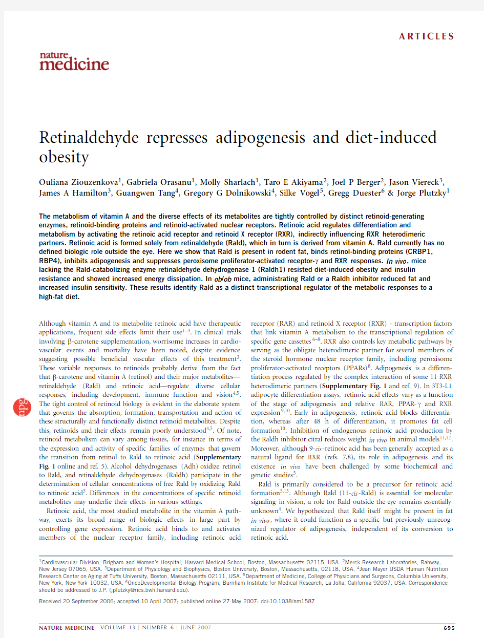
Rald is generated by the action of alcohol dehydrogenase-1(Adh1)on retinol,and its concentration is determined in large part through its subsequent catabolism by retinaldehyde dehydrogenase-1(Raldh1,the gene for which is of?cially designated Aldh1a1)to retinoic acid.We found that both Adh1and Raldh1were differentially expressed during 3T3-L1preadipocyte differentiation (Fig.1a ).Whereas preadipocytes expressed mainly Adh1,differentiated 3T3-L1cells expressed predomi-nantly Raldh1,suggesting speci?c and temporally regulated Rald production and catabolism in fat.These enzymes were also expressed in white fat from both lean (C56/BL6)and obese (ob/ob )mice (Fig.1a ).Adh1expression was signi?cantly higher in lean mice than in ob/ob mice,whereas Raldh1expression was not signi?cantly different between the groups (Fig.1a ,P o 0.05).Given the differential regulation between Adh1and Raldh1in lean versus genetically obese mice,we tested further for the presence and functional effects of Rald in fat tissue.Rald is an unstable molecule,making its detection in tissues challenging 14.T o counter this,Rald reduction to Rald oximes has been used to generate a stable biochemical Rald signature for purposes of quanti?cation 14–16.We used this approach to measure the presence of Rald in fat from C57/BL6mice on either a standard (lean)or a high-fat diet.High-fat fed mice doubled their body weight as compared with those on regular chow (Fig.1b ).We dissected equal amounts of fat tissue from lean and obese mice,immediately reduced the isolated tissues with hydroxylamine,and analyzed the concentra-tions of retinol and Rald oxime using chromatographic and spectral analysis (for comparison,we used retinol and Rald oxime standards;Supplementary Fig.2online).Our analysis revealed that not only did fat contain Rald,but that retinol and Rald were reduced approxi-mately 60%and 82%,respectively,in obese versus lean mice (Fig.1b ).Rald was also present in rabbit fat (data not shown),obviating any species-speci?c artifact.Mass spectrometric analysis con?rmed Rald oxime structures in pooled HPLC-puri?ed fractions from wild-type rodent fat extracts as evidenced by comparison with a Rald oxime standard (Fig.1c ).The presence of Rald oxime in fat was supported by the similar molecular weight of a protonated molecule at 300mass-to-charge ratio (m /z ),the loss of the oxime group at 242m /z
and the overall fragmentation pattern (94–208m /z ).Finally,our analysis showed that Rald concentration in white fat ranged from 100nM to B 1m M,based on an analysis of 13mice on regular chow (data not shown).
The cognate interaction between retinoids and retinol-binding proteins can in?uence the amounts and effects of retinoids both within and outside the cell 4.As retinol is the known binding partner for human cellular retinol-binding protein-1(CRBP1)and retinol-binding protein 4(RBP4)17,18,we used standard ?uorescence quench-ing assays to compare Rald versus retinol binding to CRBP1and RBP4.All-trans -Rald had a similar binding pro?le as all-trans -retinol to both CRBP1and RBP4(Fig.1d ).The existence of mechanisms for Rald binding and transport as well as its presence in nanomolar concentrations in fat supported further investigation into a functional role for Rald in adipogenesis at similar concentrations to those demonstrated in vivo .
Rald inhibits adipogenesis in vitro
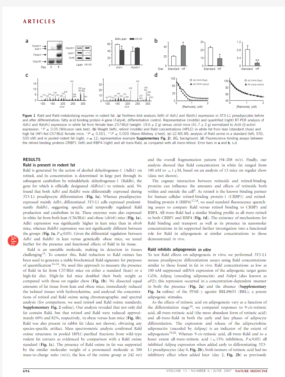
T o test Rald effects on adipogenesis in vitro ,we performed 3T3-L1mouse preadipocyte differentiation assays using Rald concentrations similar to those found in fat in vivo .Rald concentrations as low as 100nM suppressed mRNA expression of the adipogenic target genes Cd36,Adipoq (encoding adiponectin)and Fabp4(also known as aP2);this repression occurred in a concentration-dependent manner in both the presence (Fig.2a )and the absence (Supplementary Fig.3a online)of the PPAR-g agonist BRL49653(BRL),a potent adipogenic stimulus.
As the effects of retinoic acid on adipogenesis vary as a function of the differentiation stage 10,we compared responses to 9-cis -retinoic acid,all-trans -retinoic acid (the most abundant form of retinoic acid)and all-trans -Rald in both the early and late phases of adipocyte differentiation.The expression and release of the adipocytokine adiponectin (encoded by Adipoq )is an indicator of the extent of adipogenesis 19,20.Whereas 9-cis -retinoic acid,all-trans -Rald and to a lesser extent all-trans-retinoic acid (B 15%inhibition,P o 0.05)all inhibited Adipoq expression when added early to differentiating 3T3-L1preadipocytes (day 0,Fig.2b ),both isomers of retinoic acid had no inhibitory effect when added later (day 2,Fig.2b )as
previously
[Retinoids] (μM)
[Retinoids] (μM)
All-trans -retinol
m /z
m /z
300BG
BG
BG
288283
257
208
189176160
149134121
11294R a l d o x i m e S T D (i n t e n s i t y × 102)
300
BG
BG BG 298280254
208201
189176
15914713412111294
a
c
Figure 1Rald and Rald-metabolizing enzymes in rodent fat.(a )Northern blot analysis (left)of Adh1and Raldh1expression in 3T3-L1preadipocytes before and after differentiation;fatty acid binding protein 4gene (Fabp4),differentiation control.Representative (middle)and quanti?ed (right)RT-PCR analysis of Adh1and Raldh1expression in white fat from female lean C57/BL6(weight:19.6±2g)versus ob/ob mice (41.7±2g)normalized to Actb (b -actin)expression .*P o 0.05(Wilcoxon rank test).(b )Weight (left),retinol (middle)and Rald concentrations (HPLC)in white fat from lean (standard chow)and high fat (HF)-fed C57/BL6female mice.*P o 0.001,**P o 0.003(Mann-Whitney U -test).(c )LC-MS-MS analysis of Rald oxime in a standard (left,STD,500nM)and in pooled rodent fat (right,n ?12;representative example Supplementary Fig.2).BG,background.(d )Fluorescence binding assays between the retinol-binding proteins CRBP1(left)and RBP4(right)and all-trans -Rald,as compared with all-trans -retinol.Error bars in a and b ,s.d.
?2007 N a t u r e P u b l i s h i n g G r o u p h t t p ://w w w .n a t u r e .c o m /n a t u r e m e d i c i n e
reported10.In contrast to retinoic acid,Rald also inhibited Adipoq expression when added during later stages of differentiation,even at nanomolar concentrations(Fig.2a,b).In these same experiments, Rald stimulation either early or late in adipocyte differentiation also decreased adiponectin secretion,in a concentration-dependent man-ner(Fig.2c).Indeed,nanomolar concentrations of Rald mitigated the sixfold increase in adiponectin induced by BRL stimulation(Fig.2c). Rald also decreased lipid accumulation during3T3-L1preadipocyte differentiation in both the absence and the presence of BRL(Fig.2d). Rald regulates distinct nuclear receptor responses
Given the suppression of PPAR-g agonist–stimulated adipogenesis and adiponectin release by Rald,we tested whether Rald regulates RAR and RXR activity,and,if so,whether it does so in a manner distinct from that of other retinoids.We performed ligand-binding domain(LBD)-GAL4transfection assays in3T3-NIH?broblasts,in the presence and absence of known speci?c nuclear receptor agonists and Rald.As previously reported21,all-trans-Rald weakly activated the RAR-a LBD, but did not alter activation of RAR-a LBD by its known ligand,9-cis retinoic acid(Fig.3a).Rald alone had no effect on RXR-a LBD activation(Fig.3a),but,in contrast to its effect on RAR-a LBD responses,it signi?cantly inhibited RXR-a LBD activation by 9-cis-retinoic acid(Fig.3a).
Given these effects of Rald on RXR-LBD activation,we questioned whether Rald could inhibit the activation of a transfected canonical PPAR response element(PPRE)luciferase construct after transfection of RXR or PPAR-g and agonist stimulation(9-cis retinoic acid or BRL, respectively).Rald signi?cantly inhibited PPRE activation by each agonist,with the most potent effects seen after PPAR-g and RXR co-transfection and PPAR-g agonist stimulation(60%inhibition, Fig.3b).Given these results,we next investigated the direct interaction
between PPAR-g and all three major Rald
isomers(9-cis-,13-cis-and all-trans-Rald),
using cell-free radioligand displacement
assays22.All three isomers displaced high
af?nity PPAR-g agonists(K d?5.9±0.7,
9.7±1.2,and11.9±1.9m M,respectively;
mean±s.d.),consistent with direct but weak
binding of these molecules to the PPAR-g–
LBD(Fig.3b).We observed similar effects in
cell-based PPAR-g–LBD assays in the presence
of BRL(data not shown).Given Rald’s dis-
tinct effects on adipogenesis,and known
discrete roles for RXR and PPAR-g in this
process,we evaluated Rald effects in an RXR
loss-of-function model,repeating standard
3T3-L1adipogenesis assays in the presence
of Rald,but after decreasing RXR-a and RXR-
b using short interfering RNA(siRNA)to the
RNA encoding each RXR isotype.These RXR
isotypes are expressed early in adipogenesis
(48h),helping to initiate subsequent adipo-
cyte differentiation,as evident in vitro23and
in vivo24.We measured triglyceride accumula-
tion and adiponectin secretion as speci?c,
distinct indicators of adipogenesis20,25.After
siRNA exposure,total RXR was undetectable
by western blotting(Fig.3c).As expected,
3T3-L1adipocytes treated with RXR-a siRNA
and RXR-b siRNA showed decreased trigly-
ceride accumulation,which Rald decreased further,independent of RXR expression.In contrast,whereas RXR-a siRNA and RXR-b siRNA signi?cantly decreased adiponectin secretion (B92%less),Rald had no further effect(Fig.3d),consistent with an RXR-dependent Rald effect on adiponectin release.
Rald metabolism controls adipocyte biology
Endogenous concentrations of Rald are dictated by enzymes control-ling its production(Adh1)and catabolism(Raldh1)5,14.Raldh1-de?cient mice(Raldh1–/–)have been well characterized as a model for Rald overproduction14.These mice have impaired Rald oxidation, as evident in their markedly decreased retinoic acid and increased Rald concentrations while on a vitamin A–containing diet14.Raldh1–/–mice fed a standard chow diet with4IU vitamin A per g had twice the plasma Rald of those in age-and sex-matched wild-type controls (8.6±2.6and3.8±2.6nM,respectively).T o evaluate whether Raldh1 de?ciency also affects fat cell differentiation,we performed adipogen-esis assays in primary embryonic?broblasts isolated from Raldh1–/–and wild-type mice.Adipogenesis was markedly decreased in Raldh1–/–as compared with wild-type cells,as evidenced by oil-red-O staining for lipid accumulation(Fig.4a).Adiponectin secretion was 52%less in Raldh1–/–versus wild-type cells,a difference that was even more pronounced after treatment with BRL at all concentrations tested(63%less in Raldh1–/–versus wild-type cells with300nM BRL,Fig.4b).With higher BRL concentrations(10m M),these effects of Rald on adiponectin suppression and lipid accumulation approached wild-type levels(Supplementary Fig.3b).
Mass spectroscopy analyses demonstrated Rald in white fat from wild-type and Raldh1–/–mice(242.9m/z;n?12per genotype; Fig.4c).This spectral pattern was identical to that seen with a Rald oxime standard(Supplementary Fig.4a online).T o study Rald effects on adipogenesis in vivo,we placed wild-type and Raldh1–/–mice on
a a
c d
Rald, 300 nM
Rald, 100 nM
Rald, 300 nM
Rald, 100 nM
Veh
Veh
Late stage
Early stage
D
i
f
f
e
r
e
n
t
i
a
t
e
d
+
B
R
L
D
i
f
f
e
r
e
n
t
i
a
t
e
d
300
150
300
150
[Rald] (nM)
Differentiated + BRL
[Rald] (nM)
Differentiated
Early stage
Early stage
Late stage
Late stage
*
*
*
*
*
400
800
[
A
d
i
p
o
n
e
c
t
i
o
n
]
(
n
g
/
m
l
)
[
A
d
i
p
o
n
e
c
t
i
o
n
]
(
n
g
/
m
l
)
120
80
40
All-trans-Rald (μ
Fabp4
Gapdh
Adipog
Cd36
Late stage
Early stage
a
l
l
-
t
r
a
n
s
-
R
a
l
l
-
t
r
a
n
s
-
9
-
c
i
s
-
a
l
l
-
t
r
a
n
s
-
R
a
l
l
-
t
r
a
n
s
-
9
-
c
i
s
-
Figure2Rald and retinoic acid differentially regulate PPAR-g–induced adipogenic genes.(a)Northern
blotting for Cd36,Adipoq,Fabp4and Gapdh performed in3T3-L1cells differentiated with BRL in the
presence of Rald.(b)Adipoq mRNA expression after Rald,9-cis-retinoic acid or all-trans-retinoic
acid(all500nM)stimulation during either early or late stage of adipogenesis(n?3per condition).
(c)As in b but in the absence(left)or presence(right)of BRL and varying Rald concentrations before
measurement of adiponectin in media(ELISA).*P o0.05(for both early and late stages;adiponectin
concentrations after Rald stimulation versus initial levels;Mann-Whitney U-test).(d)Oil red O staining
of lipid accumulation in cells used in c.Scale bar,300m m.
?
2
7
N
a
t
u
r
e
P
u
b
l
i
s
h
i
n
g
G
r
o
u
p
h
t
t
p
:
/
/
w
w
w
.
n
a
t
u
r
e
.
c
o
m
/
n
a
t
u
r
e
m
e
d
i
c
i
n
e
high-fat diet (45%fat,standard vitamin A 4IU/g),measuring their weight weekly until tissue analysis at after 6months of experimental diets (age 8months).White fat of Raldh1–/–mice had signi?cantly higher retinol (152%)and Rald (206%)as compared with that from wild-type mice (Fig.4d ).Adipocytes from Raldh1–/–fat were half the size of those from wild-type fat (Fig.4e ,f ).In these same fat samples,adipocyte size correlated inversely with Rald concentrations (Fig.4g ).Raldh1regulates lipid and glucose metabolism
We questioned if Raldh1de?ciency would have systemic metabolic consequences.Indeed,after high-fat feeding (6months),Raldh1–/–
mice gained signi?cantly less weight (93%)than the wild-type mice (weight gain in wild-type ?26.6±1.9g;in Raldh1–/–?13.7±3.6g;Fig.5a ).Weight differences were evident beginning at 1month of high fat feeding (data not shown).Of note,Raldh1–/–females weighed signi?cantly less than males (relative to wild-type mice of the corresponding sex ,57%versus 41%less,P o 0.001).Given sex differences in metabolic parameters related to fat,we performed additional metabolic analyses in wild-type (n ?5)and Raldh1–/–females (n ?4).DEXA scanning revealed decreased whole-body fat accumulation in Raldh1–/–mice (Supplementary Fig.4b ).The decreased white fat accumulation in Raldh1–/–versus wild-type mice
siRXR β
siRNA siRXR β
siRNA
[T r i g l y c e r i d e s ] (m g /d l )
P P R E a c t i v a t i o n (%)
9-cis -RA
9-cis -RA 13-cis -Rald all-trans -Rald
*
*
*
R A R -α L B D a c t i v a t i o n (%)
Veh
a
b
c d
P < 0.03
[Rald] (nmol/g)
Raldh1
–/–
WT A d i p o c y t e s , w h i t e f a t
R e t i n o l (n m o l /g f a t )
282.9
242.9
230.8
208.9
190163176.1150.1
135.1113.295.4301.7
282.9
242.9229.8208.9
176160.1
150.1134.1113.495.3R a l d , W T (i n t e n s i t y × 104)
a c
d
f Figure 3Rald regulates nuclear receptor responses in vitro .(a )RAR-a (left)and RXR-a (right)LBD-GAL4assays performed in NIH-3T3cells in the absence (Veh)or presence of 9-cis retinoic acid (9-cis -RA)(300nM)and increasin
g Rald concentrations.n ?3measurements.*P o 0.05(RXR-a LBD activation by 9-cis -RA in the absence versus presence of Rald).(b )Left,activation of a transfected PPAR response element (PPRE)
luciferase construct transfected with constructs for RXR-a ,PPAR-g or both,followed by BRL or 9-cis -RA (1m M)stimulation in the presence of Rald (left).Data normalized to receptor activation induced by the respective
ligand (BRL or 9-cis retinoic acid alone,set at 100%.*P o 0.05(Wilcoxon rank test).Right,binding of Rald isomers to full-length PPAR-g protein in radioligand displacement assay.Results expressed as relative displacement of 3H 2-labeled PPAR-g agonist nTZD3by Rald isomers.(c )Total RXR protein abundance 48h after RXR-a siRNA and RXR-b siRNA (siRXR-a ,siRXR-b )or control siRNA transfection in 3T3-L1cells (western blotting).(d )Trigly-ceride content (left)and secreted adiponectin protein abundance (right)in cells as in c .RXR siRNA treatment decreased adiponectin concentrations in both the absence (data not shown)and presence of BRL but Rald had no further effect in these cells.NS,nonsigni?cant.For all studies,error bars represent s.d.,n ?3per condition,Wilcoxon rank test.*P o 0.001,**P o 0.01.
Figure 4Raldh1de?ciency is associated with suppressed adipogenesis in vitro and increased Rald and reduced adipocyte size in vivo .(a )Lipid accumulation in differentiated primary ?broblasts from Raldh1–/–as compared with wild-type (WT)embryos.Scale bar,200m m.(b )Left,
adiponectin concentrations in nondifferentiated (Non-D)and differentiated (D)cells as in a .Right,BRL effects on adiponectin secretion in similar Raldh1–/–versus WT cells.n ?3per condition and genotype.*P o 0.001(Mann-Whitney U -test;error bars here and below,s.d.).(c )LC-MS-MS of pooled fat from wild-type mice (left)contained less Rald oxime (242m /z fragment,arrow)than fat from Raldh1–/–mice (right).n ?4mice per
genotype.(d )Retinol (left)and Rald oxime (right)concentrations by HPLC in high fat–fed female Raldh1–/–versus wild-type mice (n ?4mice per
genotype)*P o 0.03,**P o 0.005(Mann-Whitney U -test).(e )Adipocyte size in white fat of mice studied in d .n ?3mice per genotype.*P o 0.001(Mann-Whitney U -test).(f )Representative H&E sections of fat from mice studied in d .Scale bar,200m m.(g )Correlation between Rald concentrations and adipocyte size in mice studied in d (P o 0.03,Spearman correlation test).
?2007 N a t u r e P u b l i s h i n g G r o u p h t t p ://w w w .n a t u r e .c o m /n a t u r e m e d i c i n e
was evident in both subcutaneous and visceral fat pads (76%and 74%less,respectively;Fig.5b ).Raldh1–/–mice also had signi?cantly lower plasma free fatty acids than wild-type mice (0.21±0.1mmol/l versus 0.53±0.3mmol/l,respectively;P o 0.04,Wilcoxon rank test).Livers from Raldh1–/–mice also had de-creased lipid accumulation compared with wild-type mice,as evident from H&E staining (Fig.5c )and total liver weight (Supple-mentary Fig.4c ).
The differences in fat accumulation in Raldh1–/–versus wild-type mice occurred despite similar food and water intake in both groups (Fig.5d ),indicating a shift in total energy balance in Raldh1de?ciency.Indeed,Raldh1–/–mice had a signi?cantly higher metabolic
a
d
**
*
*
*
**
*
***
*
**
*
*
–/–
WT
WT
Time (h)
Time (h)
C o n s u m p t i o n (g /d )
Figure 5Raldh1–/–mice resist high-fat diet–induced obesity.(a )Weight of wild-type and Raldh1–/–mice after high-fat diet (180d).*P o 0.001(Wilcoxon rank test).Representative X-ray images of each genotype.Scale bar,2cm.(b )Weight of subcutaneous and visceral fat pads (left)from wild-type and Raldh1–/–female mice (n ?5,4respectively;*P o 0.001,Mann-Whitney U -test).Representative isolated visceral (epididymal)and subcutaneous (proximal and scapular)fat pads (right).(c )Liver lipid area density from the same mice as in b *P o 0.03,n ?3females per genotype;a.u.,arbitrary units.Representative H&E-stained liver sections (right).Scale bar,200m m.(d –g )Food and water consumption,metabolic rates,respiratory quotients,rectal temperatures and UCP-1protein amounts in mice as in b .*P o 0.05,**P o 0.04(Wilcoxon rank test).Error bars,s.d.except in f (s.e.m.).;a.u.,arbitrary units.
Time (min)
#
*Vit A Rald Citral o b /o b
*
*
*
*
*
***
*
WT
Raldh1
–/–
Raldh1
369
A d i p o n e c t i n (μg /m l )
a
d
Figure same as 0.002,and mice 4location indicated by kidney (K)and vertebra (V)sizes.*P o 0.001,**P o 0.002,***P o 0.003(Wilcoxon rank test).
(f )Western analysis of plasma RBP4in mice as in d (one representative blot shown).*P o 0.05(treatment with vehicle
versus Rald or citral),#P o 0.05(treatment with vitamin A versus Rald or citral)(Mann-Whitney U -test).(g )Glucose tolerance in mice as in d .*P o 0.05(treatment with vitamin A versus citral),#P o 0.05(treatment with vehicle versus citral)(Wilcoxon rank test).Error bars,s.d.except in b (s.e.m.).
?2007 N a t u r e P u b l i s h i n g G r o u p h t t p ://w w w .n a t u r e .c o m /n a t u r e m e d i c i n e
rate (Fig.5e ),respiratory quotient (Fig.5f ),and body temperature (Fig.5g )compared with wild-type controls.Consistent with the role of thermogenesis in determining body weight 26,amounts of uncoupling protein-1(UCP-1)in brown fat were signi?cantly higher in Raldh1–/–mice than wild-type mice (Fig.5g ).
Fat tissue regulates whole body insulin sensitivity through various mechanisms,including adipokine release 27–30.As such,we measured changes in adiponectin,leptin and RBP4in Raldh1–/–mice.T otal adiponectin,leptin and RBP4were signi?cantly decreased in the plasma of Raldh1–/–mice as compared with wild-type mice (Fig.6a ).On high fat diet,Raldh1–/–mice were protected from the increased insulin resistance evident in wild-type mice as seen in glucose and insulin tolerance testing (Fig.6b ).Insulin concentrations did not differ between genotypes (Fig.6c ).T o further consider if Rald effects on fat accumulation and insulin resistance were due to Rald itself and not to subsequent generation of retinoic acid or other vitamin A metabolites,we used ob/ob mice that experience progressive weight gain on regular chow 28,31to compare responses to Rald,the Rald parent compound vitamin A,all-trans -retinoic acid and citral,a known inhibitor of Raldh enzymes 11.After 3weeks,we quanti?ed subcutaneous fat mass by magnetic resonance imaging (MRI).The extent of visceral fat accumulation in these mice precluded its accurate quantitative measurement.Mice receiving Rald or citral had signi?-cantly less subcutaneous fat relative to total body fat (15.5±0.6%and 14.8±0.6%,respectively),than those receiving vehicle,vitamin A or all-trans -retinoic acid (18.8±1.4%,17.3±1.3%and 19.1±1.6%)(all P o 0.05;Fig.6d ,e ).Rald administration also repressed adipogenesis in preadipocytes isolated from human visceral fat depots (data not shown).RBP4amounts also varied in ob/ob mice exposed to these different retinoids.Whereas RBP4amounts did not vary in response to retinoic acid or vehicle,they doubled in response to vitamin A (Fig.6f ).In contrast,both Rald and citral signi?cantly suppressed circulating RBP4(versus vehicle),recapitulating the RBP4pattern seen in Raldh1–/–mice (Fig.6a ).Consistent with the changes seen in adiposity and RBP4,both Rald and citral administration improved glucose tolerance in ob/ob mice (Fig.6g ).
DISCUSSION
We demonstrate here that Rald plays a distinct metabolic role in adipocyte differentiation in vitro ,and in diet-induced insulin resis-tance and obesity in vivo .Rald is present in fat at nanomolar concentrations (B 1nmol/g)and can interact with CRBP1and RBP4,binding proteins involved in intracellular and circulating retinoid transport.Rald suppresses adipogenic gene expression,adi-pocyte lipid accumulation,RXR-a and PPAR-g responses,all at concentrations (o 1m M)similar to those in rodent fat in vivo.Despite the widely held assumption that Rald outside of the eye serves mainly as a precursor for retinoic acid 4,the differences in the effects of Rald versus those of retinoic acid in vitro and in vivo argue for Rald itself as a distinct mediator in fat.When Rald concentrations were increased in vivo ,either in the absence of Raldh1or after direct Rald admin-istration,fat formation was decreased.Inhibition of Rald catabolism (citral treatment)in vivo had similar effects although again distinct from retinoic acid or vitamin A administration.T ogether this data identi?es Rald as a biologically active metabolite present in fat that may regulate adipogenesis through its action on RXR-a and PPAR-g responses and in a manner opposite of retinoic acid effects.
Rald’s unique effects on adipokine expression,adipogenesis and body weight focus attention on the parameters regulating the relative cellular concentrations of Rald and retinoic acid.The balance between Rald and retinoic acid is determined by factors such as the concentra-tion of vitamin A in the body,the expression and activity of enzymes that metabolize Rald and retinoic acid,other retinol-modifying enzymes (such as esterases and hydrolases),as well as retinol-binding proteins and the redox status in cells 4,5,32.Each of these factors may have functional consequences,as seen with the recently reported association between RBP4and diabetes in mice and humans 29,33.In terms of the enzymes metabolizing Rald,we have demonstrated the genetic absence of Raldh1results in a metabolic phenotype involving marked alterations in fat accumulation,glucose homeostasis and adipokine production after high-fat feeding.These metabolic changes are probably due at least in part to the increased concentrations of Rald in fat,as demonstrated here,especially as direct administration of Rald reproduced the metabolic pro?le evident in Raldh1–/–mice.Notably,direct administration of retinoic acid and Rald had different effects in vitro and in vivo ,further supporting a role for Rald that is distinct from its function as a retinoic acid precursor.
Rald seems to exert its effects through both RXR-dependent and RXR-independent mechanisms.Rald repressed adiponectin produc-tion,but not after RXR expression had been reduced.In contrast,Rald-mediated repression of triglyceride accumulation persisted regardless of RXR expression.Rald inhibited LBD activation and cellular responses to both RXR and PPAR-g agonists,with effects on early and late adipogenesis that are consistent with the reported temporal expression of PPAR-g and RXR 9,10.Although Rald binding to the RXR-and PPAR-g –LBDs was weak,it potently suppressed adipogenesis in vitro and in vivo.It has been reported that selective synthetic PPAR and RXR modulators also show a similar divergence between the potency of receptor binding and adipogenic effects 34,35.The ability of higher BRL concentrations to overcome the effects of Rald suggests that Rald responses are mediated at least part through PPAR-g .Various mechanisms may underlie how certain molecules in?uence nuclear receptor responses independent of receptor-binding potency,including conformational changes in the receptor,salt bridge formation,accessory molecule recruitment and release,and post-translation protein modi?cation 36.Rald’s metabolic effects make further studies on Rald derivatives,the factors determining Rald concentrations and Rald transport all of considerable interest.More broadly,these data suggest a regulatory pathway in which a given molecule (vitamin A)can yield both a nuclear receptor agonist (retinoic acid)and a molecule (Rald)capable of inhibiting speci?c nuclear receptor responses (PPAR-g ,RXR).
These studies provide a link between vitamin A metabolism and responses to a high-fat diet,including pathologic complications such as obesity and insulin resistance.The metabolic changes evident with increased Rald have several potential clinical implications.The protec-tion against diet-mediated obesity and insulin resistance identi?ed in Raldh1–/–mice suggests this inducible enzyme of vitamin A metabo-lism as a potential candidate for therapeutic targeting and/or a source of body weight variation.Similarly,Rald generation might in?uence therapeutic responses to vitamin A,retinoic acid or other retinoid-based treatments as well as the effects of PPAR-g agonists.The presence of Rald in fat and its association with RBP4suggest that Rald might contribute to the relationship between RBP4and insulin sensitivity 29,33.
In the models studied here,Rald concentrations in fat in vivo correlated tightly with changes in fat accumulation and metabolic responses,providing one probable explanation for the metabolic phenotype of Raldh1-de?cient mice.Rald has effects on both visceral and subcutaneous fat,as seen in Raldh1–/–mice.In the absence of Raldh1,energy balance appears shifted toward increased energy dissipation,as suggested by the increased body temperature,metabolic
?2007 N a t u r e P u b l i s h i n g G r o u p h t t p ://w w w .n a t u r e .c o m /n a t u r e m e d i c i n e
rate 42and UCP-1expression manifest in these mice.Interestingly,in some animal models,increased UCP-1has not been associated with an increased respiratory quotient 37.Regardless,the changes in energy balance seen with Rald could result from its actions in various Rald-and retinoic acid–sensitive tissues,including brown fat,immune cells and the central nervous system (CNS).Recent work suggests a possible role for Raldh2in dendritic cells 43,although Raldh1appears the predominant determinant of Rald metabolism in response to vitamin A intake 14.The CNS is a particularly important regulator of body weight in general and adipocyte biology speci?cally;for example,in determining rates of lipolysis 38.Our data do not speci?cally exclude the possibility of Rald exerting some effects via the CNS.Here we focused on demonstrating the presence of Rald and its effects on adipocyte responses.The linear relationship between Rald concentra-tions and adipocyte size,the lower concentrations of Rald in obese as compared with lean mice,and the impaired adipogenesis seen in Raldh1–/–preadipocytes all suggest that Rald effects in adipose tissue probably contribute to the protection against diet-induced obesity and insulin resistance evident in Rald1-de?cient mice.Certainly Rald’s actions in adipose tissue,including the regulation shown here of speci?c adipokines,can also provide feedback to other systems,including the CNS and the immune system,with subsequent effects on adipose biology.
Taken together,these ?ndings allow Rald to join retinoic acid as a distinct biologically active mediator of energy balance and insulin sensitivity.The integration of these critical metabolic pathways by a natural molecule such as Rald may provide new opportunities for understanding the complex interaction between vitamin A,its meta-bolites and the transcriptional regulation of metabolism.METHODS
Reagents.We obtained reagents and media from Sigma-Aldrich and BioWhit-taker unless otherwise indicated.All media contained amphotericin B,peni-cillin and streptomycin.BRL49653(rosiglitazone)was a gift from GlaxoSmithKline.Unless otherwise indicated,retinoids used were all-trans isomers.All diets were from Research Diet,Inc.
Animal studies.C57/BL6mice (weight:19.6±2g)versus ob/ob mice (41.7±2g),all 12-week-old females on regular chow,were used for gene expression studies and retinoid analysis (n ?3per genotype).For retinoid MS analysis,we studied pooled subcutaneous and visceral fat from male mice (129S3/SvImJ,6months of age,n ?4)and a New Zealand White rabbit.For metabolic studies,we compared age-(8weeks old)and sex-matched Raldh1–/–14and wild-type mice (?ve per sex and genotype).The D12451high-fat diet contained 45%of calories derived from fat and standard vitamin A (4IU/g).Water was ad libitum .
For MRI fat distribution studies,we administered retinoids (all-trans -Rald,vitamin A or all-trans -retinoic acid,all 500nM)or citral (10m M,equal to 240nmol/g),all in ethanol:PBS (2:200m l),by daily intraperitoneal injections for 3weeks.The Standing Committee on Animals at Harvard Medical School approved all protocols.
Cell culture.We cultured and differentiated mouse 3T3-L1preadipocytes and primary ?broblasts isolated from 16-d-old embryos using standard adipogen-esis and isolation protocols 25.Cells were differentiated (7d)with or without BRL (1m M).Rald was added at the indicated concentrations in either early (5h)or late (48h)stages of adipogenesis,as timed to initiation of differentiation.Transient transfections.For transient transfection of NIH 3T3cells (2.3?104cells,24-well plates),we used pCMX-b -galactosidase and LBD–yeast Gal 4–luciferase constructs and Fugene (Roche)as before 39.For siRNA transfections,we administered scrambled control (sequences C,D)or speci?c RXR-a and RXR-b siRNA sequences (Santa Cruz)to 3T3-L1cells (90%con?uence,antibiotic-free DMEM,10%calf serum,24-well plates)using Lipofectamine
2000(8h)and OptiMEM medium (Invitrogen).We supplemented the medium with 10%FBS 5h after transfection.We evaluated RXR amounts by Western blotting 48h after transfection.
Human recombinant RBP and ?uorescence binding assays.Human RBP4and mouse CRBP1subcloned in pET expression vectors were expressed and puri?ed as described previously 40,but without retinol.We dialyzed refolded protein against binding assay buffer (0.05M sodium phosphate,0.15M NaCl,pH 7.0)overnight,and quanti?ed proteins at 280nm (RBP440,400M –1cm –1,CRBP126,720M –1cm –1)17,41.We measured tryptophan ?uorescence (Aminco,Spectronic Unicam)by excitation (285nm and emission (335nm),both in 0.05M sodium phosphate buffer 18which indicated retinoid binding to RBP4or CRBPI (1m M).
Scintillation proximity assay.We studied Rald displacement of a 3H 2-labeled known synthetic PPAR-g agonists (nTZD3,K d ?2.5nM)from human full-length PPAR-g 2as before 22.
RNA analysis.We determined mouse Adh1,Raldh1and Actb (b -actin)mRNA (RNeasy,Qiagen)in white fat using semi-quantitative RT-PCR and the following primers:
Adh1:5¢-ATGAGCACTGCGGGAAAAGT-3¢,5¢-ACTTTATTGGCCGTGT CTCTAA-3¢;Raldh1:5¢-TGGGTTAACTGCTATATCATGTTG-3¢,5¢-GGGTG CCTTTATTAAGCTTTGCG-3¢.Results were obtained within the linear range for each gene (31and 29cycles,respectively)and normalized to Actb (b -actin)expression.We performed northern blotting with HyBond (Amersham)as before 39.
Protein and intracellular lipid analysis.We determined triglyceride content in lysed cells (RIPA buffer,complete protease inhibitor cocktail,Roche)using enzymatic colorimetric assay (Wako)and measured adiponectin using ELISA (R&D Systems).For western blotting,we performed a reducing gel separation (10%acrylamide)on cell lysates,plasma and tissue lysates,before hybridiza-tion with antibodies to RXR (Santa Cruz),to mouse RBP4(Alpco)or to UCP-1(Chemicon).
Liquid chromatography (LC),mass spectrometry (MS):We generated a Rald oxime standard using reduction of retinaldehyde (100m M,221C,2h under argon protected from light)with hydroxylamine (1M)and EDTA (25m M)in PBS as before 14,15.White fat (B 200m g)was dissected from relevant animals,immediately reduced with hydroxylamine,puri?ed by solid-phase extraction (Bakerbond amino column)and analyzed for retinol and Rald oxime content using high performance LC (HPLC)analysis (see also legend to Supplementary Fig.2).Pooled Rald oxime fractions then underwent structural analysis using LC and tandem MS (LC-MS-MS)using atmospheric pressure chemical ionization in positive mode (Bruker Daltonics,Esquire LC)to detect Rald oxime ion molecular ion peak [M+H]+at 300m /z .The ionization parameters included capillary voltage,3,000V;APCI temperature,3501C;source tem-perature,3001C;and scanning ions in the 82to 306m /z range.
Histology:We embedded fat and liver tissue in paraf?n before hematoxylin and eosin (H&E)staining followed by quanti?cation of adipocyte size or liver lipid accumulation (ImageJ software).
Magnetic resonance imaging (MRI).We obtained MRI scans (1-mm slices)using a Bruker Avance 500wide-bore spectrometer (11.7T;500MHz for proton)?tted with a gradient ampli?er and a 30mm ‘birdcage’transmitter/receiver coil before processing the data (Paravision).The spin-echo parameters for T1-weighted images were as follows:TE ?15ms,TR ?300ms,matrix ?256,FOV ?30mm;for RARE images,TE ?51ms,TR ?2,500ms,matrix ?256,FOV ?30mm.Abdominal fat measurements used axial slices (n ?8)at the level of the left renal pelvis.
Dual-energy X-ray absorptiometry (DEXA).We used the GE Lunar Corpora-tion PIXImus2Dexa Scanner,normalizing the data to a quality control plot (Charles River Laboratories).
Metabolic parameters.After mouse acclimation to a powdered high-fat diet (4d),we measured food and water intake,oxygen consumption and carbon
?2007 N a t u r e P u b l i s h i n g G r o u p h t t p ://w w w .n a t u r e .c o m /n a t u r e m e d i c i n e
dioxide production in metabolic cages (Ancare,Charles River Laboratories).The calculated metabolic rate (Weir equation)is expressed per g body weight 42.We performed insulin (ITT)and glucose tolerance tests (GTT)after fasting (16h),using intraperitoneal insulin injections (ITT,0.1U/ml,0.005ml/g body weight)or a single 25%dextrose injection (GTT,0.004ml/g body weight),and a glucometer for measurements (Accu-Chek Advantage,Roche).Note:Supplementary information is available on the Nature Medicine website.ACKNOWLEDGMENTS
We thank G.Sukhova (Brigham and Women’s Hospital),J.Kirkland and
T.Tchkonia (Boston University),N.Krinsky and R.Russell (Tufts University)for helpful discussions;P .Scherer,S.Kliewer,D.Mangelsdorf (University of T exas),C.H.Lee (Harvard University)and T.Willson for reagents;and E.Shvarz,
K.Volz,J.Qin,T.Archibald,N.Sharma,https://www.doczj.com/doc/e13511463.html,clair and R.Driscoll for technical support.This research was supported by the Boston Obesity Nutrition Research Center 5P30DK046200and K12HD051959-01NICHD BIRCWH,the American Heart Association SDG 0530101N (O.Z.);the US National Institutes of Health (R01HL071745and P01HL48743)and the Donald W.Reynolds Foundation (J.P .).
COMPETING INTERESTS STATEMENT
The authors declare no competing ?nancial interests.
Published online at https://www.doczj.com/doc/e13511463.html,/naturemedicine
Reprints and permissions information is available online at https://www.doczj.com/doc/e13511463.html,/reprintsandpermissions
1.Paydas,S.et al.Vasculitis associated with all trans retinoic acid (ATRA)in a case with
acute promyelocytic leukemia.Leuk.Lymphoma 44,547–548(2003).
2.Redlich,C.A.et al.Effect of long-term b -carotene and vitamin A on serum cholesterol
and triglyceride levels among participants in the Carotene and Retinol Ef?cacy Trial (CARET).Atherosclerosis 143,427–434(1999).
3.Rapola,J.M.et al.Randomised trial of a -tocopherol and b -carotene supplements on
incidence of major coronary events in men with previous myocardial https://www.doczj.com/doc/e13511463.html,ncet 349,1715–1720(1997).
4.Napoli,J.L.Retinoic acid:its biosynthesis and metabolism.Prog.Nucleic Acid Res.
Mol.Biol.63,139–188(1999).
5.Duester,G.,Mic,F .A.&Molotkov, A.Cytosolic retinoid dehydrogenases govern
ubiquitous metabolism of retinol to retinaldehyde followed by tissue-speci?c metabo-lism to retinoic acid.Chem.Biol.Interact.143–144,201–210(2003).
6.Chambon,P .A decade of molecular biology of retinoic acid receptors.FASEB J.10,
940–954(1996).
7.Heyman,R.A.et al.9-cis retinoic acid is a high af?nity ligand for the retinoid X
receptor.Cell 68,397–406(1992).
8.Shulman,A.I.&Mangelsdorf,D.J.Retinoid x receptor heterodimers in the metabolic
syndrome.N.Engl.J.Med.353,604–615(2005).
9.Fu,M.et al.A nuclear receptor atlas:3T3–L1adipogenesis.Mol.Endocrinol.19,
2437–2450(2005).
10.Xue,J.C.,Schwarz,E.J.,Chawla,A.&Lazar,M.A.Distinct stages in adipogenesis
revealed by retinoid inhibition of differentiation after induction of PPAR g .Mol.Cell.Biol.16,1567–1575(1996).
11.Kikonyogo,A.,Abriola,D.P .,Dryjanski,M.&Pietruszko,R.Mechanism of inhibition of
aldehyde dehydrogenase by citral,a retinoid antagonist.Eur.J.Biochem.262,704–712(1999).
12.Ress,N.B.et al.Toxicology and carcinogenesis studies of microencapsulated citral in
rats and mice.Toxicol.Sci.71,198–206(2003).
13.Duester,G.Families of retinoid dehydrogenases regulating vitamin A function:
production of visual pigment and retinoic acid.Eur.J.Biochem.267,4315–4324(2000).
14.Molotkov,A.&Duester,G.Genetic evidence that retinaldehyde dehydrogenase Raldh1
(Aldh1a1)functions downstream of alcohol dehydrogenase Adh1in metabolism of retinol to retinoic acid.J.Biol.Chem.278,36085–36090(2003).
15.von Lintig,J.&Wyss,A.Molecular analysis of vitamin A formation:cloning and
characterization of b -carotene 15,15¢-dioxygenases.Arch.Biochem.Biophys.385,47–52(2001).
https://www.doczj.com/doc/e13511463.html,kshman,M.R.,Mychkovsky,I.&Attlesey,M.Enzymatic conversion of all-trans-b -carotene to retinal by a cytosolic enzyme from rabbit and rat intestinal https://www.doczj.com/doc/e13511463.html,A 86,9124–9128(1989).
17.Vogel,S.et al.Characterization of a new member of the fatty acid-binding protein
family that binds all-trans-retinol.J.Biol.Chem.276,1353–1360(2001).
18.Berni,R.,Clerici,M.,Malpeli,G.,Cleris,L.&Formelli,F .Retinoids:in vitro interaction
with retinol-binding protein and in?uence on plasma retinol.FASEB J.7,1179–1184(1993).
19.Yu,S.et al.Adipocyte-speci?c gene expression and adipogenic steatosis in the mouse
liver due to peroxisome proliferator-activated receptor g 1(PPAR g 1)overexpression.J.Biol.Chem.278,498–505(2003).
20.Iwaki,M.et al.Induction of adiponectin,a fat-derived antidiabetic and antiatherogenic
factor,by nuclear receptors.Diabetes 52,1655–1663(2003).
21.Repa,J.J.,Hanson,K.K.&Clagett-Dame,M.All-trans-retinol is a ligand for the
retinoic acid https://www.doczj.com/doc/e13511463.html,A 90,7293–7297(1993).
22.Berger,J.P .et al.Distinct properties and advantages of a novel peroxisome proli-ferator-activated protein g selective modulator.Mol.Endocrinol.17,662–676(2003).
23.Canan Koch,S.S.et al.Synthesis of retinoid X receptor-speci?c ligands that are potent
inducers of adipogenesis in 3T3–L1cells.J.Med.Chem.42,742–750(1999).
24.Imai,T.,Jiang,M.,Chambon,P .&Metzger,D.Impaired adipogenesis and lipolysis in
the mouse upon selective ablation of the retinoid X receptor a mediated by a tamoxifen-inducible chimeric Cre recombinase (Cre-ERT2)in https://www.doczj.com/doc/e13511463.html,A 98,224–228(2001).
25.Green,H.&Meuth,M.An established pre-adipose cell line and its differentiation in
culture.Cell 3,127–133(1974).
26.Puigserver,P .et al.A cold-inducible coactivator of nuclear receptors linked to adaptive
thermogenesis.Cell 92,829–839(1998).
27.He,W.et al.Adipose-speci?c peroxisome proliferator-activated receptor g knockout
causes insulin resistance in fat and liver but not in https://www.doczj.com/doc/e13511463.html,A 100,15712–15717(2003).
28.Kubota,N.et al.Pioglitazone ameliorates insulin resistance and diabetes by both
adiponectin dependent and independent pathway.J.Biol.Chem.281,8748–8755(2006).
29.Yang,Q.et al.Serum retinol binding protein 4contributes to insulin resistance in
obesity and type 2diabetes.Nature 436,356–362(2005).
30.Lee,S.,Bacha,F .,Gungor,N.&Arslanian,S.A.Racial differences in adiponectin in
youth:relationship to visceral fat and insulin sensitivity.Diabetes Care 29,51–56(2006).
31.Nawrocki, A.R.et al.Mice lacking adiponectin show decreased hepatic insulin
sensitivity and reduced responsiveness to peroxisome proliferator-activated receptor g agonists.J.Biol.Chem.281,2654–2660(2006).
32.Maeda,K.et al.Adipocyte/macrophage fatty acid binding proteins control integrated
metabolic responses in obesity and diabetes.Cell Metab.1,107–119(2005).
33.Graham,T.E.et al.Retinol-binding protein 4and insulin resistance in lean,obese,and
diabetic subjects.N.Engl.J.Med.354,2552–2563(2006).
34.Hallsten,K.et al.Rosiglitazone but not metformin enhances insulin-and exercise-stimulated skeletal muscle glucose uptake in patients with newly diagnosed type 2diabetes.Diabetes 51,3479–3485(2002).
35.Yamauchi,T.et al.Inhibition of RXR and PPAR g ameliorates diet-induced obesity and
type 2diabetes.J.Clin.Invest.108,1001–1013(2001).
36.Cavasotto,C.N.et al.Determinants of retinoid X receptor transcriptional antagonism.
J.Med.Chem.47,4360–4372(2004).
37.Sell,H.et al.Peroxisome proliferator-activated receptor g agonism increases the
capacity for sympathetically mediated thermogenesis in lean and ob/ob mice.Endo-crinology 145,3925–3934(2004).
38.Fliers,E.et al.White adipose tissue:getting nervous.J.Neuroendocrinol.15,1005–
1010(2003).
39.Ziouzenkova,O.et al.Lipolysis of triglyceride-rich lipoproteins generates PPAR
ligands:evidence for an antiin?ammatory role for lipoprotein https://www.doczj.com/doc/e13511463.html,A 100,2730–2735(2003).
40.Xie,Y.,Lashuel,H.A.,Miroy,G.J.,Dikler,S.&Kelly,J.W.Recombinant human retinol-binding protein refolding,native disul?de formation,and characterization.Protein Expr.Purif.14,31–37(1998).
41.Cogan,U.,Kopelman,M.,Mokady,S.&Shinitzky,M.Binding af?nities of retinol
and related compounds to retinol binding proteins.Eur.J.Biochem.65,71–78(1976).
42.Weir,J.B.New methods for calculating metabolic rate with special reference to protein
metabolism.J.Physiol.(Lond.)109,1–9(1949).
43.Szatmari,I.et al.PPAR g controls CD1d expression by turning on retinoic acid
synthesis in developing human dendritic cells.J.Exp.Med.203,2351–2362(2006).
?2007 N a t u r e P u b l i s h i n g G r o u p h t t p ://w w w .n a t u r e .c o m /n a t u r e m e d i c i n e