Glycogen synthase kinase 3 (GSK3) in the heart
- 格式:pdf
- 大小:562.86 KB
- 文档页数:17
AMPK调控运动骨骼肌能量代谢的研究进展张国华【摘要】AMPK参与了运动中骨骼肌葡萄糖、糖原、脂肪酸及蛋白质等主要能源物质的代谢调节,其调节机制涉及数十种下游靶.AMPK能促使GLUT4转位入肌膜,也可调节GLUT4基因表达而促进葡萄糖的摄取和氧化;抑制糖原合成酶活性及激活磷酸果糖激酶而抑制肌糖原合成和促进分解;磷酸化并灭活ACCβ及激活MCD促进脂肪酸氧化;抑制mTOR信号通路和eEF2在翻译水平上抑制蛋白质合成.【期刊名称】《长江大学学报(自然版)理工卷》【年(卷),期】2007(004)004【总页数】5页(P152-156)【关键词】AMPK;葡萄糖转运体;蛋白质合成;脂肪酸氧化;骨骼肌【作者】张国华【作者单位】长江大学体育学院,湖北,荆州,434023;北京体育大学,北京,100084【正文语种】中文【中图分类】G804.2运动中骨骼肌能量代谢的调节对机体运动能力具有重要影响,已有越来越多的证据表明AMPK(5′-AMP activated protein kinase,5′-AMP激活的蛋白激酶)能促进骨骼肌葡萄糖摄取和氧化、抑制肌糖原合成和促进其分解、促进脂肪酸代谢及抑制蛋白合成,在调节运动时骨骼肌能量代谢具有重要作用。
笔者拟对这方面的研究成果进行综述。
1.1 运动中的葡萄糖转运大量的动物和人体试验已证明,收缩或运动能促进骨骼肌葡萄糖转运。
大鼠跑台运动时滑车上肘肌中AMPK被迅速磷酸化而激活,伴随3-甲基-D-葡萄糖(3-MG)摄取升高[1];骨骼肌离体收缩试验也有类似发现;电击α2敲除鼠比目鱼肌导致葡萄糖转运显著下降。
长期药物激活能模拟肌肉AMPK在运动训练中葡萄糖转运体4(glucose transporter 4,GLUT4)转录增加效应。
一次注射时肌肉AMPK激活可持续2h,当AMPK的激活持续5 d时,滑车上肘肌和腓肠肌总GLUT4数量分别增加100%和60%[2]。
使用AMPK激活剂5′-氨基咪唑-4-甲酰胺核苷酸(5′-aminoimidazole-4-carboxamide ribonucleoside,AICAR)的研究证明了AMPK在收缩引起的葡萄糖转运中发挥着一定的作用。

GSK3β、p-GSK3βser9、p-GSK3βTyr216在宫颈癌组织中的表达及临床意义陈良凤;张颖;王仲奇;邸曼;郑福利;王建【摘要】Objective To investigate the expressions of glycogen synthase kinase 3β( GSK3β) , p-GSK3βser9 and p-GSK3βTyr216 in cervical carcinoma and their correlation with clinical pathological factors.Methods By using immunohistochemistry, GSK3β, p-GSK3βser9 and p-GSK3βTyr216 protein expressions were detected in 80 cases of cervical carcinoma admitted in Xijing Hospital of Fourth Military Medical University during January 2012 to December 2014.The correlations between expressions of GSK3β, p-GSK3βser9 and p-GSK3βTyr216 and clinicopathological factors were analyzed.Results The positive expression rates of GSK3β, p-GSK3βser9 and p-GSK3βTyr216 in cervical carcinoma were 18.8%, 68.8% and 12.5%,resp ectively.The expressions of GSK3βand p-GSK3βTyr216 in early stage group were significantly higher than those in advanced stage group (25.5%vs 4.0%, χ2 =5.193,P=0.029;76.4% vs 52.0%,χ2 =4.749,P=0.039), but the expression rate of p-GSK3βser9 was obviously lower (5.5%vs 28.0%,χ2 =7.988,P=0.009).The positive expression rates of p-GSK3βser9 in well and moderately differentiated tumors were lower than those in poorly differentiated tumors (7.4%, 7.9%and 33.3%,χ2 =7.329, P=0.031).The expressions of GSK3βand p-GSK3βTyr216 were significantly lower in patients with positive lymph node metastasis than in those with negative lymph node status (5.9%vs 28.3%,χ2 =6.427,P=0.018;52.9%vs 80.4%,χ2=6.878, P=0.014), but the expression of p-GSK3βser9 was higher in patients with lymph node metastases (23.5% vs 4.3%, χ2=6.577,P=0.015) .Conclusion The expressions of GSK3β, p-GSK3βser9 andp-GSK3βTyr216 in cervical carcinoma are correlated with clinical TNM staging, histological grades and lymph node metastases, which suggests that GSK3βmay play a potential role in cervical tumor genesis.%目的:检测糖原合成酶激酶3β(GSK3β)、p-GSK3βser9和p-GSK3βTyr216表达水平在宫颈癌组织中的表达情况及其与临床病理因素的相关性。
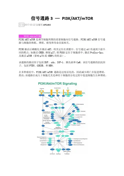
信号通路3 —PI3K/AKT/mTORAPExBIO一、PI3K/Akt/mTORPI3K/AKT/mTOR是调节细胞周期的重要细胞内信号通路。
PI3K/AKT/mTOR信号通路与细胞的休眠、增殖、癌变和寿命直接相关。
PI3K激活后磷酸化并激活AKT,将其定位在质膜中。
信号通过AKT传递到下游不同的靶点,如激活CREB,抑制p27,将FOXO定位于细胞质中,激活PtdIns-3ps,及激活mTOR(影响p70或4EBP1的转录)。
该通路的激活因子包括EGF、shh、IGF-1、胰岛素和CaM。
该信号通路的拮抗因子,包括PTEN、GSK3B、和HB9。
在多种癌症中,PI3K/AKT/mTOR通路是过度活化的,因此减少凋亡并促进增殖。
然而,该通路在成人干细胞尤其是神经干细胞的分化过程中促进细胞生长和增殖。
1. PI3KPhosphatidylinositide 3-kinases,是一种胞内磷脂酰肌醇激酶。
由调节亚基p85和催化亚基p110构成。
与v.sre和v.ras等癌基因的产物相关。
PI3K本身具有丝氨酸/苏氨酸(Ser/Thr)激酶的活性,也具有磷脂酰肌醇激酶的活性。
2. Akt又称PKB(protein kinase B)。
是一种丝氨酸/苏氨酸特异性蛋白激酶,在多种细胞生长过程中发挥关键作用,如葡萄糖代谢、凋亡、细胞增殖、转录和细胞迁移。
Akt的Ser473可以被PDK1磷酸化。
PKB与PKA和PKC均有很高的同源性,该激酶被证明是反转录病毒安基因v-akt 的编码产物,故又称Akt。
3. mTORMammalian target of rapamycin。
mTOR与其它蛋白质结合,形成两种不同蛋白质复合物,mTOR复合物1(mTORC1,)和mTOR复合物2(mTORC2),它们调节不同的细胞过程。
mTORC1由mTOR、mTOR调节相关蛋白Raptor、MLST8和非核心组分PRAS40、DEPTOR 组成。
β-Catenin的磷酸化修饰与其生物学意义刘奕君,吴军舟,于宇,张宏权(北京大学基础医学院,北京100191)5 10 15 20 25 30 35 摘要:β-Catenin 在细胞的社会性中承担了两个很关键的角色:其一是参与cadherin 介导的细胞间的粘附作用;其二是参与Wnt 介导的细胞间信号传递,调控基因转录。
β-catenin的翻译后修饰是其功能的一种重要的调控机制。
其中β-catenin 的磷酸化修饰可以调节β-catenin 自身的稳定性,并且改变其在细胞内的定位,从而实现对其自身功能的调控。
异常的β-catenin 磷酸化修饰将导致β-catenin 生物学功能失常,可能与癌症、肥胖、糖尿病等多种疾病的发生有密切联系。
关于β-catenin 磷酸化修饰在不同生物学过程中的作用和意义已经成为当今研究的热点。
关键词:分子生物学;β-catenin;磷酸化修饰;蛋白质稳定性;转录调控中图分类号:R34Phosphorylation of β-catenin and its biological function LIU Yijun, WU Junzhou, YU Yu, ZHANG Hongquan(School of Basic Medical Sciences, Peking University, Beijing 100191)Abstract:β-Catenin plays a major role in the regulation of cell adhesion and gene transcription.β-catenin is not only as a component of cell-cell adhesion complexes, but also as a key effector of canonical Wnt signalling. Post-translational modification is an important regulation mechanism ofβ-catenin. Phosphorylated state of β-catenin is significant for enhancing its own stability and inducing cellular translocation. Abnormal phosphorylation of β-catenin shows strong correlations with cancer, obesity and diabetes. It has attracted much attention to investigate the roles and significances of β-catenin phosphorylation in different biological process.Key words:Molecular biology; β-catenin; phosphorylation; protein stability; transcription regualtion0引言Wnt/β-catenin 通路作为生物进化过程中高度保守的一条信号通路,参与调控细胞增殖、存活以及命运决定,在胚胎发育中有着很重要的作用,其异常激活也和许多疾病,如恶性肿瘤的发生发展密切相关。
食管鳞癌组织中GSK3β表达的临床意义刘怡文;孙蔚;齐义军;高社干【摘要】目的:分析GSK3β在食管鳞癌组织中的表达及临床意义.方法:采用实时荧光定量PCR、Western blot、免疫组化等方法检测GSK3β在52例食管鳞癌及相应癌旁组织中的表达,并采用Cox回归分析GSK3β表达与食管鳞癌患者术后预后的关系.结果:食管鳞癌组织中GSK3β mRNA及蛋白表达水平、阳性表达率均高于相应癌旁组织(P<0.05).Cox回归分析表明GSK3β为食管鳞癌患者术后预后的独立危险因素(β=1.095,P=0.026).结论:GSK3β表达可能促进了食管鳞癌的发生发展;GSK33可能成为食管鳞癌靶向治疗的分子靶点之一.%Aim:To characterize the expression level of GSK3β and its clinical significance in esophageal squamous cell carcinoma(ESCC) tissue.Methods:The mRNA and protein expression levels of GSK3β in 52 cases of ESCC and matched adjacent tissue samples were determined by Real-time PCR,Western blot and immunohistochemistry,respectively,and the correlation between the expression of GSK3 β and the prognosis of patients was also analyzed using Cox regression.Restults:The mRNA and protein expression levels,and the positive rate of GSK3β in ESCC tissue were higher compared with those in adjacent noncancerous mucosa tissue(P < 0.05).Cox regression analysis showed that GSK3β was an independent risk factor for the prognosis of patients with ESCC (β =1.095,P =0.026).Conclusion:GSK3β expression may promote the development of ESCC,and GSK3β may be one of the molecular targets for treatment of ESCC.【期刊名称】《郑州大学学报(医学版)》【年(卷),期】2018(053)001【总页数】4页(P21-24)【关键词】食管鳞癌;GSK3β;预后【作者】刘怡文;孙蔚;齐义军;高社干【作者单位】河南科技大学第一附属医院临床医学院;河南省肿瘤表观遗传重点实验室河南洛阳471003;河南科技大学第一附属医院临床医学院;河南省肿瘤表观遗传重点实验室河南洛阳471003;河南科技大学第一附属医院临床医学院;河南省肿瘤表观遗传重点实验室河南洛阳471003;河南科技大学第一附属医院临床医学院;河南省肿瘤表观遗传重点实验室河南洛阳471003【正文语种】中文【中图分类】R735.1食管癌是世界上最常见的八大恶性肿瘤之一,而中国每年新增的食管癌患者约占全球的一半。
GSK-3β和β-catenin在弥漫性大B细胞性淋巴瘤组织中的表达及意义叶春美;孙爱宁【摘要】①目的探讨GSK-3β和β-catenin在弥漫性大B细胞性淋巴瘤组织中的表达及意义.②方法采用免疫组化二步法检测PGSK-3β(Ser9)和β-catenin蛋白在63例弥漫性大B细胞性淋巴瘤、14例反应性增生的淋巴结组织中的表达,以了解它们在弥漫性大B细胞性淋巴瘤中表达及意义.③结果β-catenin主要定位于细胞浆,弥漫性大B细胞性淋巴瘤阳性表达率(28.57%);反应性增生的淋巴结组织阳性表达率(0);两者相比较,弥漫性大B细胞性淋巴瘤阳性表达率明显高于反应性增生的淋巴结的阳性表达率,差异(P<0.05),有统计学意义;在弥漫性大B细胞性淋巴瘤可见核表达,而反应性增生的淋巴结无核表达.PGSK一3β主要定位于细胞浆,弥漫性大B细胞性淋巴瘤阳性表达率(38.10%);反应性增生的淋巴结阳性表达率于(0.07%);两者相比较,弥漫性大B细胞性淋巴瘤阳性表达率明显高于反应性增生的淋巴结的阳性表达率,差异有统计学意义(P<0.05).相关性分析,PGSK-3β和β-catenin 蛋白在弥漫性大B细胞性淋巴瘤阳性表达率存在显著的相关性(P<0.001).④结论 GSK-3β(Ser9)磷酸化使GSK-3β灭活,抑制了GSK-3β降解β-catenin的作用,导致了胞浆内游离的β-catenin 蛋白积累并进入细胞核,可能最终导致了弥漫性大B细胞性淋巴瘤的发生.【期刊名称】《河北联合大学学报(医学版)》【年(卷),期】2013(015)002【总页数】3页(P155-157)【关键词】弥漫性大B细胞性淋巴瘤;GSK-3β;β-catenin;免疫组织化学【作者】叶春美;孙爱宁【作者单位】苏州大学附属第一医院【正文语种】中文【中图分类】R73弥漫大B细胞淋巴瘤(diffuse large B-cell Ivmphoma,DLBCL)是最常见的非霍奇金淋巴瘤(nonHodgikin,S lymphomas,NHL),约占成人NHL的30% ~40%。
hedgehog信号通路简介命名由来:Nusslein-Volhard等人在筛选影响果蝇幼虫发育基因时,发现hedgehog基因突变会导致幼虫长满刚毛,因此称为hedgehog。
【1】主要功能参与发育过程中的细胞分化。
1.作为体节极性基因,在果蝇幼虫体节形成过程中发挥作用。
Wg和en受pair-rule基因调控激活。
en在even-skipped(Eve)或Fushi tarazu(Ftz)蛋白含量较高的细胞中表达,同时受到Odd-skipped, Runt,或Sloppy-paired的抑制。
Wg 在两者(Eve & Ftz)均不表达(表达sloppy-paired基因)的细胞中表达。
Wg蛋白表达后扩散到周围细胞,在表达en的细胞中,Wg和Ftz/Lrp6结合,经Wg信号通路激活en的表达。
en蛋白激活en自身及hh基因的表达,hh扩散到周围细胞,和Patch 受体结合,增强Wg基因的表达。
[正反馈]hh/wg浓度梯度确定了denticle表达的边界(hh浓度高不长毛,wg浓度高,长毛)。
若Wg/Hh通路受影响,毛会布满整个体节。
Hedgehog,Porcupine,Armadillo因此得名。
2.果蝇翅成虫盘的发育过程中参与AP方向的形态建成。
果蝇胚胎发生期到一龄幼虫初期,翅成虫盘完成AP区域分隔。
具体过程如下:engrailed在翅膀dorsal part表达,促进hedgehog的表达,同时也抑制hedgehog在dorsal part的功能(?ptc只在anterior表达)。
Hedgehog诱导下游dpp表达,然而作为短程信号蛋白,决定dpp的表达范围仅限于AP界线靠近anterior的位置。
Dpp作为长程信号蛋白,沿AP方向扩散,形成浓度梯度,组织翅膀发育。
3.脊椎动物手的发育(六指性状)在脊椎动物手的发育过程中,肢芽后端的ZPA(zone of polarizing activity)区分泌SHH,形成一个扩散的梯度。
GSK-3β在高糖环境下足细胞转分化效应中的作用
的开题报告
标题:GSK-3β在高糖环境下足细胞转分化效应中的作用
背景:
近年来,糖尿病已成为全球大流行的代表性疾病之一。
高糖环境对
细胞造成多种损伤,其中包括足细胞的功能损伤。
足细胞是肾小球滤过
屏障的重要细胞类型,其转分化被认为是糖尿病肾病发生发展的关键环节。
研究发现,GSK-3β(糖原合成激酶-3β)在足细胞转分化中扮演着重要角色。
然而,GSK-3β在高糖环境下足细胞转分化中的作用机制尚待深
入探究。
研究问题:
在高糖环境下,GSK-3β在足细胞转分化中的作用机制是什么?
研究方法:
采用体外细胞实验,使用高糖培养液处理足细胞,使用Western blot、免疫荧光染色等方法检测GSK-3β的表达及其他相关信号通路的变化,辨析其与足细胞转分化相关性。
研究意义:
深入研究GSK-3β在高糖环境下足细胞转分化效应中的作用,可以更好地理解足细胞转分化的分子机制,并为糖尿病肾病的早期诊断和治疗
提供理论依据。
此外,研究结果还可以为其他疾病的治疗提供新的思路。

b-catenin第675位氨基酸的磷酸化解释说明以及概述1. 引言1.1 概述在细胞生物学和信号转导领域,b-catenin是一个重要的蛋白质,它在细胞间粘附和Wnt信号通路中起着关键作用。
磷酸化是一种常见的细胞信号传递方式,在调控b-catenin的功能和稳定性方面也不可忽视。
本文将重点讨论b-catenin 第675位氨基酸的磷酸化事件,并探讨其对细胞功能的影响。
1.2 文章结构本文分为引言、正文、结论和参考文献四个部分。
下面将简要介绍每个部分的内容。
1.3 目的本文旨在系统地阐述b-catenin第675位氨基酸的磷酸化现象,并深入解释该磷酸化事件的意义和作用。
通过对已有研究结果进行概述和解读,我们希望能揭示b-catenin第675位氨基酸磷酸化对细胞信号转导、细胞增殖、干细胞分化等方面的重要性,并探究可能存在的应用前景和展望。
以上是“1. 引言”部分的内容,概述了文章主要讨论的内容、结构和目的。
2. 正文:2.1 b-catenin的功能和调控机制:b-catenin是一种细胞间连接蛋白,它在细胞黏附、信号转导以及胚胎发育中发挥重要作用。
作为Wnt信号通路的核心成员,b-catenin在该通路中起着关键的调节功能。
在没有Wnt信号时,b-catenin通常会被细胞质内蛋白复合体(APC、Axin和GSK-3β)引起其磷酸化并标记为降解。
而当受到Wnt信号激活后,b-catenin逃脱蛋白复合物的调控,并进入细胞核,在那里与转录因子联合并激活目标基因的表达。
2.2 磷酸化在细胞信号转导中的作用:磷酸化是一种广泛存在于生物体内的翻译后修饰, 在细胞信号转导中起着重要作用。
通过添加一个磷酸基团到特定氨基酸上,可以改变该蛋白质分子的构象和活性。
对于b-catenin来说,磷酸化状态直接影响了其稳定性和亚细胞定位,从而调节了其在Wnt信号通路中的活性。
2.3 b-catenin第675位氨基酸的重要性:b-catenin在其C端拥有许多潜在的磷酸化位点,其中第675位氨基酸就是一个关键的位置。
结直肠异型增生性异常隐窝灶中 DAB2IP、GSK-3β蛋白表达变化及意义刘芳;尚雪飞;孙鹏娟;彭燕;温实;部晶;王素云【摘要】目的:观察 DAB2互作蛋白(DAB2IP)、糖原合成酶激酶-3β(GSK-3β)在结直肠异型增生性异常隐窝灶(ACF)中的表达变化,探讨异型增生性ACF 与结直肠癌发生发展的关系。
方法收集直肠镜检查中使用放大内镜窄带成像技术获取的组织标本,其中结肠正常黏膜38例、异型增生性 ACF 40例、管状腺瘤40例、腺瘤癌变35例,均经病理确诊,采用免疫组化法检测各组织中的DAB2IP 和 GSK-3β。
结果 DAB2IP 蛋白在结直肠正常黏膜、异型增生性 ACF、管状腺瘤及腺癌中阳性表达率逐步降低,分别为94.7%、77.5%、52.5%、14.3%,P 均<0.05;GSK-3β蛋白在结直肠正常黏膜、异型增生性 ACF、管状腺瘤及腺癌中阳性表达率逐步升高,分别为13.2%、57.5%、75.0%,97.1%,P 均<0.05;在异型增生性 ACF、管状腺瘤及腺瘤癌变过程中,DAB2IP 和 GSK-3β蛋白表达呈负相关(r =-0.576,P <0.05)。
结论DAB2IP 低表达与 GSK-3β高表达可能在结直肠癌前病变中起一定作用,可能作为结直肠肿瘤早期诊断治疗的物生学指标。
【期刊名称】《山东医药》【年(卷),期】2016(056)034【总页数】3页(P50-52)【关键词】结直肠肿瘤;异常隐窝灶;管状腺瘤;DAB2 互作蛋白;糖原合成酶激酶-3β【作者】刘芳;尚雪飞;孙鹏娟;彭燕;温实;部晶;王素云【作者单位】保定市第一医院,河北保定 071000;保定市第一医院,河北保定071000;保定市第一医院,河北保定 071000;保定市第一医院,河北保定 071000;保定市第一医院,河北保定 071000;保定市第一医院,河北保定 071000;河北省人民医院【正文语种】中文【中图分类】R735.3目前,异常隐窝灶(ACF)是结直肠癌发生过程中可在光镜下观察到最小、最早期的结直肠黏膜病变,WHO分类根据是否有异型增生将ACF分为增生性ACF和异型增生性ACF。
PI3K/AKT/GSK3β通路影响糖代谢与认知过程的研究进展磷脂酰肌醇3-激酶(PI3 Ks)为酪氨酸激酶和G蛋白偶联受体的主要下游分子,其催化产生第二信使3,4,5-三磷酸磷脂酰肌醇(PIP3),并激活下游分子蛋白激酶B(PKB/AKT)、糖原合酶激酶-3β(GSK-3β)等,将多种生物信号传递到细胞内,从而对葡萄糖转运、细胞增殖、分化和凋亡等过程起调节作用,且与胰岛素抵抗相关的2型糖尿病关系密切。
本文将对国内外研究中PI3K/AKT/GSK3β信号通路在糖代谢中的作用及对认知过程的影响研究进展予以综述。
标签:2型糖尿病;糖代谢;认知;胰岛素信号通路;PI3K/AKT/GSK3β根据国际糖尿病联盟(IDF)最新数据,预计到2035年,糖尿病患病人数将达到6亿,增长至55%。
其中2型糖尿病并发症多,医疗负担重,给家庭和社会带来沉重负担。
与此同时,与认知障碍相关的阿尔茨海默病带来的问题也日益严峻。
目前两种疾病的相关机理依然存在争议,因此研究2型糖尿病和阿尔茨海默病的确切发病机制成为治疗疾病的当务之急。
1 2型糖尿病与胰岛素抵抗胰岛素抵抗是指靶细胞对胰岛素的敏感性、反应性降低,即胰岛素从周围组织摄取和清除葡萄糖的能力减低导致糖代谢紊乱,出现这种现象主要由不正常β细胞分泌产物、胰岛素拮抗剂持续供应和靶向組织缺陷三种原因造成[2]。
2型糖尿病与胰岛素抵抗和胰岛β 细胞功能缺损息息相关,其在环境或遗传等因素的影响下常表现为胰岛素抵抗[3]。
胰岛素抵抗,高胰岛素血症和葡萄糖利用障碍三者为2型糖尿病主要特征,且随胰岛素抵抗的增加,糖尿病的风险也随之增加。
2 胰岛素信号通路与糖代谢PI3K/AKT/GSK3β(Phosphoinositide-3-Kinase/Protein kinase B/Glycogen synthase kinase-3)是重要的胰岛素信号通路,调控血糖的功能主要依靠此信号通路。
胰岛素首先与细胞膜外的胰岛素受体(IR)结合使胰岛素受体构象发生改变,并在酪氨酸激酶(PTK)的作用下磷酸化,进而激活胰岛素受体底物(IRS),引起下游信号分子的级联反应,如磷脂酞肌醇激酶(phosphoinositide3-kinase,PI3 K)/蛋白激酶B(protein kinaseB,PKB/AKT)。
· 100 ·· Non-Presentation ·NP -1 三七皂苷Fe 通过诱导PVH C-Fos 表达抑制肥胖小鼠的食欲并减轻体重李鸿丽 范圣洁创新中药实验室,中药学院,上海中医药大学,201203,中国【摘要】 目的:长期机体的能量摄入和能量消耗失衡会导致肥胖,肥胖会引起一系列的并发症,如心血管疾病、二型糖尿病、高血压、高血脂、脂肪肝等,严重威胁人们的健康。
食欲的控制是目前减肥药的焦点,但是市售的减肥药作用不明显并伴有严重的副作用。
因此,我们的课题致力于从中药天然产物中筛选出减肥作用显著且副作用小的小分子化合物。
方法:在前期的药物筛选中我们发现三七皂苷Fe 能够明显抑制高脂诱导的C57 BL/6肥胖(DIO )小鼠的食欲。
为检测Fe 是否具有减肥作用,我们首先通过给DIO 小鼠腹腔注射生理盐水和Fe ,检测体重、摄食、葡萄糖耐受、脂肪和瘦肉含量,用代谢笼观测耗氧量(VO2)、二氧化碳生成量(VCO2)以及呼吸交换率(RER )等与肥胖相关的生理指标。
其次,通过侧脑室立体定位手术将Fe 直接注射入侧脑室,检测其对摄食的影响,并将全脑切片,进行C-Fos 免疫荧光染色,观察响应药物作用的神经核团。
结果:Fe 对DIO 小鼠具有明显的减肥作用,能够显著减少脂肪的体积和重量,改善肝脏脂质堆积,可能是通过降低DIO 小鼠的食欲,减少摄食量引起的。
代谢笼数据显示Fe 能够增加DIO 小鼠的能量消耗。
食欲的调控主要受中枢控制,侧脑室直接给药发现Fe 能够降低DIO 小鼠的摄食,并显著增加下丘脑室旁核(PVH ) C-Fos 表达。
结论:Fe 具有明显的减肥作用,其作用机制可能是通过改变中枢某类神经元兴奋而减少摄食引起的,提示Fe 可能是一个潜在的减肥药。
【关键词】 三七皂苷Fe ;下丘脑室旁核;减肥;摄食NP -2 GSK-3β Inhibitor Reduced Neonatal Hypoxic-Ischemic Brain Injury in Mice FENG Zhong-pingDepartment of Physiology ,Faculty of Medicine ;the Director of Collaborative Specialization Program In Neuroscience ;University of Toronto ,Toronto ,Canada【ABSTRACT 】 Hypoxia-ischemia is an important cause of brain injury and neurological morbidity in the newborn infants. Glycogen synthase kinase 3β (GSK-3β) is activated and up-regulated following hypoxic-ischemic (HI ) brain injury. We have investigated the effects of GSK-3β inhibitors ,Tideglusib and TDZD-8,on HI brain injury in neonatal mice. Specifically ,postnatal day 7(P7) mouse pups subjected to unilateral common carotid artery ligation followed by 1 h of hypoxia. HI experimental or sham control groups were administered either the GSK-3β inhibitor or vehicle intraperitoneally 20 min prior to the onset of ischemia. The brain infarct volume and· 101 ·whole brain images were used in conjunction with Nissl staining to evaluate the protective effects of GSK-3β inhibitors. Either tideglusib or TDZD-8 significantly reduced cerebral infarct volume and improved neurobehavioral outcomes following HI injury. Western blotting showed that either drug increased phosphorylated GSK-3β and Akt ,and reduced the expression of GFAP and p-STAT3. Pretreatment with tideglusib also enhanced the protein level of Notch1,and reduced the cleavage of pro-apoptotic signal caspase proteins ,including caspase 3 and caspase 9 following HI. These results indicate that these GSK-3β inhibitors show neuroprotection against hypoxic-ischemic brain injury in neonatal mice. Either Tideglusib or TDZD-8 is a potential compound for the prevention or treatment of hypoxic-ischemic brain injury in neonates.Corresponding author :FENG Zhong-ping ,Email : zp.feng@utoronto.caNP -3 Effects of Osthole Microemulsion by NasalAdministration on the Cholinergic Pathway in AD Mice HOU Xue-qin 1,RONG Cui-ping 2,HAO Ji-fu 3,ZHANG Han-ting 1,4*1. Institute of Pharmacology ,Taishan Medical University ,Shandong ,271016,China2. Guangxi Key Laboratory of Brain and Cognitive Neuroscience ,Guilin Medical University ,Guilin ,China3. Department of Pharmacy ,Taishan Medical University ,Shandong ,271016,China4. D epartments of Behavioral Medicine & Psychiatry ,Physiology & Pharmacology ,and Neuroscience ,the Rockefeller Neurosciences Institute ,West Virginia University Health Sciences Center ,Morgantown ,WV 26506,USA****************************************************************************************Hou Xue-Qin ,Ph.D.,female ,born in December ,1984. She is theInstructor at Taishan Medical University School of Pharmacy. Herresearch interest is clinical and experimental studies of encephalopathy.She has published a total of more than 20 papers ,including six SCI papersand seven first-class Chinese papers ,in all of which she was the first-author. She has been funded by two research grants as the single PI fromthe national (NNSF ) or local government ,in addition to 5 NNSF-fundedgrants as the co-investigator. She also co-authored three medical books.****************************************************************************************【ABSTRACT 】 Objectives :Fructus Cnidii is the dry ripe fruit of Cnidium monnieri (L.) Cuss.,which belongs to the umbelliferous plant. It has long been used in clinic practice and has been found to have pharmacological activity in the central nervous system. Osthole is the main active component of Fructus Cnidii. However ,it shows low bioavailability ,fast distribution and elimination ,and low concentration in the brain when given orally. In this study ,we aimed to develop a new dosage form to increase the osthole concentration in the brain and enhance its pharmacological effects in the central nervous system through reducing the dosage while improving the stability and bioavailability. Thus ,microemulsion containing osthole was prepared and the effects of osthole microemulsion were examined in the mouse model of Alzheimer’s disease (AD ).Methods :On the basis of pseudo-ternary phase diagram ,microemulsion was。
REVIEWGlycogensynthasekinase3(GSK3)intheheart:apointofintegrationinhypertrophicsignallingandatherapeutictarget?Acriticalanalysis
PHSugden,SJFuller,SCWeissandAClerkNationalHeartandLungInstitute(NHLI)Division,FacultyofMedicine,ImperialCollegeLondon,London,UKGlycogensynthasekinase3(GSK3,ofwhichtherearetwoisoforms,GSK3aandGSK3b)wasoriginallycharacterizedinthecontextofregulationofglycogenmetabolism,thoughitisnowknowntoregulatemanyothercellularprocesses.PhosphorylationofGSK3a(Ser21)andGSK3b(Ser9)inhibitstheiractivity.Intheheart,emphasishasbeenplacedparticularlyonGSK3b,ratherthanGSK3a.Importantly,catalytically-activeGSK3generallyrestrainsgeneexpressionand,intheheart,catalytically-activeGSK3hasbeenimplicatedinanti-hypertrophicsignalling.InhibitionofGSK3resultsinchangesintheactivitiesoftranscriptionandtranslationfactorsintheheartandpromoteshypertrophicresponses,anditisgenerallyassumedthatsignaltransductionfromhypertrophicstimulitoGSK3passesprimarilythroughproteinkinaseB/Akt(PKB/Akt).However,recentdatasuggestthatthesituationisfarmorecomplex.WereviewevidencepertainingtotheroleofGSK3inthemyocardiumanddiscusseffectsofgeneticmanipulationofGSK3activityinvivo.WealsodiscussthesignallingpathwayspotentiallyregulatingGSK3activityandproposethat,dependingonthestimulus,phosphorylationofGSK3isindependentofPKB/Akt.PotentialGSK3substratesstudiedinrelationtomyocardialhypertrophyincludenuclearfactorsofactivatedTcells,b-catenin,GATA4,myocardin,CREB,andeukaryoticinitiationfactor2Be.Theseandothertranscriptionfactorsubstratesputativelyimportantintheheartareconsidered.WediscusswhethercardiacpathologiescouldbetreatedbytherapeuticinterventionattheGSK3levelbutconcludethatanyinterventionwouldbeprematurewithoutgreaterunderstandingofthepreciseroleofGSK3incardiacprocesses.BritishJournalofPharmacology(2008)153,S137–S153;doi:10.1038/sj.bjp.0707659;publishedonline21January2008
Keywords:cardiacmyocytes;transgenicmousemodels;myocardialhypertrophy;glycogensynthasekinase3;proteinkinaseB/Akt;mitogen-activatedproteinkinases;transcriptionfactors;nuclearfactorofactivatedTcells;b-catenin
Abbreviations:APC,adenomatouspolyposiscoli;BW,bodyweight;eIF,eukaryoticinitiationfactor;ERK,extracellularsignal-regulatedkinase;GSK3,glycogensynthasekinase3;HDAC,histonedeacetylase;HW,heartweight;IGF1,insulin-likegrowthfactor1;LRP,lipoproteinreceptor-relatedprotein;MAPK,mitogen-activatedproteinkinase;MSK,mitogen-andstress-activatedproteinkinase;NFAT,nuclearfactorofactivatedTcell;PtdIns(4,5)P2,phosphatidylinositol4,5-bisphosphate;PtdIns(3,4,5)P3,phosphatidylinositol3,4,5-trisphosphate;PI3K,phos-
phoinositide30kinase;PKA,cyclicAMP-dependentproteinkinaseA;PKB,proteinkinaseB;PKC,proteinkinaseC;RSK,p90-ribosomalS6kinase;TAC,transverseaorticconstriction;TCF/LEF1,T-cell-specifictranscriptionfactor/lymphoidenhancerbindingfactor1;TL,tibiallength
IntroductionGlycogensynthasekinase3(GSK3)isemergingasanimportanttherapeutictargetinavarietyofpathologies(CohenandGoedert,2004).Here,wewillbeprincipallyconcernedwithitsroleincardiacgrowthandheartfailure,particularlywiththatoccurringduringmyocardialhyper-trophy.GSK3mayalsoplayanimportantroleinthe
cardioprotectionaffordedbyischaemicpreconditioning(Tongetal.,2002),butthisisbeyondthescopeofthisreview.GSK3wasoriginallyidentifiedasaSer/ThrproteinkinasethatphosphorylatesglycogensynthaseonmultiplesitesinitsC-terminalregion(Figure1a)toinhibititsactivity(Embietal.,1980).ItisakeysignallingproteinintheregulationofglycogensynthesisandtheactivityofGSK3itselfisregulatedbyphosphorylation–dephosphorylation,phosphorylationbeinginhibitory(Figure1b).Phosphoryla-tionofGSK3isincreasedbyinsulinandthisrepresentsamajormechanismbywhichinsulinpromotesglycogenaccumulation(Cohen,2006).Received26July2007;revised16November2007;accepted4December
2007;publishedonline21January2008
Correspondence:ProfessorPHSugden,NationalHeartandLungInstitute(NHLI)Division,FacultyofMedicine,ImperialCollegeLondon,FlowersBuilding(4thFloor),ArmstrongRoad,LondonSW72AZ,UK.E-mail:p.sugden@imperial.ac.uk
BritishJournalofPharmacology(2008)153,S137–S153&2008NaturePublishingGroupAllrightsreserved0007–1188/08$30.00
www.brjpharmacol.orgSinceitsdiscoveryaround25yearsago,GSK3hasbeenimplicatedintheregulationofmanyadditionalbiologicalprocessesapartfromglycogenmetabolism.Particularemphasishasbeenplacedonitsroleinthephosphorylationoftranscriptionalregulatorsandtranslationinitiationfactors,andtheconsequenteffectsongeneandproteinexpression.ItisalsonotablethatGSK3regulatesmetazoandevelopmentbymodulatingtranscriptionalactivityinthecontextofthe‘canonical’Wntpathway(LoganandNusse,2004;CadiganandLiu,2006;GordonandNusse,2006).The