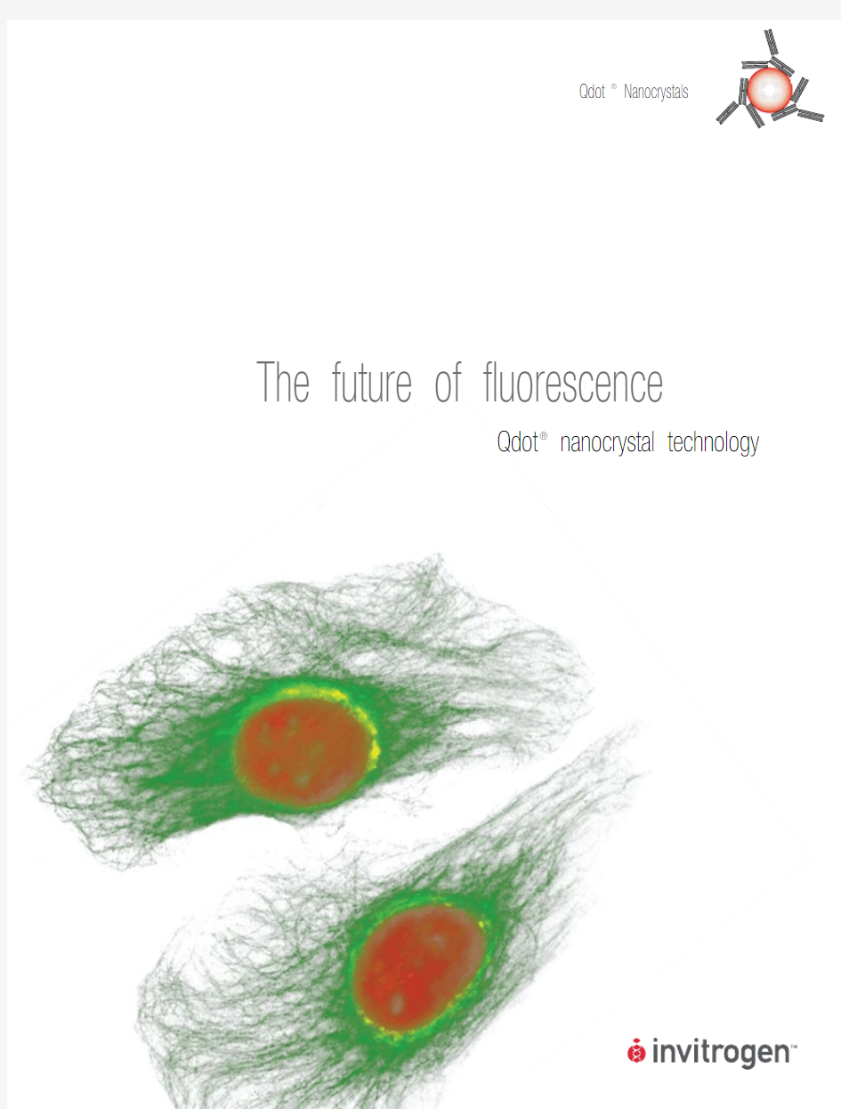
Qdot? Nanocrystals The future of fluorescence
Qdot? nanocrystal technology
2
Qdot ?
Nanocrystals
What are Qdot? nanocrystals?
Fundamentally, Qdot? nanocrystals are fluorophores—substances that absorb photons of light, then reemit photons at a different wavelength.1–3 However, simple comparisons with organic fluorescent dyes and naturally fluorescent proteins end there. Qdot? nanocrystal conjugates combine exceptional fluorescence with full biofunctionality for a wide variety of life science applications. This combination is especially important for those applications requiring excellent photostability and multicolor detection from a single excitation source.
Anatomy of a Qdot? nanocrystal
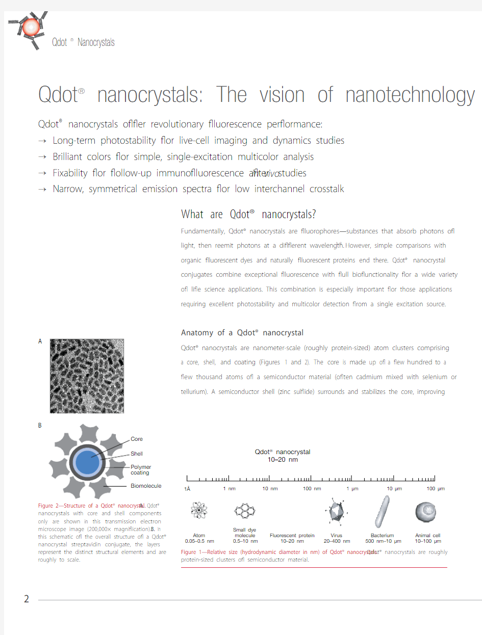
Qdot? nanocrystals are nanometer-scale (roughly protein-sized) atom clusters comprising a core, shell, and coating (Figures 1 and 2). The core is made up of a few hundred to a few thousand atoms of a semiconductor material (often cadmium mixed with selenium or tellurium). A semiconductor shell (zinc sulfide) surrounds and stabilizes the core, improving
Qdot ?
nanocrystals: The vision of nanotechnology
Qdot ? nanocrystals offer revolutionary fluorescence performance:Long-term photostability for live-cell imaging and dynamics studies →Brilliant colors for simple, single-excitation multicolor analysis →Fixability for follow-up immunofluorescence after → in vivo studies Narrow, symmetrical emission spectra for low interchannel crosstalk
→Figure 1—Relative size (hydrodynamic diameter in nm) of Qdot? nanocrystals. Qdot? nanocrystals are roughly protein-sized clusters of semiconductor material.
100 μm
10 μm
1 μm
100 nm
10 nm
1 nm
1?
Fluorescent protein
10–20 nm Small dye molecule 0.5–10 nm
Atom 0.05–0.5 nm
Animal cell 10–100 μm
Bacterium 500 nm–10 μm Virus 20–400 nm Qdot ? nanocrystal
10–20 nm
Figure 2—Structure of a Qdot? nanocrystal. A. Qdot? nanocrystals with core and shell components only are shown in this transmission electron microscope image (200,000× magnification). B. In this schematic of the overall structure of a Qdot? nanocrystal streptavidin conjugate, the layers represent the distinct structural elements and are roughly to scale.
A
B
both the optical and physical properties of the material. An amphiphilic polymer coating then encases this core and shell, providing a water-soluble surface that can be differentially modified to create Qdot? nanocrystals that meet specific assay requirements.
For most of the Qdot? nanocrystal products, this amphiphilic inner coating is cova-lently modified with a functionalized polyethylene glycol (PEG) outer coating. The PEG surface has been shown to reduce nonspecific binding in flow cytometry4 and imaging assays, thereby improving signal-to-noise ratios and providing clearer resolution of cell populations and cellular morphology. Qdot? primary and secondary antibody conjugates (Figure 3), Qdot? streptavidin conjugates, Qtracker? nontargeted quantum dots, and Qdot? ITK? amino (PEG) quantum dots, as well as the reactive nanocrystals provided in the Qdot? Antibody Conjugation Kit, all utilize this PEG chemistry.
Fluorescence of Qdot? nanocrystals
Qdot? nanocrystals are extremely efficient materials for generating fluorescence (Table 1). Their intrinsic brightness is often many times that observed for traditional organic fluoro-
phores, and their photostability is many orders of magnitude greater. These extraordinary fluorescence properties can be attributed to the unique fluo r escence mechanism of semi-conductor materials. Unlike organic fluorophores, Qdot? nanocrystals fluoresce through the formation of excitons, or Coulomb-correlated electron–hole pairs, upon absorption of a photon of light. Compared with the excited state of a fluorophore, this exciton typically exhibits a much longer lifetime (up to ~200 nanoseconds), a property that can be advanta-geous in certain types of time-gated detection studies.5Figure 3 —Multicolor immunofluorescence imaging with Qdot? secondary antibody conjugates. Actin
in a mouse intestine section was detected with mouse anti-actin antibody and Qdot? 655 goat F(ab’)
2
anti–mouse IgG antibody (red), laminin was detected with rabbit anti-laminin antibody
and Qdot? 525 goat F(ab’)
2
anti–rabbit IgG anti-body (green), and nuclei were stained with Hoechst 33342 (blue). Image contributed by Thomas Deerinck and Mark Ellisman, The National Center for Microscopy and Imaging Research, San
Diego, California, USA.
Table 1—Extinction coefficients of Qdot? streptavidin conjugates at common excitation wavelengths.
Product350 nm405 nm488 nm532 nm
Qdot? 525 nanocrystals710,000 M–1 cm–1360,000 M–1 cm–1130,000 M–1 cm–1Not applicable
Qdot? 565 nanocrystals1,900,000 M–1 cm–11,100,000 M–1 cm–1290,000 M–1 cm–1139,000 M–1 cm–1
Qdot? 585 nanocrystals3,500,000 M–1 cm–12,200,000 M–1 cm–1530,000 M–1 cm–1305,000 M–1 cm–1
Qdot? 605 nanocrystals4,400,000 M–1 cm–12,800,000 M–1 cm–11,100,000 M–1 cm–1580,000 M–1 cm–1
Qdot? 625 nanocrystals14,700,000 M–1 cm–19,900,000 M–1 cm–12,700,000 M–1 cm–1870,000 M–1 cm–1
Qdot? 655 nanocrystals9,100,000 M–1 cm–15,700,000 M–1 cm–12,900,000 M–1 cm–12,100,000 M–1 cm–1
Qdot? 705 nanocrystals12,900,000 M–1 cm–18,300,000 M–1 cm–13,000,000 M–1 cm–12,100,000 M–1 cm–1
Qdot? 800 nanocrystals12,600,000 M–1 cm–18,000,000 M–1 cm–13,000,000 M–1 cm–12,000,000 M–1 cm–1
https://www.doczj.com/doc/c418181509.html,
3
4
Qdot? Nanocrystals
Tuneability of Qdot? nanocrystals
In addition to these distinctive structural and fluorescence prop-
erties, Qdot? nanocrystals show a direct, predictable relationship
between their physical size and the energy of the exciton (and
therefore, the wavelength of emitted fluorescence) (Figure 4). This
property, referred to as “tuneability”, has allowed us to develop a
series of Qdot? nanocrystals that have a common excitation pro-
file but different fluorescent emission maxima. The tuneability of
Qdot? nanocrystals is being widely exploited in the development
of multicolor nanocrystal-based assays.
What can Qdot? nanocrystals do?
Brightness coupled with photostability for long-term
studies
The remarkable photostability of Qdot? nanocrystals enables long-
term imaging experiments under conditions that would lead to
the photoinduced deterioration of other types of fluorophores.3,6
Howarth and colleagues have achieved real-time imaging of the
complex formed between a single ligand-labeled nanocrystal and
its target receptor on live neurons,7 an approach whose success
these authors attribute in part to the brightness inherent in the
nanocrystal particles. Furthermore, cells and tissues labeled with
Qdot? nanocrystals can be archived permanently and reanalyzed
with the same level of sensitivity as achieved in the initial assay.
Nanocrystal emission series for multicolor analysis
With their broad excitation and narrow, symmetric emission prop-
erties (Figure 5), the Qdot? nanocrystals require only a single exci-
tation source (typically <450 nm), facilitating multiplex analysis of
multiple targets or events in a single sample. For example, Chat-
topadhyay and coworkers used eight different Qdot? nanocrys-
tals in combination with organic fluorophores to achieve 17-color
immunophenotyping by flow cytometry.4 Proper filter selection is
critical, however, for resolving individual fluorescent signals arising Figure 5—Absorption and emission profiles of Qdot? nanocrystals. Qdot? nanocrys-
tals are characterized by broad absorption spectra and narrow, symmetrical,
and discrete emission profiles. Note that the absorption profiles shown here
for these Qdot? nanocrystals are identical to their excitation profiles.
Figure 4—Tuneability of Qdot? nanocrystals. Seven different nanocrystal solu-
tions are shown excited with the same long-wavelength UV lamp; the size of
the nanocrystal at the angstrom scale determines the color.
525545565585605625
655
400450500550600650700750800850900
500,000
1,000,000
1,500,000
2,000,000
2,500,000
3,000,000
3,500,000
4,000,000
4,500,000
5,000,000
Wavelength (nm)
E
x
t
i
n
c
t
i
o
n
c
o
e
f
c
i
e
n
t
(
M
-
1
c
m
-
1
)
8
8
7
7
6
6
5
5
4
4
3
3
2
2
8. Qdot? 800 conjugate excitation
7. Qdot? 705 conjugate excitation
6. Qdot? 655 conjugate excitation
5. Qdot? 625 conjugate excitation
4. Qdot? 605 conjugate excitation
3. Qdot? 585 conjugate excitation
2. Qdot? 565 conjugate excitation
1. Qdot? 525 conjugate excitation
8. Qdot? 800 conjugate emission
7. Qdot? 705 conjugate emission
6. Qdot? 655 conjugate emission
5. Qdot? 625 conjugate emission
4. Qdot? 605 conjugate emission
3. Qdot? 585 conjugate emission
2. Qdot? 565 conjugate emission
1. Qdot? 525 conjugate emission
1
1
N
o
r
m
a
l
i
z
e
d
u
o
r
e
s
c
e
n
c
e
e
m
i
s
s
i
o
n
from different Qdot? nanocrystals; recommended filter sets are available from the major filter manufacturers (Table 2). Because nanocrystals are particle-based fluorophores, they have intrinsic electron and X-ray contrast, delivering powerful multimodality for correla-tive light and electron microscopy and for imaging studies that utilize both fluorescence and X-ray or computerized tomography (CT).
Diverse surface chemistries for customization
The surface chemistry dictates many of the important properties of the Qdot? nanocrys-tals in biological context. For that reason, Qdot? nanocrystals are available with a choice of surface reactivities, from the nonreactive Qtracker? nontargeted quantum dots for in vivo imaging, to the amine- and carboxyl-derivatized Qdot? Innovator’s Tool Kit (ITK?) quan-tum dots. We also prepare Qdot? ITK? nanocrystals labeled with streptavidin for creating brightly fluorescent noncovalent conjugates with biotinylated molecules, as well as Qdot? ITK? organic quantum dots for applications requiring organic solvents. The diverse surface functionalities available in the Qdot? product line offer researchers a multitude of choices for creating custom nanocrystal conjugates.
Qdot? 625 nanocrystals and conjugates
A better, brighter red nanocrystal for immunostaining, flow cytometry, and more
The first of our next-generation quantum dots, the Qdot? 625 nanocrystals, are now avail-able from Invitrogen. This new color complements our series of visible and near-infrared col-ors of Qdot? nanocrystals (Figure 6), making the possibility of true eight-color multispectral analyses a reality for many researchers. Qdot? 625 nanocrystals exhibit the same exceptional photostability and narrow emission profile as the other Qdot? nanocrystal colors but without
Table 2—Spectral properties and recommended filters for Qdot? nanocrystals.
Qdot? nanocrystal Excitation
wavelength (nm)*
Peak emission
wavelength (nm)
Recommended filter
Qdot? 565405565575/26
Qdot? 605405605605/20 Qdot? 625405625625/20 Qdot? 655405?655655/20 Qdot? 705405?705720/20 Qdot? 800405?800787/42 *Qdot? nanocrystals excite optimally in the UV and 405 nm range, but can also be excited with 488 nm lasers.
? Can also be excited with 633 nm lasers.Figure 6—Fluorescence emission spectra of Qdot? 565, Qdot? 625, and Qdot? 705 streptavidin conjugates. In the context of multiplex detection with Qdot? 565 and Qdot? 705 conjugates, the relative position of the Qdot? 625 emission envelope is superior in terms of signal crosstalk to Qdot? 605 or Qdot? 655 conjugates. In such applications, all Qdot? conju-gates can be efficiently excited at a single wave-length in the near-UV/violet region (e.g., 405 nm).
Wavelength (nm)
F
l
u
o
r
e
s
c
e
n
c
e
e
m
i
s
s
i
o
n
(
%
o
f
m
a
x
i
m
u
m
)
10
20
30
40
50
70
60
80
90
100
https://www.doczj.com/doc/c418181509.html,
5
6
Qdot? Nanocrystals
the “red shoulder” often observed with traditional dyes (Figure 7). As
evidenced by the extinction coefficients using 405 nm and 488 nm
excitation, Qdot? 625 nanocrystals are the brightest of the visible-
range quantum dots (Table 3).
Qdot? 625 nanocrystals for a multitude of applications
Our Qdot? 625 streptavidin and goat anti–mouse IgG and anti–
rabbit IgG secondary antibody conjugates provide a high level of
resolution and brightness for immunostaining of cells grown in
culture (Figure 8). For those who wish to conjugate their own anti-
bodies to these ultrabright nanocrystals, the Qdot? 625 Antibody
Conjugation Kit is available. For optimal detection of the Qdot?
625 nanocystals, we recommend the BrightLine? QD625-A Filter
Set (visit https://www.doczj.com/doc/c418181509.html,). The Qtracker? 625 Cell Labeling Kit
provides researchers with the means to nonspecifically load Qdot?
nanocrystals into live cells for long-term cell tracking. Invitrogen
has tested this new material in both CHO and HeLa cells, and from
experience with our other Qdot? colors used for cell labeling, many
other cell types should also be compatible with the 625 dots. In
addition, Qdot? 625 conjugates are compatible with flow cytome-
try and exhibit their best utility when used in instruments equipped
with violet lasers (Figure 9).
Because the electron-dense Qdot? nanocrystals have uni-
form size distribution and shape, some researchers have suc-
cessfully correlated fluorescence data with electron microscopy
data to resolve the spatial location of targets immunostained
with Qdot? conjugates.8 This utility is further exploitable because
some of the Qdot? nanocrystals themselves have distinguishable
shapes (Qdot? 605 and Qdot? 655 are rod shaped, whereas the
rest of the Qdot? nanocrystals, including Qdot? 625, are spheri-
cal (Figure 10)), making resolution of dual-stained samples at the
near-molecular level a possibility.
Figure 8—Qdot? 625 goat anti-mouse conjugate immunolabeling of microtubules in
HeLa cells. Fixed and permeabilized HeLa cells were labeled with mouse anti–
α-tubulin primary antibody and 20 nM Qdot? 625 goat anti–mouse IgG.
Figure 7—Absorption (blue) and emission (red) spectra of Qdot? 625 streptavidin
conjugate.
Table 3—Extinction coefficients of Qdot? streptavidin conjugates at common
excitation wavelengths, in M–1 cm–1. For comparison, the extinction coefficient
of fluorescein with 488 nm excitation is ~80,000 M–1 cm–1.
Product405 nm488 nm
Qdot? 525 nanocrystals360,000130,000
Qdot? 565 nanocrystals1,100,000290,000
Qdot? 585 nanocrystals2,200,000530,000
Qdot? 605 nanocrystals2,800,0001,100,000
Qdot? 625 nanocrystals9,900,0002,700,000
Qdot? 655 nanocrystals5,700,0002,900,000
Wavelength (nm)
A
b
s
o
r
p
t
i
o
n
(
%
o
f
E
3
n
m
)
F
l
u
o
r
e
s
c
e
n
c
e
e
m
i
s
s
i
o
n
(
%
o
f
m
a
x
/
m
i
n
)
10
20
30
40
50
70
60
80
90
100
10
20
30
40
50
70
60
80
90
https://www.doczj.com/doc/c418181509.html,
7
Figure 10—Qdot? 625 streptavidin transmission elec-tron microscopy (TEM) images. Courtesy of Mark Ellisman, National Center for Microscopy and Imaging Research, University of California, San
Diego, San Diego, CA.
The compatibility of Qdot? 625 nanocrystals with existing Qdot? colors makes it possible to perform multispectral analysis with all eight Qdot? nanocrystal colors using a single excitation source. The exceptional brightness exhibited by Qdot? 625 nanocrystals offers a distinct advantage when detecting low-abundance targets. These materials may also prove useful for new areas of bioresearch that involve investigating the spatial location and tracking of single molecules in live cells. Learn more about the advantages of Qdot? nanocrystals at https://www.doczj.com/doc/c418181509.html,/qdots .
Ordering information
Product
Quantity
Cat. no.
Qdot? 625 Streptavidin Conjugate, 1 μM solution 200 μl A10196Qdot? 625 Goat F(ab’)2 Anti–Mouse IgG Conjugate (H+L), 1 μM solution, highly cross-adsorbed
100 μl A10195Qdot? 625 Goat F(ab’)2 Anti–Rabbit IgG Conjugate (H+L), 1 μM solution, highly cross-adsorbed 100 μl A10194Qtracker? 625 Cell Labeling Kit
1 kit A10198Qdot? 625 ITK? Carboxyl Quantum Dots, 8 μM solution 250 μl A10200Qdot? 625 Antibody Conjugation Kit
1 kit
A10197
Figure 9—Qdot? 625 streptavidin conjugate for the characterization of human lymphocytes using flow cytometry. Human lymphocytes were first labeled with a mouse anti–human CD4 biotin conjugate. After washing with 1% BSA/PBS, the cells were then incubated with Qdot? 625 streptavidin conjugate for 15 minutes at room temperature. Cells were washed again with 1% BSA/PBS and analyzed on an LSR II flow cytometer (BD Biosciences). When gated on lymphocytes (P1 in A ), the histogram (B ) shows very good peak separation between the CD4-positive (P2) and -negative (P3) populations. The PMT setting was 500 V, and a 600 nm longpass dichroic mirror and a 630/22 nm bandpass filter were used.
A
B
Forward scatter S i d e s c a t t e r
501001500
50
100
150
250200
Biotin-CD4 + Qdot ? 625 streptavidin
N u m b e r o f c e l l s
100103104105
P1
P2
P3
8
Qdot? Nanocrystals
Qdot? nanocrystal–conjugated primary antibodies
Flexibility—excited by 405 nm or 488 nm light, allowing researchers to maximize the
→
use of violet or blue lasers
Compatibility—can be used in combination with existing organic dyes to increase the
→
number of detectable parameters
Stability—do not degrade over time like tandem conjugates, giving greater repro-
→
ducibility
Minimal single-laser compensation—narrow emission spectra require minimal com-
→
pensation when using a single excitation source
To match the ever-expanding detection capabilities available in new instrumentation,
Molecular Probes? Qdot? nanocrystals offer an exciting new array of fluorescent labels for
use in flow cytometry (Figure 11). Qdot? nanocrystals can be excited with UV or violet light
sources, as well as with longer-wavelength light sources, and exhibit long Stokes shifts and
relatively narrow emission peaks. The result is greater flexibility and precision in designing
multicolor flow cytometry panels.
As the exclusive provider of Qdot? nanocrystal technology for life science research,
Invitrogen offers a full range of tools, from our new primary antibody conjugates to sec-
ondary detection reagents, to maximize the use of your flow cytometer by combining
Qdot? nanocrystal technology with existing organic fluorophores. For more information
on this new technology, including a complete list of Qdot? conjugates available for sec-
ondary detection, please visit https://www.doczj.com/doc/c418181509.html,/qdotinflow.
Ordering information
Description Clone Isotype Qdot? 565Qdot? 605Qdot? 625Qdot? 655Qdot? 705Qdot? 800 Human CD3UCHT1Mouse IgG2a Q10054
Human CD3S4.1Mouse IgG2a Q10012
Human CD4S3.5Mouse IgG2a Q10008Q10007Q10060
Human CD83B5Mouse IgG2a Q10009Q10055Q10059
Human CD14TüK4Mouse IgG2a Q10013Q10056Q10064 Human CD27CLB-27/1Mouse IgG2a Q10065Q10066
Human CD38HIT2Mouse IgG1Q10053Q10057
Human CD45HI30Mouse IgG1Q10051Q10062
Human CD45RA MEM-56Mouse IgG2b Q10047Q10069
Mouse IgG1NA Mouse IgG1Q10073
Mouse IgG2a NA Mouse IgG2a Q10014Q10015Q10076Q10075 Mouse IgG2b NA Mouse IgG2b Q10074
Streptavidin NA NA Q10131MP Q10101MP A10196Q10121MP Q10161MP Q10171MP
All antibodies listed in this table are for Research Use Only (RUO). All antibodies and isotype controls listed are available in 0.1 ml, 100-test size (minimum). Streptavidin conjugates are available in 200 μl or 500 μl (Q10161MP) sizes. NA = Not applicable.
Qdot? 655 uorescence
(405 nm excitation, 655/20 bandpass lter)
N
u
m
b
e
r
o
f
c
e
l
l
s
c
o
u
n
t
e
d
700
50
100
103
102
101104105
CD3
A
Figure 11—Antigen detection in human peripheral
blood lymphocytes. Cells were incubated with
mouse anti–human CD3 antibody labeled with
Qdot? 655 (A) or mouse anti–human CD4 anti-
body labeled with Qdot? 605(B) and subjected to
flow cytometric analysis using LSR II flow cytom-
eters and FACSDiva? software (BD Biosciences) or
FlowJo? software (Tree Star, Inc.). The gray overlay
peaks represent a sample of unstained cells.
Qdot? 605 uorescence
(405 nm excitation, 605/20 bandpass lter)
N
u
m
b
e
r
o
f
c
e
l
l
s
c
o
u
n
t
e
d
103
102
101104105
50
100
150
CD4
B
Qdot? nanocrystal products
Qdot? secondary antibody and streptavidin conjugates
Detecting low-abundance antigens with even the best conventional dye conjugates can be a challenge when photobleaching restricts your ability to effectively observe and record staining. Although smaller in overall size and therefore better at penetrating some tissues, fluorescent dye conjugates are typically limited in their single-excitation multicolor applications by their small Stokes shift (Figure 12). The exceptional photostability of Qdot? secondary antibody and streptavidin conjugates, as well as their expansive multiplexing capabilities, can provide substantial benefits for antigen detection by fluorescence micros-copy, flow cytometry, western blot analysis, or microtiter plate–based assays.
Qdot? secondary antibody conjugates combine the spectral characteristics of Qdot? nanocrystals with the selective binding of the F(ab’)
2
fragment from affinity-purified, highly cross-adsorbed secondary antibodies, enabling multicolor analysis and long-term
body conjugates. A. The fluorescence spectra of a typical organic fluorophore (Texas Red? dye) are shown below a schematic of an organic fluorophore–labeled IgG antibody with a typical number of dyes per IgG.
B. The fluorescence spectra of the Qdot? 605 nanocrystal are shown below a schematic of a Qdot? nano-crystal–labeled IgG antibody with a typical number of antibody molecules per nanocrystal. Both antibody conjugates are drawn roughly to scale, and antibody conjugates are available containing whole antibodies,
F(ab’)
2 fragments, or Fab fragments. Organic fluorophore–labeled antibodies are smaller in size, which may
be important in some applications where accessibility of the antigen is an issue. Qdot? nanocrystal–labeled
antibodies show good separation between excitation and emission wavelengths (Stokes shift shown in red)
and are available with different fluorescent emission maxima for single-excitation multicolor analyses.
A B
https://www.doczj.com/doc/c418181509.html,
9
10
Qdot? Nanocrystals
sample stability in a wide range of immunochemical applica-
tions (Figures 13 and 14). Likewise, Qdot? streptavidin conjugates
have proven extremely useful for visualizing biotinylated probes
in fluorescence microscopy (Figures 13 and 15) and flow cytom-
etry, and for preparing noncovalent conjugates with biotinylated
proteins. Six of the seven Qdot? streptavidin conjugates are avail-
able together in the Qdot? Streptavidin Sampler Kit (525, 565, 585,
605, 655, and 705 nm emissions). As with all of the Qdot? protein
conjugates, both the Qdot? antibody and Qdot? streptavidin
conjugates utilize the PEG linker chemistry to ensure high-quality
staining with low background levels in standard physiological
buffers (pH 6–9) in a wide range of salt concentrations.
Figure 13—Multicolor immunofluorescence imaging with Qdot? secondary detec-
tion conjugates. Tubulin fibers in fixed HeLa cells were labeled with rat anti–
α-tubulin antibody, biotinylated goat anti–rat IgG antibody, and Qdot? 525
streptavidin (green); Golgi bodies were labeled with rabbit anti-giantin anti-
body and Qdot? 585 goat F(ab’)
2
anti–rabbit IgG antibody (yellow); and nuclei
were labeled with mouse anti-nucleosome antibody and Qdot? 655 goat
F(ab’)
2
anti–mouse IgG antibody (red).
Figure 15—Multicolor immunofluorescence imaging with Qdot? streptavidin conju-
gates. Multiplex labeling of mRNA and protein in mouse brain was performed
using Qdot? 525 and Qdot? 605 streptavidin conjugates. After in situ hybridiza-
tion, the same tissue sections were then processed for immunohistochemis-
try. A.Vmat2 mRNA–positive neurons in substantia nigra were probed with
a biotinylated oligonucleotide and detected with Qdot? 525 streptavidin
conjugate. B. The same cell was labeled with anti–tyrosine hydroxylase (TH)
antibody in conjunction with biotinylated secondary antibody and Qdot? 605
streptavidin conjugate. The Vmat2 mRNA signal is restricted to the cytoplasm,
whereas the labeling of TH is extended to the whole cell body and processes
(arrow). C. Cell nuclei were labeled with DAPI. D. Overlay of all three stained
images shows the different subcellular distributions of Vmat2 mRNA and TH
immunoreactivity. Scale bar = 15 μm. Images contributed by Stuart Sealfon,
Mount Sinai School of Medicine, and reprinted with permission from Nucleic
Acids Res
33:e161 (2005).
Figure 14—Multicolor immunofluorescence imaging with Qdot? secondary antibody
conjugates. Laminin in a mouse kidney section was labeled with a rabbit anti-
laminin primary antibody and visualized using green-fluorescent Qdot? 565
F(ab’)
2
anti–rabbit IgG secondary antibody. PECAM-1 (platelet/endothelial cell
adhesion molecule-1, CD31) was labeled with a rat anti–PECAM-1 primary anti-
body and visualized using red-fluorescent Qdot? 655 F(ab’)
2
anti–rat IgG sec-
ondary antibody. Nuclei were counterstained with blue-fluorescent Hoechst
33342. Image contributed by Stuart Shand, Center for Biologic Imaging,
University of Pittsburgh.
Biotin-labeled Qdot? 605 and Qdot? 655 nanocrystals containing the PEG outer coating are also available for detecting streptavidin probes and for creating noncovalent conjugates with streptavi-din-labeled molecules or with other biotinylated molecules using a streptavidin bridge.
Qdot? anti-dye conjugates
In addition to these antibody and streptavidin conjugates, we offer Qdot? 565 and Qdot? 655 conjugates of goat anti-fluores-cein antibody and a Qdot? 655 conjugate of rat anti-dinitrophenyl (anti-DNP) antibody. Although widely used as a fluorochrome, flu-orescein is also an excellent hapten that can be recognized by anti-fluorescein antibodies, providing an alternative to the traditional biotin–avidin system in applications such as in situ hybridization, enzyme-linked immunosorbent assays (ELISAs), and western blot analysis. Similarly, the DNP chromophore serves as a convenient alternative to the biotin hapten in bioconjugates because it is easy to determine the degree of substitution using the dye’s vis-ible absorption. Moreover, unlike biotin, which is an endogenous ligand in mitochondria, the fluorescein and DNP haptens allow background-free staining of cells and tissues using anti-fluorescein and anti-DNP conjugates, respectively. Many primary or second-ary detection reagents, such as proteins and nucleic acid probes, can be effectively linked to fluorescein or DNP and subsequently detected with the corresponding Qdot?anti-dye conjugates.
Qdot? primary antibody conjugate
Although secondary detection methods can provide considerable signal amplification, a directly labeled fluorescent primary anti-body often produces lower background levels and less nonspe-cific binding. The Qdot? 655 conjugate of goat anti–glutathione S-transferase (anti-GST) can be used for effective detection and localization of GST-tagged protein fusions in fluorescence micros-copy, western blot analysis, or microtiter plate–based assays.
Qdot? lectin conjugate
Fluorescent conjugates of wheat germ agglutinin (WGA), a 36 kDa protein that binds to N-acetylglucosamine and N-acetylneuraminic acid (sialic acid) residues of glycoproteins and glycolipids, are commonly used for labeling cell surfaces and for measuring retro-grade neuronal transport. Our Qdot? 655 conjugate of WGA pro-vides highly sensitive labeling of these carbo h ydrate residues with very low nonspecific binding.
Qdot? Antibody Conjugation Kits
Qdot? Antibody Conjugation Kits, which contain amine-deriva-tized, PEG-coated nanocrystals and the amine–thiol crosslinker SMCC, allow you to conjugate your own antibodies to any of eight different fluorescent colors of Qdot? nanocrystals (525, 565, 585, 605, 625, 655, 705, or 800 nm emission). The conjugation reaction can be completed in a few hours and is based on the fast and efficient coupling of thiols to reactive maleimide groups, which are present on the nanocrystals after SMCC activation. In addition to antibodies, other thiol-containing molecules can be coupled to Qdot? nanocrystals using these kits. Invitrogen also offers cus-tom conjugation services for covalently attaching your antibody or other protein of interest to Qdot? nanocrystals; please email us at probescustom@https://www.doczj.com/doc/c418181509.html, for more information.
Qdot? Western Blotting Kits
In many cases, Qdot? nanocrystal fluorescence technology offers significant advantages over colorimetric and chemilumines-cence methods traditionally used for western blotting. The Qdot? Western Blotting Kits are simple and easy to use, provide excep-
https://www.doczj.com/doc/c418181509.html,
11
12
Qdot? Nanocrystals
tional sensitivity and a broad linear range of detection, and produce two-color fluorescent
western blots that can be analyzed using a simple digital camera as well as most gel docu-
mentation imaging systems. Each kit includes two compatible Qdot? secondary antibody
conjugates (one anti–mouse IgG antibody and one anti–rabbit IgG antibody) for use with
a pair of primary antibodies (one mouse IgG and one rabbit IgG) chosen by the researcher,
along with buffers and low-autofluorescence PVDF membranes that have been optimized to
produce a highly sensitive multicolor western blot (Figure 16). Two Qdot? Western Blotting
Kits are available, containing either Qdot? 565 and Qdot? 655 secondary antibody conjugates
or Qdot? 605 and Qdot? 705 conjugates. These secondary antibody pairs have been chosen
to minimize spectral overlap between the Qdot? conjugates regardless of the imaging sys-
tem used. Optimized filter sets allow higher levels of multiplexing—including use of the two
Qdot? Western Blotting Kits simultaneously—increasing the amount of information obtained
from a single blot without any stripping and reprobing steps.
The Western Blotting Accessory Kit contains the same buffers and PVDF membranes
supplied in the Qdot? Western Blotting Kits but without the Qdot? antibody conjugate
pairs. This kit is designed for use with any Qdot? nanocrystal–labeled primary or second-
ary antibody conjugate, either purchased from Invitrogen or, for even greater flexibility,
prepared using Qdot? Antibody Conjugation Kits. On western blots, the signal amplifica-
tion achieved using secondary detection methods can be significant. Using an unlabeled
anti-GST primary antibody in conjunction with a Qdot? secondary antibody conjugate, we
have detected 4–8 pg of GST per lane on a western blot, compared with 40–70 pg per lane
using a Qdot? primary antibody conjugate (data not shown).
Qtracker? Cell Labeling Kits
Qtracker? Cell Labeling Kits, which contain the reagents needed to deliver highly fluo-
rescent Qdot? nanocrystals into the cytoplasm of live cells, provide a powerful tool for
real-time cell tracking studies. To gain access to the cell cytoplasm, the Qdot? nanocrys-
tals contain a selective targeting peptide noncovalently bound to the nanocrystal. Once Figure 16—Multicolor western blotting using Qdot?
secondary antibody conjugates. Total ERK (extra-
cellular signal–regulated kinase) and phospho-
rylated ERK were labeled on blotted cell lysates
with Qdot? 565 (green) and Qdot? 655 (red) nano-
crystal conjugates, respectively, and detected
with secondary antibody reagents.
internalized by the cells, these Qdot? nanocrystals exhibit intense, photostable fluores-cence that can be observed using continuous illumination without time constraints due to photobleaching or degradation. The Qdot? nanocrystals are distributed in vesicles throughout the cytoplasm (Figure 17) and are passed to daughter cells through at least six generations. Moreover, the Qdot? nanocrystals are not transferred to adjacent cells in the population, and their fluorescence is maintained in complex cellular environments and under various biological conditions, including changes in intracellular pH, temperature, and metabolic activity. In addition, experiments indicate that Qtracker? labeling does not significantly affect cell proliferation or cellular enzyme activity. These properties make the Qtracker? Cell Labeling Kits important tools for long-term studies of live cells and their functions, including adhesion, migration, motility, morphology, and transplantation.
Each Qtracker? Cell Labeling Kit contains Qdot? nanocrystals in one of seven brilliant fluo-rescent colors (525, 565, 585, 605, 655, 705, or 800 nm emission). These Qtracker? Cell Labeling Kits can be used together for multiplexing applications and are compatible with a variety of instrument platforms, including flow cytometry, fluorescence and confocal microscopy, fluo-rescence microplate readers, and high-content screening systems.
Qtracker? nontargeted quantum dots
Qtracker? nontargeted quantum dots are designed for small animal in vivo imaging, and especially for studying vascular structure after microinjection. These nanocrystals exhibit extremely intense fluorescence, red-shifted emission for increased tissue penetration, and a PEG surface coating specially developed to minimize nonspecific interactions and reduce any immune response by the tissue. Because the PEG surface coating does not contain reactive functional groups, the Qtracker? nontargeted quantum dots are retained in circula-tion longer and can be imaged for up to 3 months without additional injections. Qtracker? nontargeted quantum dots—available with 565, 655, 705, or 800 nm emission—can reveal highly detailed vascular structure at all levels of magnification (Figure 18).Figure 17—Distribution of Qdot? nanocrystals in cytoplasmic vesicles after labeling cells with the Qtracker? Cell Labeling Kit. HeLa cells were labeled with the Qtracker? 655 Cell Labeling Kit and then observed using a Leica TCS SP2 confocal micro-scope (excitation at 488 nm). This representative image shows the Qdot? nanocrystals distributed
in vesicles throughout the cytoplasm.
Figure 18—Chick embryo injected through the major vitelline vein with Qtracker? nontargeted quantum dots. Following a few minutes of circulation of the Qtracker? 705 nontargeted quantum dots, fluorescence images of the embryo were cap-tured at increasing magnification using 460 nm excitation and a digital imaging system equipped with appropriate emission filters. These Qtracker? reagents revealed highly detailed vascular struc-ture at all levels of magnification. Images contrib-
uted by Greg Fisher, Carnegie Mellon University.
https://www.doczj.com/doc/c418181509.html,
13
14Qdot? Nanocrystals
Qdot? ITK? quantum dots
Innovative science requires versatile tools. Qdot? Innovator’s Tool Kit (ITK?) quantum dots
enable researchers to custom-label Qdot? nanocrystals with nearly any biomolecule of
interest (Figure 19). Qdot? ITK? quantum dots are available with three different surface
chemistries—carboxyl groups, amino (PEG) groups, and organic-soluble groups—and
eight different fluorescent colors (525, 545, 565, 585, 605, 655, 705, or 800 nm emission) for
a multitude of labeling options.
The reactive Qdot? ITK? quantum dots provide a remarkable platform for the devel-
opment of nanocrystal-based assays, allowing researchers to experiment with any number
of functional surface modifications. Qdot? ITK? carboxyl quantum dots, which contain a
carboxyl-derivatized amphiphilic coating, can be coupled to amines, hydrazines, or hydrox-
ylamines in aqueous solution using an EDAC-mediated reaction. Kim and coworkers effec-
tively used Qdot? ITK? carboxyl quantum dots conjugated to a 13 amino acid peptide to
characterize the preferential binding, internalization, and localization of this peptidic ligand
and its payload in cancer cells.9 Qdot? ITK? amino (PEG) quantum dots, which contain an
amino-derivatized PEG outer coating covalently attached to the amphiphilic inner coating,
efficiently react with amine-reactive groups such as isothiocyanates, succinimidyl esters,
and other active esters. Qdot? ITK? organic quantum dots have a lipophilic surface coating
instead of an amphiphilic polymer coating. They are provided as a suspension in decane
and are specifically designed for applications requiring organic solvents.
N
H
N
H
N H
Qdot? ITK?
amino (PEG) quantum dot
Qdot? ITK?
carboxyl quantum dot
Note: With the Qdot? ITK? carboxyl quantum dots, there is typically
no linker present between the protein and the nanocrystal after coupling.
activator
protein
Figure 19—Coupling of Qdot? ITK? quantum dots. Qdot? ITK? amino (PEG) quantum dots can be coupled to
biomolecules using a wide variety of standard amine-reactive crosslinking chemistries. Qdot? ITK? car-
boxyl quantum dots can be coupled to biomolecules using standard EDAC (carbodiimide) activation and
coupling chemistries.
https://www.doczj.com/doc/c418181509.html,
15
We also offer Qdot? ITK? streptavidin quantum dots, which con-tain streptavidin covalently attached to the inner amphiphilic coating without a PEG linker, for binding biotinylated probes in applications such as fluorescence resonance energy transfer (FRET). Qdot? 605 ITK? streptavidin conjugates linked to DNA probes have been used to capture Cy?5-labeled DNA targets for the sensitive, homogeneous FRET-based detection of very low concentrations of DNA.10
In addition, the Qdot? 585 ITK? strepta-vidin conjugate has been used as a FRET donor, with Cy?5 dye as FRET acceptor, to probe single-molecule structural dynamics.11
The customizable surfaces of Qdot? ITK? quantum dots should prove particularly useful in the preparation of nanocrystals with multiple surface functionalities for powerful, data-rich assays.
Selected references
Small animal imaging
Ballou, B. et al. (2004) Noninvasive imaging of quantum dots in mice. Bioconjug Chem 15:79–86.
Live-cell labeling and assays
Lagerholm, B.C. et al. (2004) Multicolor coding of cells with cationic peptide coated quantum dots. Nano Lett 4:2019–2022.
Mattheakis, L.C. et al. (2004) Optical coding of mammalian cells using semiconductor quantum dots. Anal Biochem 327:200–208.
Ligand–receptor tracking
Lidke, D.S. et al. (2005) Reaching out for signals: filopodia sense EGF and respond by directed retrograde transport of activated receptors. J Cell Biol 170:619–626.
Dahan, M. et al. (2003) Diffusion dynamics of glycine receptors revealed by single-quantum dot tracking. Science 302:442–445.
Fluorescence and electron microscopy
Giepmans, B.N. et al. (2005) Correlated light and electron microscopic imaging of multiple endogenous proteins using Quantum dots. Nat Methods 2:743–749.
Chan, P. et al. (2005) Method for multiplex cellular detection of mRNAs using quantum dot fluorescent in situ hybridiza-tion. Nucleic Acids Res 33:e161.
Biochemical assays
Geho, D. et al. (2005) Pegylated, steptavidin-conjugated quantum dots are effective detection elements for reverse-phase protein microarrays. Bioconjug Chem 16:559–566.
Flow cytometry
Telford, W.G. (2004) Analysis of UV-excited fluorochromes by flow cytometry using near-ultraviolet laser diodes. Cytometry A 61:9–17.
Perfetto, S.P. et al. (2004) Seventeen-colour flow cytometry: unravelling the immune system. Nat Rev Immunol 4:648–655.
References
1. Bruchez, M. et al. (1998) Semiconductor nanocrystals as fluorescent biological
labels. Science 281:2013–2016. 2. Hotz, C.Z. (2005) Applications of quantum dots in biology: An overview. Meth-ods Mol Biol 303:1–17. 3. Michalet, X. et al. (2005) Quantum dots for live cells, in vivo imaging, and diag-nostics. Science 307:538–544 (2005) PN56641. 4. Chattopadhyay, P.K. et al. (2006) Quantum dot semiconductor nanocrystals for
immunophenotyping by polychromatic flow cytometry. Nat Med 12:972–977. 5. Alivisatos, P. (2004) The use of nanocrystals in biological detection. Nat Biotech-nol 22:47–52. 6. Alivisatos, A.P. et al. (2005) Quantum dots as cellular probes. Annu Rev Biomed
Eng 7:55–76. 7. Howarth, M. et al. (2005) Targeting quantum dots to surface proteins in living
cells with biotin ligase. Proc Natl Acad Sci USA 102:7583–7588. 8. Giepmans B.N. et al. (2005) Correlated light and electron microscopic
imaging of multiple endogenous proteins using Quantum dots. Nat Methods 2:743–749. 9. Kim, Y. et al. (2006) Targeting heat shock proteins on cancer cells: Selection, charac-terization, and cell-penetrating properties of a peptidic GRP78 ligand. Biochemistry 45:9434–9444.
10. Zhang, C.-Y. et al. (2005) Single-quantum-dot-based DNA nanosensor. Nat Mater 4:826–831.
11. Hohng, S. and Ha, T. (2005) Single-molecule quantum-dot fluorescence reso-nance energy transfer. Chemphyschem 6:956–960.
https://www.doczj.com/doc/c418181509.html,
Qdot? nanocrystals product selection guide
Product
Fluorescence emission maximum
525 nm
565 nm
585 nm
605 nm
625 nm
655 nm
705 nm
800 nm
Streptavidin and secondary antibody conjugates
Qdot? Streptavidin Conjugate Qdot? Streptavidin Sampler Kit (50 μl each of 6 colors)Q10141MP Q10151MP
Q10131MP Q10151MP
Q10111MP Q10151MP
Q10101MP Q10151MP A10196
Q10121MP Q10151MP Q10161MP Q10151MP
Q10171MP
Qdot? Biotin Conjugate
Q10301MP Q10321MP Qdot? Goat F(ab’)2 Anti–Mouse IgG Conjugate (200 μl)
Q11041MP
Q11031MP Q11011MP
Q11001MP A10`95Q11021MP Q11061MP Q11071MP Qdot? Goat F(ab’)2 Anti–Mouse IgG Conjugate (100 μl) *
Q11032MP Q11002MP Q11022MP Q11062MP Qdot? Goat F(ab’)2 Anti–Rabbit IgG Conjugate (200 μl)
Q11441MP Q11431MP Q11411MP Q11401MP A10194Q11421MP Q11461MP Q11471MP
Qdot? Goat F(ab’)2 Anti–Rabbit IgG Conjugate (100 μl) *
Q11432MP Q11402MP Q11422MP Q11462MP
Qdot? Goat F(ab’)2 Anti–Rat IgG Conjugate Q11631MP Q11601MP Q11621MP Qdot? Goat F(ab’)2 Anti–Human IgG Conjugate Q11231MP
Q11201MP
Q11221MP Qdot? Goat F(ab’)2 Anti–Chicken IgG Conjugate Q14421MP Qdot? Rabbit F(ab’)2 Anti–Goat IgG Conjugate
Q11821MP
Anti-dye conjugates
Qdot? Goat Anti-Fluorescein Conjugate Q15431MP
Q15421MP Qdot? Rat Anti-Dinitrophenyl (DNP) Conjugate
Q17421MP
Primary antibody conjugate
Qdot? Goat Anti–Glutathione S -Transferase (GST) Conjugate
Q14621MP
Lectin conjugate
Qdot? Wheat Germ Agglutinin
Q12021MP
Antibody conjugation kits and western blotting kits
Qdot? Antibody Conjugation Kit
Q22041MP
Q22031MP Q22011MP
Q22001MP A10197
Q22021MP
Q22061MP
Q22071MP
Qdot? Western Blotting Kits (with two Qdot? secondary antibody conjugates per kit) **
Q24011MP (anti–mouse IgG)
Q24021MP (anti–mouse IgG)
Q24011MP (anti–rabbit IgG)Q24021MP (anti–rabbit IgG)
Nanocrystals for cell, tissue, and small animal in vivo labeling
Qtracker? Cell Labeling Kit
Q25041MP
Q25031MP Q25011MP
Q25001MP
A10198
Q25021MP Q25061MP Q25071MP Qtracker? Nontargeted Quantum Dots
Q21031MP
Q21021MP
Q21061MP
Q21071MP
Nanocrystals for customizing surface properties
Qdot? ITK? Carboxyl Quantum Dots ***Q21341MP Q21331MP Q21311MP Q21301MP A10200
Q21321MP Q21361MP Q21371MP Qdot? ITK? Amino (PEG) Quantum Dots ***Q21541MP
Q21531MP Q21511MP Q21501MP Q21521MP Q21561MP Q21571MP Qdot? ITK? Organic Quantum Dots ***Q21731MP Q21711MP Q21701MP Q21721MP Q21761MP Q21771MP Qdot? ITK? Streptavidin Conjugate ***
Q10041MP Q10031MP
Q10011MP
Q10001MP
Q10021MP
Q10061MP
Q10071MP
* Specifically sized for 10 mini western blots. ** The Qdot? Western Blotting Accessory Kit (Q24001MP) contains the same buffers and PVDF membranes supplied in the Qdot? Western Blotting Kits but without the Qdot? antibody conjugates. *** These Qdot? ITK? products are also available with 545 nm emission: Qdot? 545 ITK? carboxyl quantum dots, Q21391MP; Qdot? 545 ITK? amino (PEG) quantum dots, Q21591MP; Qdot? 545 ITK? organic quantum dots, Q21791MP; Qdot? 545 ITK? streptavidin conjugate, Q10091MP.
For current prices and more information, visit https://www.doczj.com/doc/c418181509.html,/products/qdot .
For inquiries about custom or bulk Qdot? nanocrystals, please contact us by email at probescustom@https://www.doczj.com/doc/c418181509.html, .
?2008 Invitrogen Corporation. All rights reserved. These products may be covered by one or more Limited Use Label Licenses (see Invitrogen catalog or https://www.doczj.com/doc/c418181509.html,). By use of these products you accept the terms and conditions of all applicable Limited Use Label Licenses. For research use only. Not intended for any animal or human therapeutic or diagnostic use, unless otherwise stated. B-075409-r2 0608
Organic bistable devices based on core/shell CdSe/ZnS nanoparticles embedded in a conducting poly …N -vinylcarbazole …polymer layer Fushan Li,Dong-Ik Son,Seung-Mi Seo,Han-Moe Cha,Hyuk-Ju Kim,Bong-Jun Kim,Jae Hun Jung,and Tae Whan Kim a ? Advanced Semiconductor Research Center,Division of Electronics and Computer Engineering,Hanyang University,17Haengdang-dong,Seongdong-gu,Seoul 133-791,Korea ?Received 16July 2007;accepted 16August 2007;published online 21September 2007?Current-voltage measurements on the Al/?CdSe/ZnS nanoparticles embedded in a hole-transporting poly ?N -vinylcarbazole ??PVK ?layer ?/indium tin oxide ?ITO ?/glass structures at 300K showed a nonvolatile electrical bistability behavior.Capacitance-voltage ?C -V ?measurements on the Al/?CdSe/ZnS nanoparticles embedded in a PVK layer ?/ITO/glass structures at 300K showed a metal-insulator-semiconductor behavior with a ?atband voltage shift due to the existence of the CdSe/ZnS nanoparticles,indicative of trapping,storing,and emission of charges in the electronic states of the CdSe nanoparticles.Operating mechanisms for the Al/?CdSe/ZnS nanoparticles embedded in the PVK layer ?/ITO/glass devices are described on the basis of the C -V results.?2007American Institute of Physics .?DOI:10.1063/1.2783189? Organic structures containing inorganic nanoparticles have been particularly attractive due to interest in their prom-ising applications in electronic and optoelectronic devices 1–7because of their unique advantages of low-power consump-tion,high mechanical ?exibility,and chemical structural ver-satility.Such hybrid organic/inorganic devices are also excel-lent candidates for potential applications in next-generation transistor and memory devices.8,9Potential applications of memory devices utilizing nanoparticles embedded in organic layers have driven extensive effort to form various kinds of nanoparticles.10,11Even though some studies concerning the formation of metal nanoparticles embedded in an organic layer for applications such as nonvolatile organic bistable devices ?OBDs ?have been conducted,almost all of the devices were fabricated by using strin-gent high-vacuum evaporation method.12–14The memory effects of core/shell-type cadmium selenium ?CdSe ?nano-particles embedded in a conducting poly ?2-methoxy-5-?2-ethylhexyloxy ?-1,4-phenylene-vinylene ??MEH-PPV ?poly-mer fabricated by using a simple spin-coating technique were reported.15Because the narrow band gap of MEH-PPV leads to a low charge capturing ef?ciency,resulting in the realization of memory effect at a high bias voltage of 10V,a hole transport poly ?N -vinylcarbazole ??PVK ?matrix can be introduced here to obtain the memory effects in CdSe/PVK nanocomposites under an applied bias voltage as small as 2V.Furthermore,studies on the memory effects and their operating mechanisms for OBDs made of semiconductor nanoparticles embedded in a conducting polymer are very important for improving the ef?ciencies of nonvolatile ?ash memories. This letter reports data for the bistability and the operat-ing mechanisms of the memory effects of OBDs fabricated utilizing CdSe semiconductor nanoparticles embedded in a PVK polymer layer.Core/shell-type CdSe nanoparticles have become particularly attractive because of their promising ap-plications in next-generation nonvolatile ?ash memory de-vices with low-power and ultrahigh-density elements.16,17Current-voltage ?I -V ?measurements were carried out to in-vestigate the electrical bistable properties of the fabricated OBDs containing CdSe/ZnS nanoparticles embedded in the PVK layer.Capacitance-voltage ?C -V ?measurements were carried out to investigate the possibility of fabricating memory effects involving the CdSe/ZnS nanoparticles em-bedded in the PVK layer.Furthermore,the dependence of the memory effects on the thickness of the PVK layer containing CdSe/ZnS nanoparticles was also investigated. The CdSe/ZnS nanoparticles with a diameter of about 6nm were purchased commercially,and a schematic dia-gram of the core/shell-type CdSe/ZnS nanoparticles is shown in Fig.1?a ?.The device with a structure shown in Fig.1?b ?was fabricated through the following process:At ?rst,the indium tin oxide ?ITO ?coated glass acting as a hole-injection layer in the OBDs was alternately cleaned with a chemical cleaning procedure by using trichloroehylene,ac-etone,and methanol solutions.Then,the PVK layer contain-ing the CdSe/ZnS nanoparticles was formed by spin coating a chloroform solution of 1.3%by weight PVK and 0.5%by weight CdSe/ZnS nanoparticles.Finally,a top Al electrode layer with a thickness of about 800nm was deposited by thermal evaporation.The I -V and C -V measurements were performed by using an HP 4284precision LCR meter at room temperature. Figure 2shows I -V curves for the Al/?CdSe/ZnS nano-particles embedded in the PVK layer ?/ITO/glass OBD struc- a ? Author to whom correspondence should be addressed;electronic mail: twk@hanyang.ac.kr FIG.1.Schematic diagrams of the CdSe/ZnS nanoparticles and the fabri-cated device studied in this study. APPLIED PHYSICS LETTERS 91,122111?2007? 0003-6951/2007/91?12?/122111/3/$23.00?2007American Institute of Physics 91,122111-1Downloaded 22 Oct 2007 to 166.104.58.178. Redistribution subject to AIP license or copyright, see https://www.doczj.com/doc/c418181509.html,/apl/copyright.jsp
龙源期刊网 https://www.doczj.com/doc/c418181509.html, 量子点的制备及应用进展 作者:于潇张雪萍王才富倪柳松等 来源:《科技视界》2013年第29期 【摘要】本文分别从量子点的概念、特性、制备方法、表面修饰等方面对量子点进行了 描述及讨论,在此基础上,对量子点在生物传感器方面的应用进行了,最后分析了量子点生物传感器的存在的问题,对其未来发展趋势进行了展望。 【关键词】量子点;光学;生物传感器 量子点主要是由Ⅱ-Ⅵ族和Ⅲ-Ⅴ族元素组成的均一或核壳结构纳米颗粒,又称半导体纳米晶体。由于发生结构和性质发生宏观到微观的转变,其拥有独特的光、电、声、磁、催化效应,因此成为一类比较特殊的纳米材料。国内外关于量子点传感器的研究非常广泛,例如在生命科学领域,可以用于基于荧光共振能量转移原理的荧光探针检测,可以用于荧光成像,生物芯片等;在半导体器件领域,量子点可以用于激光器,发光二极管、LED等。本文对量子点 的制备方法和应用领域及前景进行了初步讨论。 1 量子点的基本特性及其制备方法 1.1 量子点的特性及优势 量子点的基本特性有:量子尺寸效应、表面效应、量子限域效应、宏观量子隧道效应,除此之外,量子点具有一些独特的光学效应,这使得量子点较传统的荧光染料用来标记生物探针具有以下优势: (1)量子点具有宽的激发光谱范围,可以用波长短于发射光的光激发,产生窄而对称的发射光谱,避免了相邻探测通道之间的干扰。 (2)量子点可以“调色”,即通过调节同一组分粒径的大小或改变量子点的组成,使其荧光发射波长覆盖整个可见光区。尺寸越小,发射光的波长越小。 (3)量子点的稳定性好,抗漂白能力强,荧光强度强,具有较高的发光效率。半导体量子点的表面上包覆一层其他的无机材料,可以对核心进行保护和提高发光效率,从而进一步提高光稳定性。正是由于量子点具有以上特性使其在生物识别及检测中具有潜在的应用前景,有望成为一类新型的生化探针和传感器的能量供体,因此备受关注。 1.2 量子点的制备方法 根据原料的不同分为无机合成路线和金属-有机物合成路线,两种合成方法各有利弊。
石墨烯调研报告(石墨烯量子点) 零维的石墨烯量子点(grapheme quantum dots, GQDs),由于其尺寸在10nm以下,同二维的石墨烯纳米片和一维的石墨烯纳米带相比,表现出更强的量子限域效应和边界效应,因此,在许多领域如太阳能光电器件,生物医药,发光二极管和传感器等有着更加诱人的应用前景。 GQDs的制备 GQDs具有特殊的结构和独特的光学性质,即有量子点的光学性质又有氧化石墨烯特殊的结构特征。GQDs的粒径大多在10 nm左右,厚度只有0.5到1.0 nm,表面含有羟基、羰基、羧基基团,使得其具有良好的水溶性。 GQDs的制备方法有自上而下法(top-down)与自下而上法(bottom-up)两种。top-down 法指将大片的石墨烯母体氧化切割成尺寸较小的石墨烯纳米片,经进一步剪切成GODs,主要有水热法、电化学法和化学剥离碳纤维法。 水热法是制备GQDs最为常见的一种方法,先将氧化石墨烯在氮气保护下热还原为GNSs,接着将GNSs置于混酸(混酸体积比VH2SO4/VHNO3 =1:3)中超声氧化,再将氧化的GNSs置于高压反应釜中200℃热切割。反应机理如图3所示,Pan等采用该方法化学切割石墨烯制备GQDs,其径主要分布在5-14 nm,并发现量子点在紫外区有较强光学吸收,吸收峰尾部扩展到可见区。光致发光光谱一般是宽峰并且与激发波长有关,当激发波长从300到407 nm变化,发射峰向长波方向移动,激发波长为60nm时,量子点发出明亮的蓝色光,此时发射峰最强。 图3. 水热法制备GQDs反应机理 Fig. 3 mechanism for the preparation of GQDs by hydrothermal method Jin等采用两步法,先用水热法制备出GQDs,再将聚乙二醇二胺修饰到GQDs 上。该法制备的胺功能化的石墨烯量子点可通过功能化物的迁移效应有效地调节石墨烯量子点的光致发光性能。
半导体激光器的发展与运用 0 引言激光器的结构从同质结发展成单异质结、双异质结、量子 阱 (单、多量子阱)等多种形式, 制作方法从扩散法发展到液相外延(LP日、气相外延(VPE)、分子束外延(MBE)、金属有机化合物气相淀积(MOCVD)、化学束外延(CBE 以及它们的各种结合型等多种工艺[5].半导体激光器的应用范围十分广泛,而且由于它的体积小,结构简单,输入能量低,寿命长,易于调制和价格低等优点, 使它已经成为当今光电子科学的核心技术,受到了世界各国的高度 重视。 1 半导体激光器的历史 半导体激光器又称激光二极管(LD)。随着半导体物理的发展,人们早在20 世纪50 年代就设想发明半导体激光器。 20 世纪60 年代初期的半导体激光器是同质结型激光器, 是一种只能以脉冲形式工作的半导体激光器。在1962 年7 月召开的固体器件研究国际会议上,美国麻省理工学院林肯实验室的两名学者克耶斯(KeyeS和奎斯特(Quist、报告了砷化镓材料的光发射现象。 半导体激光器发展的第二阶段是异质结构半导体激光器,它是由两种不同带隙的半导体材料薄层,如GaAs,GaAIAs所组成的激光器。单异质结注人型激光器(SHLD,它是利用异质结提供的势垒把注入电子限制在GaAsP 一N 结的P 区之内,以此来降低阀值电流密度的激光
器。 1970 年,人们又发明了激光波长为9 000? 在室温下连续工作的双异质结GaAs-GaAlAs(砷化稼一稼铝砷)激光器. 在半导体激光器件中,目前比较成熟、性能较好、应用较广的是具有双异质结构的电注人式GaAs 二极管激光器. 从20 世纪70 年代末开始, 半导体激光器明显向着两个方向发展,一类是以传递信息为目的的信息型激光器;另一类是以提高光功率为目的的功率型激光器。在泵浦固体激光器等应用的推动下, 高功率半导体激光器(连续输出功率在100W 以上,脉冲输出功率在5W 以上, 均可称之谓高功率半导体激光器)在20 世纪90 年代取得了突破性进展,其标志是半导体激光器的输出功率显著增加,国外千瓦级的高功率半导体激光器已经商品化,国内样品器件输出 已达到600W另外,还有高功率无铝激光器、红外半导体激光器和量子级联激光器等等。其中,可调谐半导体激光器是通过外加的电场、磁场、温度、压力、掺杂盆等改变激光的波长,可以很方便地对输出 光束进行调制。 20 世纪90 年代末,面发射激光器和垂直腔面发射激光器得到了迅速的发展。 目前,垂直腔面发射激光器已用于千兆位以太网的高速网络,为了满足21 世纪信息传输宽带化、信息处理高速化、信息存储大容量以及军用装备小型、高精度化等需要,半导体激光器的发展趋势主要是向高速宽带LD大功率LD短波长LD盆子线和量子点激光器、中红外LD
半导体材料的研究进展 摘要:随着全球科技的快速发展,当今世界已经进入了信息时代,作为信息领域的命脉,光电子技术和微电子技术无疑成为了科技发展的焦点。半导体材料凭借着自身的性能特点也在迅速地扩大着它的使用领域。本文重点对半导体材料的发展历程、性能、种类和主要的半导体材料进行了讨论,并对半导体硅材料应用概况及其发展趋势作了概述。 关键词:半导体材料、性能、种类、应用概况、发展趋势 一、半导体材料的发展历程 半导体材料从发现到发展,从使用到创新,拥有这一段长久的历史。宰二十世纪初,就曾出现过点接触矿石检波器。1930年,氧化亚铜整流器制造成功并得到广泛应用,是半导体材料开始受到重视。1947年锗点接触三极管制成,成为半导体的研究成果的重大突破。50年代末,薄膜生长激素的开发和集成电路的发明,是的微电子技术得到进一步发展。60年代,砷化镓材料制成半导体激光器,固溶体半导体此阿里奥在红外线方面的研究发展,半导体材料的应用得到扩展。1969年超晶格概念的提出和超晶格量子阱的研制成功,是的半导体器件的设计与制造从杂志工程发展到能带工程,将半导体材料的研究和应用推向了一个新的领域。90年代以来随着移动通信技术的飞速发展,砷化镓和磷化烟等半导体材料成为焦点,用于制作高速高频大功率激发光电子器件等;近些年,新型半导体材料的研究得到突破,以氮化镓为代表的先进半导体材料开始体现出超强优越性,被称为IT产业的新发动机。 新型半导体材料的研究和突破,常常导致新的技术革命和新兴产业的发展.以氮化镓为代表的第三代半导体材料,是继第一代半导体材料(以硅基半导体为代表和第二代半导体材料(以砷化镓和磷化铟为代表之后,在近10年发展起来的新型宽带半导体材料.作为第一代半导体材料,硅基半导体材料及其集成电路的发展导致了微型计算机的出现和整个计算机产业的飞跃,并广泛应用于信息处理、自动控制等领域,对人类社会的发展起了极大的促进作用.硅基半导体材料虽然在微电子领域得到广泛应用,但硅材料本身间接能带结构的特点限制了其在光电子领域的应用.随着以光
. 半导体量子点发光 一、半导体量子点的定义 当半导体的三维尺寸都小于或接近其相应物质体相材料激子的玻尔半径(约5.3nm)时,称为半导体量子点。 二、半导体量子点的原理 在光照下,半导体中的电子吸收一定能量的光子而被激发,处于激发态的电子向较低能 级跃迁,以光福射的形式释放出能量。大多数情况下,半导体的光学跃迁发生在带边,也就是说光学跃迁通常发生在价带顶和导带底附近。半导体的能带结构可以用图的简化模型来表 示。如图所示,直接带隙是指价带顶的能量位置和导带底的能量位置同处于一个K 空间,间接带隙是指价带顶位置与导带底位置的K 空间位置不同。电子从高能级向低能级跃迁,伴随着发射光子,这是半导体的发光现象。
. 对于半导体量子点,电子吸收光子而发生跃迁,电子越过禁带跃迁入空的导带,而在原来的价带中留下一个空穴,形成电子空穴对(即激子),由于量子点在三维度上对激子施加 量子限制,激子只能在三维势垒限定的势盒中运动,这样在量子点中,激子的运动完全量子 化了,只能取分立的束缚能态。激子通过不同的方式复合,从而导致发光现象。原理示意图,如图所示,激子的复合途径主要有三种形式。 (1)电子和空穴直接复合 ,产生激子态发光。由于量子尺寸效应的作用 ,所产生的发射光的波长随着颗粒尺寸的减小而蓝移。 (2)通过表面缺陷态间接复合发光。在纳米颗粒的表面存在着许多悬挂键,从而形成了许多表面缺陷态。当半导体量子点材料受光的激发后,光生载流子以极快的速度受限于表面缺 陷态而产生表面态发光。量子点的表面越完整,表面对载流子的捕获能力就越弱,从而使得表面态的发光就越弱。 (3)通过杂质能级复合发光。杂质能级发光是由于表面分子与外界分子发生化学反应生 成其它杂质,这些杂质很容易俘获导带中的电子形成杂质能级发光。 以上三种情况的发光是相互竞争的。如果量子点的表面存在着许多缺陷,对电子和空穴的俘获能力很强,电子和空穴一旦产生就被俘获,使得它们直接复合的几率很小,从而使得激子态的发光就很弱,甚至可以观察不到,而只有表面缺陷态的发光。 为了消除由于表面缺陷引起的缺陷态发光而得到激子态的发光,常常设法制备表面完整 的量子点或者通过对量子点的表面进行修饰来减少其表面缺陷,从而使电子和空穴能够有效 地直接复合发光。
石墨烯量子点的制备、表征与应用研究 氧化石墨(GO)的制备 本文采用改进的Hummers法对天然鳞片石墨进行氧化处理制备氧化石墨(GO),[20, 21] 具体如下:在干燥的三颈烧瓶中加入46 mL 98%浓硫酸,低温冷却至0-4℃。强力搅拌下加入2 g天然鳞片石墨和1 g硝酸钠,且控制水浴温度至4℃以下1小时。随后分几次缓慢加入6 g高锰酸钾,继续搅拌反应1 h,溶液呈墨绿色,然后将锥形瓶置于35℃的恒温水浴中,继续搅拌反应2 h,反应结束后搅拌下加入100 mL二次蒸馏水,控制温度在90℃继续搅拌1 h,用150 mL二次蒸馏水稀释反应液,再加入10 mL 30%双氧水,搅拌至溶液呈金黄色。趁热抽滤,用5%盐酸和去离子水充分洗涤棕黄色沉淀物至pH值≈7。将棕黄色沉淀物放置在60℃的烘箱中干燥12 h,得氧化石墨烯固体,保存备用。 还原石墨烯的制备 化学还原石墨烯是用水合肼还原氧化石墨烯制得。称取4.2.2得到的氧化石墨烯50 mg置于100 mL圆底烧瓶中,加入二次蒸馏水至100 mL,超声约0.5 h 使其完全溶解。取50 mL氧化石墨烯分散液于250 mL烧杯中,然后加入50 μL 35%水合肼溶液和350 μL浓氨水,混合均匀,剧烈搅拌几分钟。置于95℃水浴中反应1 h,溶液慢慢由棕褐色变为黑色。待溶液冷却至室温时,用0.22 μm的滤膜进行抽滤,将滤得的沉淀物于60℃干燥12 h,即得到所需的还原石墨烯薄膜。 石墨烯量子点(GQDs)的制备 石墨烯量子点(GQDs)的电化学制备是在0.01 mol L-1磷酸盐缓冲溶液(PBS)中进行的。用滴管向缓冲溶液中滴加两滴4 mg/mL巯基丙氨酸溶液作为分散剂,在±0.3v电压内以0.5 v s-1的扫描速率进行循环伏安(CV)扫描。由以上制得的石墨烯薄膜(5 mm×10 mm)作工作电极,Pt丝作辅助电极,甘汞电极作参比电极。过程中有石墨烯粒子从薄膜上剥落进入溶液中,溶液由无色变为黄色。将黄色溶液进一步用透析袋透析(透析袋截留分子量:3000道尔顿,袋外初始水体积为500 mL),每天换两次水,透析三天,得到石墨烯量子点水溶液。
科技信息2011年第29期 SCIENCE&TECHNOLOGY INFORMATION 0引言 近年来半导体材料科学主要朝两个方向发展:一方面是不断探索扩展新的半导体材料,即所谓材料工程;另一方面是逐步从高维到低维深入研究己知半导体材料体系,这就是能带工程。半导体量子点就是通过改变其尺寸实现能级的改变,达到应用的目的,这就是半导体量子点能带工程。半导体量子点是由少量原子组成的准零维纳米量子结构,原子数目通常在几个到几百个之间,三个维度的尺寸都小于100纳米。载流子在量子点的三个维度上运动受尺寸效应限制,量子效应非常显著。在量子点中,由于量子限制效应作用,其载流子的能级类似原子有不连续的能级结构,所以量子点又叫人造原子。由于特殊能级结构,使得量子点表现出独特的物理性质,如量子尺寸效应、量子遂穿效应、库仑阻塞效应、表面量子效应、量子干涉效应、多体相关和非线性光学效应等,它对于基础物理研究和新型电子和光电器件都有很重要的意义,量子点材料生长和器件应用研究一直是科学界的热点之一[1]。 1量子点制备方法 目前对量子点的制备有很多方法,主要有外延技术生长法、溶胶-凝胶法(Sol-gel 和化学腐蚀法等,下面简单介绍这几种制备方法: 1.1外延技术法 外延技术法制备半导体量子点,主要是利用当前先进的分子束外延(MBE、金属有机物分子束外延(MOCVD和化学束外延(CBE等技术通过自组装生长机理,在特定的生长条件下,在晶格失配的半导体衬底上通过异质外延来实现半导体量子点的生长,在异质外延外延中,当外延材料的生长达到一定厚度后,为了释放外延材料晶格失配产生的应力能,外延材料就会形成半导体量子点,其大小跟材料的晶格失配度、外延过程中的条件控制有很大的关系,外延技术这是目前获得高质量半导体量子点比较普遍的方法,缺点是对半导体量子点的生长都是在高真空或超高真空下进行,使得材料生长成本非常高。1.2胶体法
题目:半导体激光器的发展与应用学院:理 专业:光 姓名:刘
半导体激光器的发展与应用 摘要:激光技术自1960年面世以来便得到了飞速发展,作为激光技术中最关键的器件激光器的种类层出不穷,这其中发展最为迅速,应用作为广泛的便是半导体激光器。半导体激光器的独特性能及优点,使其获得了广泛应用。本文就简要回顾半导体激光器的发展历程,着重介绍半导体激光器在日常生活与军用等各个领域中的应用。 关键词:激光技术、半导体激光器、军事应用、医学应用
引言 激光技术最早于1960年面世,是一种因刺激产生辐射而强化的光。激光被广泛应用是因为它具有单色性好、方向性强、亮度高等特性。激光技术的原理是:当光或电流的能量撞击某些晶体或原子等易受激发的物质,使其原子的电子达到受激发的高能量状态,当这些电子要回复到平静的低能量状态时,原子就会射出光子,以放出多余的能量;而接着,这些被放出的光子又会撞击其它原子,激发更多的原子产生光子,引发一连串的“连锁反应”,并且都朝同一个方前进,形成强烈而且集中朝向某个方向的光。这种光就叫做激光。激光几乎是一种单色光波,频率范围极窄,又可在一个狭小的方向内集中高能量,因此利用聚焦后的激光束可以对各种材料进行打孔。激光因为拥有这种特性,所以拥有广泛的应用。 激光技术的核心是激光器,世界上第一台激光器是1960年由T.H.梅曼等人制成的第红宝石激光器,激光器的种类很多,可按工作物质、激励方式、运转方式、工作波长等不同方法分类。但各种激光器的基本工作原理均相同,产生激光的必不可少的条件是粒子数反转和增益大过损耗,所以装置中必不可少的组成部分有激励(或抽运)源、具有亚稳态能级的工作介质两个部分。 半导体物理学的迅速发展及随之而来的晶体管的发明,使科学家们早在50年代就设想发明半导体激光器。在1962年7月美国麻省理工学院林肯实验室的两名学者克耶斯(Keyes)和奎斯特(Quist)报告了砷化镓材料的光发射现象,通用电气研究实验室工程师哈尔(Hall)与其他研究人员一道研制出世界上第一台半导体激光器。 半导体激光器是用半导体材料作为工作物质的一类激光器,由于物质结构上的差异,产生激光的具体过程比较特殊。常用材料有砷化镓(GaAs)、硫化镉(CdS)、磷化铟(InP)、硫化锌(ZnS)等。激励方式有电注入、电子束激励和光泵浦三种形式。自1962年世界上第一只半导体激光器是问世以来,经过几十年来的研究,半导体激光器得到了惊人的发展,它的波长从红外、红光到蓝绿光,被盖范围逐渐扩大,各项性能参数也有了很大的提高!半导体激光器具有体积小、效率高等优点,因此可广泛应用于激光通信、印刷制版、光信息处理等方面。
)、纳米导线激光器 2001年,美国加利福尼亚大学伯克利分校的研究人员在只及人的头发丝千分之一的纳米光导线上制造出世界最小的激光器—纳米激光器。这种激光器不仅能发射紫外激光,经过调整后还能发射从蓝色到深紫外的激光。研究人员使用一种称为取向附生的标准技术,用纯氧化锌晶体制造了这种激光器。他们先是“培养”纳米导线,即在金层上形成直径为20nm~150nm,长度为10000nm的纯氧化锌导线。然后,当研究人员在温室下用另一种激光将纳米导线中的纯氧化锌晶体激活时,纯氧化锌晶体会发射波长只有17nm的激光。这种纳米激光器最终有可能被用于鉴别化学物质,提高计算机磁盘和光子计算机的信息存储量。 (2)、紫外纳米激光器 继微型激光器、微碟激光器、微环激光器、量子雪崩激光器问世后,美国加利福尼亚伯克利大学的化学家杨佩东及其同事制成了室温纳米激光器。这种氧化锌纳米激光器在光激励下能发射线宽小于0.3nm、波长为385nm的激光,被认为是世界上最小的激光器,也是采用纳米技术制造的首批实际器件之一。在开发的初始阶段,研究人员就预言这种ZnO纳米激光器容易制作、亮度高、体积小,性能等同甚至优于GaN蓝光激光器。由于能制作高密度纳米线阵列,所以,ZnO纳米激光器可以进入许多今天的GaAs器件不可能涉及的应用领域。为了生长这种激光器,ZnO纳米线要用催化外延晶体生长的气相输运法合成。首先,在蓝宝石衬底上涂敷一层1 nm~3.5nm厚的金膜,然后把它放到一个氧化铝舟上,将材料和衬底在氨气流中加热到880℃~905℃,产生Zn蒸汽,再将Zn蒸汽输运到衬底上,在2min~10min的生长过程内生成截面积为六边形的2μm~10μm的纳米线。研究人员发现,ZnO纳米线形成天然的激光腔,其直径为20nm~150nm,其大部分(95%)直径在70nm~100nm。为了研究纳米线的受激发射,研究人员用Nd:YAG激光器(266nm波长,3ns脉宽)的四次谐波输出在温室下对样品进行光泵浦。在发射光谱演变期间,光随泵浦功率的增大而激射,当激射超过ZnO 纳米线的阈值(约为40kW/cm)时,发射光谱中会出现最高点,这些最高点的线宽小于0.3nm,
基于石墨烯量子点的传感器在分析检测中的应用 姓名李丽娟学号 S131110042 摘要:石墨烯量子点优良的物理化学性质及石墨烯量子点边缘的羧基或者氨基基团使其易与多种有机的,聚合的,无机的或者生物种类相互作用。本文主要介绍了石墨烯量子点的制备方法以及基于(类)石墨烯量子点、(类)石墨烯材料的荧光传感器在分析检测中的应用,并详细介绍了分析检测的原理,以期为石墨烯量子点在分析检测中的应用提供相关参考与依据。 关键词:石墨烯量子点荧光检测 1 引言 最近,石墨烯获得了广泛的关注由于其独特的电子光学机械以及热学性质。大量基于石墨烯的生物传感器被开发来检测核酸,蛋白质,毒素和生物分子。石墨烯片层的形态包括它们的大小,形状以及厚度都可以有效的决定它们的性质。例如,石墨烯片层侧面尺寸小于100nm时被称为石墨烯量子点(GQDs),其许多新的化学和物理性质都是由于量子尺寸效应和边缘效应而引起的。GQDs毒性小,稳定性高,溶解性好,光致发旋光性质稳定,生物兼容性较好,使得它们在光电伏打器械,生物传感及成像上有很大的应用前景。本文着重介绍了石墨烯量子点的制备方法以及近年来基于石墨烯量子点与分析物发生作用的不同原理,如荧光共振能量转移,化学共振能量转移及石墨烯量子点表面性质的变化等来检测分析物质,并做出了展望。 2 石墨烯量子点的制备 Fei Liu等[1]成功地用化学剥离石墨纳米颗粒的方法合成了高度均匀的GQDs和GOQDs(氧化石墨烯量子点),如图1所示。该方法获得了高产率的直径在4nm 之内的单层和圆形的GQDs和GOQDs。GOQDs的表面富含各种含氧官能团,GQDs有纯粹的sp2碳晶体结构没有含氧的缺陷,因此提供了一种理想的平台来深入研究纳米尺寸的石墨烯的光致发光的起源。通过描述GQDs和GOQDs的发旋光性质,说明了GOQDs的绿色光致发光来自于含氧官能团的缺陷状态,而GQDs的蓝色发光是由高结晶结构中的内禀态所主导的。此外,GQDs中的蓝色发射显示了一个快速的复合寿命相比于GOQDs中的绿色发射的复合寿命。相比
自组装InAs GaAs 量子点材料和量子点激光器 * 王占国 刘峰奇 梁基本 徐 波 (中国科学院半导体研究所,半导体材料科学实验室,北京100083)摘要 利用分子束外延技术和Stranski _Krastanow 生长模式,系统研究了In(Ga)As Ga As,InAlAs AlGaAs Ga As,In(Ga)As InAlAs InP 材料体系应变自组装量子点的形成和演化.通过调节实验条件,可以对量子点的空间排列及有序性进行控制,并实现了InP 衬底上量子点向量子线的渡越.研制出激射波长 =960nm,条宽100 m,腔长800 m 的InAs GaAs 量子点激光器,室温连续输出功率大于1W,室温阈值电流密度218A c m 2 ,0.53W 室温连续工作寿命超过3000h. 关键词 量子点 空间有序 量子点激光器新型固态电子、光电子器件的发展依赖于半导体低维量子结构材料的发展.人们在追求更新、更小、性能更优越的量子器件的研究中发现,为了更好地按需对材料(及相应的器件)进行人工剪裁,仅在一个维度上对载流子实现限制常常是不够的.如在侧向共振隧穿器件、单电子输运以及量子干涉器件等,都要求对载流子在侧向实现限制.这要求在二个或三个维度上对载流子实现量子限制而构成一维量子线或零维量子点. 初期量子点的制备是利用光刻技术在二维异质结构材料上形成图案,通过湿法或干法刻蚀得到纳米尺寸的三维限制结构.由于该方法制备的量子点横向尺寸远比纵向尺寸大,界面损伤严重,致使相关器件的研制进展缓慢.后来,人们借助于图形衬底上的外延、解理面二次外延等方法制备量子线、量子点,但该类方法的缺点是难以制备高密度的低维结构材料,且存在严重的质量退化.近几年来,利用Stranski Krastano w (S K)应变自组织生长模式原位生长量子点取得突破性进展.类似于水蒸气在玻璃片上凝结成小水珠,在MB E 或MOC VD 外延高应变材料S K 生长模式的过程中,外延生长最初是二维层状生长,随着外延层厚度的增加产生应变积累,导致在临界厚度时外延层由层状生长转变为岛状生长以便降低系统能量(岛状结构通过弹性形变释放应力),形成了纳米量级尺寸均匀的无位错小岛.这种自发形成的小岛被用于半导体自组装量子点结构材料[1~3],它在大功率半导体激光器、光纤通讯以及光计算等方面有着广泛的应用前景.理论预言量子点激光器与量子阱激光器相比,具有更低的阈值电流密度,更高的特征温度,更高的微分增益和更宽的调制带宽[4].目前,人们已经实现In(Ga)As GaAs 量子点激光器的室温连续激射,在降低阈值电流方面已取得了很大进展,多层耦合In(Ga)As GaAs 量子点激光器的阈值电流密度已降至60A cm 2[3],然而在提高量子点激光器输出功率方 1999 09 01收稿,2000 02 03收修改稿 *国家自然科学基金资助项目(批准号:69736010)第30卷 第7期中国科学(A 辑) SCIENCE IN CHINA (Series A)2000年7月
浅谈半导体激光器及其应用 摘要:近十几年来半导体激光器发展迅速,已成为世界上发展最快的一门激光技术。由于半导体激光器的一些特点,使得它目前在各个领域中应用非常广泛,受到世界各国的高度重视。本文简述了半导体激光器的概念及其工作原理和发展历史,介绍了半导体激光器的重要特征,列出了半导体激光器当前的各种应用,对半导体激光器的发展趋势进行了预测。 关键词:半导体激光器、激光媒质、载流子、单异质结、pn结。 自1962年世界上第一台半导体激光器发明问世以来,半导体激光器发生了巨大的变化,极大地推动了其他科学技术的发展,被认为是二十世纪人类最伟大的发明之一。近十几年来,半导体激光器的发展更为迅速,已成为世界上发展最快的一门激光技术。半导体激光器的应用范围覆盖了整个光电子学领域,已成为当今光电子科学的核心技术。由于半导体激光器的体积小、结构简单、输入能量低、寿命较长、易于调制以及价格较低廉等优点,使得它目前在光电子领域中应用非常广泛,已受到世界各国的高度重视。 一、半导体激光器 半导体激光器是以直接带隙半导体材料构成的Pn 结或Pin 结为工作物质的一种小型化激光器。半导体激光工作物质有几十种,目前已制成激光器的半导体材料有砷化镓、砷化铟、锑化铟、硫化镉、碲化镉、硒化铅、碲化铅、铝镓砷、铟磷砷等。半导体激光器的激励方式主要有三种,即电注入式、光泵式和高能电子束激励式。绝大多数半导体激光器的激励方式是电注入,即给Pn 结加正向电压,以使在结平面区域产生受激发射,也就是说是个正向偏置的二极管。因此半导体激光器又称为半导体激光二极管。对半导体来说,由于电子是在各能带之间进行跃迁,而不是在分立的能级之间跃迁,所以跃迁能量不是个确定值, 这使得半导体激光器的输出波长展布在一个很宽的范围上。它们所发出的波长在0.3~34μm之间。其波长范围决定于所用材料的能带间隙,最常见的是AlGaAs双异质结激光器,其输出波长为750~890nm。 半导体激光器制作技术经历了由扩散法到液相外延法(LPE), 气相外延法(VPE),分子束外延法(MBE),MOCVD 方法(金属有机化合物汽相淀积),化学束外延(CBE)以及它们的各种结合型等多种工艺。半导体激光器最大的缺点是:激光性能受温度影响大,光束的发散角较大(一般在几度到20度之间),所以在方向性、单色性和相干性等方面较差。但随着科学技术的迅速发展, 半导体激光器的研究正向纵深方向推进,半导体激光器的性能在不断地提高。以半导体激光器为核心的半导体光电子技术在21 世纪的信息社会中将取得更大的进展, 发挥更大的作用。 二、半导体激光器的工作原理 半导体激光器是一种相干辐射光源,要使它能产生激光,必须具备三个基本条件: 1、增益条件:建立起激射媒质(有源区)内载流子的反转分布,在半导体中代表电子能量的是由一系列接近于连续的能级所组成的能带,因此在半导体中要实现粒子数反转,必须在两个能带区域之间,处在高能态导带底的电子数比处在低能态价带顶的空穴数大很多,这靠给同质结或异质结加正向偏压,向有源层内注入必要的载流子来实现, 将电子从能量较低的价带激发到能量较高的导带中去。当处于粒子数反转状态的大量电子与空穴复合时,便产生受激发射作用。 2、要实际获得相干受激辐射,必须使受激辐射在光学谐振腔内得到多次反馈而形成激光振荡,激光器的谐振腔是由半导体晶体的自然解理面作为反射镜形成的,通常在不出光的那一端镀上高反多层介质膜,而出光面镀上减反膜。对F—p 腔(法布里—珀罗腔)半导体激光器可以很方便地利用晶体的与p-n结平面相垂直的自然解理面构成F-p腔。 3、为了形成稳定振荡,激光媒质必须能提供足够大的增益,以弥补谐振腔引起的光损耗及从腔
量子点激光器 量子点是由少量原子所构成的体积很小的固体材料,量子点的尺寸一般在100纳米以下,外观恰似一极小的点状物,其三个维度的尺寸都在100纳米(nm)以下。量子点内部电子在各方向上的运动都受到局限,所以量子局限效应特别显著。量子局限效应会导致类似原子的不连续电子能阶结构,故量子点可用来作激光器的工作物质,而量子点也因此被称为“人造原子”。 在一般块材料中,电子的波长远小于块材料尺寸,因此量子局限效应不显着。如果将某一个维度的尺寸缩到小于一个波长,此时电子只能在另外两个维度所构成的二维空间中自由运动,这样的系统我们称为量子阱;如果我们再将另一个维度的尺寸缩到小于一个波长,则电子只能在一维方向上运动,我们称为量子线;当三个维度的尺寸都缩小到一个波长以下时,就成为量子点。 图1一般块材料、量子阱、量子线及量子点能级比较关系示意图 量子点激光器是由一个激光母体材料和组装在其中的量子点以及一个激发并使量子点中粒子数反转的泵浦源所构成。一个实际量子点激光器(砷化镓铟量子点激光器)的结构如下图所示。 图2量子点激光器示意图
对于不同维度的电子体系,许多独特的光学性质来源于它们的态密度。态密度是指单位体积在能量E附近单位能量间隔内的电子态数。每一个量子态可被自旋向上和向下的两个电子所占据。半导体激光器从三维到二维、再到一维、零维,这种不断发展变化的内因在于不同维度材料的态密度不同,从而激光器的性能不断改善。对于零维的量子点而言,体系在x、y、z三个方向受限,载流子的能量在三个方向上都是量子化的,不存在能量的连续分布。所以,量子点的态密度与能量的关系表示为δ函数的形式,即 ρ3D(E)=∑δ(E-E i) 其中Ei是体系的能量可取值,可表示为 由此可以得出量子点的能态为分离线,如下图所示。 图3量子点能级图 量子点有源区的高能态和基态的能级间距△足够大(即满足△E>>kBT),器件的阈值电流密度对温度的依赖就会完全消失;量子点中态密度函数的尖锐化,也使得其峰值增益变窄。 同常规的激光器相比,由于有源区为量子结构,器件特性便具有下列新特点:(1)态密度线状分布,导带中第一个电子能级E1c。高于原价带中第一个空穴能级E1,低于原价带顶Ev,因此有E1c-E1v>Eg,所产生的光子能量大于材料的禁带宽度.相应地,其发射波长出现了蓝移。 (2)量子激光器中,辐射复合主要发生在E1c和E1v之问,这是两个能级之间电子和空穴参与的复合,不同于导带底附近和价带顶附近的电子和空穴参与的辐射复合,因而量子激光器的光谱的线宽明显地变窄了。 (3)在量子激光器中,由于尺寸通常小于电子和空穴的扩散长度,电子和空穴还未来得及扩散就被势垒限制在势阱之中,产生很高的注入效率,易于实现粒子数反转,其增益大为提高,甚至可高达两个数量级. 早在80年代初,理论就已预言量子点激光器的性能与量子阶激光器或量于线激光器相比,具有更低的阂值电流密度,更高的特征温度和更高的增益等优越特性。这主要由于在量子点材料中,载流子在三个运动方向上受到限制,载流子态密度与能量关系为δ函数因而具有许多独特的物理性质,如量子效应、量子隧穿、非线性光学等,极大地改善了材料的性能。因此,不但在基础物理研究方面意义重大,而且在新型量子器件等方面显示出广阔的应用前景。目前,零维材料