Characterization of an amino acid permease from the endomycorrhizal fungus Glomus mosseae
- 格式:pdf
- 大小:383.88 KB
- 文档页数:10
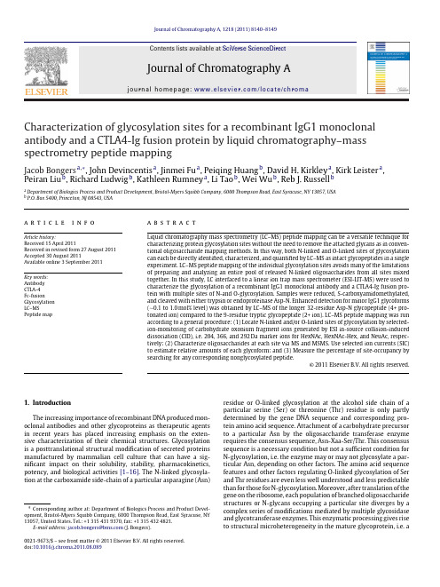
Journal of Chromatography A,1218 (2011) 8140–8149Contents lists available at SciVerse ScienceDirectJournal of ChromatographyAj o u r n a l h o m e p a g e :w w w.e l s e v i e r.c o m /l o c a t e /c h r o maCharacterization of glycosylation sites for a recombinant IgG1monoclonal antibody and a CTLA4-Ig fusion protein by liquid chromatography–mass spectrometry peptide mappingJacob Bongers a ,∗,John Devincentis a ,Jinmei Fu a ,Peiqing Huang b ,David H.Kirkley a ,Kirk Leister a ,Peiran Liu b ,Richard Ludwig b ,Kathleen Rumney a ,Li Tao b ,Wei Wu b ,Reb J.Russell ba Department of Biologics Process and Product Development,Bristol-Myers Squibb Company,6000Thompson Road,East Syracuse,NY 13057,USA bP.O.Box 5400,Princeton,NJ 08543,USAa r t i c l ei n f oArticle history:Received 15April 2011Received in revised form 27August 2011Accepted 30August 2011Available online 3 September 2011Key words:Antibody CTLA-4Fc-fusionGlycosylation LC–MSPeptide mapa b s t r a c tLiquid chromatography mass spectrometry (LC–MS)peptide mapping can be a versatile technique for characterizing protein glycosylation sites without the need to remove the attached glycans as in conven-tional oligosaccharide mapping methods.In this way,both N-linked and O-linked sites of glycosylation can each be directly identified,characterized,and quantified by LC–MS as intact glycopeptides in a single experiment.LC–MS peptide mapping of the individual glycosylation sites avoids many of the limitations of preparing and analyzing an entire pool of released N-linked oligosaccharides from all sites mixed together.In this study,LC interfaced to a linear ion trap mass spectrometer (ESI-LIT-MS)were used to characterize the glycosylation of a recombinant IgG1monoclonal antibody and a CTLA4-Ig fusion pro-tein with multiple sites of N-and O-glycosylation.Samples were reduced,S-carboxyamidomethylated,and cleaved with either trypsin or endoproteinase Asp-N.Enhanced detection for minor IgG1glycoforms (∼0.1to 1.0mol%level)was obtained by LC–MS of the longer 32-residue Asp-N glycopeptide (4+pro-tonated ion)compared to the 9-residue tryptic glycopeptide (2+ion).LC–MS peptide mapping was run according to a general procedure:(1)Locate N-linked and/or O-linked sites of glycosylation by selected-ion-monitoring of carbohydrate oxonium fragment ions generated by ESI in-source collision-induced dissociation (CID),i.e.204,366,and 292Da marker ions for HexNAc,HexNAc-Hex,and NeuAc,respec-tively;(2)Characterize oligosaccharides at each site via MS and e selected ion currents (SIC)to estimate relative amounts of each glycoform;and (3)Measure the percentage of site-occupancy by searching for any corresponding nonglycosylated peptide.© 2011 Elsevier B.V. All rights reserved.1.IntroductionThe increasing importance of recombinant DNA produced mon-oclonal antibodies and other glycoproteins as therapeutic agents in recent years has placed increasing emphasis on the exten-sive characterization of their chemical structures.Glycosylation is a posttranslational structural modification of secreted proteins manufactured by mammalian cell culture that can have a sig-nificant impact on their solubility,stability,pharmacokinetics,potency,and biological activities [1–16].The N-linked glycosyla-tion at the carboxamide side-chain of a particular asparagine (Asn)∗Corresponding author at:Department of Biologics Process and Product Devel-opment,Bristol-Myers Squibb Company,6000Thompson Road,East Syracuse,NY 13057,United States.Tel.:+13154319370,fax:+13154324821.E-mail address:jacob.bongers@ (J.Bongers).residue or O-linked glycosylation at the alcohol side chain of a particular serine (Ser)or threonine (Thr)residue is only partly determined by the gene DNA sequence and corresponding pro-tein amino acid sequence.Attachment of a carbohydrate precursor to a particular Asn by the oligosaccharide transferase enzyme requires the consensus sequence,Asn-Xaa-Ser/Thr.This consensus sequence is a necessary condition but not a sufficient condition for N-glycosylation,i.e.the enzyme may or may not glycosylate a par-ticular Asn,depending on other factors.The amino acid sequence features and other factors regulating O-linked glycosylation of Ser and Thr residues are even less well understood and less predictable than for those for N-glycosylation.Moreover,after translation of the gene on the ribosome,each population of branched oligosaccharide structures or N-glycans occupying a particular site diverges by a complex series of modifications mediated by multiple glycosidase and glycotransferase enzymes.This enzymatic processing gives rise to structural microheterogeneity in the mature glycoprotein,i.e.a0021-9673/$–see front matter © 2011 Elsevier B.V. All rights reserved.doi:10.1016/j.chroma.2011.08.089J.Bongers et al./J.Chromatogr.A1218 (2011) 8140–81498141heterogeneous population of multiple oligosaccharide structures at each site of glycosylation.To further complicate matters,the occu-pancy of a particular N-linked or O-linked site may or may not be complete[16,17].Clearly,these site-specific attributes of partial occupancy and glycan microheterogeneity place high demands on analytical methods for the structural characterization of glycopro-teins.Oligosaccharide mapping,in which an entire pool of oligosac-charides is released from the protein by hydrazinolysis or,more commonly,enzymatic hydrolysis with peptidyl N-glycosidase (PNGase)or other glycosidase treatment(PNGase F,PNGase A, EndoH,etc.),is widely used for the characterization of pro-tein glycosylation[18–25].The various analytical methods for oligosaccharide mapping include high-pH anion-exchange chro-matography with pulsed amperometric detection(HPAEC-PAD) and a number of different chromatography or electrophoresis based separations,matrix-assisted laser desorption mass spec-trometry(MALDI-TOF MS)and various other MS techniques,and combinations of these including LC–MS and other hyphenated methods.In addition,the released pool of oligosaccharides can be analyzed directly or following derivatization with chromogenic,fluorogenic,or other reagents.A notable example is the sensitive analysis of low microgram amounts offluorescent derivatives of the released carbohydrates with2-aminobenzoic acid(2-AA)or2-aminobenzamide(2-AB)by either normal-phase or reversed-phase LC and LC–MS[19,20].These various established oligosaccharide mapping methods have their own relative advantages and disad-vantages as components of comprehensive analytical strategies for glycoproteins.One limitation shared by all the various oligosaccha-ride mapping methods is that the removal and pooling together of glycans results in the loss of site-specific information on oligosac-charide occupancy and structural microheterogeneity.Of course, relinquishing the site-specific microhetrogeneity information is less of a concern for IgG monoclonal antibodies[26–34]which pos-sess only a single site of glycosylation than it is for proteins with multiple sites of glycosylation.Peptide mapping is a general technique for confirming the amino acid sequences of proteins and characterizing their covalent chemical structures,including glycosylation and other posttrans-lational modifications[35–45].These peptide map methods can be used to good advantage as an alternative or in combination with oligosaccharide mapping methods when it is desirable to preserve site-specific information for proteins with multiple sites of glyco-sylation.Sample preparation involves treatment with trypsin or other proteolytic enzymes to cleave the protein into a reproducible set of peptide fragments amenable to analysis by LC–MS and other methods.Peptide mapping can be considered a general method-ology for identifying,characterizing,quantifying,and locating the sequence site for virtually any covalent modification to a protein, including glycosylation[35–45].Ideally,the appropriate protease or proteases are chosen so as to generate a unique well resolved peptide fragment for each glycosylation site.It is worth noting that the conventional peptide mapping and oligosaccharide map-ping approaches can be used in combination either in parallel or sequentially.For instance,it is generally feasible to chromatograph-ically isolate a pooled glycopeptide fraction specific to a particular N-linked or O-linked site and then further analyze this fraction by oligosaccharide mapping,MALDI-TOF MS,and other off-line techniques.In this way,high resolution oligosaccharide mapping data can be acquired for site-specific samples prepared and iso-lated by LC peptide mapping.Another useful experiment is tofirst deglycosylate the sample with PNGase F and then analyze the deg-lycosylated protein by peptide mapping in order to identify sites of N-glycosylation and to evaluate the percent occupancy for each N-linked site[45].Asn residues with attached N-glycans(occupied) are hydrolyzed to aspartic acid(Asp)by the PNGase F treatment and can be readily distinguished from nonglycosylated Asn residues (nonoccupied).In the current study,LC interfaced to a linear ion trap mass spectrometer(ESI-LIT-MS)was used for the site-specific peptide mapping characterization of a single site of N-linked glycosylation on a recombinant human IgG1monoclonal antibody and multiple sites of N-linked and O-linked glycosylation on a CTLA4-Ig fusion protein[46].This CTLA4-Ig construct consists of a modified IgG antibody Fc[47]fused to a high affinity variant of the extracel-lular domain of cytotoxic T lymphocyte antigen-4(CTLA4).CTLA4 (CD152)is a member of the immunoglobulin(Ig)superfamily and expressed as an extracellular receptor on the surface of activated T cells.CTLA4is similar to the T cell costimulatory protein,CD28, and both molecules bind to B7-1(CD80)and B7-2(CD86)ligands on antigen-presenting cells.CD28transmits a stimulatory signal to T-cells whereas CTLA4transmits a competitive inhibitory signal [47–51].2.Experimental2.1.Materials and reagentsRecombinant DNA derived human IgG1monoclonal antibody and CTLA4-Ig fusion protein were produced in CHO cell lines and purified in-house(Bristol-Myers Squibb,East Syracuse,NY).Dithio-threitol(DTT),and iodoacetamide were purchased from Sigma Aldrich(St.Louis,MO).All solvents including water and acetoni-trile were HPLC grade and were purchased from Burdick and Jackson(Muskegon,MI).Trifluoroacetic acid(TFA)and guanidine hydrochloride were from Pierce(Rockford,IL).Trypsin,modified sequencing grade,was purchased from Promega(Madison,WI), Catalog No.V5113.Endoproteinase Asp-N,sequencing grade,was from Roche(Indianapolis,IN),Catalog No.13874622.2.2.Sample preparationIgG1antibody was reduced,S-carboxyamidomethylated, digested with trypsin,and analyzed by RP-LC according to a stan-dard quality control method for tryptic mapping(Figs.2and3). Antibody samples(500g each)and a water blank were denatured by addition of8M guanidine hydrochloride buffer,pH8.0,to a final concentration of6M guanidine hydrochloride in a volume of 600L.Next,10L of175mM DTT was added and the mixture incubated at50◦C for20min to reduce the disulfide bonds.The denatured and reduced sample was then S-alkylated by the addition of10L of350mM iodoacetamide and incubation at 50◦C in the dark for20min.The alkylated protein was buffer-exchanged(desalted)into digestion buffer(2M urea,50mM Tris, 10mM CaCl2,10mM methionine,pH8.0)by gel-permeation on NAP-5gel columns(GE Healthcare,Catalog No.17-0853-02)by loading0.5mL sample and collecting1.0mL eluate.The buffer-exchanged samples(1mL)were digested with20g each of trypsin(1:50wt:wt enzyme to substrate)at38◦C for5h after which the digest was acidified with25L1.0M HCl to halt the enzyme activity.A separate100L portion of the above reduced and S-carboxyamidomethylated antibody was also digested with 6.6g of endoproteinase Asp-N(1:150wt:wt enzyme to substrate) at37◦C for18.5h and then halted with2.5L of1.0M HCl.CTLA4-Ig fusion protein was reduced,S-carboxyamidomethylated,and digested with trypsin exactly according to a standard quality control method for tryptic peptide mapping.Samples consisting of100L of20mg/mL CTLA4-Ig (2mg)were each mixed with550L of denaturing buffer(8M guanidine hydrochloride,50M Tris/HCl,pH8.0),reduced with 35L of200mM DTT for20min at37◦C,and S-alklylated with8142J.Bongers et al./J.Chromatogr.A 1218 (2011) 8140–8149Fig.1.The IgG1model (A)is that of Padlan [52],a composite of Protein Data Dank (PDB)X-ray crystal structures,2ig2and 1fc2[53]with theoretical hinge.The glycosylation consists of a pair of neutral complex-type biantennary N-glycans (A2G1F)enclosed in the central cavity of the Fc domain.The CTLA4-Ig fusion protein model (B)shows PDB crystal structures of human CTLA4homodimer (PDB 1i85)[70]and human Fc (1fc2)[53].Models of four sialylated A2S2G2F complex-type biantennary N-glycans were added to the CTLA4arms using GlyProt [55]with arbitrary orientations and bond angles.N-Glycans are highly flexible and usually exist in a multitude of rapidly converting conformations [54,55].Complex type biantennary N-glycans are named using the abbreviation “AaSsGgF ”where “a ”is the number of antennae linked to the trimannosyl core,“s ”the number of sialic acid residues (NeuAc),“g ”the number of galactose residues (Gal),and “F ”indicates core-fucosylation (Fuc).O-linked glycans in the hinge-region of CTLA4-Ig are not shown in these models.The CTLA4-Ig tryptic peptides containing glycosylation sites are indicated,i.e.T5,T7,T9,and T14.38.5L 400mM iodoacetamide for 20min at room temperature in the dark.Removal of reagents and buffer-exchange into digestion buffer (50mM Tris,10mM CaCl 2,pH 7.6)was performed on NAP-5Sephadex G-25gel columns (GE Healthcare Catalog No.17-0853-01)by loading 0.5mL sample and collecting 1.0mL eluate.Digestions were performed with 50L of 0.5mg/mL trypsin per 1mL of 1.4mg/mL substrate at 37◦C for 120min and halted with 20L 1.0M HCl.2.3.LC–MS peptide mappingLC–MS was run on a Waters 2695Alliance LC or a Waters Acquity UPLC (Milford,MA)interfaced with a ThermoFinnigan LTQ-XL lin-ear ion-trap mass spectrometer (San Jose,CA)with electrospray ionization acquiring sequential MS full-scans with data-dependent acquisition of MSMS and high resolution zoom-scans for abun-dant ions.The LC–MS data were acquired in positive ion mode in a mass to charge ratio (m /z )range of 200–2000,capillary tem-perature of 275◦C,and a source voltage of 5000V.Tandem mass spectra (MSMS)data were acquired using 15–35%normalized col-lision energy,0.25activation Q and 100ms activation time.LC–MS runs for the CTLA4-Ig fusion protein were also made using in-source CID (SID =96V,m /z 140–380Da scans)for monitoring of carbohy-drate oxonium fragment ions [35–38].IgG1antibody tryptic digests were analyzed by C18reversed-phase HPLC–MS run on a Waters 2695Alliance LC equipped with a 2487UV detector,set at 214and 280nm,and interfaced to the above LTQ-XL linear ion-trap mass spectrometer.Injections of 100L each were made onto a Grace Vydac 218TP52column (2.1mm ×250mm),40◦C,at 0.2mL/min,with eluents (A)0.10%TFA/H 2O and (B)0.09%TFA/95%CH 3CN,and gradient:2%B isocratic for 4min,2–16%B from 5to 30min,16–19.5%B from 30to 60min,19.5–23%B from 60to 84min,23–29%B from 84to 102min,29–30%B from 102to 118min,and 30–42%B from 118to 143min,42–100%B from 143to 145min,100%B isocratic from 145to 146.1min,100–2%B from 146.1to 146.2min,and 100%B isocratic from 146.2to 166min.The flow rate was 0.2mL/min from 0to 145min,0.2to 0.3mL/min from 145to 145.1min,and 0.3mL/min from 145.1to 166min.A rotary switching-valve was used to divert the 2M urea in the digest away from the ESI source for the first 5min of the run.IgG1antibody trypsin and endoproteinase Asp-N digests were analyzed by C18reversed-phase UP LC–MS run on Waters Acquity UPLC instrument equipped with a photodiode array UV detec-tor,set at 214and 280nm,and interfaced to the above LTQ-XL linear ion-trap mass spectrometer.Injections of 50L each were made onto a Waters BEH300C18column (Part No.186003687),1.7m,2.1mm ×150mm,45◦C,at 0.2mL/min,with eluents (A)0.02%TFA/H 2O and (B)0.02%TFA/95%CH 3CN.The tryptic map-ping LC gradient was:1%B isocratic for 6min,1–35%B from 6to 80min,35–100%B from 80to 92min,100%B isocratic from 92to 95min,100–1%B from 95to 96min,and 1%B isocratic from 96to 110min,with a flow rate of 0.2mL/min throughout.The endopro-teinase Asp-N mapping LC gradient was:1%B isocratic for 6min,1–50%B from 6to 80min,50–100%B from 80to 92min,100%B isocratic from 92to 95min,100–1%B from 95to 96min,and 1%B isocratic from 96to 110min,with a flow rate of 0.2mL/min throughout.A rotary switching-valve was used to divert the 2M urea in the digest away from the ESI source for the first 3min of the runs.CTLA4-Ig tryptic digests were analyzed by C18reversed-phase HPLC–MS peptide mapping run on a Waters 2695Alliance LC equipped with a 2487UV detector,set at 215and 280nm,and interfaced to the above LTQ-XL linear ion-trap mass spectrom-eter.Injections of 25L were run on a Grace Vydac 218MS52column (2.1mm ×250mm),at 0.2mL/min,45◦C,with eluents (A)0.02%TFA/H2O and (B)0.02%TFA/95%CH 3CN,and gradient:1.7%B isocratic for 4min,1.7–30%B from 4to 64min,30–50%B from 64to 80min,and 80–100%B from 80to 90min,100%B iso-cratic from 90to 94min,100–1.7%B from 94to 95min,and 1.7%B isocratic from 95to 110min,with a flow rate of 0.2mL/min throughout.J.Bongers et al./J.Chromatogr.A 1218 (2011) 8140–81498143Fig.2.Summary of ESI linear ion-trap LC–MS tryptic peptide mapping results for a recombinant IgG1monoclonal antibody (CHO cell culture derived)showing (A)full-scale 214nm UV and MS base-peak chromatograms;(B)selected ion-current (SIC)chromatograms for the doubly protonated ions of the three main Fc domain tryptic glycopeptides (A2G0F,A2G1F,and A2G2F)and nonglycosylated heavy chain (NGHC);(C)mass spectrum summed across the 24.0–25.2min glycopeptide region with main peak assignments,(D)expanded view of 280nm UV trace of glycopeptides region showing peak area quantitation of mole percent NGHC,and (E)MS and MSMS spectra of the NGHC tryptic peptide.3.Results and discussion3.1.General procedures for LC–MS peptide mapping of glycosylation sitesComplete sequence coverage for the antibody was obtained by overlapping trypsin and endoproteinase Asp-N LC–MS pep-tide maps.The respective peptides can be readily identified by their observed monoisotopic molecular weights determined via on-line high resolution mass spectrometry and their sequences confirmed by tandem mass spectra of their collision induced disso-ciation (CID)fragmentation patterns.Samples were fully reduced with DTT,S-carboxyamidomethylated (S-alkylation of all cysteine residues),and cleaved with either trypsin or endoproteinase Asp-N and then analyzed by LC–MS according to the following general scheme:LC–MS peptide mapping was run according to a general procedure:(1)Locate N-linked and/or O-linked sites of glycosy-lation by single-ion-monitoring (SIM)of carbohydrate oxonium fragment ions generated by ESI in-source collision-induced disso-ciation (CID),i.e.204,366,and 292Da marker ions for HexNAc,HexNAc-Hex,and NeuAc,respectively [35–38];(2)Characterize oligosaccharides at each site via MS and e selected ion currents (SIC)to estimate relative amounts of each glycoform;and (3)Measure the percentage of site-occupancy by searching for any corresponding nonglycosylated peptide.3.2.LC–MS tryptic peptide mapping of an IgG1monoclonal antibodyIgG antibodies usually have a single N-linked glycosylation site in the heavy chain sequence yielding a pair of N-linked oligosac-charides enclosed in a central cavity in between the two C H 2constant-region Ig subunits of the Fc domain of the mature homod-imeric antibody structure as shown in Fig.1[4–6,52–55].The Fc N-glycans are predominantly neutral (nonsialylated)bianten-nary oligosaccharides with zero,one,or two terminal galactose units (A2G0F,A2G1F,and A2G2F).Since the Fc N-glycans are sand-wiched in between the two C H 2constant-region Ig subunits,these oligosaccharides obviously play an integral role in shaping the three dimensional structure of the Fc and modulating effector functions such as binding to the high affinity Fc ␥R receptor and mediating antibody dependent cellular cytotoxicity (ADCC)and complement fixation [4–7].For example,glycoengineering to produce afucosyl Fc N-glycans has been used to make monoclonal antibody thera-peutics with greatly enhanced ADCC [56,57].Conversely,it has been shown that the Fc N-glycosylation of IgG antibody drugs has essen-tially no effect on their pharmacokinetics and serum half-lives [11].The low microheterogeneity and lack of effect on PK clearance are reasonable considering the peculiar disposition of these N-glycans which are almost entirely enclosed and inaccessible inside the Fc domain.8144J.Bongers et al./J.Chromatogr.A 1218 (2011) 8140–8149parison of ESI linear ion-trap LC–MS glycopeptide mapping results for a recombinant IgG1monoclonal antibody obtained with (A)trypsin versus (B)endoproteinase Asp-N using the same digest concentrations,injection volumes,and MS instrument conditions.Selected ion currents are shown for the three major Fc N-glycans (A2G0F,A2G1F,and A2G2F),two minor sialylated glycoforms (A2S1G2F and A2S2G2F),and a minor oligomannose type glycoform (M5).A more comprehensive list of N-glycans observed by LC–MS Asp-N peptide mapping is given in Table 1.Peptide mapping is routinely used for confirming expres-sion of the correct amino acid sequence and characterizing the covalent chemical structures of antibodies and other recombi-nant proteins [40–43].We have found that complete amino acid sequence coverage for recombinant antibodies can usually be obtained by overlapping trypsin and endoproteinase Asp-N pep-tide maps.The respective peptides can be readily identified by their observed monoisotopic molecular weights determined via on-line high resolution mass spectrometry and their sequences confirmed by tandem mass spectra of their collision induced dis-sociation (CID)fragmentation patterns.The other well known IgG1posttranslational modifications,characteristic of IgG1antibodies,were observed for the heavy chain including N-glycosylation at the conserved Fc region Asn-Xaa-Ser/Thr consensus site,cyclization of N-terminal glutamine to pyroglutamate,and proteolytic removal of the C-terminal lysine residue.The LC–MS tryptic peptide map of the reduced/S-alkylated IgG1mAb in Fig.2was obtained according to a standard quality con-trol testing assay and the flow from the UV detector directed into a linear ion-trap mass spectrometer.In addition to provid-ing information on the amino acid sequence and covalent chemical structure of the protein,this LC–MS peptide map contains a con-siderable amount of information on the glycosylation.The three main N-glycans (A2G0F,A2G1F,and A2G2F)at the single N-linkedglycosylation site are typical for the Fc domain of an IgG1anti-body.Minor amounts of the sialylated glycans,A2S1G2F (∼0.4%)and A2S2G2F (∼0.3%)were also found as well as an M5oligoman-nose structure (∼2.0%),and nonglycosylated heavy chain (NGHC)of ∼1.0%,i.e.,an occupancy of ∼99.0%at this asparagine.These minor amounts of nonglycosylation and the immature oligomannose gly-coforms are of interest as possible indicators of cell viability during upstream cell culture processes [2].LC–MS peptide mapping of the antibody with trypsin and endo-proteinase Asp-N are compared in Fig.3and very similar amounts were obtained for this set of glycoforms.The Asp-N glycopeptide (32residues)has multiple basic residues for ionization (protona-tion)and thus tends to give higher signal-to-noise for the selected ion currents (4+ions).Enhanced detection for minor IgG1glyco-forms (∼0.1to 1.0mol%level)was obtained by LC–MS of the longer 32-residue endoproteinase Asp-N glycopeptide (4+protonated ion)compared to the 9-residue tryptic glycopeptide (2+ion).Oppo-site effects on reversed-phase retention times were observed for presence of an additional galactose (hydrophilic)versus sialic acid (hydrophobic)for both the tryptic glycopeptides and the Asp-N glycopeptides.In addition to the mass spectral peak assignments shown in Figs.2and 3,the various glycoforms identified by LC–MS Asp-N mapping are also listed in Table 1.The relative amounts of the glycopeptides as estimated by LC–MS selected ion currentJ.Bongers et al./J.Chromatogr.A 1218 (2011) 8140–81498145Fig.4.LC–MS tryptic map of a CHO cell culture derived recombinant CTLA4-Ig fusion protein showing 215nm UV chromatogram,MS base-peak chromatogram,and selected ion-current chromatograms (SIC’s)for the carbohydrate oxonium fragment marker-ions,N-acetyl hexoseamine (HexNAc,m /z =204Da),hexose N-acetyl hexoseamine disaccharide (HexNAc-Hex,m /z =366Da),and sialic acid (NeuAc,m /z =292Da)generated by in-source collision-induced dissociation (CID).The boxed-in regions indicate glycopeptides corresponding to the three specific N-linked sites and one O-linked site of glycosylation on CTLA4-Ig as shown in Fig.1.Table 1Glycopeptides found for recombinant IgG1monoclonal antibody by LC–MS endoproteinase Asp-N mapping.N-glycan a FormulaFormulaMW calc b (Da)m /z calc 4+(Da)m /z obs 4+(Da)Ret time (min)SIC rel heightRel (%)aglyco C161H258N48O52–3697.9019925.4828925.572.80.8nd A1G0C203H327N51O82C42H69N3O304793.29851199.33191199.771.60.950.20A1G0F C209H337N51O86C48H79N3O344939.35651235.84641236.271.4 4.010.86A2G0C211H340N52O87C50H82N4O354996.37781250.10171250.171.4 5.23 1.13A2G0F C217H350N52O91C56H92N4O395142.43571286.61621286.971.232269.29A2G1F C223H360N52O96C62H102N4O445304.48851327.12941327.571.110221.95A2G2F C229H370N52O101C68H112N4O495467.54141367.89261367.871.011.2 2.41A2S1G1F C234H377N53O104C73H119N5O525596.58401400.15331399.971.7 2.230.48A2S1G2F C240H387N53O109C79H129N5O575758.63681440.66651440.471.6 1.570.34A2S2G2F C251H404N54O117C90H146N6O656049.73191513.44031513.272.1 1.230.26M5C207H334N50O87C46H76N2O354914.32471229.58851229.871.38.81 1.90M6C213H344N50O92C52H86N2O405076.37751270.10171270.071.2 1.530.33M7C219H354N50O97C58H96N2O455238.43041310.61491310.771.2 2.880.62M8C225H364N50O102C64H106N2O505400.48321351.12811351.071.20.820.18M9C231H374N50O107C70H116N2O555563.53601391.89131391.871.00.230.05Sum464.69100aComplex type biantennary N-glycans are named using the abbreviation “AaSsGgFB ”where “a ”is the number of antennae linked to the trimannosyl core,“s ”the number of sialic acid residues (NeuAc),“g ”the number of galactose residues (Gal),and “F ”indicates core-fucosylation (Fuc).Oligomannose type N-glycans are named “Mm ”where m is the number of mannose residues.The amino acid sequence of the Asp-N peptide is DGVEVHNAKTKPREEQYNSTYRVVSVLTVLHQ.bCalculated masses are for the most abundant isotope.peak heights in the Asp-N map were in close agreement with rela-tive amounts found for the corresponding glycoforms in the tryptic map.However,the smaller amounts of minor glycoforms in the ∼0.1to 1.0%range that were below the limits of detection for thetryptic map were readily identified by Asp-N mapping.The rela-tive amounts of the various N-glycans estimated by LC–MS Asp-N peptide mapping selected ion currents in Table 1compare rea-sonably closely with those obtained independently by analyses of8146J.Bongers et al./J.Chromatogr.A 1218 (2011) 8140–8149Fig.5.Mass spectra summed across the four boxed-in regions of the LC–MS tryptic map for the recombinant CTLA4-Ig fusion protein in Fig.4corresponding to the three N-linked sites and one O-linked site of glycosylation on the CTLA4-Ig fusion protein molecule in Fig.1.Major peaks in the spectra are labeled with oligosaccharide structure assignments.Amino acid sequences are shown for the glycosylated tryptic peptides T5and T7(N-linked,CTLA4domain),T9(O-linked,hinge region),and T14(N-linked Fc domain)which correspond to residues 39-83,94-128,133-158,and 203-211of the CTLA4-Ig,respectively.oligosaccharides released from this antibody and similar recom-binant DNA derived monoclonal antibodies via treatment with PNGase F.3.3.LC–MS tryptic peptide mapping of a CTLA4-Ig fusion proteinThe CTLA4-Ig fusion protein shown in Fig.1consists of the extracellular portion of a disulfide-linked homodimeric glycopro-tein receptor,human cytotoxic T-lymphocyte antigen-4(CTLA4)[46–51],fused to a modified human Fc domain [47].CTLA4is a member of the CD28family of receptor/ligands involved in T-cell costimulation [48–51].CTLA4-Ig has two N-linked sites per CTLA4domain,in addition to the above Fc site,plus O-linked glycosyla-tion in the hinge region.The CTLA4domain is an immunoglobulin V (IgV)type of fold and the N-glycosylation sites at Asn 76and Asn 108are contained within tryptic peptides,T5and T7,respectively.Asn 76is located in beta-strand E of the ABED beta-sheet and the Asn 108N-glycosylation site is located in the C-terminal G strand of the CC’FG beta-sheet on the opposite face of the IgV sandwich.The “MYPPPYY loop”connecting beta-strands E and F (residues 97–103)is the key receptor-binding interface for the interaction of the CTLA4homodimer with the B7-1and B7-2ligands on antigen presenting cells.Note that,unlike the atypical case of the N-glycans constrained inside the Fc domain cavity,the N-glycans attachedto the CTLA4domain represent the more usual case of oligosac-charides projecting out from the surface of the protein into the surrounding medium (Fig.1).The four glycopeptide regions in the tryptic map can be visu-alized via single-ion monitoring of in-source CID carbohydrate marker ions in Fig.4.For the CTLA4-Ig fusion protein,the sim-plest of the three N-linked sites is that on the Fc domain T14peptide containing the three neutral biantennary complex-type core-fucosylated N-glycans (A2G0F,A2G1F,and A2G2F)which are typical for virtually all recombinant IgG1antibodies produced by mammalian cell culture.In contrast to the rather simple population of Fc N-glycans,the two N-glycosylation sites on the CTLA4domain show highly diverse microheterogeneous populations of sialylated and neutral forms of bi-,tri-,and tetra-antennary complex-type core-fucosylated glycans with variable numbers of terminal galac-tose (Fig.5).The T7N-glycans contain significantly higher levels of galactose and sialic acid,overall,versus the T5site.The T7glycopeptides are partially resolved into distinct regions on this RP-LC system (Fig.6).The first envelope within the range of approximately 67.8–68.8min consists primarily of the T7glycopep-tide with monosialylated biantennary N-glycans,A2S1G2F and A2S1G1F.The next region from approximately 68.0–70.0min con-tains T7with the disialyated biantennary N-glycan,A2S2G2F,and lesser amounts of T7with the disialylated and trisialylated species,。


2021年6月第21卷第2期廊坊师范学院学报(自然科学版)Journal of Langfang Normal University(N atural Science Edition)Jun.2021Vol.21No.2芳香族氨基酸及其衍生物的研究进展刘苹,苏卫卫(燕山大学,河北秦皇岛066004)【摘要】氨基酸是蛋白质的基本组成单元,氨基酸的缩合、衍生都与蛋白质的形成及功能相关。
芳香族氨基酸作为机体重要的氨基酸,生物学功能非常丰富。
介绍芳香族氨基酸的特征、芳香族氨基酸及其衍生物的合成及应用,并对芳香族氨基酸在营养学领域、人类医学、生物材料等方面应用进行重点阐述,对芳香族氨基酸的发展前景进行展望。
【关键词】芳香族氨基酸;生物合成法;氨基酸交联;生物材料Advances in the Study of Aromatic Amino Acids and Their DerivativesLiu Ping,Su Weiwei(Yanshan University,Qinhuangdao066004,China)[Abstract]Amino acids are the basic constituent units of proteins.The condensation and derivation of amino acids are related to the formation and function of proteins.As important amino acids in the body,aromatic amino acids have abundant biological functions.This article introduces the characteristics of aromatic amino acids,the synthesis and application of aromatic amino acids and their derivatives,and focuses on the application of aromatic amino acids in nutrition,human medicine and biological materials and so on.The development prospect of aromatic amino acids is prospected in this article.[Key words]aromatic amino acids;biosynthesis;cross-linking of amino acids;biomaterials冲图分类号〕06-1〔文献标识码〕A〔文章编号〕1674-3229(2021)02-0027-080引言氨基酸作为生物活性分子,是蛋白质的基本组成单元。

癌基因oncogene艾滋病acquired immuno-deficiency syndrome,AIDS 氨喋呤anminopterin carbamoyl phosphate synthetase Ⅰ, CPS-Ⅰ氨基甲酰磷酸合成酶Ⅰ氨基末端amino terminal氨基酸amino acid氨基酸接纳茎acceptor stem氨基酸序列amino acid sequenceδ-氨基-γ-酮戊酸δ—aminolevulinic acid,ALA氨基酰-tRNA合成酶aminoacyl-tRNA synthetase氨基酰位aminoacyl site氨基转移酶aminotransferase氨甲喋呤methotrexate, MTX氨肽酶,氨基肽酶aminopeptidase胺氧化酶amine oxidase巴士德Pastuer靶细胞target cell白化病albinism白三烯leukotrienes,LTs摆动性wobble斑点印迹bot blotting半保留复制semi-conservative replication半不连续复制semi-discontinuous replication 半胱氨酸cysteine半乳糖galactose, Gal伴侣素chaperonins包涵体inclusion body胞苷cytidine胞嘧啶cytosine,C胞质小RNA small cytoplasmic RNA,scRNA 保守性转座conservative transposition苯丙氨酸羟化酶phenylalanine hydroxylase 苯酮酸尿症phenyl ketonuria, PKU吡哆胺pyridoxamine吡哆醇pyridoxine吡哆醛pyridoxal必需氨基酸essential amino acid必需基团essential group必需激活剂essential activator编码链coding strand变构部位allosteric site变构调节allosteric regulation变构激活剂allosteric activator变构酶allosteric enzyme变构效应allosteric effect变构效应剂allosteric effector变构抑制剂allosteric inhibitor变性denaturation变性梯度胶电泳denaturing gradient gel electrophoresis, DGGE 辨认位点recognition site标志补救marker rescue表达载体expression vector表构酶epimerase表观Km值apparent Km表面效应surface effect别嘌呤醇allopurinol丙氨酸-葡萄糖循环alanine-glucose cycle 丙酮acetone丙酮酸pyruvate丙酮酸激酶pyruvate kinase丙酮酸羧化酶pyruvate carboxylase病毒癌基因virus oncogene,v-onc卟啉症porphyria补救合成途径salvage pathway不对称转录asymmetric transcription不可逆性抑制作用irreversible inhibition 参入incorporation残基residue残粒remnant操纵序列operator操纵子operon侧翼区flanking region层析chromatography层粘连蛋白laminin,Ln插入insertion插入序列insertion sequences, IS长末端重复序列long terminal repeat, LTR长萜醇dolichol肠肝循环bilinogen enterohepatic circulation,enterohepatic circulation超螺旋结构superhelix, supercoil超敏位点hypersensitive site超速离心ultra-centrifuge超速离心法ultracentrifugation超氧物歧化酶superoxide dismutase, SOD沉降系数sedimentation coefficient,S成肽peptide bond formation初级胆汁酸primary bile acids初级转录产物primary transcripts储脂细胞lipocyte串联酶tandem enzyme锤头结构hammerbead structure醇脱氢酶alcohol dehydrogenase, ADH次黄核苷inosine,I次黄嘌呤inosine次黄嘌呤核苷酸inosine monphosphate, IMP次黄嘌呤核苷酸转移酶adenine phosphoribosyl transferase, APRT 次级胆汁酸secondary bile acids从头合成途径de novo synthesis促红细胞生成素erythropoietin,EPO催化基团catalytic group催化型受体catalytic receptor催化性DNA DNAzyme催化性RNA catalytic RNA催化性小RNA small catalytic RNA脆性X综合征fragile X syndrome脆性位点fragile site错配mismatch大沟major groove代谢库metabolic pool单胺氧化酶monoamine oxidase, MAO单纯酶simple enzyme单核苷酸多态性single nucleotide polymorphisms, SNPs单链DNA结合蛋白single stranded DNA binding protein, SSB单链构象多态性single strand conformation polymorphism, SSCP 单顺反子monocistron单体酶monomeric enzyme胆chole胆钙化醇cholecalciferol胆固醇cholesterol胆固醇7a-羟化酶cholesterol 7a-hydroxylase胆固醇流出调节蛋白cholesterol-efflux regulatory protein, CERP胆固醇酯酶cholesteryl esterase胆红素bilirubin胆红素脑病bilirubin encephalopathy胆碱choline胆碱酯酶choline esterase胆绿素biliverdin胆绿素还原酶biliverdin reductase胆囊胆汁gallbladder bile胆色素bile pigment胆色素原prophobilinogen,PBG胆素bilin胆素原bilinogen胆酸cholic acid胆盐bile salts胆汁bile胆汁酸bile acids弹性蛋白酶elastase蛋白二硫键异构酶protein disulfide isomerase,PDI 蛋白激酶C protein kinase C,PKC蛋白聚糖proteoglycan蛋白聚糖聚合物aggrecan蛋白磷酸酶protein phophatase蛋白酶protease蛋白酶体proteasome,proteosome蛋白质protein蛋白质表达protein expression蛋白质-蛋白质相互作用protein-protein interaction蛋白质的靶向输送protein targeting蛋白质的凝固作用protein coagulation蛋白质数据库protein databasesDNA-蛋白质相互作用DNA-protein interaction蛋白质芯片protein chip蛋白质印迹技术Western blotting蛋白质转换protein turnover蛋白质组proteome蛋白质组学Proteomics氮平衡nitrogen balance倒位酶invertase等电点isoelectric point,pI等位基因特异寡核苷酸allele-specific ologonucleotide,ASO 低血糖diabetes mellitus低血糖症hypoglycemia底物substrate底物水平磷酸化substrate-level phosphorylation底物循环substrate cycle地心引力gravity第二信使secondary messenger点突变point mutation电穿孔electroporation电喷雾质谱electrospray ionization mass spectrometry,ESI-MS电泳electrophoresis电子传递链electron transfer chain淀粉starchα-淀粉酶α-amylase凋亡apoptosis调节部位regulatory site调节酶regulatory enzymes调节子regulon定位候选基因克隆策略positional candidate gene approaches 定位克隆positional cloning定向排列orientation arrange杜氏肌营养不良症duchenne muscular dystrophy,DMD端粒telomere端粒酶RNA human telomerase RNA,hTR端粒酶逆转录酶human telomerase reverse transcriptase,hTRT 端粒酶协同蛋白1human telomerase associated protein 1,hTP1 短串联重复序列short tandem repeat断裂基因splite gene钝性未端blunt end多胺polyamines多巴胺dopamine多不饱和脂酸polyunsaturated fatty acid多功能酶multifunctional enzyme多核糖体polyribosome多接头克隆位点polylinker cloning sites多聚核蛋白体polysome多聚核苷酸polynucleotides多聚体polymer多聚脱氧核苷酸polydeoxynucleotides多酶体系multienzyme system多顺反子polycistron多顺反子mRNA polycistronic mRNA多态性polymorphism多肽polypeptide多肽链polypeptide chain多样性片段diversity segment多元催化multielement catalysis鹅膏蕈碱amanitin鹅脱氧胆酸chenodeoxycholic acid儿茶酚胺catecholamine二次转酯反应twice transesterification二级结构secondary structure二甲基丙烯焦磷酸3,3-dimethylallyl pyrophosphate, DPP 二聚化dimerization二聚体dimer二氢叶酸合成酶dihydrofolic acid synthetase二氢叶酸还原酶dihydrofolate reductase二巯基丙醇British anti-Lewisite,proteosome,BAL二十二碳六烯酸docosahexaenoic acid, DHA二十碳五烯酸eicosapentaenoic acid, EPA二肽酶dipeptidase二硝基苯酚dinitrophenol,DNP发夹hairpin翻译起始复合物translational initiation complex反基因策略antigene strategy反竞争性抑制作用uncompetitive inhibition反密码子anticodon反式trans反式作用因子trans-acting factor反烯酰CoA异构酶trans enoyl-CoA isomerase反义寡核苷酸antioligonucleotide反转录PCR reverse transcription PCR,RT-PCR反转重复序列inverted repeat泛醌ubiquinone泛素ubiquitin泛素化ubiquuitination泛酸pantothenic acid非mRNA小RNA small non-messenger RNA,snmRNAs 非必需氨基酸non-essencial amino acid非必需激活剂non-essential activator非蛋白氮non-protein nitrogen, NPN非定位候选基因克隆策略position-independent candidate gene approaches 非竞争性抑制作用non-competitive inhibitionRNA分化加工differential RNA processing分节基因segmentation genes分解代谢阻遏catabolic repression分解物基因激活蛋白catabolite gene activation protein, CAP分支酶debranching enzyme分支移动branch migration分子伴侣chaperon,molecular chaperon分子克隆molecular clone分子医学molecular medicine粉蝶霉素A piericidin A粉液化芽苞杆菌bacillus amyloliguefaciens粪卟啉原III coproporphyrinogen III,CPGIII粪胆素stercobilin,l-urobilin粪胆素原stercobilinogen,l-urobilinogen丰富基因redundant gene佛波酯phorbol ester5-氟尿嘧啶5-fluorouracil, 5-fu脯氨酸富含域proline-rich domain辅基prosthetic group辅酶coenzyme辅酶I NAD+辅酶II NADP+辅酶Q coenzyme Q,CoQ辅脂酶colipase辅助因子cofactor辅助因子Fis factor for inversion stimulation 辅阻遏剂corepressor 腐败作用putrefaction负超螺旋negative supercoil负碳离子carbanion复合体complex复合体Ⅴcomplex V复性renaturation复制replication复制酶replicase复制体replisome复制性转座duplicative transposition复制因子replication factor,RF复制子replicon富含AT AT rich钙调蛋白calmodulin CaM干扰RNA interference RNA,RNAi RNA干涉RNA interference,RNAi 甘氨胆酸glycocholic acid甘氨鹅脱氧胆酸glycochenodeoxycholic acid 甘露糖mannose, Man 甘油激酶glycerokinase甘油磷脂phosphoglycerides甘油三酯triglyceride2-甘油一酯2-monoglyceride肝胆红素hepatobilirubin肝胆汁hepatic bile肝素heparin肝细胞性黄疸hepatocellular jaundice感染infection感受态细胞competent cell岡崎片段Okazaki fragment高保真性high fidelity高脂蛋白血症hyperlipoproteinemia高脂血症hyperlipidemia功能互补实验functional complementation assay功能基因组学functional genomics功能克隆functional cloning共价修饰covalent modification共同的偶配体SMADs common-partner-SMAD,Co-SMADs共有序列consensus sequence谷氨酰胺合成酶glutamine synthetase谷氨酰胺酶glutaminase谷丙转氨酶glutamic pyruvic transaminase, GPT, ALT谷草转氨酶glutamic oxaloacetic transaminase, GOT, AST β-谷固醇β-sitosterol 谷胱甘肽glutathione, GSH谷胱甘肽S-环氧化物转移酶glutathione S-epoxidetransferase骨形态发生蛋白bone morphogenetic protein,BMPs钴胺素cobalamin钴胺素I transcobalamin I, TC I固醇sterol固醇载体蛋白sterol carrier protein, SCP固定化酶immobilized enzyme寡核苷酸点阵oligonucleotide arrays寡核苷酸微芯片oligonucleotide micro-chip寡核苷酸杂交分析oligonucleotide hybridization analysis 寡聚酶oligomeric enzyme寡霉素oligomycin寡肽oligopeptide寡肽酶oligopeptidase关键酶key enzymes管家基因housekeeping gene光修复light repairing光修复酶photolyase胱甘肽S-转移酶glutathione S-transferase滚环复制rolling circle replication6-果糖双磷酸酶-26-phosphofructokinase-2果糖双磷酸酶-2fructose biphosphatase-2过渡态transition state过氧化酶体peroxisomes过氧化氢酶catalase,peroxidaseALA合酶ALA synthaseATP合酶ATP snthase核磁共振技术nuclear magnetic resonance, NMR核蛋白nuclear protein核蛋白体RNA ribosomal RNA,rRNA核蛋白体蛋白质ribosomal protein,rp核蛋白体结合位点ribosomal binding site, RBS核蛋白体循环ribosomal cycle核定位信号nuclear localization signal, NLS核定位序列nuclear localization sequence,NLS核苷ribonucleoside核苷二磷酸nucleoside diphosphate,NDP核苷三磷酸nucleoside triphosphate,NTP核苷酸nucleotide核苷一磷酸nucleoside monophosphate,NMP核黄素riboflavin核酶ribozyme核内不均一RNA heterogeneous nuclear RNA,hnRNA 核内小分子RNA small nuclear RNA,snRNA核仁小分子RNA small nucleolar RNA,snoRNA核输入因子nuclear importin核酸nucleic acid核酸内切酶endonuclease核酸外切酶exonuclease核糖核苷酸ribonucleotide核糖核酸ribonucleic acid,RNA核糖体ribosome核小体nucleosome核心颗粒core particle核心酶core enzyme核因子κB nuclear factor-κB,NF-κ B Pribnow盒Pribnow box黑色素melanin亨丁顿舞蹈病Huntington disease恒定片段constant segment后基因组计划post-Human Genome Project, PHGP 呼吸控制率respiratory control ratio, RCR呼吸链respiratory chainb-胡萝卜素b-carotene琥珀酸CoA succinyl CoA琥珀酸CoA合成酶succinyl-CoA synthetase琥珀酸脱氢酶succinate dehydrogenasea互补alpha complementation互补complementary互补的单链DNA complementary DNA花生四烯酸arachidonic acid化学渗透假说chemiosmotic hypothesis化学修饰chemical modificationHMG CoA还原酶HMG CoA reductase环loop环丁基环cyclobutane ringD-环复制D-loop replication缓激肽bradykinin黄疸jaundice黄嘌呤氧化酶xanthosine oxidase黄素单核苷酸flavin mononucleotide, FMN黄素蛋白flavoprotein黄素腺嘌呤二核苷酸flavin adenine dinucleotide, FAD恢复rescue回文结构palindrome混合功能氧化酶mixed-function oxidase 混合微团mixed micelles活化能activation energy活化素activin活性部位active site活性区activation domain,AD活性中心active center获得性突变gain-of-function mutation肌酸creatine肌酸酐creatinine肌酸激酶creatine kinase, CK肌营养不良蛋白dystrophin基本重组general recombination基本转录因子general transcription factors 基底层bacal laminae基膜baxal lamina基因gene基因靶向gene targeting基因沉默因子silencing factor基因工程genetic engineering基因间隔gene spacer基因矫正gene correction基因克隆gene cloning基因疗法gene therapeutics基因敲除gene knock-out基因失活gene inactivation,loss-of-function mutations 基因文库gene library 基因芯片gene chip基因芯片点阵genechip arrays基因增补gene augmentation基因诊断gene diagnosis基因治疗gene therapy基因置换gene replacement基因重组genetic recombination基因转移gene transfer基因族gene family基因组genome,genomics基因组DNA genomic DNA基因组DNA文库genomic DNA library基因作图genetic mapping激动型G蛋白stimulatory G protein,Gs激活蛋白activator激活功能activation function激活剂activator激素反应元件hormone response element激素敏感性甘油三酯脂肪酶hormone-sensitive triglyceride lipase, HSL 级联系统cascade system即刻早期反应基因immediate-early gene,IEG急性时相蛋白质acute phase protein APP急性时相反应物acute phase reactant APR己糖激酶hexokinase加单氧酶monooxygenase甲基化methylation甲基化酶methylase甲基转移酶methyl transferase甲硫氨酸循环methionine cycle甲羟戊酸mevalonic acid, MVA甲胎蛋白α-fetoproteinN-甲酰甲硫氨酸N-formyl methionine,fMet 假尿嘧啶ψ,pseudouridine假神经递质false neurotransmitter间接胆红素indirect reacting bilirubin间接体内疗法ex vivomRNA剪接mRNA splcing剪接splicing剪接接口splicing junction剪接体splicesome检查点check point简并性degeneracy碱基base碱基对base pairs, bp碱性亮氨酸拉链basic eucine zipper碱性螺旋-环-螺旋basic helix-loop-helix降解degradation交联对话cross talk胶原collagen胶原微纤维collagen fibril胶原纤维collagen fiber焦谷氨酸pyroglutamic acid焦磷酸法尼酯farnesyl pyrophosphate, FPPUDPG焦磷酸化酶UDPG pyrophosphorylase焦磷酸硫胺素thiamine pyrophosphate, TPP酵解途径glycolytic pathway酵母yeast酵母基因剔除yeast knockout project酵母人工染色体载体yeast artificial chromosome vector, YAC 酵母双杂交系统yeast two-hybrid-system阶段特异性stage specificity接合作用conjugation接受体acceptor结构基因structural gene结构基因组学structural genomicsSH2结构域Scr homology 2 domainSH3结构域Scr homology 3 domain结构域domain结合胆汁酸conjugated bile acidpoly(A结合蛋白poly(A-binding protein,PABP结合反应conjugation reactionATP结合盒转运蛋白AI ATP-binding cassette transporter A1, ABCA1 结合基团binding group结合酶conjugated enzyme结合区binding domain,BD结合体adaptorDNA结合域DNA binding domain解缠酶untwisting enzyme解毒作用detoxication解磷定pyridine aldoxime methyliodide,PAM解螺旋酶helicase解偶联蛋白uncoupling protein解偶联剂uncoupler金属激活酶metal-activated enzyme金属酶metalloenzyme紧张态tense state进位entrance茎stem茎环stem-loop精胺spermine精脒spermidine竞争性抑制作用competitive inhibition矩形双曲线rectangular hyperbola巨幼红细胞性贫血megaloblastic anemia RNA聚合酶RNA-pol聚合酶链反应polymerase chain reaction, PCR 聚糖glycan绝对特异性absolute specificity癞皮病pellagra劳氏肉瘤病毒Rous sarcoma virus,RSV酪氨酸蛋白激酶tyrosine-protein kinase,TPK 酪氨酸酶tyrosinase类核nucleoid厘摩centimorgen, cM立体异构特异性stereospecificity立早基因immediate-early gene利福平rifampicinN-连接聚糖N-linked glycanO-连接聚糖O-linked glycanDNA连接酶DNA ligase连接酶类ligases连接片段joining segment连接物蛋白adaptor protein连锁分析likage analysis连锁图genetic map,linkage map连续性commaless猎物prey,target protein裂解酶类lyases邻近效应proximity effect林-贝氏Lineweaver- Burk磷酸丙糖异构酶triose phosphate isomerase磷酸二酯键phosphodiester linkage4’-磷酸泛酰氨基乙硫醇4’-phosphopantetheine 磷酸钙转染calcium phosphate transfectionα-磷酸甘油穿梭α-glycerophosphate shuttle3-磷酸甘油醛脱氢酶glyceraldehyde 3-phosphate dehydrogenase磷酸甘油酸变位酶phosphoglycerate mutase磷酸甘油酸激酶phosphoglycerate kinase6-磷酸果糖fructose-6-phosphate , F-6-P6-磷酸果糖激酶-16-phosphofructokinase-1磷酸肌酸creatine phosphate, CP6-磷酸葡萄糖glucose-6-phosphate,G-6-P磷酸戊糖旁路pentose phosphate pathway,pentose phosphate shunt 磷酸烯醇式丙酮酸enolase,phopho-enolpyruvate,PEP3'-磷酸腺苷-5'-磷酸硫酸3'-phospho-adenosine-5'-phosphosulfate, PAPS磷脂交换蛋白phospholipid exchange proteins磷脂酶A2phospholipase A2磷脂酶类phospholipase磷脂酸phosphatidic acid领头链leading strand另一类激酶just another kinases硫胺素thiamine硫解酶thiolase硫酸角质素keratan sulfate硫酸类肝素heparan sulfate硫酸皮肤素dermatan sulfate硫酸软骨素类chordroitin sulfates硫酸转移酶sulfate transferase硫辛酸lipoic acid硫氧化还原蛋白thioredoxin硫氧化还原蛋白还原酶thioredoxin reductase路易士气Lewisite卵母细胞oocyte伦理、法律及社会问题ethical, legal and social issues, ELSI α-螺旋α-helix螺旋反稳定蛋白helix destabilizing protein,HDP麦角钙化醇ergocalciferol麦角固醇ergosterol麦芽糖maltose慢反应物质slow reacting substances of analphylaxis锚蛋白ankynin帽结构cap sequence帽结合蛋白cap binding proteins,CBPsDNA酶deoxyribonuclease,DNaseRNA酶ribonuclease, RNase酶enzyme酶促反应动力学kinetics of enzyme-catalyzed reaction酶蛋白poenzyme酶联免疫测定法enzyme-linked immunosorbent assay, ELISA 酶免检测分析enzyme immunodetection assay酶偶联测定法enzyme coupled assays酶原zymogen门管周带periportal zone靡蛋白chymotrypsin米氏方程式Michaelis equation密度梯度离心density gradient centrifugation密码子codon嘧啶pyrimidine免疫印迹immunoblotting模板template模板理论piecing theory模板链template strand模体motif末端转移酶terminal transferase母亲效应基因maternal effect genes目的基因target DNA内分泌endocrine内分泌信号endocrine signal内含子intron内化internalization内皮生长抑制蛋白vastatin内皮抑素Endostatin内肽酶endopeptidase内因子intrinsic factor, IF内在序列同源结构internal seguence homology 内酯酶lactonase内质网endoplasmic reticulum,ER尼克酸nicotinic acid逆向转运reverse cholesterol transport, RCT逆转录reverse transcription逆转录病毒retro-virus, RV逆转录酶reverse transciptase鸟氨酸脱羧酶orinithine decarboxylase鸟氨酸循环orinithine cycle鸟苷guanosine鸟苷酸结合蛋白,G蛋白guanylate binding protein鸟嘌呤guanine,G鸟嘌呤环化酶guanylyl cyclase,GC尿卟啉原I同合酶UPG I cosynthased-尿胆素d-urobilini-尿胆素i-urobilind-尿胆素原d-urobilinogen尿苷uridine尿苷二磷酸葡糖醛酸uridine diphosphate glucuronic acid, UDPGA 尿苷二磷酸葡萄糖uridine diphophate glucose, UDPG尿嘧啶uracil,U尿嘧啶核苷酸uridine monophosphate, UMP尿素循环urea cycle柠檬酸citrate柠檬酸-丙酮酸循环citrate pyruvate cycle柠檬酸合酶citrate synthase柠檬酸裂合酶citrate lyase凝集素样受体lectin-like receptor凝胶过滤gel filtration牛磺胆酸taurocholic acid牛磺鹅脱氧胆酸taurochenodeoxycholic acid 偶氮还原酶类azoreductase偶氮还原酶类azoreductase爬行模型inchworn model排出位exit site旁分泌paracrine旁分泌信号paracrine signal胚胎干细胞embryonic stem cell配体ligand配体蛋白ligandin配伍未端compatible endH片段H segmentKlenow片段Klenow fragment片段重组体patch recombinant嘌呤purine嘌呤核苷酸循环purine nucleotide cycle拼接重组体splice recombinant平行分子遗传学分析parallel molecular genetic analysis苹果酸-天冬氨酸穿梭malate-asparate shuttle苹果酸脱氢酶fumarate hydratase,malate dehydrogenase DEAE葡聚糖介导转染DEAE dextran-mediated transfection葡糖醛酸胆红素bilirubin glucuronideUDP-葡糖醛酸基转移酶UDP-glucuronyl transferases, UGT葡萄糖glucose,Glc葡萄糖-6-磷酸酶glucose-6-phophatase葡萄糖激酶glucokinase葡萄糖耐量glucose tolerence葡萄糖转运体glucose transporter,GLUT栖热水生菌thermus aquaticus启动元件initiator启动子promoter起点origin起始tRNA initiator-tRNA起始密码initiation coden起始因子initiation factor,IF前病毒provirus前导片段leader segment前列腺环素prostacyclin前列腺素prostaglandin, PG前脑forebrain前清蛋白prealbumin, PAβ-羟丁酸β-hydroxybutyrate羟化酶hydroxylase3-羟甲基戊二酸单酰3-hydroxy-3-methyl glutaryl CoA, HMG CoA3-羟甲基戊二酸单酰CoA 3-hydroxy-3-methylglutaryl CoA, HMG CoA3-羟甲基戊二酸单酰3-hydroxy-3-methylglutaryl CoA synthase, HMG CoA CoA合酶synthase鞘氨醇sphingosine鞘脂sphingolipids切除修复excision repairing切口-封闭酶nicking-closing enzyme5-氢过氧化廿碳四烯酸5-hydroperoxy-eicotetraenoic acid, 5-HPETE 清蛋白albumin清道夫受体scavenger receptor, SR球蛋白globulin区带zone去饱和酶desaturase去甲肾上腺素norepinephrine全酶holoenzyme醛缩酶aldolase醛脱氢酶aldehyde dehydrogenase, ALDH缺口nick缺失deletion染色体chromosome染色体步移chromosome walking染色体畸形chromosomal abnormality染色质chromatin热休克蛋白heat shock proteins, Hsp人类白细胞抗原human leucocyte antigen,HLA人类基因组计划human genome project, HGP人类免疫缺陷病毒human immuno-deficiency virus,HIV 溶菌生长途径lysis pathway溶血性黄疸hemolytic jaundice溶源菌生长途径lysogenic pathway融解温度elting temperature, Tm肉碱carnitine肉碱-脂酰肉碱转位酶carnitine-acylcarnitine translocase 肉碱脂酰转移酶I carnitine acyl transferase I乳酸lactate乳酸脱氢酶lactate dehydrogenase乳糖操纵子lac operon朊病毒蛋白prion protein, PrP三级结构tertiary structure三联体密码triplet codetriplex-forming oligonucleotide, TFO三链DNA形成脱氧寡核苷酸三链技术triplex approach三羧酸循环tricarboxylic acid cycle三叶草形cloverleaf patternB沙门氏菌 B Salmonella鲨烯squalene鲨烯合酶squalene synthase筛选screening上调up regulation上游因子upstream factors射线radiation,RADX射线衍射法X-ray diffraction神经鞘磷脂sphingomyelin神经肽neuropeptide肾上腺素epinephrine生长因子growth factor生糖氨基酸glucogenic amino acid生糖兼生酮氨基酸glucogenic and ketogenic amino acid 生酮氨基酸ketogenic amino acid生物胞素biocytin生物催化剂biocatalyst生物大分子biomacromolecule生物化学biochemistry生物素biotin生物信息学bioinformatics生物氧化biological oxidation生物转化biotransformationa-生育酚当量a-tocophenol equivalents, a-TE 生殖细胞germ line cell石胆酸lithocholic acid时间特异性temporal specificity视蛋白opsin视黄醇retinol视黄醇结合蛋白retinol binding protein, RBP 视黄醇磷酸retinyl phosphate视黄醛retinal视黄酸retinoic acid视网膜母细胞瘤retinoblastoma饰胶蛋白decorin释放因子release factor,RFHDL受体HDL recepter受体receptor受体调节的SMADs receptor-regulated-SMAD,R-SMADs LDL受体相关蛋白LDL receptor related protein, LRP6-疏基嘌呤6-mercaptopurine,6MP鼠伤寒沙门氏杆菌salmonella typhimurium衰减attenuation衰减区域attenuator region衰减子attenuator双倒数作图法double reciprocal plot双链duplex1,6-双磷酸果糖1,6-fructose-biphosphate,F-1,6-2P双螺旋double helix双色荧光探针杂交系统wo-color fluorescent probe hybridization 双缩脲反应biuret reaction双向复制bidirectional replication双性α-螺旋amphipathicαhelix水解酶类hydrolases水溶性维生素water-soluble vitamin顺反子cistron顺式cis顺式作用元件cis-acting element瞬时转染transient transfection丝氨酸磷脂phosphatidyl serine丝甘蛋白聚糖serglycin四级结构quaternary structure四膜虫tetrahymena四氢叶酸tetrahydrofolic acid, FH4 or THFA松弛酶relaxing enzyme松弛态relaxed state酸性激活域acidic activation domain随从链lagging strandDNA损伤DNA damage羧基末端carboxyl terminal羧基末端结构域carboxyl terminal domain, CTD羧基肽酶A carboxypeptidase A肽peptide肽单元peptide unit肽-脯氨酰顺反异构酶peptide prolyl cis-trans isomerase,PPI肽核酸peptide nucleic acid, PNA肽酶peptidase肽酰位peptidyl site肽转位复合物peptide translocation complex 糖蛋白glycoprotein糖复合物glycoconjugates糖苷键glycosidic bondDNA糖苷酶DNA glyclsidase糖酵解glycolysis糖尿病hyperglycemia and glucosuria糖型glycoform糖异生gluconeogenesis糖异生途径gluconeogenic pathway糖原glycogen糖原储存疾病glycogen storage disease糖原分解branching enzyme,glycogenolysis 糖原合酶glycogen synthase糖原累积症Calmodulin套索RNA lariat RNA特异位点重组site-specific recombination特异转录因子special transcription factors体内试验,直接体内疗法in vivo体外试验,试管内in vitro体细胞somatic cell体细胞基因治疗somatic cell gene therapy 天冬酰胺asparagine铁硫簇iron-sulfur cluster, Fe-S铁硫蛋白iron-sulfur protein通用性universal同二聚体homodimer同工酶isoenzyme同型半胱氨酸homocysteine同源homolog同源重组homologous recombination3-酮基二氢鞘氨醇3-ketodihydrosphingosine 酮体ketone bodiesα-酮戊二酸α- ketoglutatrateα-酮戊二酸脱氢酶复合α-ketoglutarate dehydrogenase complex体透明质酸hyaluronic acid透析dialysis突变mutation推荐名称recommended name退火anneal,annealing脱水酶dehydrase脱羧基作用decarboxylation脱羧酶decarboxyase脱氧胞苷deoxycytidine脱氧胞苷酸deoxycytidine monophosphate, dCMP脱氧单磷酸核苷deoxynucleotide monophosphate,dNMP脱氧胆酸deoxycholic acid脱氧核苷deoxyribonucleoside脱氧核苷酸deoxyribonucleoside monophosphate, dNMP 脱氧核酶deoxyribozyme脱氧核糖核苷酸deoxynucleotide脱氧核糖核酸deoxyribonucleic acid,DNA脱氧鸟苷deoxyguanosine脱氧鸟苷酸deoxyguanosine monophosphate, dGMP脱氧三磷酸核苷deoxynucleotide triphosphate,dNTP脱氧腺苷deoxyadenosine脱氧腺苷酸deoxyadenosine monophosphate, dAMP脱支酶glycogen phosphorylaseDNA拓扑异构酶DNA topoisomerase唾液酸酶neuraminidase外肽酶exopeptidase外显子exon网络系统networkDNA微点阵DNA microarray微粒体乙醇氧化系统microsomal ethanol oxidizing system, MEOS 微团micelles 微纤维fibrilDNA微阵列DNA microarray维生素vitamin胃蛋白酶pepsin胃蛋白酶原pepsinogencDNA文库cDNA library稳定转染stable transfection无规卷曲random coil无嘌呤/嘧啶核苷酸apurinic/apyuimidinic acids,AP 物理图physical map物理作图physical mapping吸光率absorbance,A稀有碱基rare bases系统名称systematic name细胞癌基因cellular-oncogene,c-onc细胞色素cytochrome, Cyt细胞色素C cytochrome C细胞色素P450Cytochrome P450,Cyt P450细胞特异性cell specificity细胞外基质extracellular matrixc,ECM细胞周期cell cycle细胞周期蛋白cyclin细胞周期蛋白依赖激酶cyclin dependent kinase,CDK 下调down regulation纤连蛋白fibronectin, Fn纤维蛋白fibrin纤维蛋白溶酶原plasminogen纤维蛋白原fibrinogen酰基载体蛋白acyl carrier protein, ACP显微注射microinjection线粒体DNA mitochondrial DNA, mtDNA线粒体孔蛋白mitochondrial porin限速酶limiting velocity enzymes限制性内切核酸酶restriction endonuclease限制性片段长度多态性restriction fragment length polymorphism, RFLP 限制–修饰体系restriction-modification system腺病毒adeno-virus, AV腺病毒相关病毒adeno-associated virus, AAV腺苷adenosineS-腺苷甲硫氨酸S-adenosyl methionine, SAM腺苷酸环化酶adenyl cyclase,adenylate cyclase,AC腺苷酸转运蛋白adenine nucleotide transporter腺苷脱氨酶adenosine deaminase,ADA腺嘌呤adenine,A相变异phase variation相对特异性relative specificityTBP相关因子TBP-associated factor, TAF相似性similarity相转变phase variation硝基还原酶类nitroreductase硝酸纤维素nitrocellulose , NC小分子核糖核蛋白体small nuclear ribonucleoprotein, snRNP 小沟minor groove小片段干涉RNA small interfering RNA,siRNA小叶中心带centrolobular zone校读proofread协调表达coordinate expression协调调节coordinate regulation心磷脂cardiolipin心钠素arrionatriuretic peptide, ANPcDNA芯片cDNA chipDNA芯片DNA chip锌指结构zinc finger。


周俊萍,徐玉娟,温靖,等. γ-氨基丁酸(GABA )的研究进展[J]. 食品工业科技,2024,45(5):393−401. doi: 10.13386/j.issn1002-0306.2023050004ZHOU Junping, XU Yujuan, WEN Jing, et al. Research Progress of γ-Aminobutyric Acid (GABA)[J]. Science and Technology of Food Industry, 2024, 45(5): 393−401. (in Chinese with English abstract). doi: 10.13386/j.issn1002-0306.2023050004· 专题综述 ·γ-氨基丁酸(GABA )的研究进展周俊萍1,2,徐玉娟1,温 靖1, *,吴继军1,余元善1,李楚源3,翁少全3,赵 敏3(1.广东省农业科学院蚕业与农产品加工研究所,农业农村部功能食品重点实验室,广东省农产品加工重点实验室,广东广州 510610;2.华南农业大学食品学院,广东广州 510642;3.广州王老吉大健康产业有限公司,广东广州 510623)摘 要:γ-氨基丁酸(GABA )是一种广泛分布于动、植物和微生物体内的非蛋白氨基酸,于2009年被我国卫健委批准为“新资源食品”,在食品、医药、饲料等领域具有十分广阔的应用前景,近年来有关GABA 的研究也逐渐成为热点。
本文阐述了GABA 的生物合成与代谢途径,归纳了GABA 的化学合成、植物富集方法及目前常用的GABA 检测技术,并对比分析其优缺点。
此外,本文对GABA 的主要生理功能及其作用机制进行总结,并对GABA 的未来研究和发展趋势进行展望,以期为今后GABA 的研究与应用提供参考。
关键词:γ-氨基丁酸,代谢途径,富集,检测方法,生物活性本文网刊:中图分类号:TS201.2 文献标识码:A 文章编号:1002−0306(2024)05−0393−09DOI: 10.13386/j.issn1002-0306.2023050004Research Progress of γ-Aminobutyric Acid (GABA)ZHOU Junping 1,2,XU Yujuan 1,WEN Jing 1, *,WU Jijun 1,YU Yuanshan 1,LI Chuyuan 3,WENG Shaoquan 3,ZHAO Min 3(1.Sericultural & Agri-Food Research Institute Guangdong Academy of Agricultural Sciences, Key Laboratory of Functional Foods, Ministry of Agriculture, Guangdong Key Laboratory of Agricultural Products Processing,Guangzhou 510610, China ;2.College of Food Science, South China Agricultural University, Guangzhou 510642, China ;3.Guangzhou Wanglaoji Lychee Industry Development Company Co., Ltd., Guangzhou 510623, China )Abstract :γ-aminobutyric acid (GABA) is a non-protein amino acid discovered in animals, plants, and microorganisms that was approved as the "new resource food" by the National Health Commission of the People's Republic of China (NHC) in 2009. It has a wide range of applications in food, medicine, feed, and other industries, and the research has grown increasingly popular in recent years. The paper reviews the bio-synthesis and metabolic processes of GABA, summarizes the methods of chemical synthesis, plant enrichment, and present GABA detection techniques, and discusses their advantages and limitations. Furthermore, the main physiological functions and mechanism of GABA are summarized, and GABA’s research and development trend is also presented, in order to provide reference for future research and application of GABA.Key words :γ-aminobutyric acid ;metabolic pathways ;enrichment ;detection method ;bioactivityγ-氨基丁酸(GABA )又称4-氨基丁酸,氨基取代基位于C-4位置,分子式为NH 2(CH 2)3COOH ,结构式如图1,其相对分子量为103.12,熔点202 ℃,白色至浅黄色结晶物质,易溶于水,不溶于或难溶于有收稿日期:2023−05−04基金项目:国家荔枝龙眼产业技术体系(CARS-32-13);广东省农业科技创新及推广项目(2023KJ107-3);茂名市荔枝现代贮运保鲜关键技术研究项目(2021S0061);广东荔枝跨县集群产业园(茂名)项目;广东省农业科学院学科团队建设项目(202109TD )。
氨基酸代谢的英文Amino acid metabolism is a crucial process in the human body, playing a vital role in various physiological functions. Amino acids are the building blocks of proteins, which are essential for growth, repair, and maintenance of tissues. In addition to their role in protein synthesis, amino acids are also involved in energy production, neurotransmitter synthesis, and regulation of gene expression.There are 20 standard amino acids that are commonly found in proteins. These amino acids can be classified into two categories: essential amino acids and non-essential amino acids. Essential amino acids cannot be synthesized by the body and must be obtained from the diet. Non-essential amino acids, on the other hand, can be synthesized by the body from other amino acids or metabolic intermediates.The process of amino acid metabolism involves several key steps, including protein digestion, amino acid absorption, amino acid synthesis, and amino acid degradation. Protein digestion occurs in the gastrointestinal tract, where proteins are broken down into individual amino acids by various enzymes. These amino acids are then absorbed into the bloodstream and transported to various tissues for protein synthesis or energy production.Amino acid synthesis is a complex process that involves multiple enzymes and metabolic pathways. Some amino acids can be synthesized de novo from simple precursors, while others must be obtained from the diet. Amino acid degradation, on the other hand, involves the breakdown of amino acids into metabolic intermediates that can be used for energy production or converted into other molecules.One of the key pathways in amino acid metabolism is the urea cycle, which is responsible for the detoxification of ammonia, a byproduct of amino acid degradation. Ammonia is a toxic compound that can build up in the body if not properly eliminated. The urea cycle converts ammonia into urea, which is then excreted by the kidneys in the form of urine.Another important pathway in amino acid metabolism is the transamination reaction, which involves the transfer of an amino group from one amino acid to a keto acid to form a new amino acid and a new keto acid. This reaction is catalyzed by a group of enzymes known as transaminases. Transamination plays a key role in the interconversion of different amino acids and the synthesis of non-essential amino acids.Overall, amino acid metabolism is a complex and tightly regulated process that is essential for maintaining the health and function of the human body. Imbalances in amino acid metabolism can lead to a variety of metabolic disorders and diseases. Therefore, it is important to ensure a balanced diet that provides an adequate supply of essential amino acids and supports optimal amino acid metabolism.。
生物学英语复试题及答案一、选择题1. Which of the following is not a characteristic of living organisms?A. Growth and developmentB. ReproductionC. ResponsivenessD. Inertia2. What is the basic unit of life?A. CellB. TissueC. OrganD. Organ system3. What is the process of photosynthesis?A. The conversion of light energy into chemical energyB. The conversion of chemical energy into light energyC. The conversion of heat energy into chemical energyD. The conversion of chemical energy into heat energy4. What is the primary function of chlorophyll in plants?A. To absorb light energyB. To store chemical energyC. To release oxygenD. To produce water5. What is the main component of the cell membrane?A. ProteinsB. LipidsC. CarbohydratesD. Nucleic acids二、填空题6. The genetic material of all living organisms is either__________ or __________.7. The process by which organisms adapt to their environment is called __________.8. In eukaryotic cells, the organelles that are responsible for energy production are __________.9. The basic structural and functional unit of a protein is the __________.10. The process of an organism developing from a fertilized egg into a mature individual is known as __________.三、简答题11. Explain the role of DNA in the cell.12. Describe the process of cellular respiration.13. What are the main differences between prokaryotic and eukaryotic cells?四、论述题14. Discuss the importance of biodiversity and the threats itfaces.五、翻译题15. Translate the following sentence into English:“细胞分裂是生物体生长和发育的基本过程。
氨基酸英文Amino Acids: The Building Blocks of LifeAmino acids are organic compounds that play a critical role in the structure, function, and regulation of biological systems. These simple molecules consist of an amino group (-NH2), a carboxyl group (-COOH), and a side chain (R group) that varies for each amino acid. These side chains determine the chemical and physical properties of each amino acid and its interaction with other molecules.There are twenty different amino acids commonly found in living organisms, and they can be classified based on several criteria such as their polarity, ionization state, and functional groups. Some amino acids are hydrophilic (water-loving) and interact with water molecules, while others are hydrophobic (water-fearing) and tend to avoid contact with water. The charged amino acids can be positively charged (basic) or negatively charged (acidic), depending on their ionization state at a given pH.Amino acids have a crucial role in the process of protein synthesis, where they are joined together in a linear chain through peptide bonds (-CO-NH-) to form polypeptides and ultimately proteins. The sequence and arrangement of amino acids within a protein determine its unique three-dimensional structure and function. In addition to being the building blocks of proteins, amino acids also function as neurotransmitters, precursors for hormones and metabolites, energy substrates, and regulators of gene expression.Non-protein amino acids are also found in nature, such as D-amino acids, which are used in bacterial cell walls and some antimicrobial peptides. Someamino acids have important physiological functions in their free state, such as L-glutamine, which is a major energy source for intestinal cells and immune cells, and L-arginine, which plays a role in the immune response and cardiovascular health.Amino acids are obtained through diet or synthesized de-novo by cells. Essential amino acids cannot be synthesized by the body and must be obtained through diet. Non-essential amino acids can be synthesized by the liver from other amino acids or metabolites. The requirement for amino acids varies between individuals depending on age, sex, body composition, and physical activity level.Amino acid metabolism is tightly regulated in the body to maintain a balance between the need for protein synthesis and energy production. The breakdown of proteins and amino acids produces ammonia, which is toxic to cells if it accumulates. The liver converts excess ammonia into urea, which is excreted in the urine. Amino acids can also be converted into glucose or fat for energy storage.Amino acid metabolism has also been linked to several disease states. Genetic mutations that impair amino acid metabolism can lead to inborn errors of metabolism, such as phenylketonuria (PKU), where the inability to metabolize the amino acid phenylalanine causes neurological and developmental problems. Other conditions, such as liver disease, cancer, and diabetes, alter amino acid metabolism and contribute to disease progression.In summary, amino acids are essential molecules for life and play a critical role in maintaining biological function. They serve as the building blocks of proteins, are involved in many metabolic pathways, and havediverse physiological functions. The study of amino acids continues to expand our understanding of biological processes and human health.。
矿产资源开发利用方案编写内容要求及审查大纲
矿产资源开发利用方案编写内容要求及《矿产资源开发利用方案》审查大纲一、概述
㈠矿区位置、隶属关系和企业性质。
如为改扩建矿山, 应说明矿山现状、
特点及存在的主要问题。
㈡编制依据
(1简述项目前期工作进展情况及与有关方面对项目的意向性协议情况。
(2 列出开发利用方案编制所依据的主要基础性资料的名称。
如经储量管理部门认定的矿区地质勘探报告、选矿试验报告、加工利用试验报告、工程地质初评资料、矿区水文资料和供水资料等。
对改、扩建矿山应有生产实际资料, 如矿山总平面现状图、矿床开拓系统图、采场现状图和主要采选设备清单等。
二、矿产品需求现状和预测
㈠该矿产在国内需求情况和市场供应情况
1、矿产品现状及加工利用趋向。
2、国内近、远期的需求量及主要销向预测。
㈡产品价格分析
1、国内矿产品价格现状。
2、矿产品价格稳定性及变化趋势。
三、矿产资源概况
㈠矿区总体概况
1、矿区总体规划情况。
2、矿区矿产资源概况。
3、该设计与矿区总体开发的关系。
㈡该设计项目的资源概况
1、矿床地质及构造特征。
2、矿床开采技术条件及水文地质条件。